ibra terrible triad of the elbow ligament reconstruction · terrible triad of the elbow &...
TRANSCRIPT

IBRA
Terrible Triad of the Elbow
&
Ligament Reconstruction
Mr Jason N Harvey
OrthoSport Victoria
Hand, Wrist & Elbow

3

What is it?
Elbow Dislocation
Radial Head
Fracture
Coronoid Fracture

Emergent/Urgent Reduction
Better for cartilage
Better for soft
tissues
Delineate full
spectrum of injury
JAAOS; March 2009, Vol 17, No 3
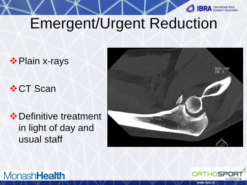
Emergent/Urgent Reduction
Plain x-rays
CT Scan
Definitive treatment
in light of day and
usual staff
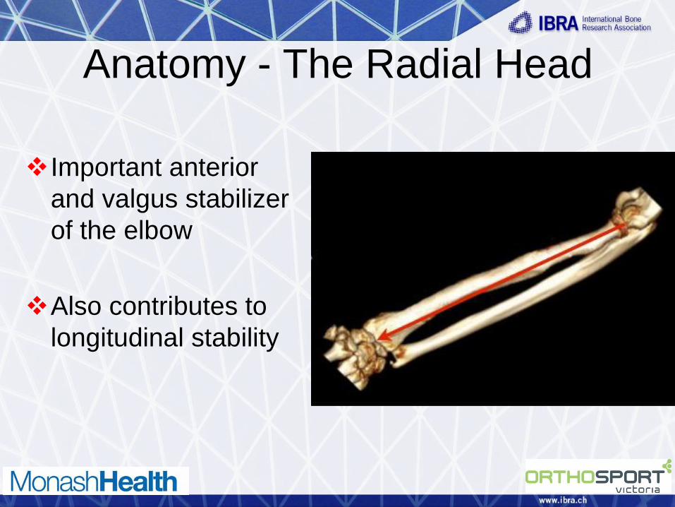
Anatomy - The Radial Head
Important anterior
and valgus stabilizer
of the elbow
Also contributes to
longitudinal stability
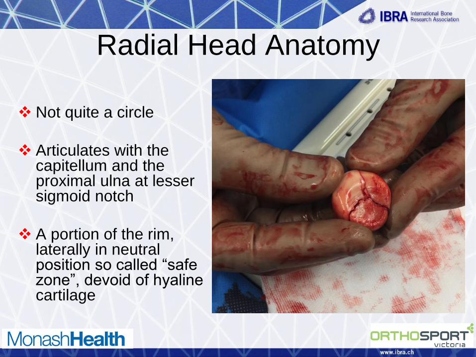
Radial Head Anatomy
Not quite a circle
Articulates with the capitellum and the proximal ulna at lesser sigmoid notch
A portion of the rim, laterally in neutral position so called “safe zone”, devoid of hyaline cartilage
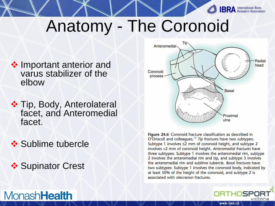
Anatomy - The Coronoid
Important anterior and
varus stabilizer of the elbow
Tip, Body, Anterolateral facet, and Anteromedial facet.
Sublime tubercle
Supinator Crest
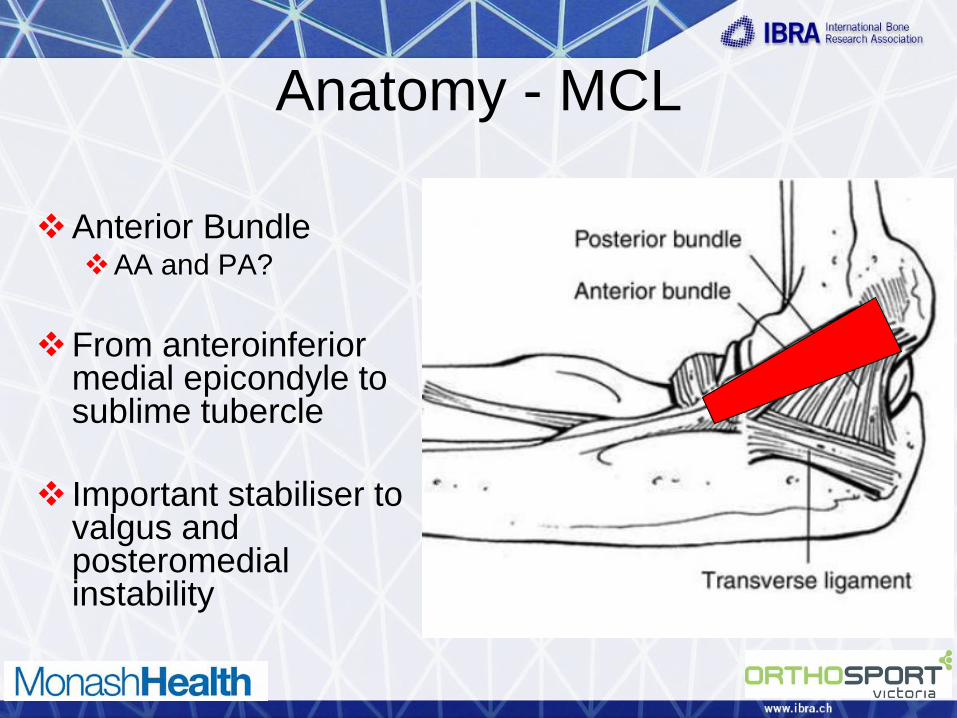
Anatomy - MCL
Anterior Bundle
AA and PA?
From anteroinferior medial epicondyle to sublime tubercle
Important stabiliser to valgus and posteromedial instability

MCL
Transverse
Posterior Bundle
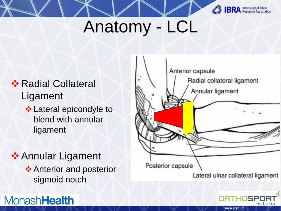
Anatomy - LCL
Radial Collateral
Ligament
Lateral epicondyle to
blend with annular
ligament
Annular Ligament
Anterior and posterior
sigmoid notch

LCL
Lateral Ulnar Collateral Ligament (LUCL) From isometric point
on lateral epicondyle to supinator crest
Important Restraint to Varus and posterolateral rotatory instability (PLRI)
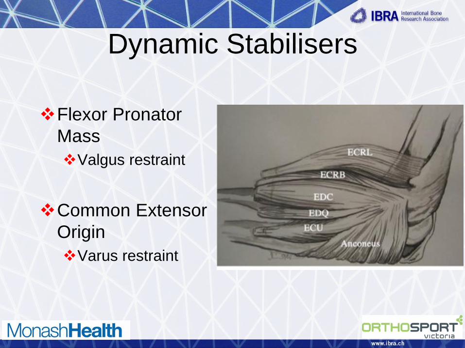
Dynamic Stabilisers
Flexor Pronator
Mass
Valgus restraint
Common Extensor
Origin
Varus restraint
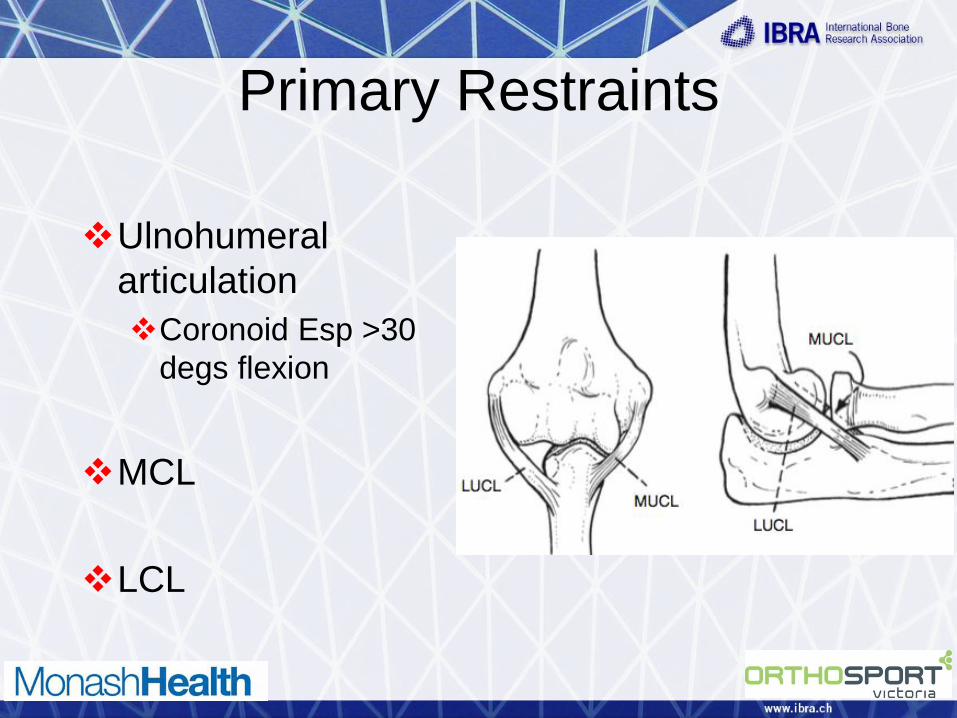
Primary Restraints
Ulnohumeral
articulation
Coronoid Esp >30
degs flexion
MCL
LCL

Secondary Restraints
Radial Head (MCL)
Both radial head and MCL required for normal stability
Joint Capsule
Flexor pronator and common extensor muscles

Mechanism of Injury
Axial load + valgus
load and rotatory
moment
Anterior capsule
tears and levering of
ulna out of
articulation
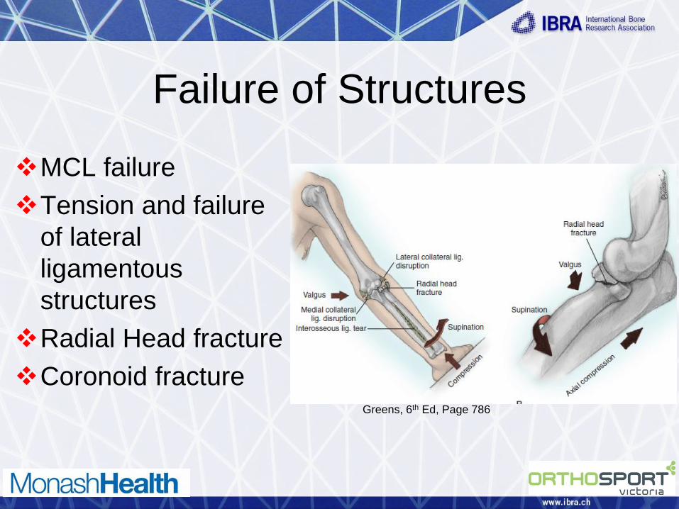
Failure of Structures
MCL failure
Tension and failure
of lateral
ligamentous
structures
Radial Head fracture
Coronoid fracture
Greens, 6th Ed, Page 786

a concentric joint
reduction
radial head fracture
that does not cause
a mechanical block
to rotation
smaller coronoid fracture
stable arc of motion to a minimum of 30° of extension to allow active motion within the first 10 days
24
12 patients

Approach
Lateral
Kocher
Direct Lateral
Medial
ECU split
Over the top
Universal Posterior

Lateral Approach
Lateral Kocher
Direct Lateral
Advantages Easy, familiar
May only need 1 approach anyway
Disadvantages May need medial
approach as well
JAAOS, May 2009, Vol 17, No 5

Medial Approach
Medial
Through floor of
cubital tunnel
Direct medial
Over the top

Radial Head - What Am I
Thinking? Is the joint any good
Are there other
injuries
Radial neck
Capitellar injury
Other
What Am I Thinking?
Is it fixable?
LASTLY but of
course not least!
WHAT IS THE
PATIENT LIKE?

ORIF with radial neck
involvement
Step cut annular
ligament (if not
ripped to shreds)
Identify lateral
ligament complex
to repair

ORIF if you can
Be aware of “safe zone”
Step cut annular ligament (if not ripped to shreds)
This allows closure afterwards if desired

Mason III/IV
Comminuted/Smashed ORIF/Replace
Use a metal
prosthesis
If young I consider a
pyrocarbon implant

Comminuted/Smashed
Make sure you don’t
overstuff
Measure head to get
size
Move immediately
post op

Coronoid Fixation
Fix from lateral side
if access adequate
If not from medially
Small fragments,
repair anterior
capsule
51

Coronoid Fixation
Larger fragments
internal fixation, can
buttress plate from
anteriorly
Retrograde screw,
use ACL guide
52
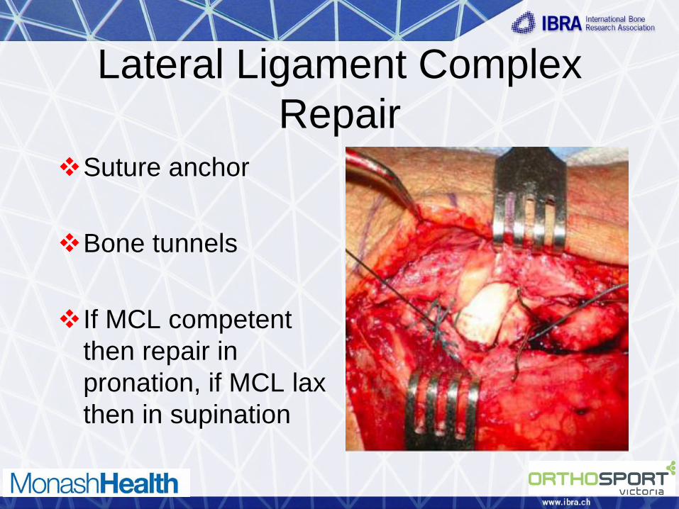
Lateral Ligament Complex
Repair Suture anchor
Bone tunnels
If MCL competent
then repair in
pronation, if MCL lax
then in supination
53

MCL Repair
If elbow remains unstable after radial head, coronoid and LCL stabilised then repair MCL
Suture anchor or tunnels
Beware the ulna nerve
54 JBJS
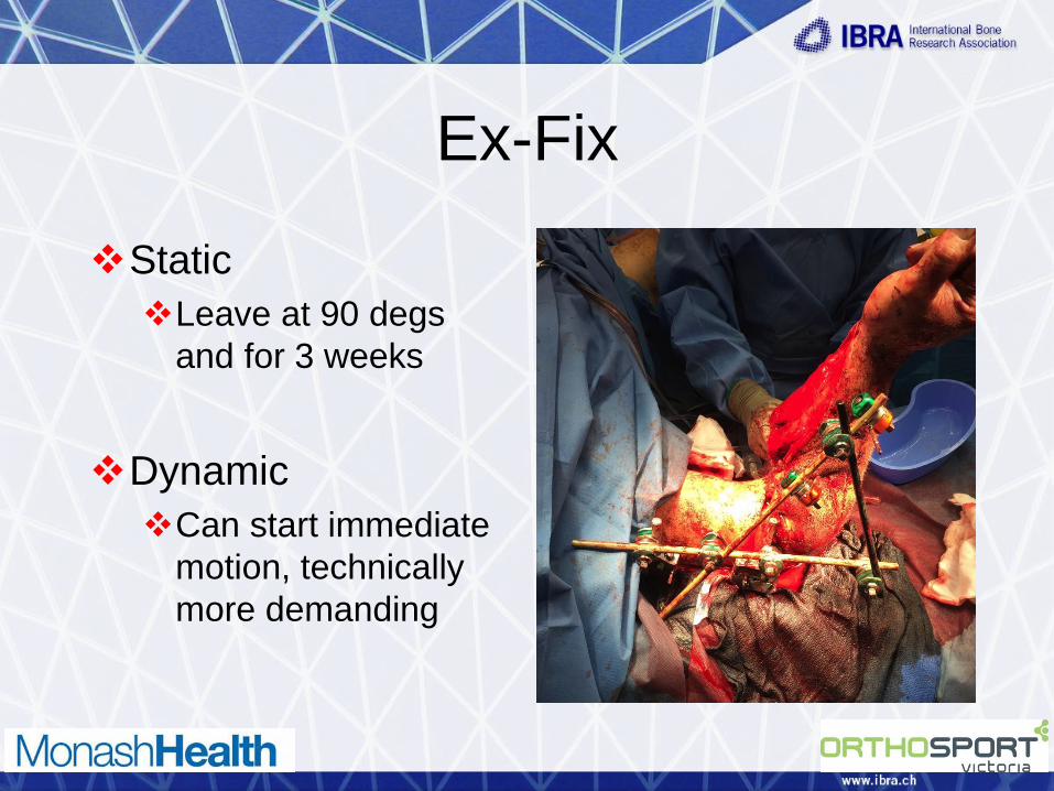
Ex-Fix
Static
Leave at 90 degs
and for 3 weeks
Dynamic
Can start immediate
motion, technically
more demanding
55

Summary
Recognise injury
Reduce joint
Adequate imaging to organise plan of attack
Choose approach
56
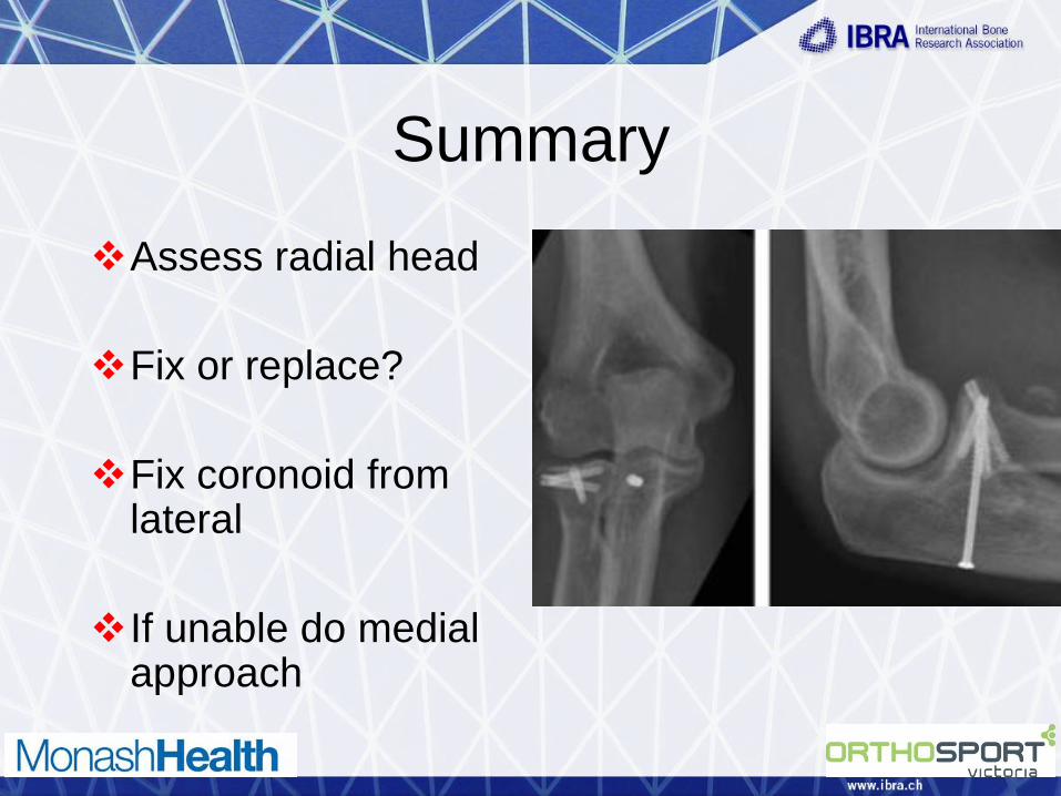
Summary
Assess radial head
Fix or replace?
Fix coronoid from lateral
If unable do medial approach
57

Summary
Repair LCL
Assess stability
Fix MCL if unstable
Assess stability
Ex fix
58 JAAOS, March 2009, Vol 17, No 3

Late Instability
In patients with a
“simple” dislocation
late instability can
occur but is
uncommon
Can be lateral,
medial or both
Lateral Instability
Pain
Locking, clicking or
snapping
Worse w supination,
extension and
valgus force

Lateral Instability
Commonly known
as PLRI =
Posterolateral
Rotatory Instability
Get subluxation of
the radial head in
extension and
supination

Lateral Instability
Also push up and chair apprehension test
Feeling of instability
• Stage 1 - subluxation of the elbow in a posterolateral direction.
• Stage 2 is subluxation of the elbow joint in which the coronoid is perched beneath the trochlea.
• Stage 3 is complete dislocation of the coronoid resting posterior to the trochlea:
• Stage 3a including a tear of posterior band of the MCL
• Stage 3b including a tear of the anterior and posterior band of the MCL

LCL/LUCL Reconstruction
Palmaris Graft
Lateral approach to
elbow
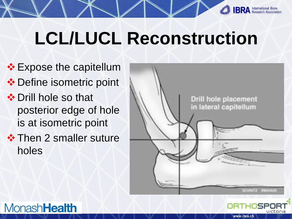
LCL/LUCL Reconstruction
Expose the capitellum
Define isometric point
Drill hole so that
posterior edge of hole
is at isometric point
Then 2 smaller suture
holes
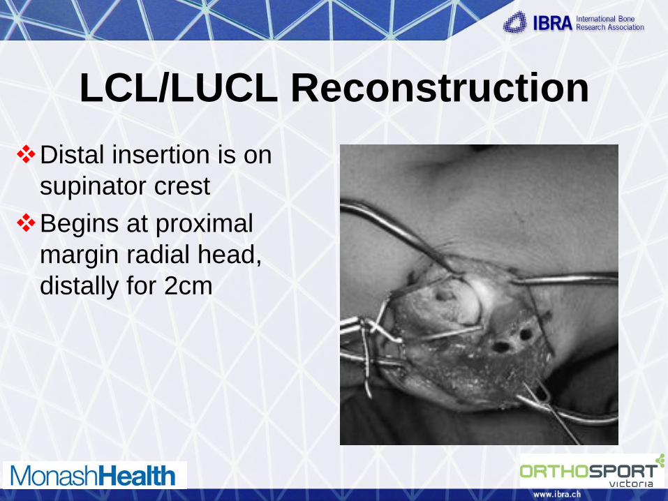
LCL/LUCL Reconstruction
Distal insertion is on
supinator crest
Begins at proximal
margin radial head,
distally for 2cm

LCL/LUCL Reconstruction
Pass graft through
the ulna
Repair native
ligament, capsule
and extensor origin
Pass sutures
through tunnels

Post Op
Start motion at 7-10 days
Avoid shoulder abduction
No supination in full extension until 6 weeks
May pronate/supinate in flexion
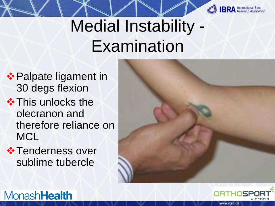
Medial Instability -
Examination
Palpate ligament in 30 degs flexion
This unlocks the olecranon and therefore reliance on MCL
Tenderness over sublime tubercle

Examination
Milking Manoeuvre
Support elbow with
contralateral
forearm and then
grasp thumb
As patient pulls
thumb apples valgus
force Hand Clin 24 (2008) 53–67
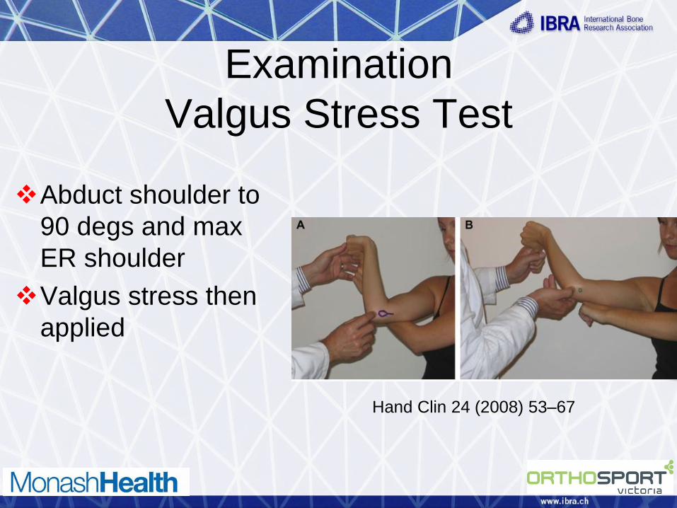
Examination
Valgus Stress Test
Abduct shoulder to
90 degs and max
ER shoulder
Valgus stress then
applied
Hand Clin 24 (2008) 53–67

Examination
Moving Valgus Stress Test
Same as valgus
stress test but then
move elbow
Pain between 70-
120 degs suggests
MCL injury
Hand Clin 24 (2008) 53–67

MCL Incompetent
Reconstruction
“Tommy John”
Procedure
Muscle split through
posterior 1/3 of
flexor/pronator mass
Protect the ulna
nerve
FCU
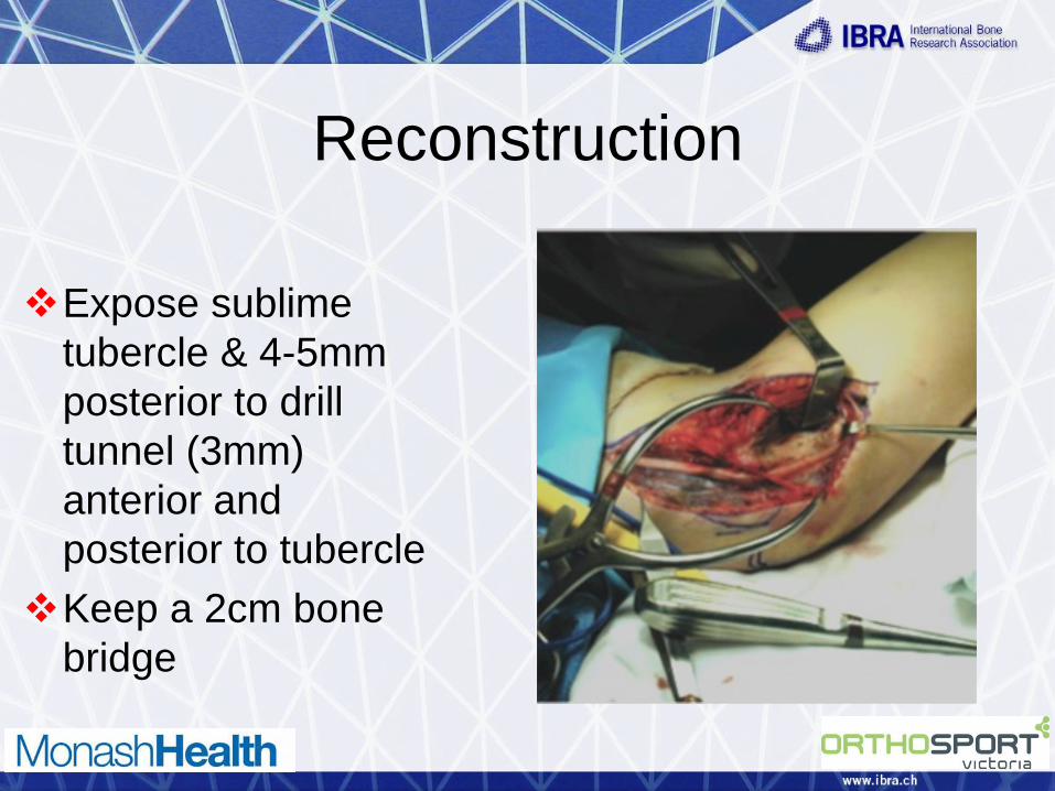
Reconstruction
Expose sublime
tubercle & 4-5mm
posterior to drill
tunnel (3mm)
anterior and
posterior to tubercle
Keep a 2cm bone
bridge

Reconstruction
Humeral exposure along line of UCL
Drill 4mm tunnel with blind end
Drill 2 smaller holes (use K-wire) 1cm apart to connect to 4mm hole

MCL Reconstruction
Usually Palmaris
graft
Pass graft from
anterior to posterior
Pass posterior limb
of graft into humeral
tunnel

MCL Reconstruction
The estimate length
needed to be in tunnel
without bottoming out
Cycle the elbow for
creep
Tension in 30 degs
and tie over bone
bridge

Post Op
Rest 1 week
Then motion 30 – 90
degs flexion for 2
weeks
Week 3-5, 15-105
degs flexion
6 weeks full motion

THANK YOU