identification of a ciliate (oligohymenophorea ...aem.asm.org/content/74/3/883.full.pdf ·...
TRANSCRIPT

APPLIED AND ENVIRONMENTAL MICROBIOLOGY, Feb. 2008, p. 883–888 Vol. 74, No. 30099-2240/08/$08.00�0 doi:10.1128/AEM.01124-07Copyright © 2008, American Society for Microbiology. All Rights Reserved.
Identification of a Ciliate (Oligohymenophorea: Scuticociliatia)Associated with Brown Band Disease on Corals of
the Great Barrier Reef�
David G. Bourne,1* Holly V. Boyett,1,2 Meegan E. Henderson,3Andrew Muirhead,1 and Bette L. Willis2
Australian Institute of Marine Science, PMB No. 3, Townsville, MC, QLD 4810, Australia1; ARC Centre of Excellence forCoral Reef Studies and School of Marine and Tropical Biology, James Cook University, Townsville, QLD 4811,
Australia2; and Centre for Marine Studies, University of Queensland, Brisbane, Australia3
Received 20 May 2007/Accepted 29 November 2007
A ciliate associated with the coral disease brown band (BrB) was identified as a new species belonging to theclass Oligohymenophorea, subclass Scuticociliatia. The ciliates were characterized by the presence of largenumbers of intracellular dinoflagellates and displayed an elongated, tube-shaped body structure. They haduniform ciliature, except for three distinct cilia in the caudal region, and were typically 200 to 400 �m in lengthand 20 to 50 �m in width.
Coral reef ecosystems have been exposed to increasing levelsof sedimentation, nutrient enrichment, and ocean warming inthe past few decades (1, 20–22), resulting in corals experienc-ing elevated levels of stress and enhanced susceptibility todisease infection (9, 19, 20, 23, 31). Coral disease epizooticshave become a major threat to reef ecosystems globally, withreports of newly emerging syndromes continuing to increase innumbers (17, 41). Identifying the microbial communities asso-ciated with coral diseases is critical to further current under-standing of how environmental and climate changes mightaffect the prevalence of diseases. To date, a wide range ofmicroorganisms, including fungi, bacteria, and cyanobacteria,have been identified in association with both healthy and dis-eased corals (10, 14, 18, 29, 30, 32, 33, 44), although microbialcommunities associated with many coral diseases remain un-known (41).
Seven coral diseases on the Great Barrier Reef (GBR) havebeen described previously (42), although their causative agentsremain largely undescribed. One disease, named brown band(BrB), was described for the first time in studies of corals inthree families (Acroporidae, Pocilloporidae, and Faviidae) inthe northern and southern sectors of the GBR (42). Macro-scopic symptoms of the disease manifest as a brown zone,which is preceded by healthy tissue and followed by exposedwhite skeleton as it progresses across the coral (see Fig. 1a). Insome cases, a white zone, comprising bleached tissue and/ordenuded skeleton, is observed between the brown band andhealthy tissue. The distinctive brown color that constitutes themacroscopic field signs of BrB is derived from a mass of un-known ciliates gliding over the exterior surface of coral sam-ples and into the coelenteron and cavities of the coral polyps.
Here we report the identification of the ciliate associated withBrB by use of microscopic and molecular approaches.
Ciliates were removed from specimens of the staghorn coral(Acropora muricata) exhibiting signs of BrB. Disease sampleswere collected from Davies Reef (n � 3) located in the centralsector of the GBR (18°49.86�S, 147°38.2�E) and from fringingreefs around Heron Island (n � 1) located in the southernsector of the GBR (23°44.17�S, 151°91.25�E). All samples weretaken from near the advancing front of the disease lesion andencompassed the brown band ciliate mass. Although poten-tially a complex microbial community involving bacteria, dia-toms, dinoflagellates, and other microscopic marine plankton,the ciliate population appeared uniform and dominated by onemorphologically distinct protozoan (Fig. 1b). Ciliates removedfrom coral specimens were processed for microscopic analysisby fixation in Bouin’s solution (13, 15) and stored in the darkat 4°C or kept at �80°C until DNA was extracted.
High densities of intracellular zooxanthellae (Symbiodiniumsp.) were observed within all ciliates examined by use of lightmicroscopy (Fig. 1c). Morphologically, the ciliate had an elon-gated, tube-like shape rounded at both the posterior and apicalends (Fig. 1c). The length of the ciliate ranged from 200 to 400�m, while the width ranged from 20 to 50 �m. Ciliation wasuniform over the surface of the organism (Fig. 1c) except forthree distinct and extended cilia in the caudal region. Scanningelectron microscopy (SEM) revealed the oral apparatus to bedifferentiated from somatic ciliature and located in the buccalcavity on the ventral side (Fig. 1d).
Total DNA from ciliate and coral tissue samples (extractedaccording to the methods described in references 6 and 43) wasamplified with conserved eukaryotic primers (18S-6-CIL-Vand 18S-1511-CIL-R) (15). PCR resulted in amplification ofthe 18S rRNA genes from protozoa and other eukaryotic or-ganisms (by PCR cycling performed at 95°C for 3 min followedby 30 cycles at 95°C for 1 min, 58°C for 1 min, and 72°C for 1min and a final extension step of 72°C for 7 min), with theproducts (�1.8 kb) cloned (TOPO TA cloning kit; Invitrogen)and the insert 18S rRNA gene reamplified from individual
* Corresponding author. Mailing address: Australian Institute ofMarine Science, PMB No. 3, Townsville, MC, QLD 4810, Australia.Phone: 61 7 4753 4139. Fax: 61 7 47725852. E-mail: [email protected].
� Published ahead of print on 14 December 2007.
883
on June 17, 2018 by guesthttp://aem
.asm.org/
Dow
nloaded from

clones. Restriction fragment length polymorphism analysis wasperformed on reamplified products (8), and clones weregrouped into operational taxonomic unit (OTU) groups.Clones sequenced from the dominant OTU groups were affil-iated with Symbiodinium species within the clade C lineage(98% to 99% sequence identity). One OTU group was affili-ated with 18S rRNA gene sequences of ciliates within thescuticociliate family and was putatively identified as derivedfrom the dominant BrB ciliate organism. This sequence dem-onstrated 95% sequence identity (over 1,749 bp) to the 18SrRNA gene sequence of the Parauronema longum ciliate. Phy-logenetic comparisons indicated that the unknown ciliate isrelated to other ciliates belonging to the class Oligohymeno-phorea, subclass Scuticociliatia (Fig. 2). This subclass includesthe scuticociliates Schizocaryum dogieli, Cohnilembus ver-minua, Anophyroides haemophila, Pseudocohnilembus marinus,and Uronema marinum. Scuticociliates often feed on bacteria,using complex morphological adaptations to create currentsand filters capable of capturing bacteria and other particlesfrom the water column or scraping them from hard surfaces(25).
Based on the retrieved 18S rRNA gene sequence, new PCRprimers were designed using the oligonucleotide primer algo-rithm of the ARB package (27). Generated primers werechecked against the GenBank database by a standard nucle-otide-nucleotide BLAST search (3). PCR primers specific forthe identified BrB ciliate included BrB-F-171 and BrB-R-1721
(Table 1) (PCR cycling was performed at 95°C for 3 minfollowed by 35 cycles at 95°C for 30 s, 45°C for 45 s, and 72°Cfor 2 min, with a final extension of 72°C for 10 min). FurtherBrB tissue samples from both Davies Reef and Heron Islandwere amplified and clones screened as described previously.Sequencing of the dominant clone types retrieved almost iden-tical ciliate-affiliated 18S rRNA gene sequences (�98% se-quence identity) as obtained with the eukaryotic-specificprimer set.
Two oligonucleotide probes, BrB-754 and BrB-1461, tar-geted to variable regions of the BrB ciliate 18S rRNA se-quence, were designed using the probe design algorithm of theARB package (27) and checked against the GenBank database(3). Alignment and comparison of closely related 18S rRNAsequences demonstrated mismatches for both probes (Fig. 3).Fixed ciliate samples (15) were filtered onto 0.22-�m-pore-sizewhite Isopore membrane filters (Millipore) by use of a gentlevacuum and washed five times with 1 ml of filtered sterileseawater. Membranes were covered with hybridization buffer(0.9 M NaCl, 20 mM Tris-HCl [pH 8], 0.01% sodium dodecylsulfate, 30% [vol/vol] formamide) and the appropriate fluoro-chrome-labeled oligonucleotide probe (50 �g). All hybridiza-tions were conducted at 46°C for 3 h, after which membraneswere floated into prewarmed wash buffer (0.102 M NaCl, 20mM Tris-HCl [pH 8], 0.01% sodium dodecyl sulfate, 5 mMEDTA) at 48°C for 10 min to remove excess and nonboundoligonucleotide probes. Air-dried filters were mounted in anantifading gel (Biomedia, ProSciTech) before being viewedand imaged on a Bio-Rad MRC-1024 confocal laser scanningmicroscope (40).
Hybridizations with the eukaryote-specific probe EUK1195(16) resulted in the presence of a fluorescence signal for boththe ciliate and the internalized Symbiodinium sp. (Fig. 4a).Comparative hybridizations of the BrB tissue with probes BrB-754 and BrB-1461 resulted in a fluorescent signal for the cili-ate, correlating the retrieved 18S rRNA gene sequence withthe distinct morphological characteristics of the BrB ciliate(Fig. 4b and c). Morphological features of the ciliates couldalso be distinguished, including the buccal cavity on the ventralside (Fig. 4b). No signal associated with Symbiodinium sp. wasobserved for the BrB ciliate-targeted probes, supporting probespecificity. Signals from the EUK1195, BrB-754, and BrB-1461probes were clearly distinguishable from autofluorescence sig-nals achieved with negative-control hybridizations (NONEUBnonsense probe) (4) (Fig. 4d).
Ciliates belonging to the Scuticociliatia subclass are abun-dant in marine habitats and often observed as endosymbiontsin marine invertebrates such as echinoids, crustaceans,polychaetes, and bivalve mollusks (25). Although the feedingbehavior of the brown band ciliate requires further study, thehigh density of Symbiodinium cells observed within its mem-branes (Fig. 1c) is the primary cause of the brown color thatcharacterizes the disease’s appearance in the field (Fig. 1a). Atpresent, it is unknown whether the ciliate ingests zooxanthellaein the course of feeding on live coral tissue (i.e., is carnivo-rous), ingests zooxanthellae in the course of feeding on deadcoral tissue (histophagous), or acquires them from elsewhere(algivorous). However, the presence of high densities of feed-ing ciliates, in combination with the retrieval of Symbiodinium18S rRNA gene sequences from DNA extracted from ciliate
FIG. 1. (a) Photograph of brown band disease on a branch ofAcropora muricata showing its macroscopic field signs. Arrow 1,healthy coral tissue; arrow 2, brown zone preceded by healthy tissue;arrow 3, exposed white skeleton following the brown zone; arrow 4, awhite, bleached zone between the brown band and healthy tissue. (b)Micrograph showing a coral polyp covered by a mass of ciliates (LeicaMZ16A; Leica Microsystems AG, Wetzlar, Germany). Arrows 1 and 2are as defined for panel a. (c) Micrograph (obtained using an OlympusVanox AH-2 compound microscope; Shinjuku-ku, Tokyo, Japan)(�100 magnification) showing morphology of the brown band ciliate.Arrow 1, uniform ciliation; arrow 2, Symbiodinium cells within a ciliate.(d) SEM micrograph (obtained using a JEOL JSM 5410LV scanningelectron microscope) showing morphology of the brown band ciliate.The arrow denotes the buccal cavity on the ventral side. Dissecting andcompound images were taken using an Olympus digital camera (C-5050Z) (5 megapixel, 3� zoom).
884 BOURNE ET AL. APPL. ENVIRON. MICROBIOL.
on June 17, 2018 by guesthttp://aem
.asm.org/
Dow
nloaded from
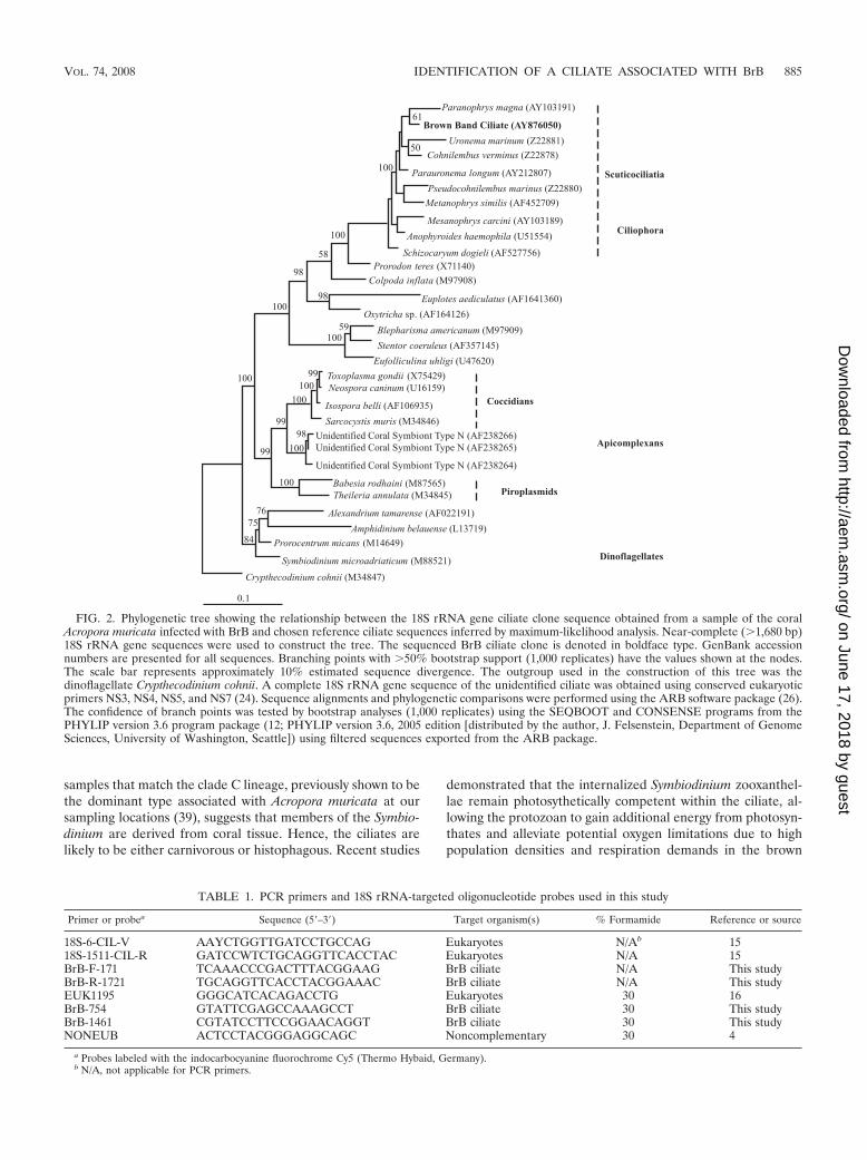
samples that match the clade C lineage, previously shown to bethe dominant type associated with Acropora muricata at oursampling locations (39), suggests that members of the Symbio-dinium are derived from coral tissue. Hence, the ciliates arelikely to be either carnivorous or histophagous. Recent studies
demonstrated that the internalized Symbiodinium zooxanthel-lae remain photosythetically competent within the ciliate, al-lowing the protozoan to gain additional energy from photosyn-thates and alleviate potential oxygen limitations due to highpopulation densities and respiration demands in the brown
FIG. 2. Phylogenetic tree showing the relationship between the 18S rRNA gene ciliate clone sequence obtained from a sample of the coralAcropora muricata infected with BrB and chosen reference ciliate sequences inferred by maximum-likelihood analysis. Near-complete (�1,680 bp)18S rRNA gene sequences were used to construct the tree. The sequenced BrB ciliate clone is denoted in boldface type. GenBank accessionnumbers are presented for all sequences. Branching points with �50% bootstrap support (1,000 replicates) have the values shown at the nodes.The scale bar represents approximately 10% estimated sequence divergence. The outgroup used in the construction of this tree was thedinoflagellate Crypthecodinium cohnii. A complete 18S rRNA gene sequence of the unidentified ciliate was obtained using conserved eukaryoticprimers NS3, NS4, NS5, and NS7 (24). Sequence alignments and phylogenetic comparisons were performed using the ARB software package (26).The confidence of branch points was tested by bootstrap analyses (1,000 replicates) using the SEQBOOT and CONSENSE programs from thePHYLIP version 3.6 program package (12; PHYLIP version 3.6, 2005 edition [distributed by the author, J. Felsenstein, Department of GenomeSciences, University of Washington, Seattle]) using filtered sequences exported from the ARB package.
TABLE 1. PCR primers and 18S rRNA-targeted oligonucleotide probes used in this study
Primer or probea Sequence (5�–3�) Target organism(s) % Formamide Reference or source
18S-6-CIL-V AAYCTGGTTGATCCTGCCAG Eukaryotes N/Ab 1518S-1511-CIL-R GATCCWTCTGCAGGTTCACCTAC Eukaryotes N/A 15BrB-F-171 TCAAACCCGACTTTACGGAAG BrB ciliate N/A This studyBrB-R-1721 TGCAGGTTCACCTACGGAAAC BrB ciliate N/A This studyEUK1195 GGGCATCACAGACCTG Eukaryotes 30 16BrB-754 GTATTCGAGCCAAAGCCT BrB ciliate 30 This studyBrB-1461 CGTATCCTTCCGGAACAGGT BrB ciliate 30 This studyNONEUB ACTCCTACGGGAGGCAGC Noncomplementary 30 4
a Probes labeled with the indocarbocyanine fluorochrome Cy5 (Thermo Hybaid, Germany).b N/A, not applicable for PCR primers.
VOL. 74, 2008 IDENTIFICATION OF A CILIATE ASSOCIATED WITH BrB 885
on June 17, 2018 by guesthttp://aem
.asm.org/
Dow
nloaded from

band zone (38). Such a mixotrophic strategy is common amongfreshwater oligotrichs, with enslaved photosynthetic compo-nents remaining functional for hours to days, thereby providingnutrients, covering respiratory demands, and increasing growth
efficiency (11, 34–36). Whether a similar relationship existsbetween BrB ciliates and internalized Symbiodinium zooxan-thellae has yet to be determined; however, symbiotic relation-ships between ciliates and zooxanthellae have previously beenreported for ciliates living in association with corals (26).
Although common in marine environments, ciliates arerarely classified as pathogenic parasites (28), especially in coralcommunities. One study has linked a GBR coral disease withthe Halofolliculina corallasia heterotrich ciliate. Known as skel-etal eroding band, this disease has been characterized by anadvancing mass of ciliates whose pericytostomial wings areencased within flask-like black loricae (5). Protozoan infec-tions have also been identified on corals held in aquaria. Forexample, the consumption of coral tissue by the ciliate Helico-stoma nonatum produces brown jelly-like symptoms in infectedaquarium corals (7). Willis et al. (42) speculated that the ciliateassociated with BrB might be related to H. nonatum, althoughresults from this study suggest that it belongs to a differentfamily. Other studies have identified a protozoan belonging tothe phylum Apicomplexa within microbial communities asso-ciated with the coral Montastraea annularis in the Caribbean,but although this protozoan is related to a group of highlyparasitic organisms, whether or not it is parasitic on corals iscurrently unknown (37).
The causative agent of the coral disease BrB remains un-known. The appearance of a white bleached zone, often ob-served between healthy coral tissue and an advancing mass ofciliates (Fig. 1a), suggests that the ciliate may invade second-arily after coral health is compromised, although it is clear that
FIG. 3. Different sequence alignments of probes BrB-754 (A) and BrB-1461 (B). Nucleotides are only identified for mispairings; pairings areindicated by dots. The bases of the mismatches refer to the sequences of the target organism and not of the probe. GenBank accession numbersof each organism are presented in parentheses.
FIG. 4. Fluorescence in situ hybridization micrographs of BrB cil-iates. (a) Ciliate probed with Cy5-labeled eukaryote-specific probeEUK1195 (16). (b) Ciliate probed with Cy5-labeled BrB-754-specificprobe. (c) Ciliates probed with Cy5-labeled BrB-1461-specific probe.(d) Ciliate probed with Cy5-labeled NONEUB nonsense probe, dem-onstrating autofluorescence of only the sample-specific probe.
886 BOURNE ET AL. APPL. ENVIRON. MICROBIOL.
on June 17, 2018 by guesthttp://aem
.asm.org/
Dow
nloaded from

the ciliate subsequently becomes responsible for macroscopicfield signs of BrB disease. A number of factors may compro-mise coral health, including bacterial or viral infections, injury,and, alternatively, apoptosis triggered by stress, injury, or in-fection (2, 18). As the health of the coral deteriorates, necros-ing tissue could attract the ciliate to feed on both bacteria andzooxanthellae associated with dead and dying coral tissue. Athigh densities, however, the ciliates may become the primarycause of tissue loss as they uptake photocompetent zooxan-thellae to alleviate potential oxygen limitations (38).
In summary, the characteristic macroscopic signs of the coraldisease BrB have been attributed to the presence of a newlyidentified ciliate species of the class Oligohymenophorea, sub-class Scuticociliatia. Future studies investigating the life cycleand taxonomic traits of the ciliate are required along withadditional microbiological studies to further clarify the natureof the causative agent(s) of this coral disease.
Nucleotide sequence accession number. The nucleotide se-quence data have been submitted to the GenBank nucleotidesequence database under accession number AY876050.
We thank Neal Cantin, Meir Sussman, and Cathie Page from JamesCook University for field and laboratory assistance, Kevin Blake fromJames Cook University for help in generating SEM images, and NeilYoung, Lone Høj, Eric Matson, and Jason Doyle from the AustralianInstitute of Marine Science and Colin Munn from the University ofPlymouth for their assistance in field and laboratory studies.
Research was supported by an ARC DP grant to B. L. Willis and theCoral Disease Working Group of the GEF CRTR Program.
REFERENCES
1. Acosta, A. 2001. Disease in zoanthids: dynamics in space and time. Hydro-biologia 460:113–130.
2. Ainsworth, T. D., E. C. Kvennefors, L. L. Blackall, M. Fine, and O. Hoegh-Gulberg. 2006. Disease and cell death in white syndrome of acroporid coralson the Great Barrier Reef. Mar. Biol. 151:19–29.
3. Altschul, S. F., T. L. Madden, A. A. Schaeffer, J. Zhang, Z. Zhang, W. Miller,and D. J. Lipman. 1997. Gapped BLAST and PSI-BLAST: a new generationof protein database search programs. Nucleic Acids Res. 25:3389–3402.
4. Amann, R. I., L. Krumholz, and D. A. Stahl. 1990. Fluorescent-oligonucle-otide probing of whole cells for determinative, phylogenetic, and environ-mental studies in microbiology. J. Bacteriol. 172:762–770.
5. Antonius, A., and D. Lipscomb. 2001. First protozoan coral-killer identifiedin the Indo-Pacific. Atoll Res. Bull. 493:1–21.
6. Asahida, T., T. Kobayashi, K. Saitoh, and I. Nakayama. 1996. Tissue pres-ervation and total DNA extraction from fish stored at ambient temperatureusing buffers containing high concentrations of urea. Fish. Sci. 62:727–730.
7. Borneman, E. H. 2001. Aquarium corals: selection husbandry and naturalhistory. TFH Publications, Neptune City, NJ.
8. Bourne, D. G., and C. B. Munn. 2005. Diversity of bacteria associated withthe coral Pocillopora damicornis from the Great Barrier Reef. Environ.Microbiol. 7:1162–1174.
9. Bruno, J. F., E. R. Selig, K. S. Casey, C. A. Page, B. L. Willis, C. D. Harvell,H. Sweatman, and A. M. Melendy. 2007. Thermal stress and coral cover asdrivers of coral disease outbreaks. PLoS Biol. 5:1–7.
10. Cooney, R. P., O. Pantos, M. D. A. Le Tissier, M. R. Barer, A. G. O’Donnell,and J. C. Bythell. 2002. Characterisation of the bacterial consortium asso-ciated with the black band disease in coral using molecular microbiologicaltechniques. Environ. Microbiol. 4:401–413.
11. Dolan, J. R., and M. T. Perez. 2000. Costs, benefits and characteristics ofmixotrophy in marine oligotrichs. Freshw. Biol. 45:227–238.
12. Felsenstein, J. 1989. PHYLIP—phylogeny inference package (version 3.2).Cladistics 5:164–166.
13. Foissner, W. 1991. Basic light and scanning electron microscopic methods fortaxonomic studies of ciliated protozoa. Eur. J. Protistol. 27:313–330.
14. Frias-Lopez, J., A. L. Zerkle, G. T. Bonheyo, and B. W. Fouke. 2002. Parti-tioning of bacterial communities between seawater and healthy, black banddiseased, and dead coral surfaces. Appl. Environ. Microbiol. 68:2214–2228.
15. Fried, J., W. Ludwig, R. Psenner, and K. H. Schleifer. 2002. Improvement ofciliate identification and quantification: a new protocol for fluorescence insitu hybridisation (FISH) in combination with silver stain techniques. Syst.Appl. Microbiol. 25:555–571.
16. Giovannoni, S. J., E. F. DeLong, G. J. Oslen, and N. R. Pace. 1988. Phylo-
genetic group-specific oligonucleotide probes for identification of single mi-crobial cells. J. Bacteriol. 170:720–726.
17. Green, E., and A. Bruckner. 2000. The significance of coral disease epizooti-ology for coral reef conservation. Biol. Conserv. 96:347–361.
18. Harvell, C. D., E. Jordan-Dahlgren, S. Merkel, E. Rosenberg, L. Raymundo,G. Smith, E. Weil, and B. L. Willis. 2007. Coral disease, environmentaldrivers and the balance between coral and microbial associates. Oceanogra-phy 20:58–81.
19. Harvell, C. D., K. Kim, J. Burkholder, R. Colwell, P. Epstein, D. Grimes, E.Hofmann, E. Lipp, A. Osterhaus, R. Overstreet, J. Porter, G. Smith, and G.Vasta. 1999. Emerging marine diseases—climate links and anthropogenicfactors. Science 285:1505–1510.
20. Harvell, C. D., C. E. Mitchell, J. R. Ward, S. Altizer, A. P. Dobson, R. S.Ostfeld, and M. D. Samuel. 2002. Climate warming and disease risks forterrestrial and marine biota. Science 296:2158–2162.
21. Hoegh-Guldberg, O. 1999. Climate change, coral bleaching and the future ofthe world’s coral reefs. Mar. Freshw. Res. 50:839–866.
22. Hughes, T. P., A. H. Baird, D. R. Bellwood, M. Card, S. R. Connolly, C.Folke, R. Grosberg, O. Hoegh-Guldberg, J. B. C. Jackson, J. Kleypas, J. M.Lough, P. Marshall, M. Nystrom, S. R. Palumbi, J. M. Pandolfi, B. Rosen,and J. Roughgarden. 2003. Climate change, human impacts and the resil-ience of coral reefs. Science 301:929–933.
23. Lafferty, K. D., J. Porter, and S. E. Ford. 2004. Are diseases increasing in theoceans? Annu. Rev. Ecol. Evol. Syst. 35:31–54.
24. Lane, D. J. 1991. 16S/23S rRNA sequencing, p. 115–175. In E. Stackebrandtand M. Goodfellow (ed.), Nucleic acid techniques in bacterial systematics.John Wiley and Sons, New York, NY.
25. Lee, J. J., and G. M. Capriulo. 1990. The ecology of marine protozoa: anoverview, p. 1–15. In G. M. Capriulo (ed.), Ecology of marine protozoa.Oxford University Press, New York, NY.
26. Lobban, C. S., M. Schefter, A. G. B. Simpson, X. Pochon, J. Pawlowski, andW. Foissner. 2002. Maristentor dinoferus n. gen., n. sp., a giant heterotrichciliate (Spirotrichea: Heterotrichida) with zooxanthellae, from coral reefs onGuam, Mariana Islands. Mar. Biol. 141:207–208.
27. Ludwig, W., O. Strunk, R. Westram, L. Richter, H. Meier, Yadhukumar, A.Buchner, T. Lai, S. Steppi, G. Jobb, W. Forster, I. Brettske, S. Gerber, A. W.Ginhart, O. Gross, S. Grumann, S. Hermann, R. Jost, A. Konig, T. Liss, R.Lu�mann, M. May, B. Nonhoff, B. Reichel, R. Strehlow, A. Stamatakis, N.Struckmann, A. Vilbig, M. Lenke, T. Ludwig, A. Bode, and K.-H. Schleifer.2004. ARB: a software package environment for sequence data. NucleicAcids Res. 32:1363–1371.
28. Lynn, D. H., and J. O. Corliss. 1991. Ciliophora, p. 333–467. In F. W.Harrison (ed.), Microscopic anatomy of invertebrates, vol. 1: protozoa.Wiley-Liss, Inc., New York, NY.
29. Patterson, K. L., J. W. Porter, K. B. Ritchie, S. W. Polson, E. Mueller, E. C.Peters, D. L. Santavvy, and G. W. Smith. 2002. The etiology of white pox, alethal disease of the Caribbean elkhorn coral, Acropora palmata. Proc. Natl.Acad. Sci. USA 99:8725–8730.
30. Peters, E. C. 1997. Diseases of coral-reef organisms, p. 114–139. In C.Birkeland (ed.), Life and death of coral reefs. Chapman and Hall Publishers,London, United Kingdom.
31. Porter, J., P. Dunstan, W. Jaap, K. Patterson, V. Kosmynin, O. Meier, M.Patterson, and M. Parsons. 2001. Patterns of spread of coral disease in theFlorida Keys. Hydrobiologia 460:1397–1407.
32. Rohwer, F., M. Breitbart, J. Jara, F. Azam, and N. Knowlton. 2001. Diversityof bacteria associated with the Caribbean coral Montastraea franksi. CoralReefs 20:85–91.
33. Rohwer, F., V. Seguritan, F. Azam, and N. Knowlton. 2002. Diversity anddistribution of coral-associated bacteria. Mar. Ecol. Prog. Ser. 243:1–10.
34. Stoecker, D. K., and M. W. Silver. 1990. Replacement and aging of chloro-plasts in Strombidium capitatum (Ciliophora: Oligotrichida). Mar. Biol. 107:491–502.
35. Stoecker, D. K., M. W. Silver, A. E. Michaels, and L. H. Davis. 1989.Enslavement of algal chloroplasts by four Strombidium spp. Mar. Microb.Food Webs 3:79–100.
36. Stoecker, D. K., M. W. Silver, A. E. Michaels, and L. H. Davis. 1988. Obligatemixotrophy in Laboea strobila, a ciliate which retains chloroplasts. Mar. Biol.99:415–423.
37. Toller, W. W., R. Rowan, and N. Knowlton. 2002. Genetic evidence for aprotozoan (phylum Apicomplexa) associated with corals of the Montastraeaannularis species complex. Coral Reefs 21:143–146.
38. Ulstrup, K. E., M. Kuhl, and D. G. Bourne. 2007. Zooxanthellae harvestedby ciliates associated with brown band syndrome of coral remain photo-synthetically competent. Appl. Environ. Microbiol. 73:1968–1975.
39. van Oppen, M. J. H., F. Palstra, A. M.-T. Piquet, and D. J. Miller. 2001.Patterns of coral-dinoflagellate associations in Acropora: significance of localavailability and physiology of Symbiodinium strains and host-symbiont selec-tivity. Proc. R. Soc. Lond. B 268:1759–1767.
40. Webster, N., D. G. Bourne, and M. Hall. 2006. Vibrio infection in phylloso-mas of the tropical rock lobster Panulirus ornatus as detected by fluorescencein situ hybridisation. Aquaculture 255:173–178.
41. Weil, E. 2004. Coral reef diseases in the wider Caribbean: status and prog-
VOL. 74, 2008 IDENTIFICATION OF A CILIATE ASSOCIATED WITH BrB 887
on June 17, 2018 by guesthttp://aem
.asm.org/
Dow
nloaded from

nosis, p. 35–68. In E. Rosenberg and Y. Loya (ed.), Coral health and disease.Springer-Verlag, New York, NY.
42. Willis, B. L., C. A. Page, and E. A. Dinsdale. 2004. Coral diseases on the greatbarrier reef, p. 69–104. In E. Rosenberg and Y. Loya (ed.), Coral health anddisease. Springer-Verlag, New York, NY.
43. Wilson, K. J., Y. Li, V. Whan, S. A. Lehnert, K. Byrne, S. S. Moore, S.Pongsomboon, A. Tassanakajon, G. Rosenberg, E. Ballment, Z. Fayazi, J.
Swan, M. J. Kenway, and J. A. H. Benzie. 2002. Genetic mapping of the blacktiger shrimp Penaeus monodon with amplified fragment length polymor-phisms. Aquaculture 204:297–309.
44. Yarden, O., T. D. Ainsworth, G. Roff, W. Leggat, M. Fine, and O. Hoegh-Guldberg. 2007. Increased prevalence of ubiquitous Ascomycetes in an acro-poid coral (Acropora formosa) exhibiting symptoms of brown band syndromeand skeletal eroding band disease. Appl. Environ. Microbiol. 73:2755–2757.
888 BOURNE ET AL. APPL. ENVIRON. MICROBIOL.
on June 17, 2018 by guesthttp://aem
.asm.org/
Dow
nloaded from