identification of a μ δ opioid receptor heteromer- biased ... · biased agonist with...
TRANSCRIPT
Corrections
MEDICAL SCIENCESCorrection for “Myc and mTOR converge on a common nodein protein synthesis control that confers synthetic lethality inMyc-driven cancers,” by Michael Pourdehnad, Morgan L. Truitt,Imran N. Siddiqi, Gregory S. Ducker, Kevan M. Shokat, andDavide Ruggero, which appeared in issue 29, July 16, 2013, of
Proc Natl Acad Sci USA (110:11988–11993; first published June26, 2013; 10.1073/pnas.1310230110).The authors note that Fig. 5 appeared incorrectly. The cor-
rected figure and its legend appear below.
www.pnas.org/cgi/doi/10.1073/pnas.1317701110
NEUROSCIENCECorrection for “Identification of a μ-δ opioid receptor heteromer-biased agonist with antinociceptive activity,” by Ivone Gomes,Wakako Fujita, Achla Gupta, Adrian S. Saldanha, Ana Negri,Christine E. Pinello, Edward Roberts, Marta Filizola, Peter Hodder,and Lakshmi A. Devi, which appeared in issue 29, July 16, 2013,of Proc Natl Acad Sci USA (110:12072–12077; first published July 1,2013; 10.1073/pnas.1222044110).The authors note that Christina Eberhart should be added to
the author list between Christine E. Pinello and Edward Roberts.Christina Eberhart should be credited with having performedresearch and analyzed data.The authors also note that the author name Adrian S. Saldanha
should instead appear as S. Adrian Saldanha.The corrected author line, affiliation line, and author con-
tributions appear below. The online version has been corrected.
Ivone Gomesa, Wakako Fujitaa, Achla Guptaa,S. Adrian Saldanhab, Ana Negric, Christine E. Pinellob,Christina Eberhartb, Edward Robertsd, Marta Filizolac,Peter Hodderb, and Lakshmi A. Devia
Departments of aPharmacology and Systems Therapeutics and cStructuraland Chemical Biology, Icahn School of Medicine at Mount Sinai, New York,NY 10029; bScripps Research Institute Molecular Screening Center, LeadIdentification Division, Translational Research Institute, Jupiter, FL 33458;and dDepartment of Chemistry, The Scripps Research Institute, La Jolla,CA 92037
Author contributions: P.H. and L.A.D. designed research; I.G., W.F., A.G., S.A.S.,A.N., C.E.P., C.E., and E.R. performed research; E.R. and M.F. contributednew reagents/analytic tools; I.G., W.F., S.A.S., C.E.P., C.E., M.F., P.H., and L.A.D.analyzed data; and I.G. and L.A.D. wrote the paper.
www.pnas.org/cgi/doi/10.1073/pnas.1317238110
NEUROSCIENCECorrection for “Transient, afferent input-dependent, postnatalniche for neural progenitor cells in the cochlear nucleus,”by Stefan Volkenstein, Kazuo Oshima, Saku T. Sinkkonen,C. Eduardo Corrales, Sam P. Most, Renjie Chai, Taha A. Jan,Alan G. Cheng, and Stefan Heller, which appeared in issue 35,August 27, 2013, of Proc Natl Acad Sci USA (110:14456–14461;first published August 12, 2013; 10.1073/pnas.1307376110).The authors note that Renée van Amerongen should be added
to the author list between Taha A. Jan and Alan G. Cheng. Renéevan Amerongen should be credited with having performed re-search and having contributed new reagents/analytic tools. Thecorrected author line, affiliation line, and author contributionsappear below. The online version has been corrected.
Stefan Volkensteina,b, Kazuo Oshimaa,b, Saku T.Sinkkonena,b, C. Eduardo Corralesa, Sam P. Mosta,Renjie Chaia, Taha A. Jana, Renée van Amerongenc,Alan G. Chenga, and Stefan Hellera,b
Departments of aOtolaryngology–Head and Neck Surgery and bMolecularand Cellular Physiology, Stanford University School of Medicine, Stanford,CA 94305; and cDivision of Molecular Oncology, Netherlands CancerInstitute, 1066 CX, Amsterdam, The Netherlands
Author contributions: S.V., K.O., T.A.J., and S.H. designed research; S.V., K.O.,S.T.S., C.E.C., S.P.M., R.C., T.A.J., and R.v.A. performed research; R.v.A.contributed new reagents/analytic tools; S.V., K.O., S.T.S., C.E.C., S.P.M., T.A.J.,A.G.C., and S.H. analyzed data; and S.V., K.O., S.P.M., and S.H. wrote the paper.
www.pnas.org/cgi/doi/10.1073/pnas.1317787110
A B C
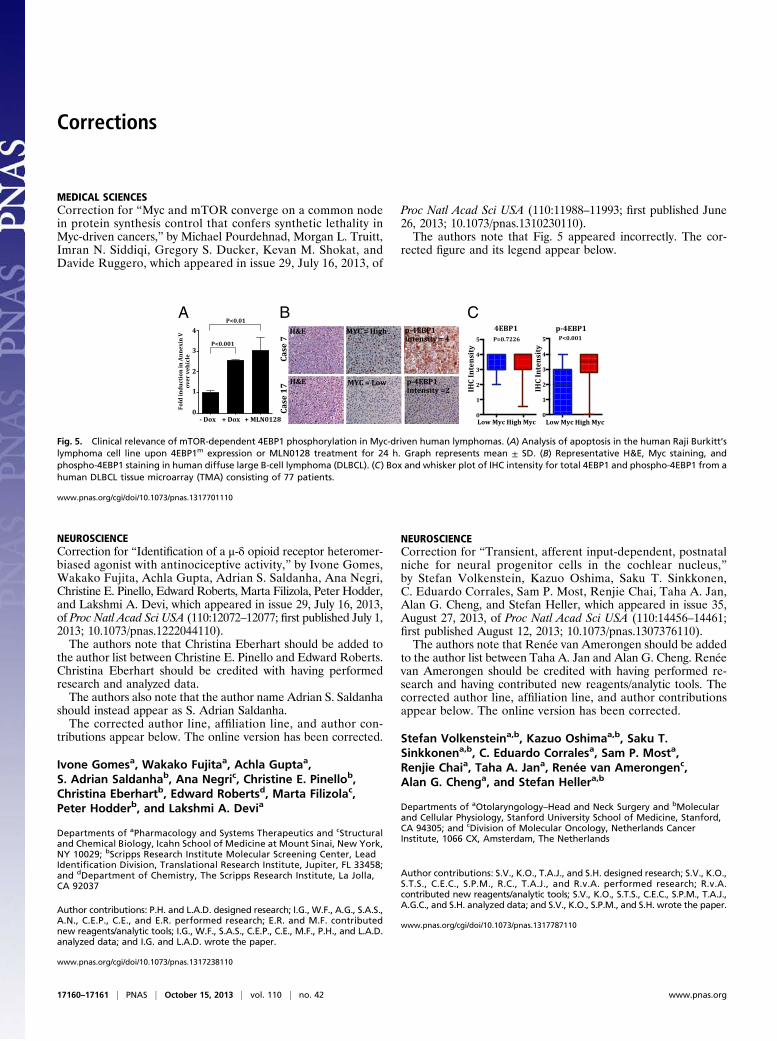
Fig. 5. Clinical relevance of mTOR-dependent 4EBP1 phosphorylation in Myc-driven human lymphomas. (A) Analysis of apoptosis in the human Raji Burkitt’slymphoma cell line upon 4EBP1m expression or MLN0128 treatment for 24 h. Graph represents mean ± SD. (B) Representative H&E, Myc staining, andphospho-4EBP1 staining in human diffuse large B-cell lymphoma (DLBCL). (C) Box and whisker plot of IHC intensity for total 4EBP1 and phospho-4EBP1 from ahuman DLBCL tissue microarray (TMA) consisting of 77 patients.
17160–17161 | PNAS | October 15, 2013 | vol. 110 | no. 42 www.pnas.org
NEUROSCIENCECorrection for “The role of long-range connections on the speci-ficity of the macaque interareal cortical network,” by Nikola T.Markov, Maria Ercsey-Ravasz, Camille Lamy, Ana Rita RibeiroGomes, Loïc Magrou, Pierre Misery, Pascale Giroud, PascalBarone, Colette Dehay, Zoltán Toroczkai, Kenneth Knoblauch,David C. Van Essen, and Henry Kennedy, which appeared inissue 13, March 26, 2013, of Proc Natl Acad Sci USA (110:5187–5192; first published March 11, 2013; 10.1073/pnas.1218972110).The authors note that Fig. 4 appeared incorrectly. The correct
figure and its legend appear below.Additionally, on page 5190, right column, first full paragraph,
lines 21–22, “These values contrast with the interregion graph, inwhich the density is 50%” should instead appear as “These valuescontrast with the interregion graph, in which the density is 61%.”
www.pnas.org/cgi/doi/10.1073/pnas.1316840110
Experimental valuesRandomized values
Num
ber o
f com
mon
are
as
Front Occ Par Pref Temp
A intra target regions
0
5
10
15
20
25
Front Occ Par Pref Temp
inter target regions
Distance (mm)
Sim
ilarit
y sc
ore (0
,10]
(10,
20]
(20,
30]
(30,
40]
(40,
50]
(50,
60] 0
20
40
60
80
100
Den
sity
in %
AbsentPresent
Distance intervals (mm)
Num
ber o
f are
as
020
040
060
080
010
00C
D
0 10 20 30 40 50 60-0.2
0.0
0.2
0.4 p < 0.001
PrefFront TempParOcc Inter G29x29
Den
sity
0
0.2
0.4
0.6
0.8
1.0B
0
5
10
15
20
25
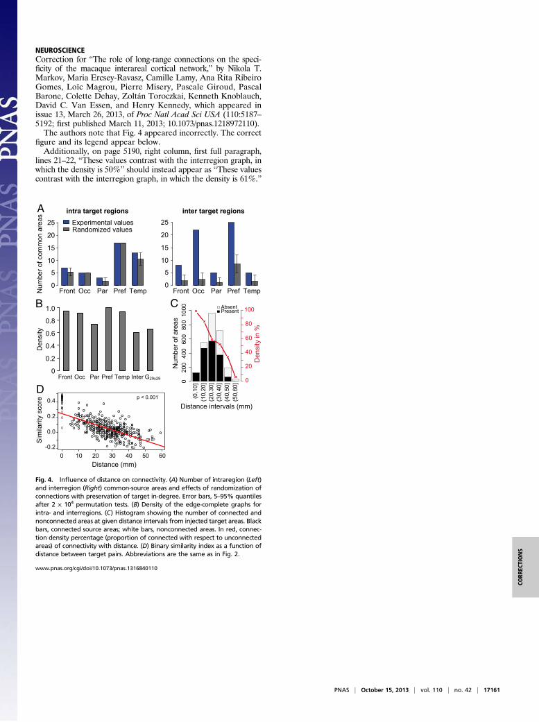
Fig. 4. Influence of distance on connectivity. (A) Number of intraregion (Left)and interregion (Right) common-source areas and effects of randomization ofconnections with preservation of target in-degree. Error bars, 5–95% quantilesafter 2 × 10⁴ permutation tests. (B) Density of the edge-complete graphs forintra- and interregions. (C) Histogram showing the number of connected andnonconnected areas at given distance intervals from injected target areas. Blackbars, connected source areas; white bars, nonconnected areas. In red, connec-tion density percentage (proportion of connected with respect to unconnectedareas) of connectivity with distance. (D) Binary similarity index as a function ofdistance between target pairs. Abbreviations are the same as in Fig. 2.
PNAS | October 15, 2013 | vol. 110 | no. 42 | 17161
CORR
ECTIONS
Identification of a μ-δ opioid receptor heteromer-biased agonist with antinociceptive activityIvone Gomesa, Wakako Fujitaa, Achla Guptaa, S. Adrian Saldanhab, Ana Negric, Christine E. Pinellob,Christina Eberhartb, Edward Robertsd, Marta Filizolac, Peter Hodderb, and Lakshmi A. Devia,1
Departments of aPharmacology and Systems Therapeutics and cStructural and Chemical Biology, Icahn School of Medicine at Mount Sinai, New York,NY 10029; bScripps Research Institute Molecular Screening Center, Lead Identification Division, Translational Research Institute, Jupiter, FL 33458;and dDepartment of Chemistry, The Scripps Research Institute, La Jolla, CA 92037
Edited* by Susan G. Amara, National Institute of Mental Health, Bethesda, MD, and approved June 4, 2013 (received for review December 19, 2012)
Gprotein-coupled receptors play a pivotal role inmany physiologicalsignaling pathways. Mounting evidence suggests that G protein-coupled receptors, including opioid receptors, form dimers, anddimerization is necessary for receptor maturation, signaling, andtrafficking. However, the physiological role of dimerization invivo has not been well-explored because of the lack of tools tostudy these dimers in endogenous systems. To address this prob-lem, we previously generated antibodies to μ-δ opioid receptor(μOR-δOR) dimers and used them to study the pharmacology andsignaling by this heteromer. We also showed that the heteromerexhibits restricted distribution in the brain and that its abundanceis increased in response to chronic morphine administration.Thus, the μOR-δOR heteromer represents a potentially uniquetarget for the development of therapeutics to treat pain. Here, wereport the identification of compounds targeting μOR-δOR hetero-mers through high-throughput screening of a small-molecule li-brary. These compounds exhibit activity in μOR-δOR cells but notμOR or δOR cells alone. Among them, CYM51010 was found tobe a μOR-δOR–biased ligand, because its activity is blocked bythe μOR-δOR heteromer antibody. Notably, systemic administra-tion of CYM51010 induced antinociceptive activity similar tomorphine, and chronic administration of CYM51010 resulted inlesser antinociceptive tolerance comparedwith morphine. Takentogether, these results suggest that CYM51010, a μOR-δOR–biasedligand, could serve as a scaffold for the development of a uniquetype (heteromer-biased) of drug that ismore potent andwithout thesevere side effects associated with conventional clinical opioids.
Studies with mice lacking opioid receptors show that theantinociceptive actions of clinically administered opioids,
such as morphine or fentanyl, involve the activation of μ-opioidreceptors (μORs) (1). However, continued opioid use leads toundesired side effects, including respiratory depression, con-stipation, immunosuppression, and development of toleranceand addiction (2). In an effort to identify novel compounds thatare as effective as morphine in the treatment of chronic pain butwithout the associated side effects, our group, among others, hasinvestigated the modulation of μOR function by receptor het-eromerization. We found that μOR can form interacting com-plexes with δ-opioid receptors (δORs), that both receptors are inclose proximity to interact in live cells, and that, in heterologoussystems, low nonsignaling doses of some δOR ligands can po-tentiate the binding and signaling of μOR agonists (3–5). Therecently reported crystal structure of μOR (6), in which receptorswere crystallized as parallel dimers, is consistent with the ideathat μOR can associate in complexes.We also generated mAbs selective to μOR-δOR heteromers;
we showed that the latter can be detected in the brains of WTbut not KO mice and that heteromer levels are increased in brainregions involved in pain processing after chronic morphine ad-ministration under a paradigm that leads to the development oftolerance (7). The idea that μOR-δOR heteromers may playa role in the development of tolerance to morphine is furthersupported by studies showing that genetic deletion of either δOR
or β-arrestin or possible disruption of μOR-δOR heteromersleads to an enhancement of morphine-mediated antinociceptionand attenuation in the development of tolerance (8–10). Nota-bly, we observed that a δOR antagonist, H-Tyr-Tic[CH2NH]-Phe-Phe-OH (TIPPψ), can potentiate morphine-mediated an-algesia (4), and studies using bivalent ligands targeting μOR-δOR heteromers showed that these ligands induce anti-nociception with attenuated development of tolerance as well asconditioned place preference (11, 12). Taken together, thesedata suggest that occupancy of δOR by an antagonist coulddissociate the antinociceptive effects of μOR agonists from thedevelopment of tolerance and addiction. Therefore, there isa need for ligands that selectively interact with μOR-δOR het-eromers to understand their role in antinociception and de-velopment of tolerance to morphine.In an attempt to identify μOR-δOR heteromer-selective ago-
nists, we used a β-arrestin recruitment assay and screened smallmolecules available through the Molecular Libraries Probe Pro-duction Centers Network. This screen identified 94 compoundsthat were biased to μOR-δOR heteromers compared with μOR,δOR, or serotonin 5HT5A receptors. Among a dozen compoundsthat were repurchased and tested using secondary screens, one,which we named CYM51010 [PubChem compound identifier(CID)23723457; Probe Report ID ML335], exhibited a strongμOR-δOR–biased activity that was blocked by μOR-δOR het-eromer-selective mAb (μ-δ mAb). Furthermore, systemic ad-ministration of CYM51010 led to antinociceptive activity similarto morphine but with a lower antinociceptive tolerance onchronic administration. Notably, although the intrathecal (i.t.)antinociceptive activity of CYM51010 could be significantlyblocked by i.t. administration of μ-δ mAb, the i.t. antinociceptiveactivity ofmorphinewas not. These results suggest that CYM51010could serve as a scaffold for the development of unique thera-peutics acting at the μOR-δOR heteromer for the effective man-agement of pain.
Results and DiscussionTo screen for μOR-δOR heteromer-biased ligands, we used aβ-arrestin recruitment assay that is based on an enzyme frag-ment complementation technology. Specifically, receptor acti-vation-mediated β-arrestin recruitment leads to reconstitutionof β-gal activity (Fig. S1). This strategy was used to engineercell lines stably expressing μβgalOR-δOR, δβgalOR, or μβgalOR(DiscoverX). Based on the finding that these cells bind
Author contributions: P.H. and L.A.D. designed research; I.G., W.F., A.G., S.A.S., A.N.,C.E.P., C.E., and E.R. performed research; E.R. and M.F. contributed new reagents/analytictools; I.G., W.F., S.A.S., C.E.P., C.E., M.F., P.H., and L.A.D. analyzed data; and I.G. and L.A.D.wrote the paper.
The authors declare no conflict of interest.
*This Direct Submission article had a prearranged editor.1To whom correspondence should be addressed. E-mail: [email protected].
This article contains supporting information online at www.pnas.org/lookup/suppl/doi:10.1073/pnas.1222044110/-/DCSupplemental.
12072–12077 | PNAS | July 16, 2013 | vol. 110 | no. 29 www.pnas.org/cgi/doi/10.1073/pnas.1222044110
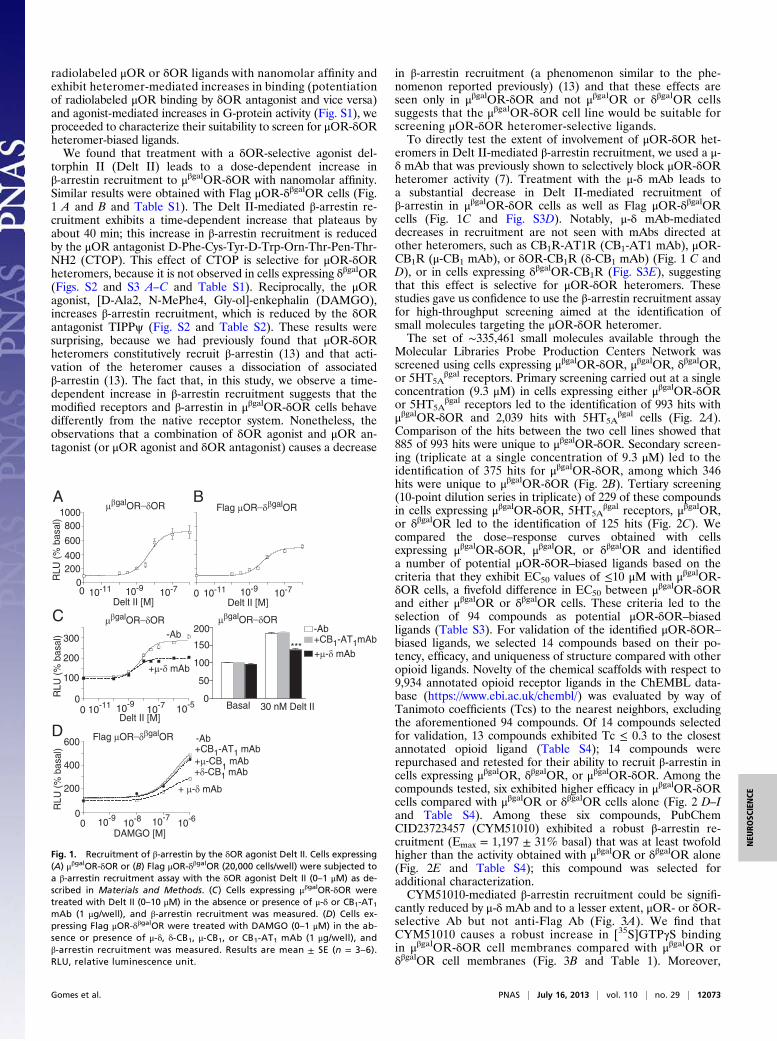
radiolabeled μOR or δOR ligands with nanomolar affinity andexhibit heteromer-mediated increases in binding (potentiationof radiolabeled μOR binding by δOR antagonist and vice versa)and agonist-mediated increases in G-protein activity (Fig. S1), weproceeded to characterize their suitability to screen for μOR-δORheteromer-biased ligands.We found that treatment with a δOR-selective agonist del-
torphin II (Delt II) leads to a dose-dependent increase inβ-arrestin recruitment to μβgalOR-δOR with nanomolar affinity.Similar results were obtained with Flag μOR-δβgalOR cells (Fig.1 A and B and Table S1). The Delt II-mediated β-arrestin re-cruitment exhibits a time-dependent increase that plateaus byabout 40 min; this increase in β-arrestin recruitment is reducedby the μOR antagonist D-Phe-Cys-Tyr-D-Trp-Orn-Thr-Pen-Thr-NH2 (CTOP). This effect of CTOP is selective for μOR-δORheteromers, because it is not observed in cells expressing δβgalOR(Figs. S2 and S3 A–C and Table S1). Reciprocally, the μORagonist, [D-Ala2, N-MePhe4, Gly-ol]-enkephalin (DAMGO),increases β-arrestin recruitment, which is reduced by the δORantagonist TIPPψ (Fig. S2 and Table S2). These results weresurprising, because we had previously found that μOR-δORheteromers constitutively recruit β-arrestin (13) and that acti-vation of the heteromer causes a dissociation of associatedβ-arrestin (13). The fact that, in this study, we observe a time-dependent increase in β-arrestin recruitment suggests that themodified receptors and β-arrestin in μβgalOR-δOR cells behavedifferently from the native receptor system. Nonetheless, theobservations that a combination of δOR agonist and μOR an-tagonist (or μOR agonist and δOR antagonist) causes a decrease
in β-arrestin recruitment (a phenomenon similar to the phe-nomenon reported previously) (13) and that these effects areseen only in μβgalOR-δOR and not μβgalOR or δβgalOR cellssuggests that the μβgalOR-δOR cell line would be suitable forscreening μOR-δOR heteromer-selective ligands.To directly test the extent of involvement of μOR-δOR het-
eromers in Delt II-mediated β-arrestin recruitment, we used a μ-δ mAb that was previously shown to selectively block μOR-δORheteromer activity (7). Treatment with the μ-δ mAb leads toa substantial decrease in Delt II-mediated recruitment ofβ-arrestin in μβgalOR-δOR cells as well as Flag μOR-δβgalORcells (Fig. 1C and Fig. S3D). Notably, μ-δ mAb-mediateddecreases in recruitment are not seen with mAbs directed atother heteromers, such as CB1R-AT1R (CB1-AT1 mAb), μOR-CB1R (μ-CB1 mAb), or δOR-CB1R (δ-CB1 mAb) (Fig. 1 C andD), or in cells expressing δβgalOR-CB1R (Fig. S3E), suggestingthat this effect is selective for μOR-δOR heteromers. Thesestudies gave us confidence to use the β-arrestin recruitment assayfor high-throughput screening aimed at the identification ofsmall molecules targeting the μOR-δOR heteromer.The set of ∼335,461 small molecules available through the
Molecular Libraries Probe Production Centers Network wasscreened using cells expressing μβgalOR-δOR, μβgalOR, δβgalOR,or 5HT5A
βgal receptors. Primary screening carried out at a singleconcentration (9.3 μM) in cells expressing either μβgalOR-δORor 5HT5A
βgal receptors led to the identification of 993 hits withμβgalOR-δOR and 2,039 hits with 5HT5A
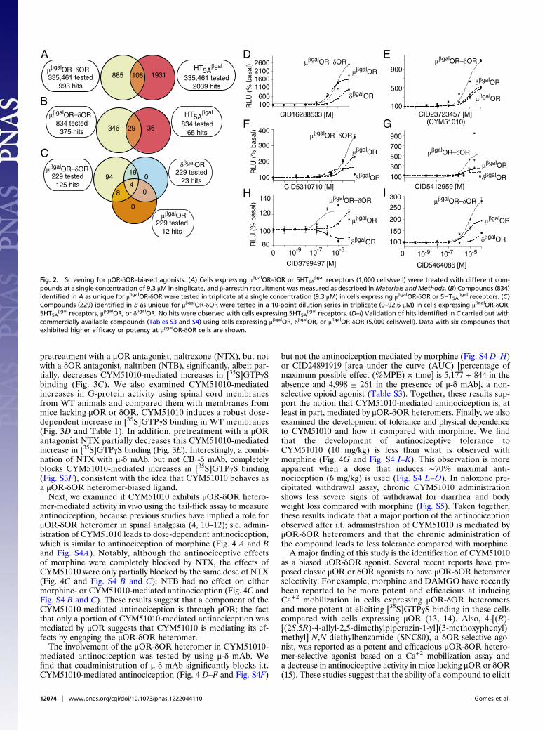
βgal cells (Fig. 2A).Comparison of the hits between the two cell lines showed that885 of 993 hits were unique to μβgalOR-δOR. Secondary screen-ing (triplicate at a single concentration of 9.3 μM) led to theidentification of 375 hits for μβgalOR-δOR, among which 346hits were unique to μβgalOR-δOR (Fig. 2B). Tertiary screening(10-point dilution series in triplicate) of 229 of these compoundsin cells expressing μβgalOR-δOR, 5HT5A
βgal receptors, μβgalOR,or δβgalOR led to the identification of 125 hits (Fig. 2C). Wecompared the dose–response curves obtained with cellsexpressing μβgalOR-δOR, μβgalOR, or δβgalOR and identifieda number of potential μOR-δOR–biased ligands based on thecriteria that they exhibit EC50 values of ≤10 μM with μβgalOR-δOR cells, a fivefold difference in EC50 between μβgalOR-δORand either μβgalOR or δβgalOR cells. These criteria led to theselection of 94 compounds as potential μOR-δOR–biasedligands (Table S3). For validation of the identified μOR-δOR–
biased ligands, we selected 14 compounds based on their po-tency, efficacy, and uniqueness of structure compared with otheropioid ligands. Novelty of the chemical scaffolds with respect to9,934 annotated opioid receptor ligands in the ChEMBL data-base (https://www.ebi.ac.uk/chembl/) was evaluated by way ofTanimoto coefficients (Tcs) to the nearest neighbors, excludingthe aforementioned 94 compounds. Of 14 compounds selectedfor validation, 13 compounds exhibited Tc ≤ 0.3 to the closestannotated opioid ligand (Table S4); 14 compounds wererepurchased and retested for their ability to recruit β-arrestin incells expressing μβgalOR, δβgalOR, or μβgalOR-δOR. Among thecompounds tested, six exhibited higher efficacy in μβgalOR-δORcells compared with μβgalOR or δβgalOR cells alone (Fig. 2 D–Iand Table S4). Among these six compounds, PubChemCID23723457 (CYM51010) exhibited a robust β-arrestin re-cruitment (Emax = 1,197 ± 31% basal) that was at least twofoldhigher than the activity obtained with μβgalOR or δβgalOR alone(Fig. 2E and Table S4); this compound was selected foradditional characterization.CYM51010-mediated β-arrestin recruitment could be signifi-
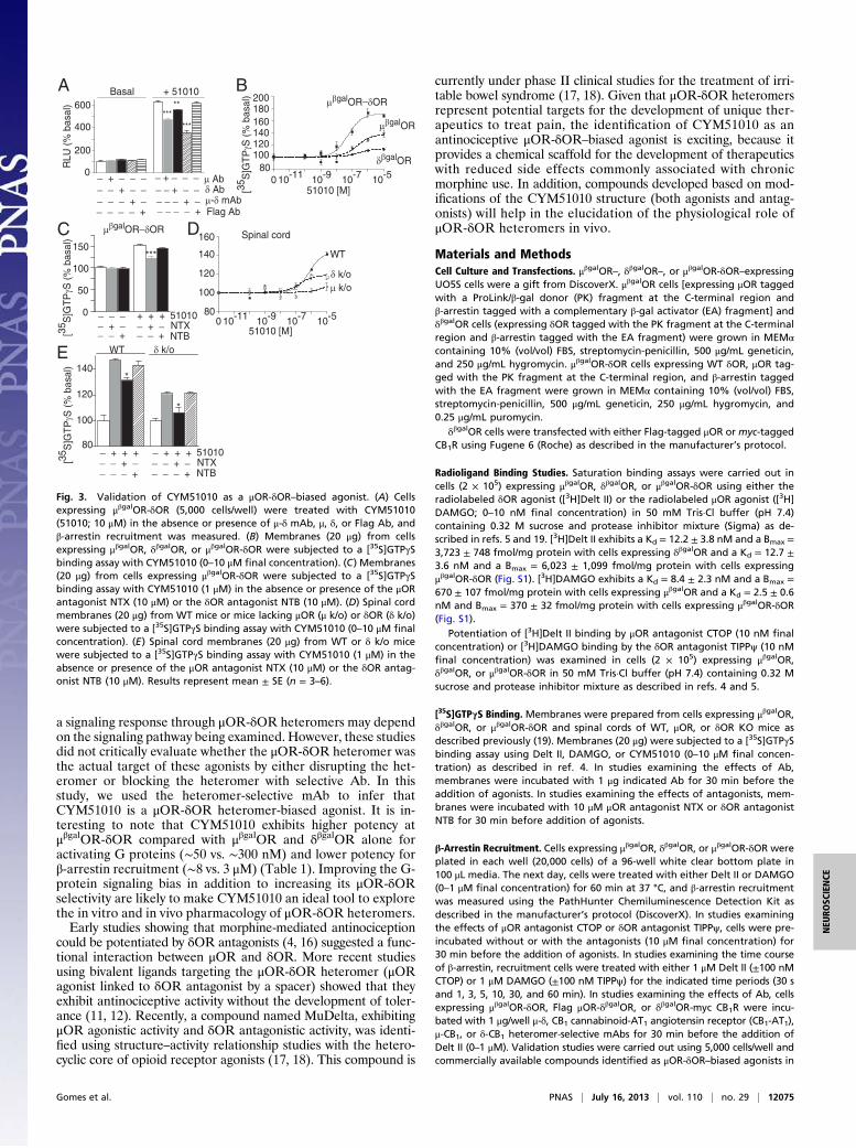
cantly reduced by μ-δ mAb and to a lesser extent, μOR- or δOR-selective Ab but not anti-Flag Ab (Fig. 3A). We find thatCYM51010 causes a robust increase in [35S]GTPγS bindingin μβgalOR-δOR cell membranes compared with μβgalOR orδβgalOR cell membranes (Fig. 3B and Table 1). Moreover,
μβgalOR_δOR Flag μOR_δβgalORA B
0 10-11 10-9 10-7
Delt II [M]0 10-11 10-9 10-7
Delt II [M]
RLU
(%
bas
al)
200400
600
8001000
0
0
100
200
300
RLU
(%
bas
al)
0 10-11 10-910-7
Delt II [M]10-5
-Ab
+μ-δ mAb
Basal 30 nM Delt II0
50
100
150
200 -Ab+CB1-AT1mAb
+μ-δ mAb
C
0
200
400
600
0 10-8 10-710-6
DAMGO [M]10-9
-Ab+CB1-AT1 mAb
+ μ-δ mAb
+μ-CB1 mAb
+δ-CB1 mAb
RLU
(%
bas
al)
D
***
μβgalOR_δOR
Flag μOR_δβgalOR
μβgalOR_δOR
Fig. 1. Recruitment of β-arrestin by the δOR agonist Delt II. Cells expressing(A) μβgalOR-δOR or (B) Flag μOR-δβgalOR (20,000 cells/well) were subjected toa β-arrestin recruitment assay with the δOR agonist Delt II (0–1 μM) as de-scribed in Materials and Methods. (C) Cells expressing μβgalOR-δOR weretreated with Delt II (0–10 μM) in the absence or presence of μ-δ or CB1-AT1mAb (1 μg/well), and β-arrestin recruitment was measured. (D) Cells ex-pressing Flag μOR-δβgalOR were treated with DAMGO (0–1 μM) in the ab-sence or presence of μ-δ, δ-CB1, μ-CB1, or CB1-AT1 mAb (1 μg/well), andβ-arrestin recruitment was measured. Results are mean ± SE (n = 3–6).RLU, relative luminescence unit.
Gomes et al. PNAS | July 16, 2013 | vol. 110 | no. 29 | 12073
NEU
ROSC
IENCE
pretreatment with a μOR antagonist, naltrexone (NTX), but notwith a δOR antagonist, naltriben (NTB), significantly, albeit par-tially, decreases CYM51010-mediated increases in [35S]GTPγSbinding (Fig. 3C). We also examined CYM51010-mediatedincreases in G-protein activity using spinal cord membranesfrom WT animals and compared them with membranes frommice lacking μOR or δOR. CYM51010 induces a robust dose-dependent increase in [35S]GTPγS binding in WT membranes(Fig. 3D and Table 1). In addition, pretreatment with a μORantagonist NTX partially decreases this CYM51010-mediatedincrease in [35S]GTPγS binding (Fig. 3E). Interestingly, a combi-nation of NTX with μ-δ mAb, but not CB1-δ mAb, completelyblocks CYM51010-mediated increases in [35S]GTPγS binding(Fig. S3F), consistent with the idea that CYM51010 behaves asa μOR-δOR heteromer-biased ligand.Next, we examined if CYM51010 exhibits μOR-δOR hetero-
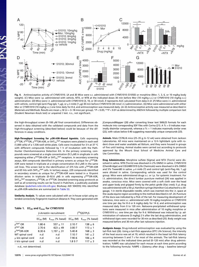
mer-mediated activity in vivo using the tail-flick assay to measureantinociception, because previous studies have implied a role forμOR-δOR heteromer in spinal analgesia (4, 10–12); s.c. admin-istration of CYM51010 leads to dose-dependent antinociception,which is similar to antinociception of morphine (Fig. 4 A and Band Fig. S4A). Notably, although the antinociceptive effectsof morphine were completely blocked by NTX, the effects ofCYM51010 were only partially blocked by the same dose of NTX(Fig. 4C and Fig. S4 B and C); NTB had no effect on eithermorphine- or CYM51010-mediated antinociception (Fig. 4C andFig. S4 B and C). These results suggest that a component of theCYM51010-mediated antinociception is through μOR; the factthat only a portion of CYM51010-mediated antinociception wasmediated by μOR suggests that CYM51010 is mediating its ef-fects by engaging the μOR-δOR heteromer.The involvement of the μOR-δOR heteromer in CYM51010-
mediated antinociception was tested by using μ-δ mAb. Wefind that coadministration of μ-δ mAb significantly blocks i.t.CYM51010-mediated antinociception (Fig. 4 D–F and Fig. S4F)
but not the antinociception mediated by morphine (Fig. S4 D–H)or CID24891919 [area under the curve (AUC) [percentage ofmaximum possible effect (%MPE) × time] is 5,177 ± 844 in theabsence and 4,998 ± 261 in the presence of μ-δ mAb], a non-selective opioid agonist (Table S3). Together, these results sup-port the notion that CYM51010-mediated antinociception is, atleast in part, mediated by μOR-δOR heteromers. Finally, we alsoexamined the development of tolerance and physical dependenceto CYM51010 and how it compared with morphine. We findthat the development of antinociceptive tolerance toCYM51010 (10 mg/kg) is less than what is observed withmorphine (Fig. 4G and Fig. S4 I–K). This observation is moreapparent when a dose that induces ∼70% maximal anti-nociception (6 mg/kg) is used (Fig. S4 L–O). In naloxone pre-cipitated withdrawal assay, chronic CYM51010 administrationshows less severe signs of withdrawal for diarrhea and bodyweight loss compared with morphine (Fig. S5). Taken together,these results indicate that a major portion of the antinociceptionobserved after i.t. administration of CYM51010 is mediated byμOR-δOR heteromers and that the chronic administration ofthe compound leads to less tolerance compared with morphine.A major finding of this study is the identification of CYM51010
as a biased μOR-δOR agonist. Several recent reports have pro-posed classic μOR or δOR agonists to have μOR-δOR heteromerselectivity. For example, morphine and DAMGO have recentlybeen reported to be more potent and efficacious at inducingCa+2 mobilization in cells expressing μOR-δOR heteromersand more potent at eliciting [35S]GTPγS binding in these cellscompared with cells expressing μOR (13, 14). Also, 4-[(R)-[(2S,5R)-4-allyl-2,5-dimethylpiperazin-1-yl](3-methoxyphenyl)methyl]-N,N-diethylbenzamide (SNC80), a δOR-selective ago-nist, was reported as a potent and efficacious μOR-δOR hetero-mer-selective agonist based on a Ca+2 mobilization assay anda decrease in antinociceptive activity in mice lacking μOR or δOR(15). These studies suggest that the ability of a compound to elicit
885 108 1931
346 29 36
HT5Aβgal
9419
0
δβgalOR
4
μβgalOR
0
0
8
A
B
C
229 tested125 hits
229 tested23 hits
229 tested12 hits
834 tested375 hits
834 tested65 hits
335,461 tested993 hits
HT5Aβgal
335,461 tested2039 hits
80
100
120
140
CID3799497 [M]
D
100300500700900
CID5412959 [M]
E
100
150
200
250
300
CID5464086 [M]
F
100
200
300
400
CID5310710 [M]
G (CYM51010)
H
100600
1100160021002600
CID16288533 [M]
10-9 10-7 10-50
I
RLU
(%
bas
al)
RLU
(%
bas
al)
100
500
900
CID23723457 [M]
μβgalOR_δOR
μβgalOR_δOR
μβgalOR_δOR
μβgalOR_δOR
μβgalOR_δOR
μβgalOR_δOR
μβgalOR_δOR
μβgalOR_δORμβgalOR_δOR
μβgalOR
μβgalOR
μβgalOR
μβgalOR
μβgalOR
μβgalOR
δβgalOR
δβgalOR
δβgalOR
δβgalOR
δβgalOR
δβgalOR
10-9 10-7 10-50R
LU (
% b
asal
)
Fig. 2. Screening for μOR-δOR–biased agonists. (A) Cells expressing μβgalOR-δOR or 5HT5Aβgal receptors (1,000 cells/well) were treated with different com-
pounds at a single concentration of 9.3 μM in singlicate, and β-arrestin recruitment was measured as described inMaterials and Methods. (B) Compounds (834)identified in A as unique for μβgalOR-δOR were tested in triplicate at a single concentration (9.3 μM) in cells expressing μβgalOR-δOR or 5HT5A
βgal receptors. (C)Compounds (229) identified in B as unique for μβgalOR-δOR were tested in a 10-point dilution series in triplicate (0–92.6 μM) in cells expressing μβgalOR-δOR,5HT5A
βgal receptors, μβgalOR, or δβgalOR. No hits were observed with cells expressing 5HT5Aβgal receptors. (D–I) Validation of hits identified in C carried out with
commercially available compounds (Tables S3 and S4) using cells expressing μβgalOR, δβgalOR, or μβgalOR-δOR (5,000 cells/well). Data with six compounds thatexhibited higher efficacy or potency at μβgalOR-δOR cells are shown.
12074 | www.pnas.org/cgi/doi/10.1073/pnas.1222044110 Gomes et al.
a signaling response through μOR-δOR heteromers may dependon the signaling pathway being examined. However, these studiesdid not critically evaluate whether the μOR-δOR heteromer wasthe actual target of these agonists by either disrupting the het-eromer or blocking the heteromer with selective Ab. In thisstudy, we used the heteromer-selective mAb to infer thatCYM51010 is a μOR-δOR heteromer-biased agonist. It is in-teresting to note that CYM51010 exhibits higher potency atμβgalOR-δOR compared with μβgalOR and δβgalOR alone foractivating G proteins (∼50 vs. ∼300 nM) and lower potency forβ-arrestin recruitment (∼8 vs. 3 μM) (Table 1). Improving the G-protein signaling bias in addition to increasing its μOR-δORselectivity are likely to make CYM51010 an ideal tool to explorethe in vitro and in vivo pharmacology of μOR-δOR heteromers.Early studies showing that morphine-mediated antinociception
could be potentiated by δOR antagonists (4, 16) suggested a func-tional interaction between μOR and δOR. More recent studiesusing bivalent ligands targeting the μOR-δOR heteromer (μORagonist linked to δOR antagonist by a spacer) showed that theyexhibit antinociceptive activity without the development of toler-ance (11, 12). Recently, a compound named MuDelta, exhibitingμOR agonistic activity and δOR antagonistic activity, was identi-fied using structure–activity relationship studies with the hetero-cyclic core of opioid receptor agonists (17, 18). This compound is
currently under phase II clinical studies for the treatment of irri-table bowel syndrome (17, 18). Given that μOR-δOR heteromersrepresent potential targets for the development of unique ther-apeutics to treat pain, the identification of CYM51010 as anantinociceptive μOR-δOR–biased agonist is exciting, because itprovides a chemical scaffold for the development of therapeuticswith reduced side effects commonly associated with chronicmorphine use. In addition, compounds developed based on mod-ifications of the CYM51010 structure (both agonists and antag-onists) will help in the elucidation of the physiological role ofμOR-δOR heteromers in vivo.
Materials and MethodsCell Culture and Transfections. μβgalOR–, δβgalOR–, or μβgalOR-δOR–expressingUO5S cells were a gift from DiscoverX. μβgalOR cells [expressing μOR taggedwith a ProLink/β-gal donor (PK) fragment at the C-terminal region andβ-arrestin tagged with a complementary β-gal activator (EA) fragment] andδβgalOR cells (expressing δOR tagged with the PK fragment at the C-terminalregion and β-arrestin tagged with the EA fragment) were grown in MEMαcontaining 10% (vol/vol) FBS, streptomycin-penicillin, 500 μg/mL geneticin,and 250 μg/mL hygromycin. μβgalOR-δOR cells expressing WT δOR, μOR tag-ged with the PK fragment at the C-terminal region, and β-arrestin taggedwith the EA fragment were grown in MEMα containing 10% (vol/vol) FBS,streptomycin-penicillin, 500 μg/mL geneticin, 250 μg/mL hygromycin, and0.25 μg/mL puromycin.
δβgalOR cells were transfected with either Flag-tagged μOR or myc-taggedCB1R using Fugene 6 (Roche) as described in the manufacturer’s protocol.
Radioligand Binding Studies. Saturation binding assays were carried out incells (2 × 10⁵) expressing μβgalOR, δβgalOR, or μβgalOR-δOR using either theradiolabeled δOR agonist ([3H]Delt II) or the radiolabeled μOR agonist ([3H]DAMGO; 0–10 nM final concentration) in 50 mM Tris·Cl buffer (pH 7.4)containing 0.32 M sucrose and protease inhibitor mixture (Sigma) as de-scribed in refs. 5 and 19. [3H]Delt II exhibits a Kd = 12.2 ± 3.8 nM and a Bmax =3,723 ± 748 fmol/mg protein with cells expressing δβgalOR and a Kd = 12.7 ±3.6 nM and a Bmax = 6,023 ± 1,099 fmol/mg protein with cells expressingμβgalOR-δOR (Fig. S1). [3H]DAMGO exhibits a Kd = 8.4 ± 2.3 nM and a Bmax =670 ± 107 fmol/mg protein with cells expressing μβgalOR and a Kd = 2.5 ± 0.6nM and Bmax = 370 ± 32 fmol/mg protein with cells expressing μβgalOR-δOR(Fig. S1).
Potentiation of [3H]Delt II binding by μOR antagonist CTOP (10 nM finalconcentration) or [3H]DAMGO binding by the δOR antagonist TIPPψ (10 nMfinal concentration) was examined in cells (2 × 10⁵) expressing μβgalOR,δβgalOR, or μβgalOR-δOR in 50 mM Tris·Cl buffer (pH 7.4) containing 0.32 Msucrose and protease inhibitor mixture as described in refs. 4 and 5.
[35S]GTPγS Binding. Membranes were prepared from cells expressing μβgalOR,δβgalOR, or μβgalOR-δOR and spinal cords of WT, μOR, or δOR KO mice asdescribed previously (19). Membranes (20 μg) were subjected to a [35S]GTPγSbinding assay using Delt II, DAMGO, or CYM51010 (0–10 μM final concen-tration) as described in ref. 4. In studies examining the effects of Ab,membranes were incubated with 1 μg indicated Ab for 30 min before theaddition of agonists. In studies examining the effects of antagonists, mem-branes were incubated with 10 μM μOR antagonist NTX or δOR antagonistNTB for 30 min before addition of agonists.
β-Arrestin Recruitment. Cells expressing μβgalOR, δβgalOR, or μβgalOR-δOR wereplated in each well (20,000 cells) of a 96-well white clear bottom plate in100 μL media. The next day, cells were treated with either Delt II or DAMGO(0–1 μM final concentration) for 60 min at 37 °C, and β-arrestin recruitmentwas measured using the PathHunter Chemiluminescence Detection Kit asdescribed in the manufacturer’s protocol (DiscoverX). In studies examiningthe effects of μOR antagonist CTOP or δOR antagonist TIPPψ, cells were pre-incubated without or with the antagonists (10 μM final concentration) for30 min before the addition of agonists. In studies examining the time courseof β-arrestin, recruitment cells were treated with either 1 μM Delt II (±100 nMCTOP) or 1 μM DAMGO (±100 nM TIPPψ) for the indicated time periods (30 sand 1, 3, 5, 10, 30, and 60 min). In studies examining the effects of Ab, cellsexpressing μβgalOR-δOR, Flag μOR-δβgalOR, or δβgalOR-myc CB1R were incu-bated with 1 μg/well μ-δ, CB1 cannabinoid-AT1 angiotensin receptor (CB1-AT1),μ-CB1, or δ-CB1 heteromer-selective mAbs for 30 min before the addition ofDelt II (0–1 μM). Validation studies were carried out using 5,000 cells/well andcommercially available compounds identified as μOR-δOR–biased agonists in
51010 [M][35S
]GT
PγS
(%
bas
al)
A
C D
μ Abδ Abμ-δ mAb Flag Ab
_ + _ _ _ _ + _ _ __ _ + _ _ _ _ + _ __ _ _ + _ _ _ _ + __ _ _ _ + _ _ _ _ +
Basal + 51010
******
**
E
0
200
400
600
80100120140160180200
51010 [M]
B
0
50
100
150
51010NTXNTB
***
+ + +_ _ _ _ + _ _ + __ _ + _ _ +
100
120
140
51010NTXNTB
_ + + +_ _ + __ _ _ +
_ + + +_ _ + __ _ _ +
WT δ k/o
80
100
120
140
160
80
WT
δ k/oμ k/o
Spinal cord
*
*
[35S
]GT
PγS
(%
bas
al)
RLU
(%
bas
al) μβgalOR_δOR
μβgalOR_δOR
μβgalOR
δβgalOR
[35S
]GT
PγS
(%
bas
al)
10-9 10-7 10-50 10-11
10-9 10-7 10-50 10-11
Fig. 3. Validation of CYM51010 as a μOR-δOR–biased agonist. (A) Cellsexpressing μβgalOR-δOR (5,000 cells/well) were treated with CYM51010(51010; 10 μM) in the absence or presence of μ-δ mAb, μ, δ, or Flag Ab, andβ-arrestin recruitment was measured. (B) Membranes (20 μg) from cellsexpressing μβgalOR, δβgalOR, or μβgalOR-δOR were subjected to a [35S]GTPγSbinding assay with CYM51010 (0–10 μM final concentration). (C) Membranes(20 μg) from cells expressing μβgalOR-δOR were subjected to a [35S]GTPγSbinding assay with CYM51010 (1 μM) in the absence or presence of the μORantagonist NTX (10 μM) or the δOR antagonist NTB (10 μM). (D) Spinal cordmembranes (20 μg) from WT mice or mice lacking μOR (μ k/o) or δOR (δ k/o)were subjected to a [35S]GTPγS binding assay with CYM51010 (0–10 μM finalconcentration). (E) Spinal cord membranes (20 μg) from WT or δ k/o micewere subjected to a [35S]GTPγS binding assay with CYM51010 (1 μM) in theabsence or presence of the μOR antagonist NTX (10 μM) or the δOR antag-onist NTB (10 μM). Results represent mean ± SE (n = 3–6).
Gomes et al. PNAS | July 16, 2013 | vol. 110 | no. 29 | 12075
NEU
ROSC
IENCE
the high-throughput screen (0–100 μM final concentration). Differences ob-served in data obtained with the validated compounds and data from thehigh-throughput screening (described below) could be because of the dif-ferences in assay conditions.
High-Throughput Screening for μOR-δOR–Biased Agonists. Cells expressingμβgalOR, δβgalOR, μβgalOR-δOR, or 5HT5A
βgal receptors were plated in each well(1,000 cells) of a 1,536-well white plate. Cells were incubated for 3 h at 37 °Cwith different compounds followed by 1 h of incubation with the Path-Hunter Chemiluminescence Detection Kit. In the primary screening, com-pounds were screened at a single concentration (9.3 μM) in singlicate in cellsexpressing either μβgalOR-δOR or 5HT5A
βgal receptors. In secondary screeningassays, 834 compounds identified in primary screens as unique for μβgalOR-δOR were tested in triplicate at a single concentration (9.3 μM) in the samecell lines. This screen led to the identification of 375 hits with μβgalOR-δORand 65 hits with 5HT5A
βgal cells. In tertiary assays, 229 compounds identifiedin secondary screens as unique for μβgalOR-δOR were tested in a 10-pointdilution series in triplicate (0–92.6 μM) in cells expressing μβgalOR-δOR,5HT5A
βgal receptors, μβgalOR, or δβgalOR. Detailed screening assay protocols aswell as all screening results can be found in PubChem, a publically availabledatabase (pubchem.ncbi.nlm.nih.gov; BioAssay AID 504355) Hits identifiedas μOR-δOR–selective are summarized in Table S3.
Similarity Analysis. Tc values were calculated with an in-house script using ex-tended connectivityfingerprint maximumdistance 4. Theywere generatedwith
jCompoundMapper (20) after converting linear text SMILES formats for eachmolecule into corresponding SDF files with Corina (21). A Tc = 0 indicates max-imally dissimilar compounds, whereas a Tc = 1 indicates maximally similar ones(22), with values below 0.40 suggesting reasonably unique compounds (23).
Animals. Male C57BL/6 mice (25–35 g; 6–12 wk) were obtained from JacksonLaboratories. All mice were maintained on a 12-h light/dark cycle with ro-dent chow and water available ad libitum, and they were housed in groupsof five until testing. Animal studies were carried out according to protocolsapproved by the Mount Sinai School of Medicine Animal Care andUse Committee.
Drug Administration. Morphine sulfate (Sigma) and NTX (Tocris) were dis-solved in saline. NTB (Tocris) was dissolved in 2% DMSO in saline. CYM51010(ChemBridge) and CID24891919 (Life Chemicals) were dissolved in 6% DMSOand 5% Tween80 in saline. μ-δ mAb (7) and control IgG (anti-Flag Ab; 1 μg)were diluted in saline. Corresponding vehicle was used for the controlgroup. Mice were administered drugs s.c. or i.p. for systemic treatment. Fori.t. administration, the direct lumbar puncture method (24) was applied inawake, conscious mice. Mice were covered with a soft cloth over the headand upper body and gripped firmly by the pelvic girdle (iliac crest); 5 μL drugwas administeredwith a 50-μL Hamilton syringe (Hamilton Co) attached to a 30-gauge, 1-in sterile disposable needle, which was inserted into the i.t. space atthe cauda equine region according to themethod described in ref. 24. Punctureof the dura was indicated by a flick of the tail. For measuring development oftolerance, mice were s.c. administered with 10 mg/kg morphine or CYM51010one time per day for 8 d or 6 mg/kg daily for 14 d, and antinociception wasmeasured daily from 0 to 120 min. Naloxone-precipitated withdrawal symp-toms were measured in mice after administration of 10 mg/kg morphine orCYM51010 s.c. one time per day for 9 d. Withdrawal was induced by i.p. ad-ministration of naloxone (5 mg/kg) 2 h after the last drug administration, andwithdrawal signs were recorded for 30 min as described (25). Body weight wasmeasured before and 30 min after last naloxone injection.
Analgesia Assays. Drug-induced antinociception was evaluated by using thetail-flick test (26). Using a tail-flick apparatus (IITC Life Science), the intensityof the heat source was set at 10, which resulted in the basal tail-flick latencyoccurring between 5 and 7 s for most of the animals. The tail-flick latencywas recorded at the indicated time period (0–120 min) after drug adminis-tration; %MPE was calculated for each mouse at each time point accordingto the following formula: %MPE = [(latency after drug − baseline latency)/
30 60 90
Time (min)
% M
PE
A
A
UC
(%
MP
E x
tim
e)
B
0
4000
8000
12000
Mor
0.01 0.1 1 1
5101
0
+NTX +NTB 0.
01 0.1 1 1
+NTX +NTB
mg/kg
***
*
****
n.s.
C
-20
20
60
100
D
0
1000
2000
3000
4000
vehi
cle 3 10 30 nmol
51010
*E F
01000
20003000
4000
5000
+IgG +μ-δ
mAb
5101
0
**
0
4000
8000
12000
d1 d2 d3 d4 d5 d8
Mor51010
** **
G
0
40
80
1200 2 4 6 8 10-2000
2000
6000
10000 Mor51010
Drug (mg/kg)
% M
PE
A
UC
(%
MP
E x
tim
e)
vehicle51010; 1mg/kg51010; 3mg/kg51010; 6mg/kg
51010; 10mg/kgMor; 10mg/kg
30 60 90
120
Time (min)
vehicle51010; 3nmol
51010; 10nmol
51010; 30nmol
120
Fig. 4. Antinociceptive activity of CYM51010. (A and B) Mice were s.c. administered with CYM51010 (51050) or morphine (Mor; 1, 3, 6, or 10 mg/kg bodyweight). (C) Mice were i.p. administered with vehicle, NTX, or NTB at the indicated doses 30 min before Mor (10 mg/kg s.c.) or CYM51010 (10 mg/kg s.c.)administration. (D) Mice were i.t. administered with CYM51010 (3, 10, or 30 nmol). E represents AUC calculated from data in D. (F) Mice were i.t. administeredwith vehicle, control IgG (anti-Flag IgG; 1 μg), or μ-δmAb (1 μg) 30 min before CYM51010 (30 nmol i.t.) administration. (G) Mice were administered with eitherMor or CYM51010 (10 mg/kg s.c.) one time daily for 8 d, and antinociception was measured daily. (A–G) Antinociceptive activity was measured as described inMaterials and Methods. Results are mean ± SE (n = 3–18 mice per group). *P < 0.05; **P < 0.01 as determined by ANOVA followed by multiple comparison test(Student Newman–Keuls test) or unpaired t test. n.s., not significant.
Table 1. EC50 and Emax for CYM51010
β-Arrestin recruitment [35S]GTPγS
EC50 (M) Emax (% basal) EC50 (M) Emax (% basal)
μβgal OR 1.8E-6 557 ± 11 2.1E-7 138 ± 6δβgal OR 2.7E-6 423 ± 49 3.0E-7 113 ± 1μβgalOR-δOR 8.3E-6 1,197 ± 31 5.4E-8 168 ± 3WT spinal cord n.d n.d 7.1E-7 141 ± 2μ k/o spinal cord n.d n.d 7.9E-6 106 ± 4δ k/o spinal cord n.d n.d 1.8 E-7 117 ± 3
n.d., not determined.
12076 | www.pnas.org/cgi/doi/10.1073/pnas.1222044110 Gomes et al.
(20 − baseline latency)] × 100. Cutoff latency was selected at 20 s to mini-mize tissue damage. The area under the %MPE vs. time curves (AUCs) foreach treatment condition is shown in Fig. 4 and Fig. S4.
Statistical Analyses. The data were expressed as means ± SEMs. One-wayANOVA and multiple comparison tests (Student Newman–Keuls tests) wereused to analyze the data. Tolerance and withdrawal data were analyzed byunpaired t tests. A difference was considered to be significant at P < 0.05.
ACKNOWLEDGMENTS. We thank Dr. John Pintar for the gift of brains frommice lacking μOR or δOR, DiscoverX for the μβgalOR, δβgalOR, and μβgalOR-δORcells, and PathHunter Assay kits and Pierre Baillargeon, Lina DeLuca, andLouis Scampavia for compound management and analysis. These studieswere supported by National Institutes of Health Molecular Library ProbeProduction Center Grant U54 MH084512 (to E.R. and P.H.) and NationalInstitutes of Health Grants DA034049 (to M.F.), DA026434 (to M.F.),DA008863 (to L.A.D.), and DA019521 (to L.A.D.).
1. Matthes HW, et al. (1996) Loss of morphine-induced analgesia, reward effect and
withdrawal symptoms in mice lacking the mu-opioid-receptor gene. Nature 383
(6603):819–823.2. Waldhoer M, Bartlett SE, Whistler JL (2004) Opioid receptors. Annu Rev Biochem 73:
953–990.3. Gomes I, et al. (2000) Heterodimerization of mu and delta opioid receptors: A role in
opiate synergy. J Neurosci 20(22):RC110.4. Gomes I, et al. (2004) A role for heterodimerization of mu and delta opiate receptors
in enhancing morphine analgesia. Proc Natl Acad Sci USA 101(14):5135–5139.5. Gomes I, Ijzerman AP, Ye K, Maillet EL, Devi LA (2011) G protein-coupled receptor
heteromerization: A role in allosteric modulation of ligand binding. Mol Pharmacol
79(6):1044–1052.6. Manglik A, et al. (2012) Crystal structure of the μ-opioid receptor bound to a mor-
phinan antagonist. Nature 485(7398):321–326.7. Gupta A, et al. (2010) Increased abundance of opioid receptor heteromers after
chronic morphine administration. Sci Signal 3(131):ra54.8. Zhu Y, et al. (1999) Retention of supraspinal delta-like analgesia and loss of morphine
tolerance in delta opioid receptor knockout mice. Neuron 24(1):243–252.9. Bohn LM, et al. (1999) Enhanced morphine analgesia in mice lacking beta-arrestin 2.
Science 286(5449):2495–2498.10. He SQ, et al. (2011) Facilitation of μ-opioid receptor activity by preventing δ-opioid
receptor-mediated codegradation. Neuron 69(1):120–131.11. Daniels DJ, et al. (2005) Opioid-induced tolerance and dependence in mice is modu-
lated by the distance between pharmacophores in a bivalent ligand series. Proc Natl
Acad Sci USA 102(52):19208–19213.12. Lenard NR, Daniels DJ, Portoghese PS, Roerig SC (2007) Absence of conditioned place
preference or reinstatement with bivalent ligands containing mu-opioid receptor
agonist and delta-opioid receptor antagonist pharmacophores. Eur J Pharmacol
566(1–3):75–82.13. Rozenfeld R, Devi LA (2007) Receptor heterodimerization leads to a switch in sig-
naling: Beta-arrestin2-mediated ERK activation by mu-delta opioid receptor hetero-
dimers. FASEB J 21(10):2455–2465.
14. Yekkirala AS, Kalyuzhny AE, Portoghese PS (2010) Standard opioid agonists activateheteromeric opioid receptors: Evidence for morphine and [d-Ala(2)-MePhe(4)-Glyol(5)]enkephalin as selective μ-δ agonists. ACS Chem Neurosci 1(2):146–154.
15. Metcalf MD, et al. (2012) The δ opioid receptor agonist SNC80 selectively activatesheteromeric μ-δ opioid receptors. ACS Chem Neurosci 3(7):505–509.
16. Abul-Husn NS, Sutak M, Milne B, Jhamandas K (2007) Augmentation of spinal mor-phine analgesia and inhibition of tolerance by low doses of mu- and delta-opioidreceptor antagonists. Br J Pharmacol 151(6):877–887.
17. Wade PR, et al. (2012) Modulation of gastrointestinal function by MuDelta, a mixedμ opioid receptor agonist/ μ opioid receptor antagonist. Br J Pharmacol 167(5):1111–1125.
18. Breslin HJ, et al. (2012) Identification of a dual δ OR antagonist/μ OR agonist asa potential therapeutic for diarrhea-predominant Irritable Bowel Syndrome (IBS-d).Bioorg Med Chem Lett 22(14):4869–4872.
19. Gomes I, Filipovska J, Devi LA (2003) Opioid receptor oligomerization. Detection andfunctional characterization of interacting receptors. Methods Mol Med 84:157–183.
20. Hinselmann G, Rosenbaum L, Jahn A, Fechner N, Zell A (2011) jCompoundMapper: Anopen source Java library and command-line tool for chemical fingerprints. J Chem-inform 3(1):3.
21. Gasteiger JR, Rudolph C, Sadowski J (1990) Automatic generation of 3D-atomic co-ordinates for organic molecules. Tetrahedron Comp Method 3(6):537–547.
22. Rogers D, Brown RD, Hahn M (2005) Using extended-connectivity fingerprints withLaplacian-modified Bayesian analysis in high-throughput screening follow-up. J Bio-mol Screen 10(7):682–686.
23. Wawer M, Bajorath J (2010) Similarity-potency trees: A method to search for SARinformation in compound data sets and derive SAR rules. J Chem Inf Model 50(8):1395–1409.
24. Hylden JL, Wilcox GL (1980) Intrathecal morphine in mice: A new technique. Eur JPharmacol 67(2–3):313–316.
25. Abul-Husn NS, et al. (2011) Chronic morphine alters the presynaptic protein profile:Identification of novel molecular targets using proteomics and network analysis. PLoSOne 6(10):e25535.
26. D’Amour FE, Smith DL (1941) A method for determining loss of pain sensation. JPharmacol Exp Ther 72(1):74–79.
Gomes et al. PNAS | July 16, 2013 | vol. 110 | no. 29 | 12077
NEU
ROSC
IENCE