identification of type 3 fimbriae in uropathogenic
TRANSCRIPT

JOURNAL OF BACTERIOLOGY, Feb. 2008, p. 1054–1063 Vol. 190, No. 30021-9193/08/$08.00�0 doi:10.1128/JB.01523-07Copyright © 2008, American Society for Microbiology. All Rights Reserved.
Identification of Type 3 Fimbriae in Uropathogenic Escherichia coliReveals a Role in Biofilm Formation�
Cheryl-Lynn Y. Ong,1 Glen C. Ulett,1 Amanda N. Mabbett,1 Scott A. Beatson,1 Richard I. Webb,2Wayne Monaghan,3 Graeme R. Nimmo,3 David F. Looke,4
Alastair G. McEwan,1 and Mark A. Schembri1*School of Molecular and Microbial Sciences1 and Centre for Microscopy and Microanalysis,2 University of Queensland,
Brisbane, Australia, and Queensland Health Pathology Service3 and Infection Management Services,Princess Alexandra Hospital, Brisbane, Australia4
Received 21 September 2007/Accepted 17 November 2007
Catheter-associated urinary tract infection (CAUTI) is the most common nosocomial infection in the UnitedStates. Uropathogenic Escherichia coli (UPEC), the most common cause of CAUTI, can form biofilms onindwelling catheters. Here, we identify and characterize novel factors that affect biofilm formation by UPECstrains that cause CAUTI. Sixty-five CAUTI UPEC isolates were characterized for phenotypic markers ofurovirulence, including agglutination and biofilm formation. One isolate, E. coli MS2027, was uniquelyproficient at biofilm growth despite the absence of adhesins known to promote this phenotype. Mini-Tn5mutagenesis of E. coli MS2027 identified several mutants with altered biofilm growth. Mutants containinginsertions in genes involved in O antigen synthesis (rmlC and manB) and capsule synthesis (kpsM) possessedenhanced biofilm phenotypes. Three independent mutants deficient in biofilm growth contained an insertionin a gene locus homologous to the type 3 chaperone-usher class fimbrial genes of Klebsiella pneumoniae. Thesetype 3 fimbrial genes (mrkABCDF), which were located on a conjugative plasmid, were cloned from E. coliMS2027 and could complement the biofilm-deficient transconjugants when reintroduced on a plasmid. Primerstargeting the mrkB chaperone-encoding gene revealed its presence in CAUTI strains of Citrobacter koseri,Citrobacter freundii, Klebsiella pneumoniae, and Klebsiella oxytoca. All of these mrkB-positive strains caused type3 fimbria-specific agglutination of tannic acid-treated red blood cells. This is the first description of type 3fimbriae in E. coli, C. koseri, and C. freundii. Our data suggest that type 3 fimbriae may contribute to biofilmformation by different gram-negative nosocomial pathogens.
Catheter-associated urinary tract infection (CAUTI) is themost common nosocomial infection in the United States,where it accounts for approximately 40% of all nosocomialinfections (49). Although CAUTI is usually asymptomatic, it isa frequent cause of bacteremia and infected patients can alsoexperience fever, acute pyelonephritis, and death (59). The riskof bacteriuria following urethral catheterization is estimated tobe 5 to 10% per day (60). Most patients with indwelling urinarycatheters for 30 days or longer develop bacteriuria (49).
Nosocomial CAUTI is caused by a range of gram-negativeand gram-positive organisms, including Escherichia coli, Pro-teus mirabilis, Pseudomonas aeruginosa, Providencia stuartii,Staphylococcus epidermidis, and Enterococcus faecalis (60).These infections are often polymicrobial and can last fromseveral days to months (29). E. coli is one of the most commonorganisms isolated from the urine of CAUTI patients. Likeuropathogenic E. coli (UPEC) strains that cause cystitis andpyelonephritis, CAUTI E. coli strains possess a range of viru-lence factors, including adhesins (e.g., P and type 1 fimbriae)and toxins (e.g., hemolysin), and express certain O antigen andcapsule (K) types (29). Adherence is important for the colo-nization of the urinary tract, and the best-characterized ad-
hesins of UPEC are P and type 1 fimbriae from the chaperone-usher subclass. P fimbriae are associated most strongly withpyelonephritis and contribute to the establishment of bacteri-uria by binding to the �-D-galactopyranosyl-(1-4)-�-D-galacto-pyranoside receptor epitope in the globoseries of glycolipids(22, 27). Type 1 fimbriae are produced by most E. coli strainsand contribute to the colonization of the bladder by binding to�-D-mannosylated proteins, such as uroplakins (62). Both Pand type 1 fimbriae recognize their receptor targets by virtue oforganelle tip-located adhesins, namely PapG and FimH, re-spectively (25).
CAUTI results from the growth of bacterial biofilms on theinner surface of the urinary catheter. Biofilm formation pro-motes encrustation and protects bacteria from the hydrody-namic forces of urine flow, host defenses, and antibiotics (58).The removal and replacement of the infected catheter is oftenthe only option for patients with symptomatic CAUTI. Treat-ment with antibiotics is thought to merely postpone the onsetof bacteriuria and may result in the emergence of resistantbacteria in the patient and in the medical unit (58). Indeed, inintensive care units, CAUTI can be caused by bacteria that areresistant to all known antibiotics (34).
The mechanisms by which CAUTI E. coli strains adhere toand form biofilms on the surfaces of urinary catheters have notbeen well described. Several different factors have been asso-ciated with biofilm formation by E. coli, including type 1 andF9 fimbriae, flagella, curli, and antigen 43 (24, 29, 37, 38, 53).Here we examined in detail the biofilm-forming properties of
* Corresponding author. Mailing address: School of Molecular andMicrobial Sciences, University of Queensland, Brisbane QLD 4072,Australia. Phone: 61 7 3365 3306. Fax: 61 7 3365 4699. E-mail: [email protected].
� Published ahead of print on 30 November 2007.
1054
on October 13, 2015 by U
niversity of Queensland Library
http://jb.asm.org/
Dow
nloaded from
the E. coli strain MS2027, which was isolated from a patientwith nosocomial CAUTI. Genes associated with the formationof O antigen, capsule, and type 3 fimbriae were found toinfluence biofilm growth. This is the first report to describe theproduction and functional role of type 3 fimbriae in E. coli.
MATERIALS AND METHODS
Bacterial strains, plasmids, and growth conditions. The strains and plasmidsused in this study are described in Table 1. Clinical UTI isolates were obtainedfrom urine samples of patients at the Princess Alexandra Hospital (Brisbane,Australia). The E. coli reference (ECOR) collection was obtained from theSTEC Center, Michigan State University. E. coli MS427 (MG1655 flu) and E.coli MS528 (MG1655 fim flu) have previously been described (23, 37). E. coliMS661 is E. coli MS427 recA::tet. E. coli MS1355 is a spontaneous rifampin-resistant mutant of E. coli MS2027. Cells were routinely grown at 37°C on solidor in liquid Luria-Bertani (LB) medium supplemented with appropriate anti-biotics unless otherwise stated. M9 minimal medium consisted of 42 mMNa2HPO4, 22 mM KH2O4, 9 mM NaCl, 18 mM NH4Cl, 1 mM MgSO4, 0.1 mMCaCl2, and 0.2% glucose (41) supplemented with appropriate antibiotics.
DNA manipulations and genetic techniques. Plasmid DNA was isolated byusing the QIAprep Spin Miniprep kit (Qiagen, Australia). Restriction endo-nucleases were used according to the manufacturer’s specifications (New En-gland Biolabs). Chromosomal DNA was purified by using the GenomicPrep celland tissue DNA isolation kit (Amersham Pharmacia Biotech, Inc., Australia).PCR was performed by using the Expand Long Template PCR system (foramplification of the mrk gene cluster) or Taq polymerase (for screening assays)according to the manufacturer’s instructions (Roche, Australia). DNA sequenc-ing was performed by the Australian Genome Research Facility. The mrk gene
cluster was amplified from E. coli MS2027 by PCR using primers 871 (5�-CGCGCGATATCGCAGCATAACCGAACAAATG) and 872 (5�-CCGGGGATATCTAAATTTTCTGCGGCAAACC). The PCR product was digested withEcoRV and ligated to the EcoRV-digested plasmid pBR322 to construct plasmidpCO12.
Phenotypic assays. Type 1 fimbria expression was assayed by the ability ofbacterial cells to cause mannose-sensitive (MS) agglutination of yeast (Saccharo-myces cerevisiae) cells on glass slides (46). Bacterial strains were grown overnightas shaking cultures in LB broth. Those strains with negative results by this assaywere retested after three successive rounds of 48 h of static growth in LB broth.Mannose-resistant (MR) agglutination was assessed as described previously (15).Briefly, a 5% suspension (10 �l) of human type A red blood cells (RBC) washedin phosphate-buffered saline (PBS) was mixed with a 10-�l bacterial suspensionon glass slides in the presence and in the absence of D-mannose. The bacterialsuspension was prepared by transferring cells from a freshly grown LB agarcolony into 50 �l PBS. Bacterial agglutination of tannic acid-treated human RBC(MR/K agglutination) was performed as described previously (10). Curli pro-duction was detected by the ability of colonies to stain with Congo red (63).
Transposon mutagenesis. Transposon mutagenesis was performed via filterpaper bacterial conjugation (7, 9). An overnight culture of the donor strain wasconcentrated 10-fold and left to stand at 37°C for 30 min to allow growth of thesex pili. The donor and recipient were then mixed in a ratio of 1:10 and left toincubate on filter paper for 3 to 4 h. The filter paper mixture was then resus-pended in LB and plated out on selective antibiotic medium. Colonies confirmedas kanamycin resistant and ampicillin sensitive were tested for biofilm growth inthe microtiter assay. Transposon insertion sites of transconjugants with alteredbiofilm abilities were identified by using inverse PCR as described previously(57). Primers 390 (5�-GGTTCTTTTTGTCAAGACCGACCTGT) and 391 (5�-CAGTCTAGCTATCGTCATGTAAGCCCACT) were used in combinationwith Taq�I digestion and religation; primers 395 (5�-AAGCTTGCTCAATCAATCACC) and 465 (5�-CGCCAACTATTGCGATAACA) were used in combi-nation with HhaI digestion and religation.
Biofilm study. Biofilm formation on polyvinyl chloride (PVC) surfaces wasmonitored by using 96-well microtiter plates (Falcon) essentially as describedpreviously (45). Briefly, cells were grown for 24 h in M9 minimal medium(containing 0.2% glucose) at 37°C, washed to remove unbound cells and stainedwith crystal violet. The quantification of bound cells was performed by theaddition of acetone-ethanol (20:80) and measurement of the dissolved crystalviolet at an optical density of 595 nm (OD595). Flow chamber biofilm experi-ments were performed as described previously (23, 44), with the exception thatcells were detected by using BacLight green fluorescent stain (MolecularProbes). Briefly, biofilms were allowed to form on glass surfaces in a multichan-nel flow system that permitted online monitoring of community structures. Flowcells were inoculated with OD600-standardized pregrown overnight cultures inM9 medium. BacLight green fluorescent stain was used at a concentration of 0.1mM, according to the manufacturer’s instructions. Biofilm development wasmonitored by confocal scanning laser microscopy at 20 to 24 h after inoculation.All experiments were performed in triplicate.
Scanning electron microscopy (SEM). Cells were grown as described for thebiofilm study on polystyrene surfaces, with the exception that the experiment wasperformed by using a 12-well microtiter plate (Greiner Bio-One) with a poly-styrene disc placed at the bottom. The disc was fixed with 3% glutaraldehyde in0.1 M cacodylate buffer and postfixed with 1% osmium tetroxide in 0.1 Mcacodylate buffer. The sample was then infiltrated with glycerol and frozen inliquid nitrogen. The sample was freeze-substituted in 100% ethanol containing amolecular sieve and left at �80°C for 10 h, and then the temperature wasincreased from �80°C to �20°C over a 10-h period and critical point dried. Thesample was then mounted on carbon tabs and sputter coated with platinum 15mA for 120 s.
Phylogenetic analysis. PCR products obtained from screening for the presenceof mrkB were sequenced from 36 strains. Sequences were trimmed to obtain 130nucleotides of high-quality sequence corresponding to the central region of mrkB(i.e., nucleotides 179 to 308 of mrkB in K. pneumoniae MGH78578). Phylogeneticanalyses of 36 aligned mrkB sequences were carried out by using PHYLIP (8, 12,16, 20). Consensus trees of bootstrap analyses were prepared by using theconsensus network method (8, 12, 16, 20) as implemented by SplitsTree, version4 (8, 12, 16, 20). Evidence for recombination was assessed by using the pairwisehomoplasy index recombination test (8, 12, 16, 20).
Statistical analysis. Differences in comparison of phenotypes from CAUTI E.coli and E. coli from other UTI syndromes were determined by using a chi-squaretest for the differences between two groups. Differences in biofilm phenotypes(mean optical density values) were compared by using a t test with a linear mixedmodel; each microtiter plate well was treated as a random effect, and each gene
TABLE 1. Bacterial strains and plasmids
Strains/plasmids Description Reference
E. coli strainsMS427 K-12 MG1655 flu 39MS528 K-12 MG1655 fim flu 25MS661 MS427recA::tet 39MS1219 E. coli S17-1 � pUT(mini-Tn5kan) 30MS1355 Rifampin resistant MS2027 This studyMS1520 E. coli DH5� containing pCO10 This studyMS1486 MS2027 transconjugant P4-6E
(mrkD::Tn5kan)This study
MS1488 MS2027 transconjugant P20-5B(mrkB::Tn5kan)
This study
MS1489 MS2027 transconjugant P20-11A(mrkA::Tn5kan)
This study
MS1502 MS2027 transconjugant P17-9H(rmlC::Tn5kan)
This study
MS1505 MS2027 transconjugant P20-11B(manB::Tn5kan)
This study
MS1506 MS2027 transconjugant P22-3H(kpsM::Tn5kan)
This study
MS1998 MS528 � pCO12 This studyMS2000 MS528 � pBR322 (no insert) This studyMS2001 MS1486 � pCO12 This studyMS2003 MS1486 � pBR322 (no insert) This studyMS2004 MS1488 � pCO12 This studyMS2006 MS1488 � pBR322 (no insert) This studyMS2007 MS1505 � pCO12 This studyMS2009 MS1505 � pBR322 (no insert) This studyMS2027 E. coli CAUTI isolate This study
PlasmidspCO10 Plasmid from MS1486 (mrkD::Tn5kan) This studypCO12 mrkABCDF amplified from E. coli
MS2027 with primers 871 and 872and ligated into EcoRV site ofpBR322
This study
pBR322 Cloning vector 63
VOL. 190, 2008 TYPE 3 FIMBRIAE IN UROPATHOGENIC E. COLI 1055
on October 13, 2015 by U
niversity of Queensland Library
http://jb.asm.org/
Dow
nloaded from

modification was treated as a fixed effect. All comparisons were against thevalues for E. coli MS2027. Both analyses were performed by using the statisticalanalysis program R (36a).
Nucleotide sequence accession numbers. The mrkB sequence fragments from34 strains were deposited in GenBank under accession numbers EU109428 toEU109460. The complete 9.3-kb mrk cluster (and adjacent regions) from E. coliMS2027 was deposited in GenBank under accession number EU105468.
RESULTS
Biofilm formation by CAUTI E. coli strains. Sixty-fiveCAUTI E. coli strains from our UTI collection were examinedfor the ability to cause MS agglutination of yeast cells, MRagglutination of human RBC, and biofilm formation. Thesephenotypes were compared to those of strains collected frompatients with asymptomatic bacteriuria (ABU), cystitis, andpyelonephritis from the same geographic location (Table 2).Sixty-two percent of the CAUTI E. coli strains were positive bythe biofilm assay, while 42% of ABU (P � 0.05) and 15% ofpyelonephritis strains were positive (P � 0.05). There was nocorrelation between biofilm formation and MR agglutinationof human RBC. Biofilm analysis of the 65 CAUTI E. coli
strains revealed a range of phenotypes (Fig. 1). To identifynovel factors associated with biofilm formation by CAUTI E.coli, we studied strains that exhibited strong biofilm growth forthe expression of type 1 fimbriae (by MS agglutination of yeastcells) and curli production (by Congo red staining). E. coliMS2027 was identified as a unique strong biofilm former thatdid not express type 1 fimbriae or curli under the growthconditions employed in this study. We therefore chose E. coliMS2027 for an in-depth molecular analysis.
Generation of E. coli MS2027 mini-Tn5 mutants altered inbiofilm formation. A collection of approximately 2,000 Kmr
transposon insertion mutants were screened for their ability toform biofilms. Two types of biofilm mutants were obtained: (i)mutants displaying an enhanced biofilm phenotype and (ii)mutants displaying a decreased biofilm phenotype. SEM wasperformed to examine the structure of the biofilms producedby E. coli MS2027 and selected mutants representing eachphenotype (Fig. 2). The enhanced biofilm growth of E. coliMS1502 and E. coli MS1506 correlated with a more denselypacked arrangement of cells than the growth of E. coli
FIG. 1. Biofilm formation by CAUTI E. coli strains. Strains were grown at 37°C in PVC microtiter plates containing M9 medium (supplementedwith 0.2% glucose) for 16 h under shaking conditions, washed to remove unbound cells, and stained with 0.1% crystal violet. Biofilm formationwas quantified by resuspending adhered cells in ethanol-acetate (80:20) and measuring the absorbance at 595 nm. The results are presented as theaverage of eight individual replicates ( standard deviation [error bars]). An arbitrary cutoff of OD595 at 0.5 was used, and strains were scored aseither positive or negative for biofilm formation. The black bar highlights E. coli MS2027.
TABLE 2. Comparison of phenotypes from CAUTI E. coli and E. coli from other UTIs
Source of isolates
Value for phenotypea
MS agglutination MR agglutination Biofilm
No. of isolates (%) PR P No. of isolates (%) PR P No. of isolates (%) PR P
CAUTI (n � 65) 54 (83) 10 (15%) 40 (62%)Pyelonephritis (n � 26) 24 (92) 1.11 0.42 13 (50%) 3.33 0.00 4 (15%) 0.24 0.00Cystitis (n � 19) 19 (100) 1.20 0.12 5 (26%) 1.73 0.45 8 (42%) 0.68 0.21ABU (n � 57) 42 (74) 0.89 0.30 14 (25%) 1.67 0.30 24 (42%) 0.68 0.05
a Where the prevalence ratios (PR) and P values are relative to CAUTI isolates.
1056 ONG ET AL. J. BACTERIOL.
on October 13, 2015 by U
niversity of Queensland Library
http://jb.asm.org/
Dow
nloaded from
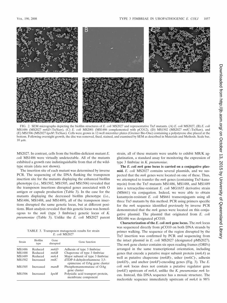
MS2027. In contrast, cells from the biofilm-deficient mutant E.coli MS1486 were virtually undetectable. All of the mutantsexhibited a growth rate indistinguishable from that of the wild-type strain (data not shown).
The insertion site of each mutant was determined by inversePCR. The sequencing of the DNA flanking the transposoninsertion site for the mutants displaying the enhanced biofilmphenotype (i.e., MS1502, MS1505, and MS1506) revealed thatthe transposon insertions disrupted genes associated with Oantigen or capsule production (Table 3). In the case for themutants displaying the decreased biofilm phenotype (i.e.,MS1486, MS1488, and MS1489), all of the transposon inser-tions disrupted the same genetic locus, but at different posi-tions. Blast analysis revealed that this genetic locus was homol-ogous to the mrk (type 3 fimbriae) genetic locus of K.pneumoniae (Table 3). Unlike the E. coli MS2027 parent
strain, all of these mutants were unable to exhibit MR/K ag-glutination, a standard assay for monitoring the expression oftype 3 fimbriae in K. pneumoniae.
The E. coli mrk gene locus is carried on a conjugative plas-mid. E. coli MS2027 contains several plasmids, and we sus-pected that the mrk genes were located on one of these. Thus,we attempted to transfer the mrk genes (containing Tn5-kana-mycin) from the Tn5 mutants MS1486, MS1488, and MS1489into a tetracycline-resistant E. coli MG1655 derivative strain(MS661) via conjugation. Indeed, we were able to obtainkanamycin-resistant E. coli MS661 transconjugants using allthree Tn5 mutants by this method. PCR using primers specificfor the mrk sequence identified previously by inverse PCRdemonstrated that the mrk genes were located on this conju-gative plasmid. The plasmid that originated from E. coliMS1486 was designated pCO10.
Characterization of the E. coli mrk gene locus. The mrk locuswas sequenced directly from pCO10 on both DNA strands byprimer walking. The sequence of the region disrupted by theTn5 insertion was confirmed by PCR and sequencing fromthe intact plasmid in E. coli MS2027 (designated pMS2027).The mrk gene cluster contains six open reading frames (ORFs)arranged in the same transcriptional orientation, includinggenes that encode a putative major subunit protein (mrkA) aswell as putative chaperone (mrkB)-, usher (mrkC)-, adhesin(mrkD)-, and anchor (mrkF)-encoding genes (Fig. 3). The E.coli mrk locus does not contain a putative regulator gene(mrkE) upstream of mrkA, unlike the K. pneumoniae mrk lo-cus. Instead, this DNA sequence has a mosaic structure. Thenucleotide sequence immediately upstream of mrkA is 98%
FIG. 2. SEM micrographs depicting the biofilm structures of E. coli MS2027 and representative Tn5 mutants. (A) E. coli MS2027, (B) E. coliMS1486 (MS2027 mrkD::Tn5kan), (C) E. coli MS2001 (MS1486 complemented with pCO12), (D) MS1502 (MS2027 rmlC::Tn5kan), and(E) MS1506 (MS2027 kpsM::Tn5kan). Cells were grown in 12-well microtiter plates (Greiner Bio-One) containing a polystyrene disc placed at thebottom. Following overnight growth, the disc was removed, fixed, stained, and examined by SEM as described in Materials and Methods. Scale bar,10 �m.
TABLE 3. Transposon mutagenesis results for strainE. coli MS2027
Strain Biofilmtype
Genedisrupted Gene function
MS1486 Reduced mrkD Adhesin of type 3 fimbriaeMS1488 Reduced mrkB Chaperone of type 3 fimbriaeMS1489 Reduced mrkA Major subunit of type 3 fimbriaeMS1502 Increased rmlC dTDP-4-dehydrorhamnose 3,5-
epimerase of OAg gene clusterMS1505 Increased manB Phosphomannomutase of OAg
gene clusterMS1506 Increased kpsM Polysialic acid transport protein,
membrane component
VOL. 190, 2008 TYPE 3 FIMBRIAE IN UROPATHOGENIC E. COLI 1057
on October 13, 2015 by U
niversity of Queensland Library
http://jb.asm.org/
Dow
nloaded from
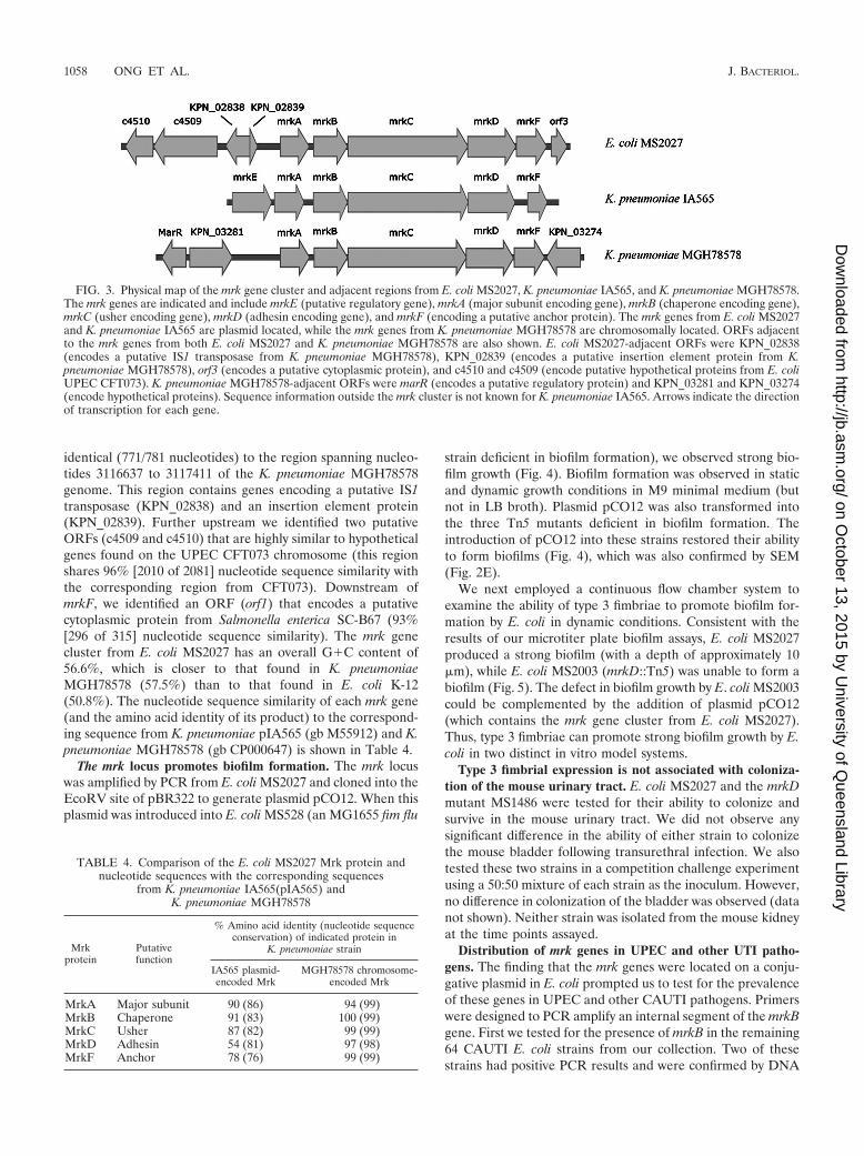
identical (771/781 nucleotides) to the region spanning nucleo-tides 3116637 to 3117411 of the K. pneumoniae MGH78578genome. This region contains genes encoding a putative IS1transposase (KPN_02838) and an insertion element protein(KPN_02839). Further upstream we identified two putativeORFs (c4509 and c4510) that are highly similar to hypotheticalgenes found on the UPEC CFT073 chromosome (this regionshares 96% [2010 of 2081] nucleotide sequence similarity withthe corresponding region from CFT073). Downstream ofmrkF, we identified an ORF (orf1) that encodes a putativecytoplasmic protein from Salmonella enterica SC-B67 (93%[296 of 315] nucleotide sequence similarity). The mrk genecluster from E. coli MS2027 has an overall G�C content of56.6%, which is closer to that found in K. pneumoniaeMGH78578 (57.5%) than to that found in E. coli K-12(50.8%). The nucleotide sequence similarity of each mrk gene(and the amino acid identity of its product) to the correspond-ing sequence from K. pneumoniae pIA565 (gb M55912) and K.pneumoniae MGH78578 (gb CP000647) is shown in Table 4.
The mrk locus promotes biofilm formation. The mrk locuswas amplified by PCR from E. coli MS2027 and cloned into theEcoRV site of pBR322 to generate plasmid pCO12. When thisplasmid was introduced into E. coli MS528 (an MG1655 fim flu
strain deficient in biofilm formation), we observed strong bio-film growth (Fig. 4). Biofilm formation was observed in staticand dynamic growth conditions in M9 minimal medium (butnot in LB broth). Plasmid pCO12 was also transformed intothe three Tn5 mutants deficient in biofilm formation. Theintroduction of pCO12 into these strains restored their abilityto form biofilms (Fig. 4), which was also confirmed by SEM(Fig. 2E).
We next employed a continuous flow chamber system toexamine the ability of type 3 fimbriae to promote biofilm for-mation by E. coli in dynamic conditions. Consistent with theresults of our microtiter plate biofilm assays, E. coli MS2027produced a strong biofilm (with a depth of approximately 10�m), while E. coli MS2003 (mrkD::Tn5) was unable to form abiofilm (Fig. 5). The defect in biofilm growth by E. coli MS2003could be complemented by the addition of plasmid pCO12(which contains the mrk gene cluster from E. coli MS2027).Thus, type 3 fimbriae can promote strong biofilm growth by E.coli in two distinct in vitro model systems.
Type 3 fimbrial expression is not associated with coloniza-tion of the mouse urinary tract. E. coli MS2027 and the mrkDmutant MS1486 were tested for their ability to colonize andsurvive in the mouse urinary tract. We did not observe anysignificant difference in the ability of either strain to colonizethe mouse bladder following transurethral infection. We alsotested these two strains in a competition challenge experimentusing a 50:50 mixture of each strain as the inoculum. However,no difference in colonization of the bladder was observed (datanot shown). Neither strain was isolated from the mouse kidneyat the time points assayed.
Distribution of mrk genes in UPEC and other UTI patho-gens. The finding that the mrk genes were located on a conju-gative plasmid in E. coli prompted us to test for the prevalenceof these genes in UPEC and other CAUTI pathogens. Primerswere designed to PCR amplify an internal segment of the mrkBgene. First we tested for the presence of mrkB in the remaining64 CAUTI E. coli strains from our collection. Two of thesestrains had positive PCR results and were confirmed by DNA
FIG. 3. Physical map of the mrk gene cluster and adjacent regions from E. coli MS2027, K. pneumoniae IA565, and K. pneumoniae MGH78578.The mrk genes are indicated and include mrkE (putative regulatory gene), mrkA (major subunit encoding gene), mrkB (chaperone encoding gene),mrkC (usher encoding gene), mrkD (adhesin encoding gene), and mrkF (encoding a putative anchor protein). The mrk genes from E. coli MS2027and K. pneumoniae IA565 are plasmid located, while the mrk genes from K. pneumoniae MGH78578 are chromosomally located. ORFs adjacentto the mrk genes from both E. coli MS2027 and K. pneumoniae MGH78578 are also shown. E. coli MS2027-adjacent ORFs were KPN_02838(encodes a putative IS1 transposase from K. pneumoniae MGH78578), KPN_02839 (encodes a putative insertion element protein from K.pneumoniae MGH78578), orf3 (encodes a putative cytoplasmic protein), and c4510 and c4509 (encode putative hypothetical proteins from E. coliUPEC CFT073). K. pneumoniae MGH78578-adjacent ORFs were marR (encodes a putative regulatory protein) and KPN_03281 and KPN_03274(encode hypothetical proteins). Sequence information outside the mrk cluster is not known for K. pneumoniae IA565. Arrows indicate the directionof transcription for each gene.
TABLE 4. Comparison of the E. coli MS2027 Mrk protein andnucleotide sequences with the corresponding sequences
from K. pneumoniae IA565(pIA565) andK. pneumoniae MGH78578
Mrkprotein
Putativefunction
% Amino acid identity (nucleotide sequenceconservation) of indicated protein in
K. pneumoniae strain
IA565 plasmid-encoded Mrk
MGH78578 chromosome-encoded Mrk
MrkA Major subunit 90 (86) 94 (99)MrkB Chaperone 91 (83) 100 (99)MrkC Usher 87 (82) 99 (99)MrkD Adhesin 54 (81) 97 (98)MrkF Anchor 78 (76) 99 (99)
1058 ONG ET AL. J. BACTERIOL.
on October 13, 2015 by U
niversity of Queensland Library
http://jb.asm.org/
Dow
nloaded from

sequencing. Next, we tested for the presence of mrkB in 70CAUTI pathogens representing different gram-negative organ-isms isolated from UTI patients from the same location (Table5). The mrk genes were detected from K. pneumoniae, K.
oxytoca, C. koseri, and C. freundii CAUTI isolates (Table 5).The identity of each PCR product was confirmed by DNAsequencing. To determine whether the presence of the mrkgenes was specific to CAUTI strains, we also tested for theirprevalence in 45 E. coli strains isolated from cystitis and py-elonephritis patients. Among these strains, 2 of 45 containedthe mrkB gene as determined by PCR amplification and DNAsequencing. Finally, we tested for the prevalence of mrkB instrains from the ECOR collection; three strains had positivePCR products for these genes, with results confirmed as cor-rect by DNA sequencing. All of the organisms that containedthe mrkB gene displayed a positive MR/K agglutination phe-notype following growth in M9 minimal medium. We note thatthe C. koseri and C. freundii strains required growth in M9
FIG. 4. (A) Biofilm formation by E. coli MS2027 and derivatives. Strains were grown at 37°C in PVC microtiter plates containing M9 medium(supplemented with 0.2% glucose) for 16 h under shaking conditions, washed to remove unbound cells, and stained with 0.1% crystal violet. Biofilmformation was quantified by resuspending adhered cells in ethanol-acetate (80:20) and measuring the absorbance at 595 nm. The results arepresented as the average of eight individual replicates ( standard deviation). Shown are the results for MS1486 (MS2027 mrkD::Tn5kan), MS1488(MS2027 mrkB::Tn5kan), MS1489 (MS2027 mrkA::Tn5kan), MS2003, MS2006, and MS2009, mrk mutants containing pBR322; MS2001, MS2004, andMS2007, mrk mutants containing pCO12 (mrk�); MS1502 (rmlC mutant); MS1505 (manB mutant); and MS1506 (kpsM mutant). (B) Biofilm formationby E. coli MS528 and E. coli MS528 containing pCO12. Cells were grown and analyzed for biofilm formation as described above. The introduction ofplasmid pCO12 (containing the mrk gene cluster from MS2027) into MS528 promoted strong biofilm growth. �, absence of; �, presence of.
FIG. 5. Flow chamber biofilm formation of E. coli MS2027 (A), E.coli MS2003 (B), and E. coli MS2001 (C). Biofilm development wasmonitored by confocal scanning laser microscopy 24 h after inocula-tion. Micrographs represent horizontal sections. Depicted to the rightand below are vertical sections through the biofilm collected at thepositions indicated by the lines.
TABLE 5. Prevalence of mrkB gene and MR/K agglutinationphenotype among UTI strains
Bacterial speciesand strain type
Total no. ofstrains
Presencea of:
mrkB MR/KHA
Escherichia coliCAUTI 93 3 (3.2) 3 (3.2)ABU 23 0 (0) 0 (0)Cystitis 19 1 (5.3) 1 (5.3)Pyelonephritis 26 1 (3.8) 1 (3.8)ECOR 72 3 (4.2) 3 (4.2)
Other speciesC. freundii 7 1 (14.3) 1 (14.3)C. koseri 9 9 (100) 9 (100)K. oxytoca 2 2 (100) 2 (100)K. pneumoniae 15 13 (86.7) 13 (86.7)
a Values are shown as no. (%).
VOL. 190, 2008 TYPE 3 FIMBRIAE IN UROPATHOGENIC E. COLI 1059
on October 13, 2015 by U
niversity of Queensland Library
http://jb.asm.org/
Dow
nloaded from
minimal medium for 72 h under static conditions to induceMR/K agglutination.
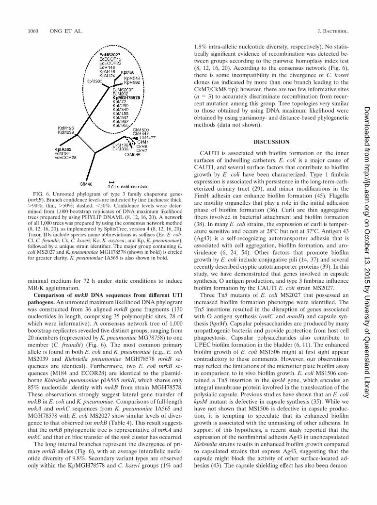
Comparison of mrkB DNA sequences from different UTIpathogens. An unrooted maximum likelihood DNA phylogramwas constructed from 36 aligned mrkB gene fragments (130nucleotides in length, comprising 35 polymorphic sites, 28 ofwhich were informative). A consensus network tree of 1,000bootstrap replicates revealed five distinct groups, ranging from20 members (represented by K. pneumoniae MG78758) to onemember (C. freundii) (Fig. 6). The most common primaryallele is found in both E. coli and K. pneumoniae (e.g., E. coliMS2039 and Klebsiella pneumoniae MGH78578 mrkB se-quences are identical). Furthermore, two E. coli mrkB se-quences (M184 and ECOR28) are identical to the plasmid-borne Klebsiella pneumoniae pIA565 mrkB, which shares only85% nucleotide identity with mrkB from strain MGH78578.These observations strongly suggest lateral gene transfer ofmrkB in E. coli and K. pneumoniae. Comparisons of full-lengthmrkA and mrkC sequences from K. pneumoniae IA565 andMGH78578 with E. coli MS2027 show similar levels of diver-gence to that observed for mrkB (Table 4). This result suggeststhat the mrkB phylogenetic tree is representative of mrkA andmrkC and that en bloc transfer of the mrk cluster has occurred.
The long internal branches represent the divergence of pri-mary mrkB alleles (Fig. 6), with an average interallelic nucle-otide diversity of 9.8%. Secondary variant types are observedonly within the KpMGH78578 and C. koseri groups (1% and
1.8% intra-allelic nucleotide diversity, respectively). No statis-tically significant evidence of recombination was detected be-tween groups according to the pairwise homoplasy index test(8, 12, 16, 20). According to the consensus network (Fig. 6),there is some incompatibility in the divergence of C. kosericlones (as indicated by more than one branch leading to theCkM7/CkM8 tip); however, there are too few informative sites(n � 3) to accurately discriminate recombination from recur-rent mutation among this group. Tree topologies very similarto those obtained by using DNA maximum likelihood wereobtained by using parsimony- and distance-based phylogeneticmethods (data not shown).
DISCUSSION
CAUTI is associated with biofilm formation on the innersurfaces of indwelling catheters. E. coli is a major cause ofCAUTI, and several surface factors that contribute to biofilmgrowth by E. coli have been characterized. Type 1 fimbriaexpression is associated with persistence in the long-term-cath-eterized urinary tract (29), and minor modifications in theFimH adhesin can enhance biofilm formation (45). Flagellaare motility organelles that play a role in the initial adhesionphase of biofilm formation (36). Curli are thin aggregativefibers involved in bacterial attachment and biofilm formation(38). In many E. coli strains, the expression of curli is temper-ature sensitive and occurs at 28°C but not at 37°C. Antigen 43(Ag43) is a self-recognizing autotransporter adhesin that isassociated with cell aggregation, biofilm formation, and uro-virulence (6, 24, 54). Other factors that promote biofilmgrowth by E. coli include conjugative pili (14, 37) and severalrecently described cryptic autotransporter proteins (39). In thisstudy, we have demonstrated that genes involved in capsulesynthesis, O antigen production, and type 3 fimbriae influencebiofilm formation by the CAUTI E. coli strain MS2027.
Three Tn5 mutants of E. coli MS2027 that possessed anincreased biofilm formation phenotype were identified. TheTn5 insertions resulted in the disruption of genes associatedwith O antigen synthesis (rmlC and manB) and capsule syn-thesis (kpsM). Capsular polysaccharides are produced by manyuropathogenic bacteria and provide protection from host cellphagocytosis. Capsular polysaccharides also contribute toUPEC biofilm formation in the bladder (6, 11). The enhancedbiofilm growth of E. coli MS1506 might at first sight appearcontradictory to these comments. However, our observationsmay reflect the limitations of the microtiter plate biofilm assayin comparison to in vivo biofilm growth. E. coli MS1506 con-tained a Tn5 insertion in the kpsM gene, which encodes anintegral membrane protein involved in the translocation of thepolysialic capsule. Previous studies have shown that an E. colikpsM mutant is defective in capsule synthesis (35). While wehave not shown that MS1506 is defective in capsule produc-tion, it is tempting to speculate that its enhanced biofilmgrowth is associated with the unmasking of other adhesins. Insupport of this hypothesis, a recent study reported that theexpression of the nonfimbrial adhesin Ag43 in unencapsulatedKlebsiella strains results in enhanced biofilm growth comparedto capsulated strains that express Ag43, suggesting that thecapsule might block the activity of other surface-located ad-hesins (43). The capsule shielding effect has also been demon-
FIG. 6. Unrooted phylogram of type 3 family chaperone genes(mrkB). Branch confidence levels are indicated by line thickness: thick,90%; thin, 50%; dashed, �50%. Confidence levels were deter-mined from 1,000 bootstrap replicates of DNA maximum likelihoodtrees prepared by using PHYLIP DNAML (8, 12, 16, 20). A networkof all 1,000 trees was prepared by using the consensus network method(8, 12, 16, 20), as implemented by SplitsTree, version 4 (8, 12, 16, 20).Taxon IDs include species name abbreviations as suffixes (Ec, E. coli;Cf, C. freundii; Ck, C. koseri; Ko, K. oxytoca; and Kp, K. pneumoniae),followed by a unique strain identifier. The major group containing E.coli MS2027 and K. pneumoniae MGH78578 (shown in bold) is circledfor greater clarity. K. pneumoniae IA565 is also shown in bold.
1060 ONG ET AL. J. BACTERIOL.
on October 13, 2015 by U
niversity of Queensland Library
http://jb.asm.org/
Dow
nloaded from

strated in adherence studies of other organisms, including E.coli (40, 43), Neisseria meningitidis (56), and Haemophilus in-fluenzae (50). Furthermore, the function of type 1 fimbriae hasbeen shown to be impeded by the presence of a capsule on thebacterial cell surface (42) and thus reduced capsule synthesisby E. coli MS1506 may enhance the contribution of fimbriae tobiofilm formation. We note that soluble polysaccharide se-creted by UPEC strains that produce a group II capsule wasrecently shown to inhibit biofilm growth by preventing adhe-sion (55). Therefore, we cannot rule out the possibility that theenhanced biofilm growth of MS1506 is due to reduced secre-tion of antiadhesive polysaccharide material. The enhancedbiofilm growth by E. coli MS1508 and E. coli MS1509 might beassociated with a similar mechanism, since many UPEC strainsare known to produce large O antigen structures. We arecurrently attempting to elucidate the role of the capsule and Oantigen in UPEC colonization and biofilm formation in an invivo model of CAUTI.
The decreased biofilm growth by Tn5 mutants of E. coliMS2027 was due to the disruption of genes encoding type 3fimbriae. Three biofilm-deficient mutants were identified, all ofwhich contained a Tn5 insertion in the mrk operon. The role oftype 3 fimbriae in biofilm formation was confirmed by thecomplementation of each of these mutants with a plasmid(pCO12) containing the mrk genes. Type 3 fimbriae are thin,filamentous structures (4 to 5 nm wide and 0.5 to 2 �m long)that extend from the surface of the cell (10) and are morpho-logically similar to K88 and K99 fimbriae (26). Type 3 fimbriaeare characterized by their ability to mediate MR agglutinationof tannic acid-treated RBC (which is referred to as MR Kleb-siella-like or MR/K agglutination) (26). MR/K agglutination isconferred by the MrkD adhesin (5, 19). MrkD also mediatesbinding to the basolateral surface of renal tubular, tracheal,and bronchial cells via a high-affinity interaction with type Vcollagen (17, 51, 52). Type 3 fimbriae from K. pneumoniae havealso been shown to mediate biofilm formation (21).
Type 3 fimbriae are most commonly associated with Kleb-siella spp. (13). However, they are also produced by othermembers of the Enterobacteriaceae family, including Entero-bacter, Morganella, Proteus, Providencia, Serratia, Salmonella,and Yersinia species (1–4, 13, 30–33, 48). Here we demonstratethat type 3 fimbriae are also produced by E. coli, C. koseri, andC. freundii. In E. coli MS2027, the mrk genes are located on aconjugative plasmid of approximately 45 kb (C.-L. Y. Ong,A. G. McEwan, and M. A. Schembri, unpublished data). Plas-mid-carried mrk genes have previously been identified for K.pneumoniae and Y. enterocolitica (5, 13). K. pneumoniae IA565possesses both chromosomal and plasmid-carried mrk genes;the plasmid pIA565 contains a functional copy of mrkA andmrkD, while only mrkA has been detected on the K. pneu-moniae IA565 chromosome. The mrk genes from pIA565 havebeen well characterized and possess the same genetic arrange-ment as do the mrk genes on plasmid pMS2027. However, thesequence upstream of mrkA is different between the two geneclusters (the sequence downstream of mrkF on pIA565 has notbeen reported). On plasmid pIA565, a gene (mrkE) encodinga putative regulator protein is located immediately upstream ofmrkA (5). This gene is not present on pMS2027. Instead, weidentified a putative transposase-encoding gene upstream ofmrkA and the entire cluster is flanked by two putative insertion
sequence elements. Thus, it seems likely that the mrk clusteron plasmid pMS2027 is associated with a mobile genetic ele-ment. Importantly, the presence of mrk genes in E. coli was notunique to strains in our UTI collection, as three strains fromthe ECOR collection also contained mrkABC and causedMR/K hemagglutination. The location of the mrk genes fromthese strains remains to be determined.
We observed that the expression of type 3 fimbriae wasdependent on the growth medium. E. coli MS2027 produced astrong biofilm and caused characteristic MR/K agglutinationwhen grown in M9 minimal medium supplemented with glu-cose but not when grown in LB medium. This finding is con-sistent with the results of previous reports of type 3 fimbriaexpression in K. pneumoniae, where bacteria grown in minimalmedium in the presence of glycerol or glucose resulted in astronger MR/K hemagglutination reaction than did bacteriagrown in complex medium (18, 47). This result suggests asimilar method of regulation of the type 3 fimbrial genes of K.pneumoniae IA565 and E. coli MS2027, despite the absence ofthe putative mrkE regulator gene on pMS2027. It is interestingthat all of the mrkABC-positive strains identified in this studycaused MR/K agglutination, since previous studies have shownthat not all Klebsiella spp. possess the adhesin-encoding mrkDgene (19).
We compared the mrk cluster from E. coli MS2027 with themrk clusters from K. pneumoniae MGH78578 and pIA565 (Ta-ble 4). All five mrkABCDE genes showed remarkable sequencesimilarity to the chromosomally located MGH78578 sequences(98.8% 0.4%) compared to that shown by the respectivepIA565 sequences (81.6% 3.6%). The presence of insertionsequences adjacent to the plasmid-borne E. coli MS2027 mrkcluster and the G�C content suggest that there was relativelyrecent lateral transfer from a K. pneumoniae strain, althoughwe cannot rule out the possibility that both strains acquired thecluster independently from a third species. To assess the dis-tribution of this cluster among UTI organisms, we amplifiedand sequenced a fragment of the chaperone gene (mrkB),which is typically the most highly conserved gene within chap-erone/usher fimbrial clusters. Phylogenetic analyses indicatedthere were five primary alleles which, with the exception of thetwo K. oxytoca strains, are strongly supported by long internalbranches (Fig. 6). The most common allele is that shared by K.pneumoniae MGH78578 and E. coli MS2027. This allele is alsoshared by E. coli CAUTI strains MS2039 and M148 and cystitisstrain M202. Interestingly, mrkB from the pyelonephritis E.coli strain M184 is identical to that found in K. pneumoniaepIA565. The observation that two alleles (represented bystrains MGH78578 and IA565) contain sequences that areidentical in both E. coli and K. pneumoniae species, but sub-stantially divergent from each other, is strong evidence of re-current and recent lateral gene transfer of the mrk clusteramong K. pneumoniae and E. coli UTI strains.
In conclusion, we have identified the capsule, O antigen, andtype 3 fimbriae as factors that affect biofilm growth by CAUTIE. coli. Type 3 fimbriae are produced by many members of theEnterobacteriaceae family that are associated with opportunis-tic infections. Biofilm growth mediated by type 3 fimbriae maybe important for the survival of these organisms on the sur-faces of urinary catheters and within the hospital environment.We speculate that the mrk gene cluster in E. coli MS2027 may
VOL. 190, 2008 TYPE 3 FIMBRIAE IN UROPATHOGENIC E. COLI 1061
on October 13, 2015 by U
niversity of Queensland Library
http://jb.asm.org/
Dow
nloaded from

have originated from K. pneumoniae, and we are currentlyinvestigating this possibility.
ACKNOWLEDGMENTS
This work was supported by grants from the National Health andMedical Research Council (NHMRC) of Australia (grant no. 455914)and the Australian Research Council (grant no. DP0666852). C.-L.Y.O. was supported by an International Postgraduate ResearchScholarship from the University of Queensland. S.A.B. was supportedby an NHRMC Howard Florey Centenary fellowship.
The K. pneumoniae genome sequence data were produced by theGenome Sequencing Center at the Washington University School ofMedicine in St. Louis, MO.
REFERENCES
1. Adegbola, R. A., and D. C. Old. 1985. Fimbrial and non-fimbrial hemagglu-tinins in enterobacter-aerogenes. J. Med. Microbiol. 19:35–43.
2. Adegbola, R. A., and D. C. Old. 1983. Fimbrial hemagglutinins in entero-bacter species. J. Gen. Microbiol. 129:2175–2180.
3. Adegbola, R. A., and D. C. Old. 1982. New fimbrial hemagglutinin in Serratiaspecies. Infect. Immun. 38:306–315.
4. Adegbola, R. A., D. C. Old, and S. Aleksic. 1983. Rare Mr/K-like hemagglu-tinins (and type-3-like fimbriae) of salmonella strains. FEMS Microbiol.Lett. 19:233–238.
5. Allen, B. L., G. F. Gerlach, and S. Clegg. 1991. Nucleotide sequence andfunctions of mrk determinants necessary for expression of type 3 fimbriae inKlebsiella pneumoniae. J. Bacteriol. 173:916–920.
6. Anderson, G. G., J. J. Palermo, J. D. Schilling, R. Roth, J. Heuser, and S. J.Hultgren. 2003. Intracellular bacterial biofilm-like pods in urinary tract in-fections. Science 301:105–107.
7. Blaesing, F., A. Muhlenweg, S. Vierling, G. Ziegelin, S. Pelzer, and E. Lanka.2005. Introduction of DNA into Actinomycetes by bacterial conjugationfrom E-coli—an evaluation of various transfer systems. J. Biotechnol. 120:146–161.
8. Bruen, T. C., H. Philippe, and D. Bryant. 2006. A simple and robust statis-tical test for detecting the presence of recombination. Genetics 172:2665–2681.
9. Delorenzo, V., M. Herrero, U. Jakubzik, and K. N. Timmis. 1990. Mini-Tn5transposon derivatives for insertion mutagenesis, promoter probing, andchromosomal insertion of cloned DNA in gram-negative eubacteria. J. Bac-teriol. 172:6568–6572.
10. Duguid, J. P. 1959. Fimbriae and adhesive properties in Klebsiella strains.J. Gen. Microbiol. 21:271–286.
11. Eto, D. S., J. L. Sundsbak, and M. A. Mulvey. 2006. Actin-gated intracellulargrowth and resurgence of uropathogenic Escherichia coli. Cell. Microbiol.8:704–717.
12. Felsenstein, J. 2004. PHYLIP (phylogeny inference package), version 3.6.Department of Genome Sciences, Seattle, WA.
13. Gerlach, G. F., B. L. Allen, and S. Clegg. 1989. Type 3 fimbriae amongenterobacteria and the ability of spermidine to inhibit Mr/K hemagglutina-tion. Infect. Immun. 57:219–224.
14. Ghigo, J. M. 2001. Natural conjugative plasmids induce bacterial biofilmdevelopment. Nature 412:442–445.
15. Hagberg, L., U. Jodal, T. K. Korhonen, G. Lidin-Janson, U. Lindberg, andC. Svanborg Eden. 1981. Adhesion, hemagglutination, and virulence of Esch-erichia coli causing urinary tract infections. Infect. Immun. 31:564–570.
16. Holland, B., and V. Moulton. 2003. Consensus networks: a method forvisualizing incompatibilities in collections of trees, p. 165–176. In G. Bensonand R. Page (ed.), Algorithms in bioinformatics, WABI 2003. Springer-Verlag, Berlin, Germany.
17. Hornick, D. B., B. L. Allen, M. A. Horn, and S. Clegg. 1992. Adherence torespiratory epithelia by recombinant Escherichia coli expressing Klebsiellapneumoniae type 3 fimbrial gene products. Infect. Immun. 60:1577–1588.
18. Hornick, D. B., B. L. Allen, M. A. Horn, and S. Clegg. 1991. Fimbrial typesamong respiratory isolates belonging to the family Enterobacteriaceae.J. Clin. Microbiol. 29:1795–1800.
19. Hornick, D. B., J. Thommandru, W. Smits, and S. Clegg. 1995. Adherenceproperties of an mrkD-negative mutant of Klebsiella pneumoniae. Infect.Immun. 63:2026–2032.
20. Huson, D. H., and D. Bryant. 2006. Application of phylogenetic networks inevolutionary studies. Mol. Biol. Evol. 23:254–267.
21. Jagnow, J., and S. Clegg. 2003. Klebsiella pneumoniae MrkD-mediatedbiofilm formation on extracellular matrix- and collagen-coated surfaces. Mi-crobiology 149:2397–2405.
22. Kallenius, G., R. Mollby, S. B. Svenson, I. Helin, H. Hultberg, B. Cedergren,and J. Winberg. 1981. Occurrence of P-fimbriated Escherichia coli in urinarytract infections. Lancet ii:1369–1372.
23. Kjaergaard, K., M. A. Schembri, C. Ramos, S. Molin, and P. Klemm. 2000.
Antigen 43 facilitates formation of multispecies biofilms. Environ. Microbiol.2:695–702.
24. Klemm, P., L. Hjerrild, M. Gjermansen, and M. A. Schembri. 2004. Struc-ture-function analysis of the self-recognizing antigen 43 autotransporter pro-tein from Escherichia coli. Mol. Microbiol. 51:283–296.
25. Klemm, P., and M. A. Schembri. 2000. Bacterial adhesins: function andstructure. Int. J. Med. Microbiol. 290:27–35.
26. Korhonen, T. K., E. Tarkka, H. Ranta, and K. Haahtela. 1983. Type 3fimbriae of Klebsiella sp.: molecular characterization and role in bacterialadhesion to plant roots. J. Bacteriol. 155:860–865.
27. Leffler, H., and C. Svanborg-Eden. 1981. Glycolipid receptors for uropatho-genic Escherichia coli on human erythrocytes and uroepithelial cells. Infect.Immun. 34:920–929.
28. Mahapatra, N. R., S. Ghosh, P. K. Sarkar, and P. C. Banerjee. 2003. Gen-eration of novel plasmids in Escherichia coli S17-1(pSUP106). Curr. Micro-biol. 46:318–323.
29. Mobley, H. L., G. R. Chippendale, J. H. Tenney, R. A. Hull, and J. W.Warren. 1987. Expression of type 1 fimbriae may be required for persistenceof Escherichia coli in the catheterized urinary tract. J. Clin. Microbiol. 25:2253–2257.
30. Old, D. C., and R. A. Adegbola. 1985. Antigenic relationships among type-3fimbriae of Enterobacteriaceae revealed by immunoelectronmicroscopy.J. Med. Microbiol. 20:113–121.
31. Old, D. C., and R. A. Adegbola. 1982. Hemagglutinins and fimbriae ofMorganella, Proteus and Providencia. J. Med. Microbiol. 15:551–564.
32. Old, D. C., and R. A. Adegbola. 1984. Relationships among broad-spectrumand narrow-spectrum mannose-resistant fimbrial hemagglutinins in differentYersinia species. Microbiol. Immunol. 28:1303–1311.
33. Old, D. C., and S. S. Scott. 1981. Hemagglutinins and fimbriae of Providenciaspp. J. Bacteriol. 146:404–408.
34. Paterson, D. L., and J. Lipman. 2007. Returning to the pre-antibiotic era inthe critically ill: the XDR problem. Crit. Care Med. 35:1789–1791.
35. Pigeon, R. P., and R. P. Silver. 1997. Analysis of the G93E mutant allele ofKpsM, the membrane component of an ABC transporter involved in poly-sialic acid translocation in Escherichia coli K1. FEMS Microbiol. Lett. 156:217–222.
36. Pratt, L. A., and R. Kolter. 1998. Genetic analysis of Escherichia coli biofilmformation: roles of flagella, motility, chemotaxis and type I pili. Mol. Micro-biol. 30:285–293.
36a.R Development Core Team. 2007. R: a language and environment for sta-tistical computing. R Foundation for Statistical Computing, Vienna, Austria.
37. Reisner, A., J. A. Haagensen, M. A. Schembri, E. L. Zechner, and S. Molin.2003. Development and maturation of Escherichia coli K-12 biofilms. Mol.Microbiol. 48:933–946.
38. Romling, U. 2005. Characterization of the rdar morphotype, a multicellularbehaviour in Enterobacteriaceae. Cell. Mol. Life Sci. 62:1234–1246.
39. Roux, A., C. Beloin, and J. M. Ghigo. 2005. Combined inactivation andexpression strategy to study gene function under physiological conditions:application to identification of new Escherichia coli adhesins. J. Bacteriol.187:1001–1013.
40. Runnels, P. L., and H. W. Moon. 1984. Capsule reduces adherence ofenterotoxigenic Escherichia coli to isolated intestinal epithelial cells of pigs.Infect. Immun. 45:737–740.
41. Sambrook, J., and D. W. Russell. 2001. Molecular cloning: a laboratorymanual, 3rd ed., vol. 1. Cold Spring Harbor Laboratory Press, Cold SpringHarbor, NY.
42. Schembri, M. A., J. Blom, K. A. Krogfelt, and P. Klemm. 2005. Capsule andfimbria interaction in Klebsiella pneumoniae. Infect. Immun. 73:4626–4633.
43. Schembri, M. A., D. Dalsgaard, and P. Klemm. 2004. Capsule shields thefunction of short bacterial adhesins. J. Bacteriol. 186:1249–1257.
44. Schembri, M. A., K. Kjaergaard, and P. Klemm. 2003. Global gene expres-sion in Escherichia coli biofilms. Mol. Microbiol. 48:253–267.
45. Schembri, M. A., and P. Klemm. 2001. Biofilm formation in a hydrodynamicenvironment by novel FimH variants and ramifications for virulence. Infect.Immun. 69:1322–1328.
46. Schembri, M. A., E. V. Sokurenko, and P. Klemm. 2000. Functional flexibilityof the FimH adhesin: insights from a random mutant library. Infect. Immun.68:2638–2646.
47. Schurtz, T. A., D. B. Hornick, T. K. Korhonen, and S. Clegg. 1994. The type3 fimbrial adhesin gene (mrkD) of Klebsiella species is not conserved amongall fimbriate strains. Infect. Immun. 62:4186–4191.
48. Sebghati, T. A. S., T. K. Korhonen, D. B. Hornick, and S. Clegg. 1998.Characterization of the type 3 fimbrial adhesins of Klebsiella strains. Infect.Immun. 66:2887–2894.
49. Stamm, W. E. 1991. Catheter-associated urinary tract infections: epidemiol-ogy, pathogenesis, and prevention. Am. J. Med. 91:65S–71S.
50. St. Geme, J. W., III, and S. Falkow. 1991. Loss of capsule expression byHaemophilus influenzae type b results in enhanced adherence to and invasionof human cells. Infect. Immun. 59:1325–1333.
51. Tarkkanen, A. M., B. L. Allen, B. Westerlund, H. Holthofer, P. Kuusela, L.Risteli, S. Clegg, and T. K. Korhonen. 1990. Type V collagen as the target for
1062 ONG ET AL. J. BACTERIOL.
on October 13, 2015 by U
niversity of Queensland Library
http://jb.asm.org/
Dow
nloaded from
type-3 fimbriae, enterobacterial adherence organelles. Mol. Microbiol.4:1353–1361.
52. Tarkkanen, A. M., R. Virkola, S. Clegg, and T. K. Korhonen. 1997. Bindingof the type 3 fimbriae of Klebsiella pneumoniae to human endothelial andurinary bladder cells. Infect. Immun. 65:1546–1549.
53. Ulett, G. C., A. N. Mabbett, K. C. Fung, R. I. Webb, and M. A. Schembri.2007. The role of F9 fimbriae of uropathogenic Escherichia coli in biofilmformation. Microbiology 153:2321–2331.
54. Ulett, G. C., J. Valle, C. Beloin, O. Sherlock, J. M. Ghigo, and M. A.Schembri. 2007. Functional analysis of antigen 43 in uropathogenic Esche-richia coli reveals a role in long-term persistence in the urinary tract. Infect.Immun. 75:3233–3244.
55. Valle, J., S. Da Re, N. Henry, T. Fontaine, D. Balestrino, P. Latour-Lambert,and J. M. Ghigo. 2006. Broad-spectrum biofilm inhibition by a secretedbacterial polysaccharide. Proc. Natl. Acad. Sci. USA 103:12558–12563.
56. Virji, M., K. Makepeace, D. J. Ferguson, M. Achtman, and E. R. Moxon.1993. Meningococcal Opa and Opc proteins: their role in colonization andinvasion of human epithelial and endothelial cells. Mol. Microbiol. 10:499–510.
57. Vrionis, H. A., A. J. Daugulis, and A. M. Kropinski. 2002. Identification and
characterization of the AgmR regulator of Pseudomonas putida: role inalcohol utilization. Appl. Microbiol. Biotechnol. 58:469–475.
58. Warren, J. W. 2001. Catheter-associated urinary tract infections. Int. J.Antimicrob. Agents 17:299–303.
59. Warren, J. W., D. Damron, J. H. Tenney, J. M. Hoopes, B. Deforge, andH. L. Muncie, Jr. 1987. Fever, bacteremia, and death as complications ofbacteriuria in women with long-term urethral catheters. J. Infect. Dis. 155:1151–1158.
60. Warren, J. W., J. H. Tenney, J. M. Hoopes, H. L. Muncie, and W. C.Anthony. 1982. A prospective microbiologic study of bacteriuria in patientswith chronic indwelling urethral catheters. J. Infect. Dis. 146:719–723.
61. Watson, N. 1988. A new revision of the sequence of plasmid pBR322. Gene70:399–403.
62. Wu, X. R., T. T. Sun, and J. J. Medina. 1996. In vitro binding of type1-fimbriated Escherichia coli to uroplakins Ia and Ib: relation to urinary tractinfections. Proc. Natl. Acad. Sci. USA 93:9630–9635.
63. Zogaj, X., M. Nimtz, M. Rohde, W. Bokranz, and U. Romling. 2001. Themulticellular morphotypes of Salmonella typhimurium and Escherichia coliproduce cellulose as the second component of the extracellular matrix. Mol.Microbiol. 39:1452–1463.
VOL. 190, 2008 TYPE 3 FIMBRIAE IN UROPATHOGENIC E. COLI 1063
on October 13, 2015 by U
niversity of Queensland Library
http://jb.asm.org/
Dow
nloaded from