identification and characterization of grim-1, a cell ... · isoforms suppress ribosomal rna (rrna)...
TRANSCRIPT
Research Article 2781
IntroductionCytokines belonging to the interferon (IFN) group potently suppresscell growth and promote apoptosis (Kimchi, 1992). They exertantiviral, immuno-regulatory and anti-proliferative effectsemploying the JAK (Janus tyrosine kinase)-STAT (signal transducerand activator of transcription) pathways (Schindler et al., 2007).Certain IFN-insensitive tumors become sensitive to IFNs when inthe presence of all-trans retinoic acid (RA) (Moore et al., 1994).RA is a major physiological retinoid that binds to specific nuclearreceptors (Chambon, 1996). The RA receptors act as transcriptionfactors to drive expression of genes involved in cell differentiationand growth control. Retinoids inhibit growth of certain leukemias,skin dysplasias in vivo and tumor cell lines in vitro (Altucci andGronemeyer, 2001). We have demonstrated earlier that thecombination of both IFN and RA (IFN/RA) is a highly effectiveinhibitor of tumor growth in vivo and in vitro (Kalvakolanu, 2004;Lindner et al., 1997). Although IFN/RA combination inducesapoptosis, the exact molecular mechanisms involved are stillunclear.
Apoptosis eliminates superfluous and/or potentially dangerouscells in mammals. It is regulated by cytokines, survival factors,cell-cell and/or cell-ECM interactions, oncogenes, DNA damageand viral proteins (Ashkenazi and Dixit, 1998; Green and Reed,1998; Stennicke and Salvesen, 2000; Youle and Strasser, 2008).Loss of apoptotic response seems to cause drug resistance in tumorcells (Ashkenazi and Dixit, 1998; Green and Reed, 1998; Logueand Martin, 2008; Lowe et al., 1994; Stennicke and Salvesen,2000; Youle and Strasser, 2008). Although the roles of centralplayers – such as caspases, Bcl2-like proteins and death receptors– in apoptotic responses have been well defined over the lastdecade, it is unclear how these proteins control cell death in a
signal-specific and cell-type-specific manner. Via a positive-growthselection in the presence of cytotoxic agents, genome-wideexpression-knockdown strategies permit the identification of geneproducts that are indispensable for cell-growth suppression or celldeath. Such strategies do not require a prior knowledge about thegene(s) or their product(s) and allow an unbiased identification ofactivators of apoptosis (Deiss et al., 1995; Hofmann et al., 1998).We have employed one such approach, the antisense technicalknockout (TKO), to isolate the GRIM (genes associated withretinoid-IFN-induced mortality) genes. In this approach,endogenous gene expression is knocked down by a library ofantisense complementary DNAs (cDNAs) expressing from anepisome. In this study, we have characterized a newly identifiedgene, GRIM-1, whose antisense expression protected cells fromIFN/RA-induced cell death. The GRIM-1 mRNA produces threeprotein isoforms, designated , and , from a single open readingframe (ORF); these isoforms induce cell death. Whereas GRIM-1can readily activate cell death, GRIM-1 and GRIM-1 requireda proteolytic activation by caspase-9 to induce cell death. Theseisoforms suppress ribosomal RNA (rRNA) maturation to ablatecell growth. Together, these results identify a novel mediator anda novel cell-death-regulatory pathway.
ResultsIdentification of GRIM-1 using a genetic approachWe isolated GRIM genes using the antisense TKO strategy (Angellet al., 2000; Hofmann et al., 1998). Briefly, HeLa cells weretransfected with an antisense cDNA library and selected withhygromycin B (100 g/ml) and a lethal dose of human IFN (5000U/ml) and RA (5 M) for 4 weeks. The surviving colonies wereexpanded and the episomal DNA bearing the death-associated gene
Identification and characterization of GRIM-1, a cell-death-associated gene productEdward R. Hofmann1,*, Shreeram C. Nallar1, Limei Lin1, Jonathan D’Cunha1, Daniel J. Lindner2, Xiao Weihua3
and Dhananjaya V. Kalvakolanu1,‡
1Greenebaum Cancer Center, Department of Microbiology and Immunology, Molecular and Cellular Cancer Biology Track, GPILS,University of Maryland School of Medicine, Baltimore, MD 21201, USA2Taussig Cancer Center, Lerner Research Institute, Cleveland, OH 44195, USA3Hefei National Laboratory of Physical Sciences at Microscale, University of Science and Technology, Hefei, Anhui 230027, China*Present address: Smith-Kline Beecham, Collegeville, PA 19426, USA‡Author for correspondence ([email protected])
Accepted 26 April 2010Journal of Cell Science 123, 2781-2791 © 2010. Published by The Company of Biologists Ltddoi:10.1242/jcs.070250
SummaryUsing a genome-wide technical knockout, we isolated a newly identified set of GRIM (genes associated with retinoid-interferon-induced mortality) genes; GRIM genes mediate IFN- and retinoic-acid (RA)-induced cell death. Here, we describe the isolation andcharacterization of one such gene, GRIM-1. Three proteins, with identical C-termini, were produced from the GRIM-1 open readingframe when this gene was transcribed and translated in vitro. These protein isoforms, designated GRIM-1, GRIM-1 and GRIM-1,differentially suppressed growth via apoptosis in various cell lines. We also show that a caspase-dependent mechanism generates theproapoptotic GRIM-1 isoforms. Lastly, GRIM-1 isoforms differentially blocked maturation of 18S ribosomal RNA, consistent withtheir respective growth-suppressive ability. Together, these studies identified a novel protein involved in growth suppression and celldeath.
Key words: Apoptosis, Cell growth, Cytokines
Jour
nal o
f Cel
l Sci
ence

was isolated. One such episome carried an insert of 1.9 kb and wasdesignated GRIM-1. Stable cell-line pools (n~150 colonies) weregenerated after transfecting pTKO1 or the same vector expressingantisense GRIM-1 (pTKO1–GRIM-1) and selecting withhygromycin B. Upon exposure of the individual cells to IFN/RAfor 4 weeks, the antisense-GRIM-1-episome-transfected but notthe empty-vector-transfected cells formed viable colonies (Fig.1A). Similar results were obtained with two other cell lines, MCF-7 and T47D (data not shown). In a separate experiment, equalnumbers of pTKO1 cells and pTKO1–GRIM-1-expressing cellswere seeded into 96-well plates, and were grown in the presenceof a sub-lethal but growth-inhibitory concentration of human IFN- (1000 U/ml) and RA (1 M). In contrast to the pTKO1 cells,cells harboring pTKO1–GRIM-1 continued to grow in the presenceof IFN/RA (Fig. 1B). Thus, antisense GRIM-1 blocked IFN/RA-induced growth suppression and cell death.
To determine whether cell protection was indeed due to theproduction of antisense GRIM-1 message, total RNA from the cellswas subjected to a northern blot analysis with 32P-labeled GRIM-1 probe. In pTKO1 cells and pTKO1–GRIM-1-expressing cells,two GRIM-1 RNAs, of ~2.7 and 3.0 kb, were detected. The basalexpression of these RNAs was induced (~fivefold) upon IFN/RAtreatment (Fig. 1C). These two RNAs correspond to endogenousGRIM-1 transcripts. In pTKO1–GRIM-1-transfected cells, a newRNA band of 1.9 kb was present. This seems to be the antisenseGRIM-1 RNA, given its absence in the empty-vector-transfectedcells. Because the antisense construct in the pTKO1 vector isunder the control of an IFN-inducible promoter, antisense GRIM-1 message was observed only in IFN/RA-treated cells. Consistentwith these results, a loss of GRIM-1 protein expression occurred(see below). Thus, GRIM-1 seems to be a growth suppressor.
IFN/RA induces GRIM-1 RNA levels in multiple cell typesBecause antisense GRIM-1 expression conferred resistance toIFN/RA-induced death and two GRIM-1 transcripts were seen inHeLa cells, we next examined the effects of IFN/RA on GRIM-1gene expression in the MCF-7 cell line. Initially, a time course ofIFN/RA response in MCF-7 cells was used to determine a suitabletime point for analyzing GRIM-1 expression. Total RNA, fromuntreated and IFN/RA-treated cells, was extracted and subjected tonorthern blot analysis with GRIM-1 as probe. Similar to HeLacells, MCF-7 cells also expressed two GRIM-1 transcripts (sizes~2.7 and 3.0 kb), the expressions of which were highly induced byIFN/RA (Fig. 1D). However, after 72 hours of exposure to IFN/RA,GRIM-1 RNA levels declined (data not shown). This blot wassubsequently hybridized to GAPDH probe (Fig. 1D) to ensure acomparable amount of RNA across lanes. This increase in GRIM-1 transcripts also correlated with the time of significant cell deathin these cell lines (data not shown). Consistent with theseobservations, northern blot analyses revealed two IFN/RA-inducibletranscripts in five other cell lines (T47D, BT-20, A375, Colo-320and ACHN) (Fig. 1E). In some cell lines, a higher steady-statelevel of GRIM-1 was observed. In all cases, IFN/RA induced theexpression of these RNAs, although the magnitude of inductionwas variable among these cell lines.
Analysis of GRIM-1 cDNABecause the antisense GRIM-1 (1.9 kb) did not represent the full-length message(s), using a combination of cDNA-library screeningand 5�RACE we cloned a cDNA of 2.7 kb, which corresponded tothe smaller of the two RNAs detected in northern blots. Attempts
to clone the 3.0-kb transcript were not successful. Sequence analysisof the 2.7-kb clone revealed the presence of a 1.7-kb ORF thatencodes a 577-amino-acid polypeptide with a theoretical weight of64.8 kDa. Database searches revealed that GRIM-1 exhibited
2782 Journal of Cell Science 123 (16)
Fig. 1. GRIM-1 is an IFN/RA-inducible gene that is essential for growthsuppression mediated by IFN/RA. (A)HeLa cells transfected with pTKO1or pTKO1–GRIM-1 and treated with IFN/RA for 4 weeks. Colonies areobserved only in cells transfected with antisense GRIM-1 (pTKO1–GRIM-1)because it confers a growth advantage to cells during IFN/RA treatment.(B)HeLa cells were stably transfected with pTKO1 or pTKO1–GRIM-1 andtreated with IFN/RA for the indicated durations. A pool of 150 individualclones was used for this study. Growth assay was performed as mentioned inthe Materials and Methods section. (C)Northern blot analysis of HeLa cellstransfected with pTKO1 or pTKO1–GRIM-1 and treated with IFN/RA (+) for48 hours were probed with 32P-labeled GRIM-1-specific sequence (upperpanel). Endogenous GRIM-1 RNA (3.0 and 2.7 kb) levels are high followingstimulation with IFN/RA compared with steady state (–). The 1.9-kb bandrepresents antisense GRIM-1 expressed from pTKO1. The blot was striped andreprobed with 32P-labeled GAPDH-specific sequence (lower panel) to ensurecomparable loading of RNA. (D)Northern blot analysis of GRIM-1 inductionin naive MCF-7 cells upon IFN/RA treatment. RNA prepared from MCF-7cells at the indicated time points was probed with a 32P-labeled GRIM-1-specific sequence (upper panel). The levels of GRIM-1 RNA increase withtime, indicative of transcriptional induction. The blot was striped and reprobedwith 32P-labeled GAPDH-specific sequence (lower panel) to ensurecomparable loading of RNA. (E)GRIM-1 is an IFN/RA-inducible gene innumerous cancer cell lines. Northern blot analysis of GRIM-1 expression(upper panel) in the indicated cell lines treated with IFN/RA for 72 hours;GAPDH levels (lower panel) was used to access comparable loading of RNA.The cell lines are as follows: T47D and BT-20, estrogen-independent humanbreast carcinoma cell lines; A375, human melanoma; Colo-320, humancolonic adenocarcinoma; ACHN, human renal cell carcinoma. Numbersadjacent to the blots indicate fragment size in kilobases.
Jour
nal o
f Cel
l Sci
ence

homology to numerous human SHQ1 entries (see supplementarymaterial Table S1), although no protein(s) have been characterizedto date. Despite its similarity with yeast Shq1p (Table 1), GRIM-1 exhibited a significant sequence divergence at its C-terminus.Thus, GRIM-1 seems to be a distant ortholog of yeast shq1.Depletion of Shq1p in yeast causes severe growth retardation,owing to low ribosomal levels (Yang et al., 2002). A modularrepresentation of GRIM-1 with putative domains (predictions madeusing Expasy, BLOCKS and MOTIF tools) is shown in Fig. 2A.Salient features are: (1) many leucine/isoleucine repeats that aresix to eight amino acids apart are found between residues V358and A438, indicating a potential leucine zipper-like motif; (2) aserine-rich C-terminus, suggesting potential regulation byserine/threonine kinases; and (3) actin crosslinking-like domainbetween residues S233 and P306. Other regions of interest areputative caspase-cleavage sites and multiple tyrosine residues that,if phosphorylated, could play an important role in the regulation ofGRIM-1 function. A detailed characterization of the functionalsignificance of these domains is underway.
The cloned human GRIM-1 encodes three polypeptidesIn order to ascertain whether GRIM-1 actually encodes a protein,we performed coupled in vitro transcription and translation(Promega). The RNA from the full-length GRIM-1 clone gavethree polypeptides of 85, 75 and 45 kDa (Fig. 2B), much higherthan the predicted weights of 65, 55 and 39 kDa, respectively. Allthree proteins were produced from a single ORF and the sizes ofthe proteins agreed with translation from three potential start siteslocated within the ORF. This was confirmed by cloning the threerespective ORFs into pGEM-7zf and repeating the in vitro analysis(Fig. 2B). We have termed these three peptides as GRIM-1,GRIM-1 and GRIM-1. Thus, GRIM-1 mRNA can produce threeproteins with identical C-termini in mammalian cells.
Multiple GRIM-1 peptides are induced by IFN/RA treatmentBecause two GRIM-1 transcripts were induced by IFN/RA (Fig.2), we next determined whether the same was true at the proteinlevel. A rabbit polyclonal IgG, raised against the C-terminal 203amino acids of GRIM-1, was used in western blot analyses todetermine the expression of GRIM-1 proteins in MCF-7 (Fig. 2C)and HeLa (data not shown) cells. In whole-cell extracts from bothMCF-7 and HeLa cells, four to six bands ranging from 30 to 85kDa were detected (Fig. 2C, upper panel), and all these proteinswere induced upon IFN/RA treatment by 72 hours and continuedto accumulate beyond 72 hours (data not shown). We arbitrarilynamed these bands 1, 2, 1, 2, 1 and 2 on the basis of theirmolecular sizes. The sizes of these bands were different from thepredicted molecular weights of , or or their corresponding invitro translated proteins, suggesting potential post-translational
modification(s). The blot was stripped and reprobed with actin-specific antibody to ensure comparable protein loading (Fig. 2C).
To confirm the effect of antisense GRIM-1 on these proteins,extracts from control and IFN/RA-treated (72 hours) HeLa cellscarrying pTKO1 or pTKO1–GRIM-1 were subject to a westernblot analysis. This experiment revealed that expression of themajority of GRIM-1-like polypeptides was knocked down in thepresence of antisense GRIM-1 (Fig. 2D, upper panel). This blotwas stripped and re-probed with actin-specific antibody to ensurecomparable protein loading (Fig. 2D, lower panel). Thus, theantisense-mediated knockdown of GRIM-1 proteins correlated withresistance to IFN/RA-mediated growth inhibition (see Fig. 1A,B).
Because the rabbit antibody produced a lot of backgroundstaining, it was not useful for in situ studies. To determine thelocation of endogenous GRIM-1, we analyzed cells by confocalmicroscopy using a mouse polyclonal GRIM-1-specific antibody.This antibody recognized the , and isoforms of GRIM-1, andyielded similar patterns to the rabbit polyclonal antibody in westernblots (data not shown). GRIM-1-specific staining was observed inthe cytoplasm and nucleus (Fig. 2E). To determine the effect ofIFN/RA on GRIM-1, cells were treated with IFN/RA for 24 hoursbefore processing. Upon IFN/RA treatment there was an increasein nuclear and cytoplasmic levels of GRIM-1 (Fig. 2E). In bothcases, a focal staining was seen in the nuclei, indicating theassociation of GRIM-1 with sub-nuclear complexes and/orstructures. The isotypic control IgG did not produce any signals inuntreated and/or IFN/RA-treated cells (Fig. 2E). Because theantibody recognizes all GRIM-1 isoforms and isoform-specificantibodies are not available at this stage, we employed epitope-tagged GRIM-1 constructs for determining their cellularlocalization. Cells were transfected with an empty vector (pCXN2)or individual Myc-tagged GRIM-1 isoforms. Cells werepermeabilized, fixed and incubated with Myc-tag-specific antibody24 hours post transfection. They were then incubated with an anti-mouse IgG labeled with Alexa Fluor 488. Like the native antibodies,the anti-Myc-tag antibody detected nuclear and cytoplasmic GRIM-1 staining. Interestingly, the GRIM-1-expressing cells had lobingof nuclei, which is an early sign of apoptosis. The empty-vector-transfected cells did not show any signals, indicating the specificityof detection (Fig. 2F). Although GRIM-1 was present in thecytoplasm diffusely, in the nucleus it localized primarily as distinctfoci.
Overexpression of GRIM-1 isoforms sensitizes cells toIFN/RA-induced deathAs mentioned earlier, antisense-mediated downregulation ofGRIM-1 conferred resistance to IFN/RA-induced death (see Figs1 and 2). Because three GRIM-1 peptides can be produced froma single RNA (see Fig. 2B), we next determined whether there
2783Newly identified cell-death-associated gene product
Table 1. Comparison of human GRIM-1 (577 aa) protein with other species
Species Size (aa) Identity Similarity Gaps
Pan troglodytes (Chimpanzee) 577 575/577 (99%) 575/577 (99%) 0/577 (0%)Macaca mulatta (Monkey) 577 548/577 (94%) 561/577 (97%) 0/577 (0%)Bos taurus (Cow) 579 460/581 (79%) 502/581 (86%) 6/581 (1%)Mus musculus (Mouse) 569 423/570 (74%) 478/570 (83%) 13/570 (2%)Rattus norvegicus (Rat) 603 432/606 (71%) 481/606 (79%) 44/606 (7%)Caenorhabditis elegans 431 149/438 (34%) 227/438 (51%) 0/438 (6%)Drosophila melanogaster 608 96/345 (27%) 156/345 (45%) 32/345 (9%)Schizosaccharomyces pombe 451 155/445 (34%) 238/445 (53%) 33/445 (7%)Saccharomyces cerevisiae 507 117/445 (26%) 212/445 (47%) 64/445 (14%)
Jour
nal o
f Cel
l Sci
ence

were differences in their death-inductive properties. MCF-7 cellswere transfected to express individual GRIM-1 isoforms as Myc-tagged proteins. Significantly fewer stable GRIM-1 (P<0.05),GRIM-1 (P<0.01) and GRIM-1 (P<0.01) colonies formed,compared with the control-vector transfectant (Fig. 3A). Cellsexpressing moderate levels of GRIM-1 isoforms grew slowercompared with control-vector-transfected cells (Fig. 3B). We nextexamined the sensitivity of GRIM-1-expressing cells to IFN/RA-induced apoptosis by using TRITC-labeled-annexin-V binding asa tool. Indeed, cells expressing GRIM-1 isoforms were
hypersensitive to IFN/RA-induced apoptosis compared with thoseexpressing the control vector alone (Fig. 3C). Importantly, a highbaseline apoptosis occurred upon GRIM-1 overexpressioncompared with vector control. In all three experiments above, thegrowth-suppressive and/or apoptotic effects of GRIM-1 isoformswere in the order of >>. The finding that IFN/RA furtherenhances GRIM-1-dependent death suggested that post-translationmodifications and/or activation of other pathways that controlGRIM-1 activity might play a role in promoting cell death. Wealso determined the expression of GRIM-1 isoforms by a western
2784 Journal of Cell Science 123 (16)
Fig. 2. Expression of GRIM-1 in cells. (A)Schematic representation of putative domains and motifs in GRIM-1 protein. LZ, leucine zipper-like domain; S-rich,serine-rich region; arrowheads, putative caspase-cleavage sites; N, N-terminus; C, C-terminus. (B)In vitro translation of GRIM-1 ORFs resolved by SDS-PAGE.Lane marked as cDNA contained full-length RNA, whereas lanes marked GRIM-1, GRIM-1 and GRIM-1 contained only putative ORFs of GRIM-1. pGEM-7Zf, empty cloning vector. Nucleotides adjacent to the putative translational start codon (AUG) are indicated on the right for all three ORFs and compared with anideal Kozak sequence (K) required for efficient translation. (C)Western blot profile of GRIM-1 proteins in naive MCF-7 cells. Purified rabbit anti-GRIM-1 IgGwas used. Hours of treatment are indicated above the blots. (D)Western blot profile of GRIM-1 proteins in HeLa cells transfected with the indicated plasmids atsteady state (–) and after 3 days of IFN/RA stimulation (+). Note the appearance of distinctly sized proteins upon IFN/RA treatment in cells harboring empty vector(pTKO1), whereas the same bands are markedly decreased in size for cells harboring antisense GRIM-1 (pTKO1–GRIM-1). Numbers adjacent to the blots indicatethe motilities of the molecular-weight markers in kDa. (E)Cellular localization of GRIM-1 protein. Confocal images following incubation with a control IgG or amouse polyclonal GRIM-1-specific IgG. MCF-7 cells were stained with DAPI to detect nuclei. GRIM-1 localizes as distinct foci in the nucleus. GRIM-1 levels areelevated upon IFN/RA treatment and the protein accumulates more in the cytoplasm, compared with control. (F)Cellular localization of GRIM-1 isoforms. pCXN2vector (EV; empty vector) or the same vector expressing C-terminally Myc-epitope-tagged GRIM-1 isoforms were electroporated into MCF-7 cells. Twenty-fourhours later cells were fixed and incubated with Myc-epitope-tag-specific antibodies. They were then stained with DAPI and anti-mouse IgG-tagged with AlexaFluor 488. Images were captured using a confocal microscope.
Jour
nal o
f Cel
l Sci
ence

blot analysis of cellular lysates with a Myc-tag-specific antibody(Fig. 3D, upper panel). All three isoforms were expressed inMCF-7 cells. However, GRIM-1 expressed at the lowest levelcompared with the others. The blot was stripped and re-probedwith actin-specific antibody to ensure comparable protein loading(Fig. 3D, lower panel). We consistently observed a lower level ofGRIM-1 expression in other cell lines compared with GRIM-1and GRIM-1 (data not shown). Although we have used annexinV as the marker for apoptosis, other characteristics such ascaspase activation and nuclear fragmentation were noted in thesecells (data not shown). However, MCF-7 cells do not undergonuclear fragmentation, owing to a lack of caspase-3 gene (Janickeet al., 1998).
Cellular sensitivity to GRIM-1-induced cell death was assessedusing another transient assay. For this purpose, we infected HeLacells with lentiviral particles coding for GFP-tagged GRIM-1isoforms. Twenty-four hours later, cells were fixed, stained withDAPI and observed, using a fluorescent microscope, for dyingcells. In initial studies, we observed that cells expressing GFP–GRIM-1 isoforms rounded up overtime, unlike those expressingGFP alone (supplementary material Fig. S1). These cells also hadfragmented and/or condensed nuclei, whereas no such changeswere observed in control cells (supplementary material Fig. S2).We also ensured the expression of these constructs by performinga western blot analysis with a GFP-specific antibody (Fig. 3E); thiscorresponded to a shift towards higher molecular size. We countedgreen fluorescent cells from multiple independent fields withfragmented and/or condensed chromatin and expressed them asfraction of total GFP-positive cells (Fig. 3F, white bars). Thepotency of GRIM-1 isoforms to induce cell death by this criterionwas in the order of >>. This experiment was further supportedby a FACS analysis of GFP–GRIM-1-expressing cells stained withTRITC-labeled annexin V. GFP and TRITC double-positive cellswere calculated from total GFP-positive cells (Fig. 3F, black bars).The data obtained using these two methods were very similar. Insummary, all three GRIM-1 isoforms induced apoptosis, withGRIM-1 being the most potent.
Deletion of an N-terminal region in GRIM-1 enables it todrive GRIM-1-like cell deathBecause GRIM-1 and GRIM-1 were weaker inducers ofapoptosis compared with GRIM-1, we next checked whether therewas an inhibitory region in these two proteins that prevents theirspontaneous apoptotic activity. Given that GRIM-1 and GRIM-1 shared similar apoptotic properties and a common N-terminalregion that is absent in GRIM-1, we generated serial N-terminaldeletions to the GRIM-1 ORF (Fig. 4A) and cloned themdownstream of the GFP tag in the pEGFP-C2 vector. We firstensured the expression of these deletion mutants. All chimericproteins expressed with the expected sizes (Fig. 4B). The GRIM-1 deletions N97 and N121 expressed to a lower extentcompared with GRIM-1. Upon expression in MCF-7 cells, N82,N97 and N121 robustly induced apoptosis, compared with theGRIM-1 and N27 constructs (Fig. 4C). Similar results wereobtained in HeLa and Cos-7 cells (data not shown). Thus, it seemsthat the proapoptotic activity of GRIM-1 is restrained by a domainlocated between amino acids 27 and 82 of its N-terminus.Interestingly, this region harbored potential caspase-cleavage sites.Therefore, in the next set of experiments, we examined the impactof caspase activities in stimulating the death-promoting activity ofGRIM-1.
2785Newly identified cell-death-associated gene product
Fig. 3. GRIM-1 isoforms exert differential growth-suppressive potency.The indicated GRIM-1 ORFs, expressed as C-terminal Myc-tagged proteins,transfected into MCF-7 cells were used to assess the effect of individualGRIM-1 isoforms on (A) colony formation, (B) growth and (C) sensitivity toIFN/RA treatment. (A)MCF-7 cells were transfected to express individualGRIM-1 isoforms. Twenty-four hours post transfection, cells were selectedwith G418 to kill non-transfected cells in the population. Cells were allowed togrow for 3 weeks before colony numbers were determined. (B)MCF-7 cellswere manipulated as in A and cellular content, as a function of growth, wasmeasured using a colorimetric assay. Only pools of colonies (~90) were usedfor these assays. (C)Expression of GRIM-1 isoforms sensitizes cells toIFN/RA treatment. MCF-7 cells were transfected with individual GRIM-1isoforms. One set of cells was processed 24-hours post transfection (–) andanother set was treated with IFN/RA (+) for 24 hours before processing. Cellswere fixed, stained with TRITC-labeled annexin-V antibody and analyzed byFACS. (D)Western blot profile showing expression levels of Myc-taggedGRIM-1 isoforms (upper panel) in MCF-7 cells; actin (lower panel) was usedas a control for comparable protein loading. Numbers adjacent to the blotsindicate the mobilities of the molecular-weight markers in kDa. (E)Westernblot showing expression levels of GFP-tagged GRIM-1 isoforms. (F)Celldeath induced by GRIM-1 isoforms. HeLa cells were manipulated as in A andone set of cells were processed 24 hours post transfection for fluorescencemicroscopy (white bars), and another set was trypsinized and analyzed byFACS following incubation with TRITC-labeled annexin-V antibody (blackbars). For microscopic analysis, GFP-positive cells from multiple (n10) fields(~60 cells per field) were counted. GFP-positive cells with a roundedappearance and smaller nucleus (supplementary material Fig. S1) werecounted as cells in the process of undergoing death (apoptosis). For FACSanalysis, GFP-TRITC double-positive cells were counted as dead and expressedas a percentage of total GFP-positive cells (n5/cell type) in this assay.
Jour
nal o
f Cel
l Sci
ence

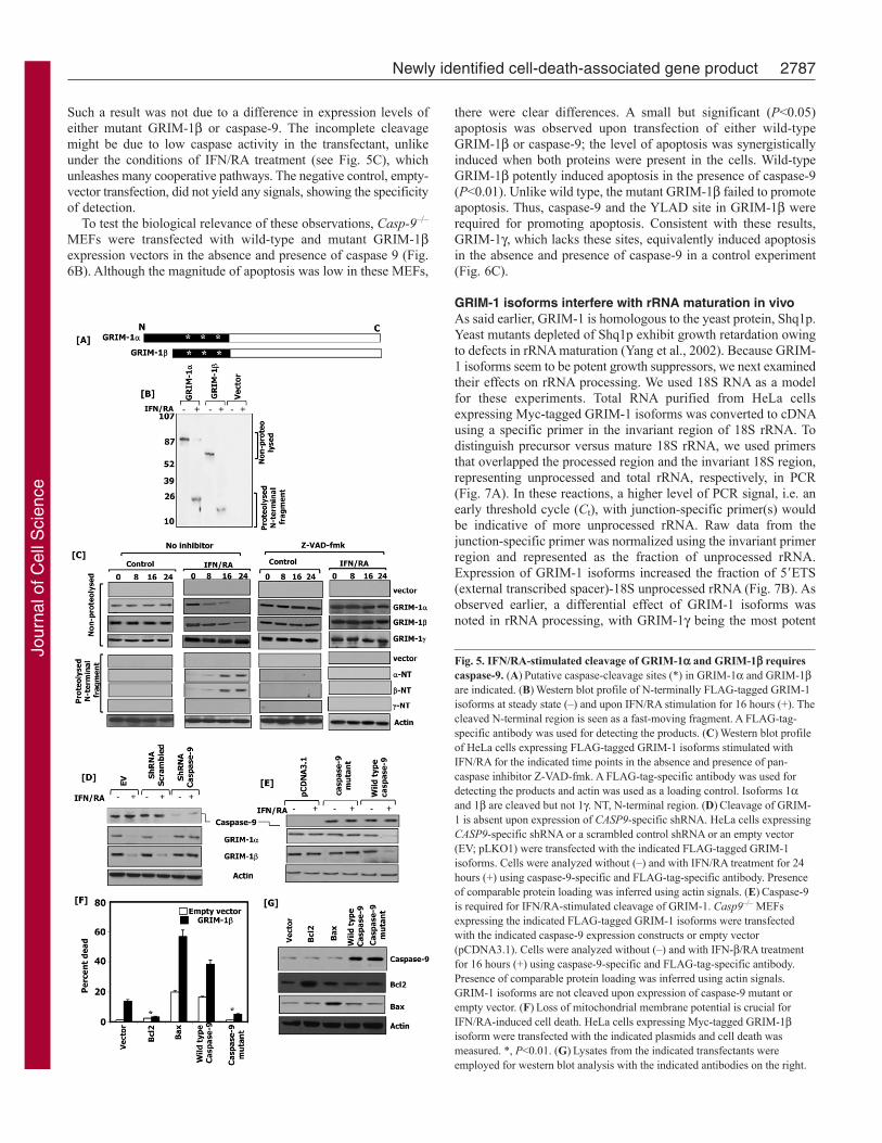
GRIM-1 and GRIM-1 undergo a caspase-dependentprocessingTo determine the role of caspases in regulating GRIM-1 cleavage,we expressed GRIM-1 and GRIM-1 as N-terminally FLAG-tagged proteins (Fig. 5A), and then treated cells with IFN/RA. Fig.5B shows a typical IFN/RA-induced cleavage of GRIM-1 andGRIM-1 proteins. IFN/RA treatment caused a decline of full-length protein level, which was accompanied by an appearance ofa short N-terminal fragment that corresponds to the N-terminus.No such cleavage was observed in steady state. Because the N-terminal fragment, which bears the FLAG tag, is cleaved off, thelarger processed GRIM-1 product could not be seen in these blots.In the next set of experiments, we investigated whether Z-VAD-fmk, a pan-caspase inhibitor, could inhibit IFN/RA-inducedcleavage of GRIM-1 and GRIM-1. As controls, we used emptyvector and FLAG–GRIM-1. Because there are no other cleavageproducts (see Fig. 5B), we showed only the relevant portions of theblots in these experiments. As expected, in all cases no cleavageof GRIM-1 and GRIM-1 occurred in the untreated controls orin the presence of Z-VAD-fmk (Fig. 5C). However, IFN/RAtreatment activated cleavage of GRIM-1 and GRIM-1, whichwas inhibited in presence of Z-VAD-fmk. As expected, IFN/RAtreatment did not induce the cleavage of GRIM-1. GRIM-1cleavage occurred in a delayed manner, indicating the activation ofadditional processes, such as mitochondrial damage, prior to GRIM-1 activation.
Caspase-9 is important for the cleavage of GRIM-1 andGRIM-1Our previous studies indicated a role for caspases, in particularcaspase-9, in the regulation of IFN/RA-induced cell death (Angellet al., 2000). Therefore, we next checked whether knockdown ofcaspase-9 affected IFN/RA-induced cleavage of GRIM-1 andGRIM-1. Using specific lentiviral short hairpin RNAs (shRNAs),we knocked down the expression of caspase-9 in HeLa cells andmeasured its effects on IFN/RA-induced cleavage of GRIM-1and GRIM-1. The CASP9-specific shRNA knocked down >85%of endogenous protein, compared with the controls (Fig. 5D, toppanel). IFN/RA treatment activated a normal cleavage of GRIM-1 and GRIM-1 in the control cells but it failed to do so in cellsexpressing CASP9-specific shRNA (Fig. 5D, middle panels). Acomparable protein loading was confirmed by probing the blotswith an actin-specific antibody (Fig. 5D, bottom panel). Theimportance of caspase-9 for the cleavage of GRIM-1 andGRIM-1 was further ascertained in a complementary experiment.Casp9–/– MEFs were transfected with expression vectors codingwild-type caspase-9 or a catalytically inactive mutant along withGRIM-1 or GRIM-1. First, the expression of caspase-9 wasconfirmed by a western blot analysis (Fig. 5E, top panel). NoIFN/RA-induced cleavage of GRIM-1 and GRIM-1 occurred incells complemented with empty vector and/or mutant caspase-9(Fig. 5E, middle panels). However, expression of wild-type caspase-9 complemented this defect and restored IFN/RA-induced cleavageof GRIM-1 and GRIM-1. A comparable protein loading wasascertained by probing these blots with an actin-specific antibody(Fig. 5E, bottom panel). The above experiments indicated theinvolvement of mitochondrial damage in regulating the cleavageof GRIM-1. Therefore, we transfected GRIM-1-expressing HeLacells individually with expression vectors coding for Bcl2 (whichblocks mitochondrial damage), Bax (which activates mitochondrialdamage), wild-type caspase-9 or a catalytically inactive mutantand measured cell death. Cell death via GRIM-1 was robustlyinduced in the presence of Bax and caspase-9 (Fig. 5F, bars 3 and4), whereas it was significantly lower in the presence of Bcl2 andmutant caspase-9 (Fig. 5F, bars 2 and 5). Similar results wereobtained with GRIM-1 (data not shown). We also ensured theexpression of the transfected Bcl2, Bax and caspase-9 byperforming a western blot analysis of the extracts with specificantibodies. In all cases, corresponding protein bands were intense(compared with the controls) when the proteins were expressed(Fig. 5G). These results suggested that caspase-9 and mitochondrialdamage are important for the induction of GRIM-1 and IFN/RA-driven apoptosis.
Because caspase activation seems to be a crucial step, we nextexamined whether mutation of potential caspase-cleavage siteswould lead to suppression of GRIM-1-induced apoptosis. On thebasis of the observation that the N97 mutant maximally inducedcell death (see Fig. 4), similar to GRIM-1, and of the size of N-terminal protein generated by caspase cleavage (see Fig. 5), wedecided to mutate the most probable caspase-cleavage site, locatedbetween positions 110 and 113, in the GRIM-1 protein: YLAD toYLAA. The effect of caspase-9 on the cleavage of the N-terminusof this mutant was determined by performing a western blotanalysis with FLAG-tag-specific antibodies (Fig. 6A). EmptypCXN2 vector or the same vector carrying wild-type or mutantGRIM-1 were transfected along with wild-type caspase-9 intoCasp-9–/– MEFs. As expected, wild-type GRIM-1, but not themutant GRIM-1, was cleaved only in the presence of caspase-9.
2786 Journal of Cell Science 123 (16)
Fig. 4. The N-terminus of GRIM-1 harbors a death-inhibitory domain.(A)Modular representation of GRIM-1 deletions cloned in pEGFP-C2. TheGFP tag is at the N-terminus of GRIM-1 isoforms. GRIM-1 and GRIM-1are shown for comparison. Putative caspase-cleavage sites (*) are indicated.(B)Western blot profile showing the expression of GFP-tagged GRIM-1 N-terminal deletions in MCF-7 cells. (C)MCF-7 cells were infected withlentiviral GFP-tagged GRIM-1 N-terminal-deletion constructs and cell deathwas quantified using annexin-V-stained samples on a FACS system. Double-positive cells (GFP and annexin V) were scored as dead.
Jour
nal o
f Cel
l Sci
ence
Such a result was not due to a difference in expression levels ofeither mutant GRIM-1 or caspase-9. The incomplete cleavagemight be due to low caspase activity in the transfectant, unlikeunder the conditions of IFN/RA treatment (see Fig. 5C), whichunleashes many cooperative pathways. The negative control, empty-vector transfection, did not yield any signals, showing the specificityof detection.
To test the biological relevance of these observations, Casp-9–/–
MEFs were transfected with wild-type and mutant GRIM-1expression vectors in the absence and presence of caspase 9 (Fig.6B). Although the magnitude of apoptosis was low in these MEFs,
there were clear differences. A small but significant (P<0.05)apoptosis was observed upon transfection of either wild-typeGRIM-1 or caspase-9; the level of apoptosis was synergisticallyinduced when both proteins were present in the cells. Wild-typeGRIM-1 potently induced apoptosis in the presence of caspase-9(P<0.01). Unlike wild type, the mutant GRIM-1 failed to promoteapoptosis. Thus, caspase-9 and the YLAD site in GRIM-1 wererequired for promoting apoptosis. Consistent with these results,GRIM-1, which lacks these sites, equivalently induced apoptosisin the absence and presence of caspase-9 in a control experiment(Fig. 6C).
GRIM-1 isoforms interfere with rRNA maturation in vivoAs said earlier, GRIM-1 is homologous to the yeast protein, Shq1p.Yeast mutants depleted of Shq1p exhibit growth retardation owingto defects in rRNA maturation (Yang et al., 2002). Because GRIM-1 isoforms seem to be potent growth suppressors, we next examinedtheir effects on rRNA processing. We used 18S RNA as a modelfor these experiments. Total RNA purified from HeLa cellsexpressing Myc-tagged GRIM-1 isoforms was converted to cDNAusing a specific primer in the invariant region of 18S rRNA. Todistinguish precursor versus mature 18S rRNA, we used primersthat overlapped the processed region and the invariant 18S region,representing unprocessed and total rRNA, respectively, in PCR(Fig. 7A). In these reactions, a higher level of PCR signal, i.e. anearly threshold cycle (Ct), with junction-specific primer(s) wouldbe indicative of more unprocessed rRNA. Raw data from thejunction-specific primer was normalized using the invariant primerregion and represented as the fraction of unprocessed rRNA.Expression of GRIM-1 isoforms increased the fraction of 5�ETS(external transcribed spacer)-18S unprocessed rRNA (Fig. 7B). Asobserved earlier, a differential effect of GRIM-1 isoforms wasnoted in rRNA processing, with GRIM-1 being the most potent
2787Newly identified cell-death-associated gene product
Fig. 5. IFN/RA-stimulated cleavage of GRIM-1 and GRIM-1 requirescaspase-9. (A)Putative caspase-cleavage sites (*) in GRIM-1 and GRIM-1are indicated. (B)Western blot profile of N-terminally FLAG-tagged GRIM-1isoforms at steady state (–) and upon IFN/RA stimulation for 16 hours (+). Thecleaved N-terminal region is seen as a fast-moving fragment. A FLAG-tag-specific antibody was used for detecting the products. (C)Western blot profileof HeLa cells expressing FLAG-tagged GRIM-1 isoforms stimulated withIFN/RA for the indicated time points in the absence and presence of pan-caspase inhibitor Z-VAD-fmk. A FLAG-tag-specific antibody was used fordetecting the products and actin was used as a loading control. Isoforms 1and 1 are cleaved but not 1. NT, N-terminal region. (D)Cleavage of GRIM-1 is absent upon expression of CASP9-specific shRNA. HeLa cells expressingCASP9-specific shRNA or a scrambled control shRNA or an empty vector(EV; pLKO1) were transfected with the indicated FLAG-tagged GRIM-1isoforms. Cells were analyzed without (–) and with IFN/RA treatment for 24hours (+) using caspase-9-specific and FLAG-tag-specific antibody. Presenceof comparable protein loading was inferred using actin signals. (E)Caspase-9is required for IFN/RA-stimulated cleavage of GRIM-1. Casp9–/– MEFsexpressing the indicated FLAG-tagged GRIM-1 isoforms were transfectedwith the indicated caspase-9 expression constructs or empty vector(pCDNA3.1). Cells were analyzed without (–) and with IFN-/RA treatmentfor 16 hours (+) using caspase-9-specific and FLAG-tag-specific antibody.Presence of comparable protein loading was inferred using actin signals.GRIM-1 isoforms are not cleaved upon expression of caspase-9 mutant orempty vector. (F)Loss of mitochondrial membrane potential is crucial forIFN/RA-induced cell death. HeLa cells expressing Myc-tagged GRIM-1isoform were transfected with the indicated plasmids and cell death wasmeasured. *, P<0.01. (G)Lysates from the indicated transfectants wereemployed for western blot analysis with the indicated antibodies on the right.
Jour
nal o
f Cel
l Sci
ence
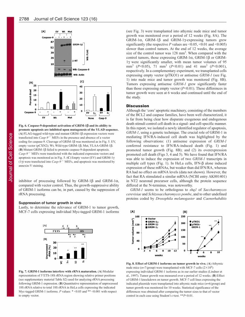
inhibitor of processing followed by GRIM-1 and GRIM-1,compared with vector control. Thus, the growth-suppressive abilityof GRIM-1 isoforms can be, in part, caused by the suppression ofrRNA processing.
Suppression of tumor growth in vivoLastly, to determine the relevance of GRIM-1 to tumor growth,MCF-7 cells expressing individual Myc-tagged GRIM-1 isoforms
(see Fig. 3) were transplanted into athymic nude mice and tumorgrowth was monitored over a period of 12 weeks (Fig. 8A). TheGRIM-1, GRIM-1 and GRIM-1-expressing tumors grewsignificantly (the respective P values are <0.05, <0.01 and <0.005)slower than control tumors. At the end of 12 weeks, the averagesize of the control tumor was 128 mm3. When compared with thecontrol tumors, those expressing GRIM-1, GRIM-1 or GRIM-1 were significantly smaller, with mean tumor volumes of 95mm3 (P<0.05), 71 mm3 (P<0.01) and 41 mm3 (P<0.001),respectively. In a complementary experiment, we transplanted cellsexpressing empty vector (pTKO1) or antisense GRIM-1 (see Fig.1) into nude mice and tumor growth was monitored (Fig. 8B).Tumors expressing antisense GRIM-1 grew significantly fasterthan those expressing empty vector (P<0.01). These differences intumor growth were seen at 6 weeks and continued until the end ofthe study.
DiscussionAlthough the ‘core’ apoptotic machinery, consisting of the membersof the BCL2 and caspase families, have been well characterized, itis far from being clear how disparate exogenous and endogenousdeath stimuli control cell death in a signal- and cell-specific manner.In this report, we isolated a newly identified regulator of apoptosis,GRIM-1, using a genetic technique. The crucial role of GRIM-1 inmediating IFN/RA-induced cell death was highlighted by thefollowing observations: (1) antisense expression of GRIM-1conferred resistance to IFN/RA-induced death (Fig. 1) andpromoted tumor growth (Fig. 8B); and (2) its overexpressionpromoted cell death (Figs 3, 4 and 5). We have found that IFN/RAwas able to induce the expression of two GRIM-1 transcripts inmultiple cell types (Fig. 1). In HeLa cells, IFN- alone inducedexpression of these mRNAs, but weaker than did IFN/RA, whereasRA had no effect on mRNA levels (data not shown). However, thefact that RA stimulated a similar mRNA (NCBI entry AK001401)in NT2 neuronal precursor cells, although the protein sequencediffered at the N-terminus, was noteworthy.
GRIM-1 seems to be orthologous to shq1 of Saccharomycescerevisiae and Schizosaccharomyces pombe, and to other undefinedproteins coded by Drosophila melanogaster and Caenorhabditis
2788 Journal of Cell Science 123 (16)
Fig. 6. Caspase-9-dependent activation of GRIM-1 and its ability topromote apoptosis are inhibited upon mutagenesis of the YLAD sequence.(A)FLAG-tagged wild-type and mutant GRIM-1 expression vectors weretransfected into Casp-9–/– MEFs in the presence and absence of a vectorcoding for caspase-9. Cleavage of GRIM-1 was monitored as in Fig. 5. EV,empty vector (pCXN2); Wt, Wild-type GRIM-1; Mut, YLAA-GRIM-1.(B)Mutant GRIM-1 failed to promote caspase-9-dependent apoptosis.Casp-9–/– MEFs were transfected with the indicated expression vectors andapoptosis was monitored as in Fig. 5. (C)Empty vector (EV) and GRIM-1(1) were transfected into Casp-9–/– MEFs, and apoptosis was monitored byannexin-V staining.
Fig. 7. GRIM-1 isoforms interfere with rRNA maturation. (A)Modularrepresentation of 5�ETS-18S rRNA region showing relative primer positions(see supplementary material Table S2) used for analyzing rRNA processingfollowing GRIM-1 expression. (B)Quantitative representation of unprocessed18S rRNA relative to total 18S rRNA in HeLa cells expressing the indicatedMyc-tagged GRIM-1 isoforms. P values: * <0.05 and ** <0.001 with respectto empty vector.
Fig. 8. Effect of GRIM-1 isoforms on tumor growth in vivo. (A)Athymicnude mice (n7/group) were transplanted with MCF-7 cells (2�106)expressing individual GRIM-1 isoforms as in our earlier studies (Lindner etal., 1997). Tumor growth was measured over a period of 12 weeks. (B)Effectof GRIM-1 knockdown on tumor growth. MCF-7 cell lines expressing theindicated plasmids were transplanted into athymic nude mice (n6/group) andtumor growth was monitored for 10 weeks. Statistical significance of thedifferences was obtained after comparing the tumor sizes to that of vectorcontrol in each case using Student’s t-test. **P<0.01.
Jour
nal o
f Cel
l Sci
ence

elegans (Table 1). The depletion of Shq1p in yeast causes growthretardation due to a defect in rRNA processing, thus highlightingits importance for cell growth (Yang et al., 2002). By contrast, wefound that human GRIM-1 isoforms differentially suppress rRNAprocessing by acting as inhibitors (Fig. 7). The differential behaviorof these two proteins could be due to intrinsic differences in theirstructure and/or regulation by other factors. These issues arecurrently being investigated.
Surprisingly, translation of a transcript derived from GRIM-1cDNA yielded three proteins in vitro (Fig. 2C). These observationsare consistent with the expression of multiple proteins in cells asdetected by the polyclonal antibodies (Fig. 2). Analysis of theDNA sequences around the putative start codons corresponding toGRIM-1 isoforms revealed the presence of a suboptimal Kozaksequences. Two crucial bases required for optimal initiation are theA in the –3 position and the G in the +4 position (Kozak, 1999).For GRIM-1, neither the A–3 nor the G+4 were present (Fig. 2);although GRIM-1 and GRIM-1 both have an A–3, neither havethe G+4 base. Therefore, multiple GRIM-1 proteins observed invivo can be produced either by translational control and/or bydifferential post-translational modification(s).
Cells expressing GRIM-1 isoforms grew significantly slowerthan the controls in vitro (Fig. 3) and in vivo (Fig. 8), thushighlighting their anti-tumor property. The anti-tumor activity inGRIM-1- and GRIM-1-expressing tumors might not be due tothe production of GRIM-1 as we did not observe a form consistentwith GRIM-1 in these tumors (data not shown). Such lack ofexpression of GRIM-1 from GRIM-1 and GRIM-1 mRNAscould also be due to the presence of an ideal Kozak sequenceupstream of the translational start site and/or the 3�UTR of -globin in these expression vectors, unlike the native mRNA. Insummary, each of these proteins seems to exhibit differential anti-tumor properties. GRIM-1 seemed to activate apoptosisindependently of other death regulators, such as p53, and this wassupported by several observations. HeLa and Cos-7 cells lackendogenous p53 protein owing to degradation or physical bindingby the viral proteins HPV-E6 and SV40 T-antigen, respectively(Finlay et al., 1989; Levine, 2009; Scheffner et al., 1990). AlthoughMCF-7 cells possessed a wild-type p53, its inactivation by anoverexpressed HPV-E6 did not substantially affect GRIM-1-drivenapoptosis (data not shown). MCF-7 cells also lack caspase-3 owingto a genetic deletion (Janicke et al., 1998). On the basis of theseobservations, we propose that GRIM-1 induces apoptosisindependently of p53 and caspase-3. Therefore, we investigated amechanism of its activation.
Another mechanism that generates high levels of GRIM-1isoforms in vivo occurs via a caspase-9-dependent conversion ofhigh-molecular-weight forms of GRIM-1 to shorter death-activating forms in response to IFN/RA (Figs 5 and 6). An N-terminal sequence, present in GRIM-1 and GRIM-1, seems toact as a negative regulator of its apoptotic activity. Deletion of theN-terminal sequences dramatically induced apoptosis (Fig. 4). Itis possible that this domain folds back on to the C-terminus toprevent the apoptosis-inducing capacity of GRIM-1 and GRIM-1. Indeed, it was recently shown that yeast Shq1p folds into twoindependent domains that contain sites of casein kinase 1phosphorylation (Godin et al., 2009). Alternatively, it mightassociate with other undefined protein(s), which holds GRIM-1or GRIM-1 in an inhibitory conformation. This region harboredthree potential caspase-cleavage sites. It seems that one of thethree potential cleavage sites present in this area, the third site
(YLAD), is preferred for cleavage. Mutation of this site not onlyresulted in a failure to undergo caspase-9-dependent cleavage ofGRIM-1 but also suppressed apoptosis (Fig. 6). Although it istheoretically possible for caspase-9 to play a role in GRIM-1-induced apoptosis, currently we do not have any data to thatextent.
The role of caspases was based on our additional observationsthat, in the presence of Z-VAD-fmk and/or mutant caspase-9, celldeath and IFN/RA-induced cleavage of GRIM-1 and GRIM-1was blocked (Fig. 5). Consistent with these observations, Bcl2blocked and Bax enhanced IFN/RA-induced and GRIM-1-dependent apoptosis. Indeed, our earlier studies have showncaspase-9 activation and release of cytochrome c in response toIFN/RA (Angell et al., 2000; Ma et al., 2001). Caspases specificallycleave peptides after aspartic-acid residues (Nicholson andThornberry, 1997). Recognition of at least four amino acids N-terminal to this site is also required. Several putative caspase-cleavage sites are present in GRIM-1 protein. Interestingly, manyof the putative caspase-cleavage sites are conserved only inmammalian and C. elegans proteins, but not in yeast proteins. Thisis consistent with the fact that yeast does not have a known caspase(Kang et al., 1999). The presence of putative protein-interactiondomains, phosphorylation sites and caspase-cleavage sites in theGRIM-1 protein suggests a highly ordered regulation of cellexecution by GRIM-1, wherein several regulators converge on asingle substrate. Indeed, deletion of an N-terminal region in GRIM-1 converts it into a GRIM-1-like death activator (Fig. 4).Activation of pro-death proteins via caspase-dependent cleavagehas been reported in other cases, e.g. death-promoting activity ofmitochondrial regulator BID occurs via caspase-8-dependentcleavage (Li et al., 1998; Luo et al., 1998) and apoptotic activityof CAD, an endonuclease that fragments nuclear DNA, occurs viaa caspase-3-dependent mechanism (Enari et al., 1998; Sakahira etal., 1998). Importantly, we have shown a newly identified IFN-inducible gene product that promotes IFN action by slowing rRNAmaturation. However, a complete understanding of this regulationrequires additional studies, which are currently being pursued.
We have previously reported that antisense-mediated inactivationof two other proteins, GRIM-12 (also known as TR; thioredoxinreductase) (Hofmann et al., 1998) and GRIM-19 (Angell et al.,2000), also suppressed cell death in response to IFN/RA. GRIM-12 was required for keeping the active sites of caspases in reducedstate through its substrate thioredoxin (Ma et al., 2001). The secondprotein, GRIM-19, inhibits transcription factor STAT3 (Zhang etal., 2003), a protein known to upregulate the expression ofmitochondrial antiapoptotic regulators Bcl2, Bcl-XL and Mcl-1. Italso represses the cell-death inducer FAS (Ivanov et al., 2001). Wehave shown that GRIM-19 antagonizes these functions of STAT3to promote tumor suppression (Kalakonda et al., 2007). Indeed, wehave recently documented loss of GRIM-19 in a number of primarytumors (Alchanati et al., 2006; Zhang et al., 2007). Loss ofpermeability of the mitochondrial membrane results in a release ofapoptogenic proteins – including cytochrome c (Kluck et al., 1997;Liu et al., 1996), which is required for the activation of caspase-9.In our earlier studies, we have shown activation of caspase-9(Angell et al., 2000; Ma et al., 2007) and cytochrome-c release inresponse to IFN/RA (Ma et al., 2001). In this study, we haveshown that mutant caspase-9 and Bcl2 block the processing ofGRIM-1. On the basis of our current data, we propose that GRIM-19 and GRIM-12 act upstream of GRIM-1 in the cell-death pathwayregulated in response to IFN/RA treatment (Fig. 9). In summary,
2789Newly identified cell-death-associated gene product
Jour
nal o
f Cel
l Sci
ence

we identified a novel death-regulatory protein, whose activation bycaspase-dependent mechanism(s) seems to contribute to apoptosis.
Materials and MethodsReagentsHuman IFN (Biogen), murine Ifn (R&D Systems), RA (Sigma), Z-VAD-fmk(Calbiochem), Ni-chelation sepharose (Novagen), ECL reagents (Pierce), HRP-coupled secondary antibodies (Amersham), hygromycin B (Boehringer Mannheim),DAPI (Sigma), and GFP-specific and caspase-9-specific antibodies (SantacruzBiotech) were employed in these studies. Lipofectamine-Plus (Invitrogen) was usedfor routine transfections as per the manufacturer’s recommendation. Fresh stocks ofRA were prepared in ethanol and added to cultures under subdued light.
PlasmidsIndividual GRIM-1 isoforms were expressed as Myc-tagged (at the C-terminus)proteins using pCXN2-Myc vector (Hofmann et al., 1998), as FLAG-tagged (N-terminus) proteins from pCXN2-FLAG vector and/or as GFP-tagged proteins usingpEGFP-C2 (Clontech); expression vectors for wild-type and catalytically inactivecaspase-9 were provided by Srinivasa M. Srinivasula (NCI, Bethesda, MD), and Baxand Bcl2 were provided by Richard J. Youle (NIH-Bethesda, MD). To knock downendogenous GRIM-1 and caspase-9, antisense GRIM-1 in pTKO1 (Hofmann et al.,1998) and CASP9-specific shRNA in pLKO1 (Open Biosystems) were used,respectively. Lentiviral particles coding for GFP-tagged GRIM-1 isoforms werecloned into pLVX-Puro (Clontech) and shRNA constructs were produced as in ourearlier publication (Gade et al., 2008). Deletion and site-directed mutagenesis, andnorthern and western blot analyses were performed as described earlier (Hofmannet al., 1998).
Cell cultureHeLa cells were cultured in DMEM containing 5% charcoal-stripped FBS, non-essential amino acids, L-glutamine and antibiotics. MCF-7 and T47D cell lines
were cultured in Phenol-Red-free EMEM containing 5% charcoal-stripped FBS,non-essential amino acids, L-glutamine and 10–11 M estradiol during IFN/RAtreatment. The BT-20 cell line was cultured in similar media with Phenol Red butsupplemented with 5% charcoal-stripped FBS prior to IFN/RA treatment. BecausePhenol Red in culture media exerts estrogenic effects, cells were grown in Phenol-Red-free media 24 hours before treatments were initiated. Casp9–/– MEFs, providedby S. M. Srinivasula (NCI, Bethesda, MD), were grown in DMEM containing 5%FBS.
Cell growth assaysCell growth was measured using a colorimetric assay (Skehan et al., 1990). Briefly,cells (2000 cells/well) were treated with human IFN- (1000 U/ml) and RA (1 M)in EMEM with 2.5% charcoal-stripped FBS in 96-well plates, and fixed with 10%tri-chloro acetic acid (TCA) at the indicated time points. One control plate was fixed8 hours after plating to determine the starting cell numbers. Plates were stained with0.4% Sulforhodamine B (SRB; Sigma) prepared in 1% acetic acid for 1 hour;washed, dried and bound dye was eluted by adding 10 mM Tris-Cl (pH 10). Theabsorbance at 570 nm was quantified using a micro plate reader.
Isolation of GRIM-1A cDNA library was generated using poly-A+ RNA derived from the BT-20 cell linetreated with human IFN (500 U/ml) and RA (1 M) for 0, 2, 4, 8, 16, 24, 48 and72 hours, pooled, converted to cDNA and inserted in antisense orientation andexpressed from an episomal vector, pTKO1. This library was electroporated intocells and transfectants were selected with IFN/RA as described (Hofmann et al.,1998). The surviving colonies were expanded, and episomal DNA was extracted andtransformed into Escherichia coli XL-10 to isolate the potential GRIM genes.Individual antisense GRIM genes were transfected into several cell lines to ensureprotection against IFN/RA-induced apoptosis. One antisense clone identified in thismanner contained a 1.9-kb fragment corresponding to the 3� region of GRIM-1cDNA. This fragment was labeled with 32P and used as a probe to screen a phagelibrary. After three rounds of screening, two clones (~2.1 kb) were isolated. Theseclones, however, did not contain the 5� end of the cDNA. Therefore, a 5�RACE wasperformed using a commercially available kit (Life Technologies). The RACEproduct (560 bp) was sequenced and then ligated to the 2.1-kb clone to generate thenear-full-length cDNA (~2.7 kb).
In vitro transcription and translationGRIM-1 cDNA and the indicated ORFs were subcloned into pGEM-7zf (Promega)under the control of T7 promoter. After linearizing the plasmid DNA (1 g), withHindIII, it was programmed into rabbit reticulocyte lysate in a coupled in vitrotranscription-translation system (Promega) in the presence of 35S-methionine. Theresultant products were separated by SDS-PAGE, dried and fluorographed.
Bacterial expression of GRIM-1 for polyclonal-antibody productionInitial attempts to express full-length GRIM-1 ORF did not yield sufficient quantityof the protein. Hence, a cDNA fragment corresponding to the C-terminal 203 aminoacids was cloned into pET-32b (Novagen) to generate the recombinant protein inE. coli BL21(DE3). Cells were lysed by sonication and GRIM-1 protein waspurified from clarified supernatant using Ni-chelation Sepharose (Novagen) asrecommended by the manufacturer. The purified GRIM-1 protein was digested withenterokinase, to remove the tag, and resolved by SDS-PAGE. The bandcorresponding to purified GRIM-1 peptide was used for antibody production inrabbits and mice.
Immunofluorescent and confocal microscopyCells cultured on cover glass in a 24-well tissue-culture plate were fixed for 15minutes using 4% paraformaldehyde, permeabilized with 0.5% Triton X-100 in PBSand blocked in 5% BSA before additional processing. DAPI was used to visualizenuclei. Direct or indirect fluorescence was employed to visualize tagged GRIM-1isoforms. Images were captured using a fluorescence microscope (Olympus BX-FLA, Osaka) fitted with a digital camera (QICAM), and processed by Q-capture Pro5.1 (Q-Imaging Corporation) or using a confocal microscope (Zeiss LSM 510). Cellnumbers were determined using immunofluorescent images from ten randomlyselected fields, with each field containing ~60 cells and subjected to statisticalanalysis with Student’s t-test.
Tumorigenic assaysThree- to four-week-old athymic nude (nu/nu) NCr mice (Taconic) were used in thestudy (Lindner et al., 1997). Procedures involving animals and their care wereconducted in conformity with the institutional guidelines that comply with nationaland international laws and policies (EEC Council Directive 86/609, OJL 358, 1 Dec.1987, and the National Institutes of Health Guide for the Care and Use of LaboratoryAnimals, NIH Publication No. 85-23, 1985). Cells (2�106) were injected into flanksin the mid-axillary line and tumor growth was monitored over a period of 12 weeks.Tumor volume (V) was calculated using caliper measurements and the formula:V(4/3)a2b, where 2a minor axis, 2b major axis of the prolate spheroid.Student’s t-test was used to assess the statistical significance of difference betweenpairs of samples.
2790 Journal of Cell Science 123 (16)
Fig. 9. A model for the anti-tumor actions of IFN/RA using GRIMs. Allblack and gray-colored objects represent growth-promoting and growth-suppressing factors, respectively. In tumor cells, oncogenic growth factors oractivated oncogenes promote growth by upregulating anti-apoptotic factors,such as Bcl2 and Bcl-XL. IFN/RA employs three GRIMs, GRIM-1, GRIM-12and GRIM-19. Of these, GRIM-19 blocks STAT3, an oncogenic transcriptionfactor (Zhang et al., 2003) to lower the levels of antiapoptotic factors. GRIM-19 also associates with a mitochondrial serine protease HtrA2 (Omi) topromote the degradation of XIAP (Ma et al., 2007), an inhibitor of caspase-9.Mitochondrial damage followed by apoptosome formation promotes thecleavage of GRIM-1. GRIM-12 activity maintains caspase-9 in an active stateby providing the reducing power (Ma et al., 2001), promoting caspase-9activity during IFN/RA treatment.
Jour
nal o
f Cel
l Sci
ence

Reverse transcriptase PCR analysis of rRNATotal RNA was converted to cDNA using an rRNA-specific primer (supplementarymaterial Table S2). Unprocessed 5�ETS-18S and processed 18S rRNA fraction wereobtained using specific primer pairs (supplementary material Table S2) as shown inFig. 8. Unprocessed 5�ETS-18S rRNA was represented as fraction of total 18S rRNApool using the Ct method (Nolan et al., 2006).
D.V.K. is supported by NIH grants CA105005 and CA78282.Deposited in PMC for release after 12 months.
Supplementary material available online athttp://jcs.biologists.org/cgi/content/full/123/16/2781/DC1
ReferencesAlchanati, I., Nallar, S. C., Sun, P., Gao, L., Hu, J., Stein, A., Yakirevich, E., Konforty,
D., Alroy, I., Zhao, X. et al. (2006). A proteomic analysis reveals the loss of expressionof the cell death regulatory gene GRIM-19 in human renal cell carcinomas. Oncogene25, 7138-7147.
Altucci, L. and Gronemeyer, H. (2001). The promise of retinoids to fight against cancer.Nat. Rev. Cancer 1, 181-193.
Angell, J. E., Lindner, D. J., Shapiro, P. S., Hofmann, E. R. and Kalvakolanu, D. V.(2000). Identification of GRIM-19, a novel cell death-regulatory gene induced by theinterferon-beta and retinoic acid combination, using a genetic approach. J. Biol. Chem.275, 33416-33426.
Ashkenazi, A. and Dixit, V. M. (1998). Death receptors: signaling and modulation.Science 281, 1305-1308.
Chambon, P. (1996). A decade of molecular biology of retinoic acid receptors. FASEB J.10, 940-954.
Deiss, L. P., Feinstein, E., Berissi, H., Cohen, O. and Kimchi, A. (1995). Identificationof a novel serine/threonine kinase and a novel 15-kD protein as potential mediators ofthe gamma interferon-induced cell death. Genes Dev. 9, 15-30.
Enari, M., Sakahira, H., Yokoyama, H., Okawa, K., Iwamatsu, A. and Nagata, S.(1998). A caspase-activated DNase that degrades DNA during apoptosis, and its inhibitorICAD. Nature 391, 43-50.
Finlay, C. A., Hinds, P. W. and Levine, A. J. (1989). The p53 proto-oncogene can act asa suppressor of transformation. Cell 57, 1083-1093.
Gade, P., Roy, S. K., Li, H., Nallar, S. C. and Kalvakolanu, D. V. (2008). Critical rolefor transcription factor C/EBP-beta in regulating the expression of death-associatedprotein kinase 1. Mol. Cell. Biol. 28, 2528-2548.
Godin, K. S., Walbott, H., Leulliot, N., van Tilbeurgh, H. and Varani, G. (2009). Thebox H/ACA snoRNP assembly factor Shq1p is a chaperone protein homologous toHsp90 cochaperones that binds to the Cbf5p enzyme. J. Mol. Biol. 390, 231-244.
Green, D. R. and Reed, J. C. (1998). Mitochondria and apoptosis. Science 281, 1309-1311.
Hofmann, E. R., Boyanapalli, M., Lindner, D. J., Weihua, X., Hassel, B. A., Jagus, R.,Gutierrez, P. L. and Kalvakolanu, D. V. (1998). Thioredoxin reductase mediates celldeath effects of the combination of beta interferon and retinoic acid. Mol. Cell. Biol. 18,6493-6504.
Ivanov, V. N., Bhoumik, A., Krasilnikov, M., Raz, R., Owen-Schaub, L. B., Levy, D.,Horvath, C. M. and Ronai, Z. (2001). Cooperation between STAT3 and c-junsuppresses Fas transcription. Mol. Cell 7, 517-528.
Janicke, R. U., Sprengart, M. L., Wati, M. R. and Porter, A. G. (1998). Caspase-3 isrequired for DNA fragmentation and morphological changes associated with apoptosis.J. Biol. Chem. 273, 9357-9360.
Kalakonda, S., Nallar, S. C., Lindner, D. J., Hu, J., Reddy, S. P. and Kalvakolanu, D.V. (2007). Tumor-suppressive activity of the cell death activator GRIM-19 on aconstitutively active signal transducer and activator of transcription 3. Cancer Res. 67,6212-6220.
Kalvakolanu, D. V. (2004). The GRIMs: a new interface between cell death regulationand interferon/retinoid induced growth suppression. Cytokine Growth Factor Rev. 15,169-194.
Kang, J. J., Schaber, M. D., Srinivasula, S. M., Alnemri, E. S., Litwack, G., Hall, D.J. and Bjornsti, M. A. (1999). Cascades of mammalian caspase activation in the yeastSaccharomyces cerevisiae. J. Biol. Chem. 274, 3189-3198.
Kimchi, A. (1992). Cytokine triggered molecular pathways that control cell cycle arrest.J. Cell. Biochem. 50, 1-9.
Kluck, R. M., Bossy-Wetzel, E., Green, D. R. and Newmeyer, D. D. (1997). The releaseof cytochrome c from mitochondria: a primary site for Bcl-2 regulation of apoptosis.Science 275, 1132-1136.
Kozak, M. (1999). Initiation of translation in prokaryotes and eukaryotes. Gene 234, 187-208.
Levine, A. J. (2009). The common mechanisms of transformation by the small DNAtumor viruses: The inactivation of tumor suppressor gene products: p53. Virology 384,285-293.
Li, H., Zhu, H., Xu, C. J. and Yuan, J. (1998). Cleavage of BID by caspase 8 mediatesthe mitochondrial damage in the Fas pathway of apoptosis. Cell 94, 491-501.
Lindner, D. J., Borden, E. C. and Kalvakolanu, D. V. (1997). Synergistic antitumoreffects of a combination of interferons and retinoic acid on human tumor cells in vitroand in vivo. Clin. Cancer Res. 3, 931-937.
Liu, X., Kim, C. N., Yang, J., Jemmerson, R. and Wang, X. (1996). Induction ofapoptotic program in cell-free extracts: requirement for dATP and cytochrome c. Cell86, 147-157.
Logue, S. E. and Martin, S. J. (2008). Caspase activation cascades in apoptosis. Biochem.Soc. Trans. 36, 1-9.
Lowe, S. W., Bodis, S., McClatchey, A., Remington, L., Ruley, H. E., Fisher, D. E.,Housman, D. E. and Jacks, T. (1994). p53 status and the efficacy of cancer therapyin vivo. Science 266, 807-810.
Luo, X., Budihardjo, I., Zou, H., Slaughter, C. and Wang, X. (1998). Bid, a Bcl2interacting protein, mediates cytochrome c release from mitochondria in response toactivation of cell surface death receptors. Cell 94, 481-490.
Ma, X., Karra, S., Guo, W., Lindner, D. J., Hu, J., Angell, J. E., Hofmann, E. R.,Reddy, S. P. and Kalvakolanu, D. V. (2001). Regulation of interferon and retinoicacid-induced cell death activation through thioredoxin reductase. J. Biol. Chem. 276,24843-24854.
Ma, X., Kalakonda, S., Srinivasula, S. M., Reddy, S. P., Platanias, L. C. andKalvakolanu, D. V. (2007). GRIM-19 associates with the serine protease HtrA2 forpromoting cell death. Oncogene 26, 4842-4849.
Moore, D. M., Kalvakolanu, D. V., Lippman, S. M., Kavanagh, J. J., Hong, W. K.,Borden, E. C., Paredes-Espinoza, M. and Krakoff, I. H. (1994). Retinoic acid andinterferon in human cancer: mechanistic and clinical studies. Semin. Hematol. 31, 31-37.
Nicholson, D. W. and Thornberry, N. A. (1997). Caspases: killer proteases. TrendsBiochem. Sci. 22, 299-306.
Nolan, T., Hands, R. E. and Bustin, S. A. (2006). Quantification of mRNA using real-time RT-PCR. Nat. Protoc. 1, 1559-1582.
Sakahira, H., Enari, M. and Nagata, S. (1998). Cleavage of CAD inhibitor in CADactivation and DNA degradation during apoptosis. Nature 391, 96-99.
Scheffner, M., Werness, B. A., Huibregtse, J. M., Levine, A. J. and Howley, P. M.(1990). The E6 oncoprotein encoded by human papillomavirus types 16 and 18 promotesthe degradation of p53. Cell 63, 1129-1136.
Schindler, C., Levy, D. E. and Decker, T. (2007). JAK-STAT signaling: from interferonsto cytokines. J. Biol. Chem. 282, 20059-20063.
Skehan, P., Storeng, R., Scudiero, D., Monks, A., McMahon, J., Vistica, D., Warren,J. T., Bokesch, H., Kenney, S. and Boyd, M. R. (1990). New colorimetric cytotoxicityassay for anticancer-drug screening. J. Natl. Cancer Inst. 82, 1107-1112.
Stennicke, H. R. and Salvesen, G. S. (2000). Caspases-controlling intracellular signalsby protease zymogen activation. Biochim. Biophys. Acta 1477, 299-306.
Yang, P. K., Rotondo, G., Porras, T., Legrain, P. and Chanfreau, G. (2002). TheShq1p.Naf1p complex is required for box H/ACA small nucleolar ribonucleoproteinparticle biogenesis. J. Biol. Chem. 277, 45235-45242.
Youle, R. J. and Strasser, A. (2008). The BCL-2 protein family: opposing activities thatmediate cell death. Nat. Rev. Mol. Cell Biol. 9, 47-59.
Zhang, J., Yang, J., Roy, S. K., Tininini, S., Hu, J., Bromberg, J. F., Poli, V., Stark, G.R. and Kalvakolanu, D. V. (2003). The cell death regulator GRIM-19 is an inhibitorof signal transducer and activator of transcription 3. Proc. Natl. Acad. Sci. USA 100,9342-9347.
Zhang, L., Gao, L., Zhao, L., Guo, B., Ji, K., Tian, Y., Wang, J., Yu, H., Hu, J.,Kalvakolanu, D. V. et al. (2007). Intratumoral delivery and suppression of prostatetumor growth by attenuated Salmonella enterica serovar typhimurium carrying plasmid-based small interfering RNAs. Cancer Res. 67, 5859-5864..
2791Newly identified cell-death-associated gene product
Jour
nal o
f Cel
l Sci
ence