identification and characterization of nagnag alternative splicing in the moss physcomitrella patens
TRANSCRIPT
Sinha et al. BMC Plant Biology 2010, 10:76http://www.biomedcentral.com/1471-2229/10/76
Open AccessR E S E A R C H A R T I C L E
Research articleIdentification and characterization of NAGNAG alternative splicing in the moss Physcomitrella patensRileen Sinha1,5, Andreas D Zimmer2,3, Kathrin Bolte2,4,7, Daniel Lang2,3, Ralf Reski3,4,5, Matthias Platzer6, Stefan A Rensing*2,4,5 and Rolf Backofen*1,4,5
AbstractBackground: Alternative splicing (AS) involving tandem acceptors that are separated by three nucleotides (NAGNAG) is an evolutionarily widespread class of AS, which is well studied in Homo sapiens (human) and Mus musculus (mouse). It has also been shown to be common in the model seed plants Arabidopsis thaliana and Oryza sativa (rice). In one of the first studies involving sequence-based prediction of AS in plants, we performed a genome-wide identification and characterization of NAGNAG AS in the model plant Physcomitrella patens, a moss.
Results: Using Sanger data, we found 295 alternatively used NAGNAG acceptors in P. patens. Using 31 features and training and test datasets of constitutive and alternative NAGNAGs, we trained a classifier to predict the splicing outcome at NAGNAG tandem splice sites (alternative splicing, constitutive at the first acceptor, or constitutive at the second acceptor). Our classifier achieved a balanced specificity and sensitivity of ≥ 89%. Subsequently, a classifier trained exclusively on data well supported by transcript evidence was used to make genome-wide predictions of NAGNAG splicing outcomes. By generation of more transcript evidence from a next-generation sequencing platform (Roche 454), we found additional evidence for NAGNAG AS, with altogether 664 alternative NAGNAGs being detected in P. patens using all currently available transcript evidence. The 454 data also enabled us to validate the predictions of the classifier, with 64% (80/125) of the well-supported cases of AS being predicted correctly.
Conclusion: NAGNAG AS is just as common in the moss P. patens as it is in the seed plants A. thaliana and O. sativa (but not conserved on the level of orthologous introns), and can be predicted with high accuracy. The most informative features are the nucleotides in the NAGNAG and in its immediate vicinity, along with the splice sites scores, as found earlier for NAGNAG AS in animals. Our results suggest that the mechanism behind NAGNAG AS in plants is similar to that in animals and is largely dependent on the splice site and its immediate neighborhood.
BackgroundEukaryotic primary mRNAs consist of protein-codingregions (exons) and intervening non-coding regions(introns). The mature mRNA transcript, which acts assubstrate for translation into protein, is produced byremoving introns in a process called splicing. Splicing canbe either constitutive, always producing the same mRNA,
or alternative, via variable inclusion of parts of the pri-mary transcript. Alternative splicing (AS) is thus a mech-anism that enables multiple transcripts and proteins to beencoded by the same gene, thereby promoting transcriptand protein diversity [1]. Furthermore, events of AS canprovide an additional level of post-transcriptional generegulation, e.g. by the production of mRNA isoforms withtruncated open reading frames that are subject to degra-dation by the nonsense mediated decay pathway [2,3]. ASis particularly widespread in higher eukaryotes, especiallyin mammals - it has been estimated that up to 94% of allmulti-exonic H. sapiens genes are alternatively spliced [4].Large-scale detection of AS usually involves expressed
* Correspondence: [email protected], [email protected] Faculty of Biology, University of Freiburg, Hauptstrasse 1, 79104 Freiburg, Germany2 Bioinformatics group, University of Freiburg, Georges-Koehler-Allee 106, 79110 Freiburg, GermanyFull list of author information is available at the end of the article
BioMed Central© 2010 Sinha et al; licensee BioMed Central Ltd. This is an Open Access article distributed under the terms of the Creative CommonsAttribution License (http://creativecommons.org/licenses/by/2.0), which permits unrestricted use, distribution, and reproduction inany medium, provided the original work is properly cited.
Sinha et al. BMC Plant Biology 2010, 10:76http://www.biomedcentral.com/1471-2229/10/76
Page 2 of 14
sequence tags (ESTs), microarray, or RNA-seq analysis.However, not all AS events can be detected by thesemethods. Moreover, nowadays genomic sequence data isbeing churned out at a much faster rate than transcriptdata, that is, many genomes have low transcript coverage.Thus, there is a need for independent methods of detect-ing AS.
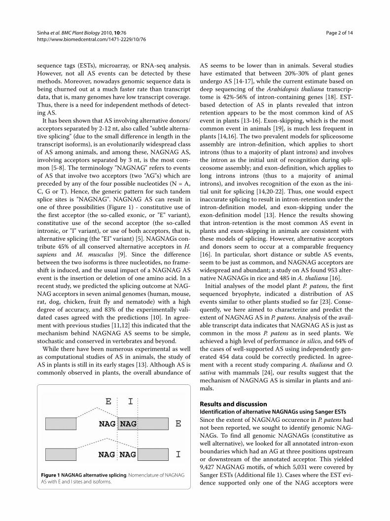
It has been shown that AS involving alternative donors/acceptors separated by 2-12 nt, also called "subtle alterna-tive splicing" (due to the small difference in length in thetranscript isoforms), is an evolutionarily widespread classof AS among animals, and among these, NAGNAG AS,involving acceptors separated by 3 nt, is the most com-mon [5-8]. The terminology "NAGNAG" refers to eventsof AS that involve two acceptors (two "AG"s) which arepreceded by any of the four possible nucleotides (N = A,C, G or T). Hence, the generic pattern for such tandemsplice sites is "NAGNAG". NAGNAG AS can result inone of three possibilities (Figure 1) - constitutive use ofthe first acceptor (the so-called exonic, or "E" variant),constitutive use of the second acceptor (the so-calledintronic, or "I" variant), or use of both acceptors, that is,alternative splicing (the "EI" variant) [5]. NAGNAGs con-tribute 45% of all conserved alternative acceptors in H.sapiens and M. musculus [9]. Since the differencebetween the two isoforms is three nucleotides, no frame-shift is induced, and the usual impact of a NAGNAG ASevent is the insertion or deletion of one amino acid. In arecent study, we predicted the splicing outcome at NAG-NAG acceptors in seven animal genomes (human, mouse,rat, dog, chicken, fruit fly and nematode) with a highdegree of accuracy, and 83% of the experimentally vali-dated cases agreed with the predictions [10]. In agree-ment with previous studies [11,12] this indicated that themechanism behind NAGNAG AS seems to be simple,stochastic and conserved in vertebrates and beyond.
While there have been numerous experimental as wellas computational studies of AS in animals, the study ofAS in plants is still in its early stages [13]. Although AS iscommonly observed in plants, the overall abundance of
AS seems to be lower than in animals. Several studieshave estimated that between 20%-30% of plant genesundergo AS [14-17], while the current estimate based ondeep sequencing of the Arabidopsis thaliana transcrip-tome is 42%-56% of intron-containing genes [18]. EST-based detection of AS in plants revealed that intronretention appears to be the most common kind of ASevent in plants [13-16]. Exon-skipping, which is the mostcommon event in animals [19], is much less frequent inplants [14,16]. The two prevalent models for spliceosomeassembly are intron-definition, which applies to shortintrons (thus to a majority of plant introns) and involvesthe intron as the initial unit of recognition during spli-ceosome assembly; and exon-definition, which applies tolong introns introns (thus to a majority of animalintrons), and involves recognition of the exon as the ini-tial unit for splicing [14,20-22]. Thus, one would expectinaccurate splicing to result in intron-retention under theintron-definition model, and exon-skipping under theexon-definition model [13]. Hence the results showingthat intron-retention is the most common AS event inplants and exon-skipping in animals are consistent withthese models of splicing. However, alternative acceptorsand donors seem to occur at a comparable frequency[16]. In particular, short distance or subtle AS events,seem to be just as common, and NAGNAG acceptors arewidespread and abundant; a study on AS found 953 alter-native NAGNAGs in rice and 485 in A. thaliana [16].
Initial analyses of the model plant P. patens, the firstsequenced bryophyte, indicated a distribution of ASevents similar to other plants studied so far [23]. Conse-quently, we here aimed to characterize and predict theextent of NAGNAG AS in P. patens. Analysis of the avail-able transcript data indicates that NAGNAG AS is just ascommon in the moss P. patens as in seed plants. Weachieved a high level of performance in silico, and 64% ofthe cases of well-supported AS using independently gen-erated 454 data could be correctly predicted. In agree-ment with a recent study comparing A. thaliana and O.sativa with mammals [24], our results suggest that themechanism of NAGNAG AS is similar in plants and ani-mals.
Results and discussionIdentification of alternative NAGNAGs using Sanger ESTsSince the extent of NAGNAG occurence in P. patens hadnot been reported, we sought to identify genomic NAG-NAGs. To find all genomic NAGNAGs (constitutive aswell alternative), we looked for all annotated intron-exonboundaries which had an AG at three positions upstreamor downstream of the annotated acceptor. This yielded9,427 NAGNAG motifs, of which 5,031 were covered bySanger ESTs (Additional file 1). Cases where the EST evi-dence supported only one of the NAG acceptors were
Figure 1 NAGNAG alternative splicing. Nomenclature of NAGNAG AS with E and I sites and isoforms.
Sinha et al. BMC Plant Biology 2010, 10:76http://www.biomedcentral.com/1471-2229/10/76
Page 3 of 14
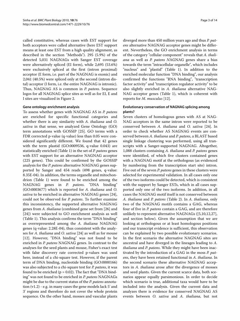
called constitutive, whereas cases with EST support forboth acceptors were called alternative (here EST supportmeans at least one EST from a high quality alignment, asdescribed in the section "Methods"). 295 (5.9%) of thedetected 5,031 NAGNAGs with Sanger EST coveragewere alternatively spliced (EI form), while 2,695 (53.6%)were exclusively spliced at the first (intron proximal)acceptor (E form, i.e. part of the NAGNAG is exonic) and2,041 (40.5%) were spliced only at the second (intron dis-tal) acceptor (I form, i.e. the entire NAGNAG is intronic).Thus, NAGNAG AS is common in P. patens. Sequencelogos for all NAGNAG splice sites as well as for EI, E andI sites are visualized in Figure 2.
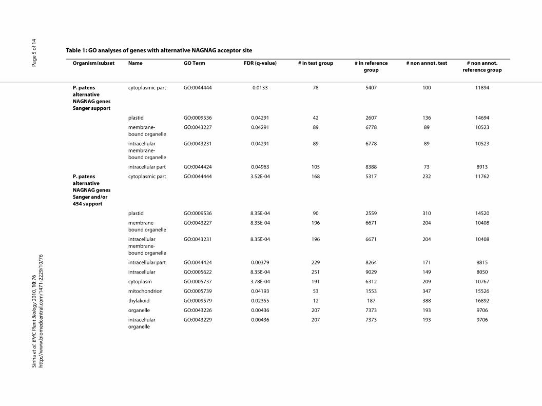
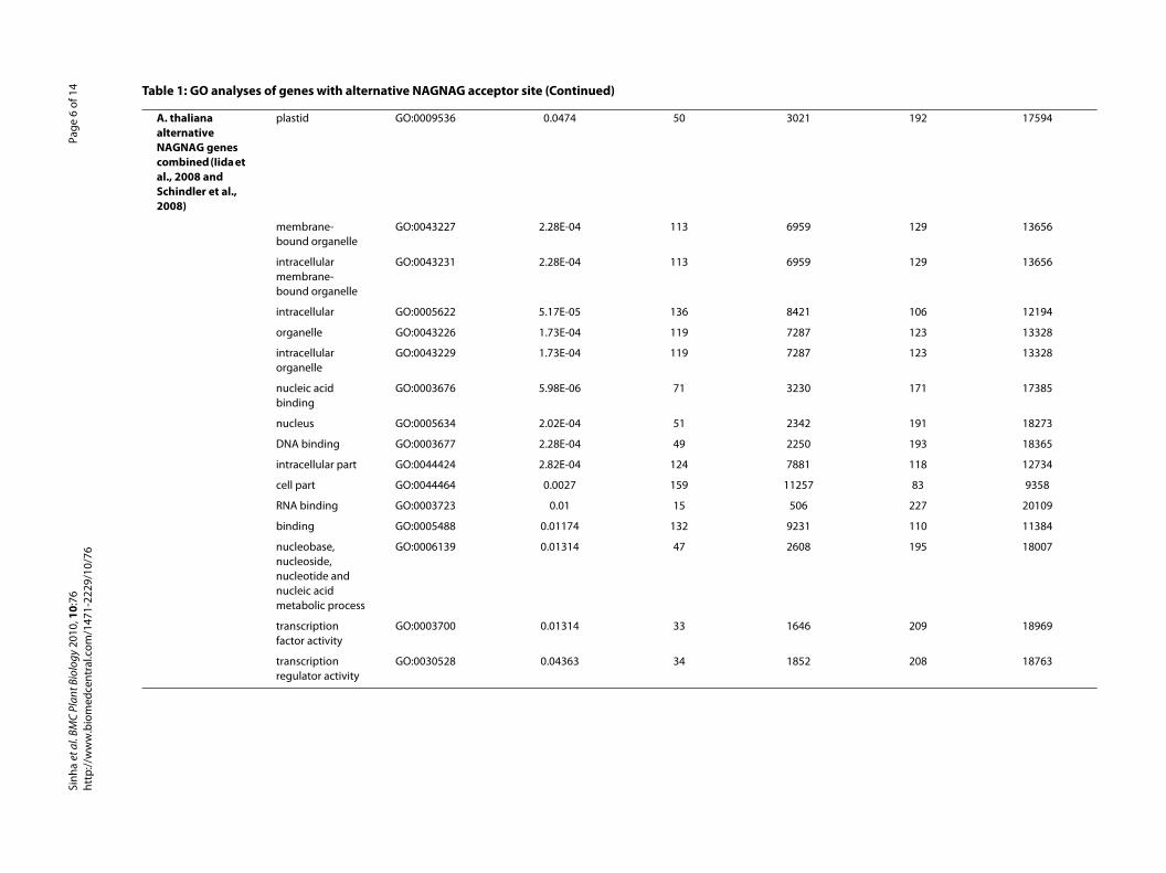
Gene ontology enrichment analysisTo assess whether genes with NAGNAG AS in P. patensare enriched for specific functional categories andwhether there is any similarity with A. thaliana and O.sativa in that sense, we analyzed Gene Ontology (GO)term annotations with GOSSIP [25]. GO terms with aFDR corrected p-value (q-value) less than 0.05 were con-sidered significantly different. We found that 42 geneswith the term plastid (GO:0009536, q-value 0.043) arestatistically enriched (Table 1) in the set of P. patens geneswith EST support for an alternative NAGNAG acceptor(225 genes). This could be confirmed by the GOSSIPanalysis for the P. patens alternative NAGNAG genes sup-ported by Sanger and 454 reads (498 genes, q-value:8.35E-04). In addition, the terms organelle and mitochon-drion (Table 1) were found to be enriched among theNAGNAG genes in P. patens. "DNA binding"(GO:0003677) which is reported for A. thaliana and O.sativa to be enriched in alternative NAGNAG genes [24],could not be observed for P. patens. To further examinethis inconsistency, the supported alternative NAGNAGgenes from A. thaliana (combined gene set from [26] and[24]) were subjected to GO enrichment analysis as well(Table 1). This analysis confirms the term "DNA binding"as overrepresented among the A. thaliana NAGNAGgenes (q-value: 2.28E-04), thus consistent with the analy-ses for A. thaliana and O. sativa [24] as well as for mouse[12]. However, "DNA binding" was not found to beenriched in P. patens NAGNAG genes. In contrast to theanalyses for the seed plants and mouse, Fisher's exact testwith false discovery rate corrected p-values was usedhere, instead of a chi-square test. However, if the parentterm of DNA binding, nucleotide binding (GO:0000166)was also subjected to a chi-square test for P. patens, it wasfound to be enriched (p = 0.02). The fact that "DNA bind-ing" was not found to be enriched in P. patens NAGNAGsmight be due to the current status of the P. patens annota-tion (v1.2) - e.g. in many cases the gene models lack 5' and3' regions and therefore do not cover the whole proteinsequence. On the other hand, mosses and vascular plants
diverged more than 450 million years ago and thus P. pat-ens alternative NAGNAG acceptor genes might be differ-ent. Nevertheless, the GO enrichment analysis in termsof the category "cellular component" reveals that A. thali-ana as well as P. patens NAGNAG genes share a biastowards the term "intracellular organelle", which includes"nucleus" and "plastid" (Table 1). In addition to theenriched molecular function "DNA binding", our analysisconfirmed the functions "RNA binding", "transcriptionfactor activity" and "transcription regulator activity" to bealso slightly enriched in A. thaliana alternative NAG-NAG acceptor genes (Table 1), which is coherent withreports for M. musculus [12].
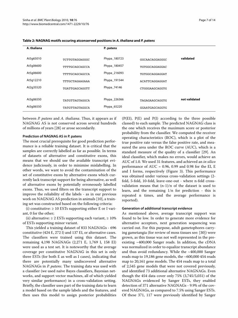
Evolutionary conservation of NAGNAG splicing among plants?Seven clusters of homologous genes with AS at NAG-NAG acceptors in the same intron were reported to beconserved between A. thaliana and O. sativa [24]. Inorder to check whether AS NAGNAG events are con-served between A. thaliana and P. patens, a BLAST basedsingle linkage clustering was performed, using all tran-scripts with a Sanger-supported NAGNAG. Altogether,1,088 clusters containing A. thaliana and P. patens geneswere identified, of which five clusters contained geneswith a NAGNAG motif at the orthologous (as evidencedby numbering from the transcription start site) intron.Five out of the seven P. patens genes in these clusters wereselected for experimental validation. In all cases only oneof the two isoforms could be detected, which is consistentwith the support by Sanger ESTs, which in all cases sup-ported only one of the two isoforms. In addition, in allcases the NAGNAG motif itself is not conserved betweenA. thaliana and P. patens (Table 2). In A. thaliana, onlytwo of the NAGNAG motifs contains a GAG, whereasfour of five in P. patens contain a GAG, and are thereforeunlikely to represent alternative NAGNAGs ([5,10,12,27],and section below). Given the assumption that we arelooking at orthologous or at least homologous positionsand our transcript evidence is sufficient, this observationcan be explained by two possible evolutionary scenarios.In the first scenario the alternative NAGNAG sites areancestral and have diverged in the lineages leading to A.thaliana and P. patens. While they might have been inac-tivated by the introduction of a GAG in the moss P. pat-ens, they have been retained functional in A. thaliana. Inthe second scenario these alternative NAGNAG accep-tors in A. thaliana arose after the divergence of mossesand seed plants. Given the current scarce data, both sce-narios appear equally parsimonious. In order to decidewhich scenario is true, additional taxa would have to beincluded into the analysis. Given the current data andanalyzes there is evidence for conserved NAGNAG ASevents between O. sativa and A. thaliana, but not
Sinha et al. BMC Plant Biology 2010, 10:76http://www.biomedcentral.com/1471-2229/10/76
Page 4 of 14
Figure 2 Sequence logos of NAGNAG splice sites. The first three positions represent the last 3 nucleotides (nt) of the upstream exon, followed by the 30 nt upstream of the NAGNAG, the NAGNAG motif itself, and the 10 nt downstream of the NAGNAG (total 49 positions). A: all splice sites; B: EI sites, C: E sites; D: I sites.
rence p
# non annot. test # non annot. reference group
100 11894
136 14694
89 10523
89 10523
73 8913
232 11762
310 14520
204 10408
204 10408
171 8815
149 8050
209 10767
347 15526
388 16892
193 9706
193 9706
Sinh
a et
al.
BMC
Plan
t Bio
logy
201
0, 1
0:76
http
://w
ww
.bio
med
cent
ral.c
om/1
471-
2229
/10/
76Pa
ge 5
of 1
4
Table 1: GO analyses of genes with alternative NAGNAG acceptor site
Organism/subset Name GO Term FDR (q-value) # in test group # in refegrou
P. patens alternative NAGNAG genes Sanger support
cytoplasmic part GO:0044444 0.0133 78 5407
plastid GO:0009536 0.04291 42 2607
membrane-bound organelle
GO:0043227 0.04291 89 6778
intracellular membrane-bound organelle
GO:0043231 0.04291 89 6778
intracellular part GO:0044424 0.04963 105 8388
P. patens alternative NAGNAG genes Sanger and/or 454 support
cytoplasmic part GO:0044444 3.52E-04 168 5317
plastid GO:0009536 8.35E-04 90 2559
membrane-bound organelle
GO:0043227 8.35E-04 196 6671
intracellular membrane-bound organelle
GO:0043231 8.35E-04 196 6671
intracellular part GO:0044424 0.00379 229 8264
intracellular GO:0005622 8.35E-04 251 9029
cytoplasm GO:0005737 3.78E-04 191 6312
mitochondrion GO:0005739 0.04193 53 1553
thylakoid GO:0009579 0.02355 12 187
organelle GO:0043226 0.00436 207 7373
intracellular organelle
GO:0043229 0.00436 207 7373
192 17594
129 13656
129 13656
106 12194
123 13328
123 13328
171 17385
191 18273
193 18365
118 12734
7 83 9358
227 20109
110 11384
195 18007
209 18969
208 18763
Sinh
a et
al.
BMC
Plan
t Bio
logy
201
0, 1
0:76
http
://w
ww
.bio
med
cent
ral.c
om/1
471-
2229
/10/
76Pa
ge 6
of 1
4
A. thaliana alternative NAGNAG genes combined (Iida et al., 2008 and Schindler et al., 2008)
plastid GO:0009536 0.0474 50 3021
membrane-bound organelle
GO:0043227 2.28E-04 113 6959
intracellular membrane-bound organelle
GO:0043231 2.28E-04 113 6959
intracellular GO:0005622 5.17E-05 136 8421
organelle GO:0043226 1.73E-04 119 7287
intracellular organelle
GO:0043229 1.73E-04 119 7287
nucleic acid binding
GO:0003676 5.98E-06 71 3230
nucleus GO:0005634 2.02E-04 51 2342
DNA binding GO:0003677 2.28E-04 49 2250
intracellular part GO:0044424 2.82E-04 124 7881
cell part GO:0044464 0.0027 159 1125
RNA binding GO:0003723 0.01 15 506
binding GO:0005488 0.01174 132 9231
nucleobase, nucleoside, nucleotide and nucleic acid metabolic process
GO:0006139 0.01314 47 2608
transcription factor activity
GO:0003700 0.01314 33 1646
transcription regulator activity
GO:0030528 0.04363 34 1852
Table 1: GO analyses of genes with alternative NAGNAG acceptor site (Continued)
Sinha et al. BMC Plant Biology 2010, 10:76http://www.biomedcentral.com/1471-2229/10/76
Page 7 of 14
between P. patens and A. thaliana. Thus, it appears as ifNAGNAG AS is not conserved across several hundredsof millions of years [28] or arose secondarily.
Prediction of NAGNAG AS in P. patensThe most crucial prerequisite for good prediction perfor-mance is a reliable training dataset. It is critical that thesamples are correctly labelled as far as possible. In termsof datasets of alternative and constitutive exons, thismeans that we should use the available transcript evi-dence judiciously, in order to minimise mislabelling. Inother words, we want to avoid the contamination of theset of constitutive exons by alternative exons which cur-rently lack transcript support for being alternative, as wellof alternative exons by potentially erroneously labelledexons. Thus, we used filters on the transcript support toimprove the reliability of the labels - as in our previouswork on NAGNAG AS prediction in animals [10], a train-ing set was constructed based on the following criteria:
(i) constitutive: ≥ 10 ESTs supporting either E or I vari-ant, 0 for the other;
(ii) alternative: ≥ 2 ESTs supporting each variant, ≥ 10%of ESTs supporting minor variant.
This yielded a training dataset of 833 NAGNAGs - 696constitutive (424 E, 272 I) and 137 EI, or alternative cases.The classifiers were trained using this dataset. Theremaining 4,198 NAGNAGs (2,271 E, 1,769 I, 158 EI)were used as a test set. It is noteworthy that the averagecoverage per constitutive NAGNAG in this set is onlythree ESTs (for both E as well as I cases), indicating thatthere are potentially many undiscovered alternativeNAGNAGs in P. patens. The training data was used witha classifier (we used naïve Bayes classifiers, Bayesian net-works, and support vector machines, all of which yieldedvery similar performance) in a cross-validation setting.Briefly, the classifier uses part of the training data to learna model based on the sample labels and the features, andthen uses this model to assign posterior probabilities
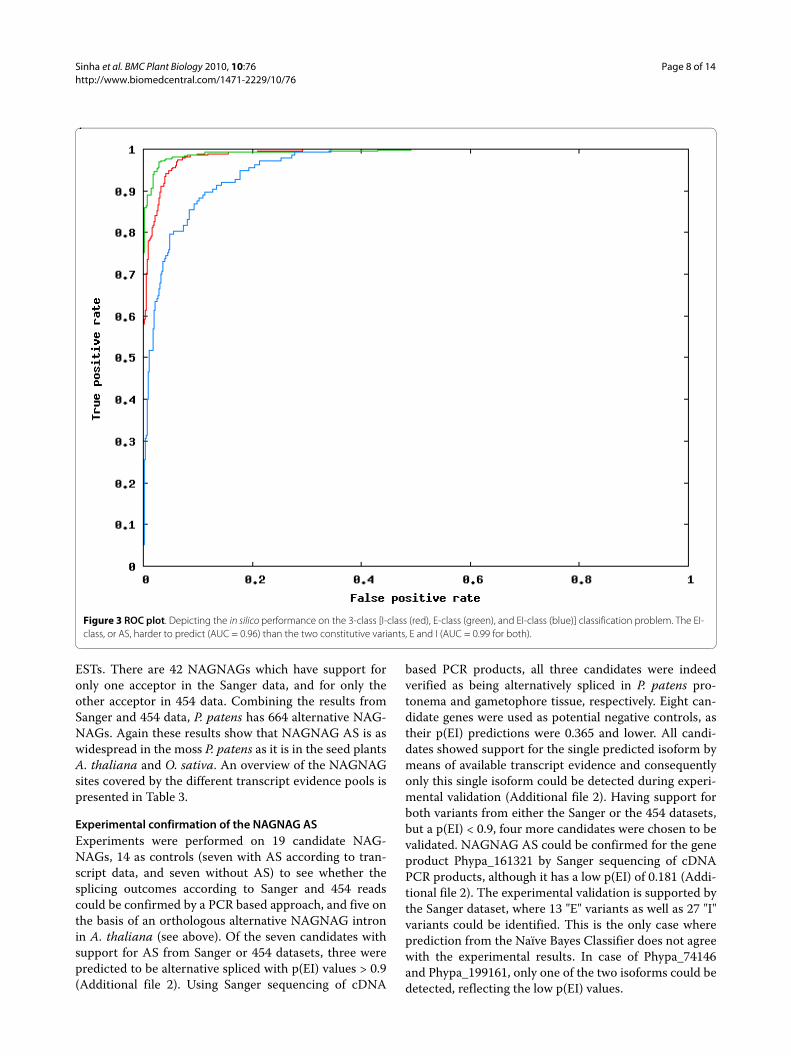
(P(EI), P(E) and P(I) according to the three possibleclassed) to each sample. The predicted NAGNAG class isthe one which receives the maximum score or posteriorprobability from the classifier. We computed the receiveroperating characteristics (ROC), which is a plot of thetrue positive rate versus the false positive rate, and mea-sured the area under the ROC curve (AUC), which is astandard measure of the quality of a classifier [29]. Anideal classifier, which makes no errors, would achieve anAUC of 1.0. We used 31 features, and achieved an in silicoperformance of AUC = 0.96, 0.99 and 0.98 for the EI, Eand I forms, respectively (Figure 3). This performancewas obtained under various cross-validation settings (2-fold, 5-fold, 10-fold, leave-one-out - where n-fold cross-validation means that (n-1)/n of the dataset is used tolearn, and the remaining 1/n for prediction - this isrepeated n times, and the average performance isreported).
Generation of additional transcript evidenceAs mentioned above, average transcript support wasfound to be low. In order to generate more evidence foralternative acceptors, next generation sequencing wascarried out. For this purpose, adult gametophores carry-ing gametangia (for review of moss tissues see: [30]) weregrown, as this tissue was not well represented in the pre-existing ~400,000 Sanger reads. In addition, the cDNAwas normalized in order to equalize transcript abundanceand thus avoid redundancy. While the ~400,000 Sangerreads map to 19,186 gene models, the ~600,000 454 readsmap to 20,161 gene models. The 454 reads map to a totalof 2,545 gene models that were not covered previously,and identified 73 additional alternative NAGNAGs. Eventhough the 454 data cover only 75% (3,745/5,031) of theNAGNAGs evidenced by Sanger ESTs, they enableddetection of 371 alternative NAGNAGs - 9.9% of the cov-ered NAGNAGs, as compared to 7.5% using Sanger ESTs.Of these 371, 117 were previously identified by Sanger
Table 2: NAGNAG motifs occuring atconserved positions in A. thaliana and P. patens
A. thaliana P. patens
At5g65010 TCTTGTAGGAGGGC Phypa_180723 GGCAACAGGAGGGC validated
At5g06600 TTTTGCAGCAGCCA Phypa_180457 TGTGGCAGGAGGAC
At5g06600 TTTTGCAGCAGCCA Phypa_216093 TGTGGCAGGAGGAT
At5g12210 TTTGCTAGAAGAAA Phypa_191544 ACATTCAGGAGGAT
At2g35520 TGATTGAGCAGGTT Phypa_74146 CTGGGAAGCAGGTG
At3g06550 TATGTTAGTAGGCA Phypa_226366 TAGAGAAGCAGGTG not validated
At3g06550 TATGTTAGTAGGCA Phypa_65220 GGAATGAGCAGGTG
Sinha et al. BMC Plant Biology 2010, 10:76http://www.biomedcentral.com/1471-2229/10/76
Page 8 of 14
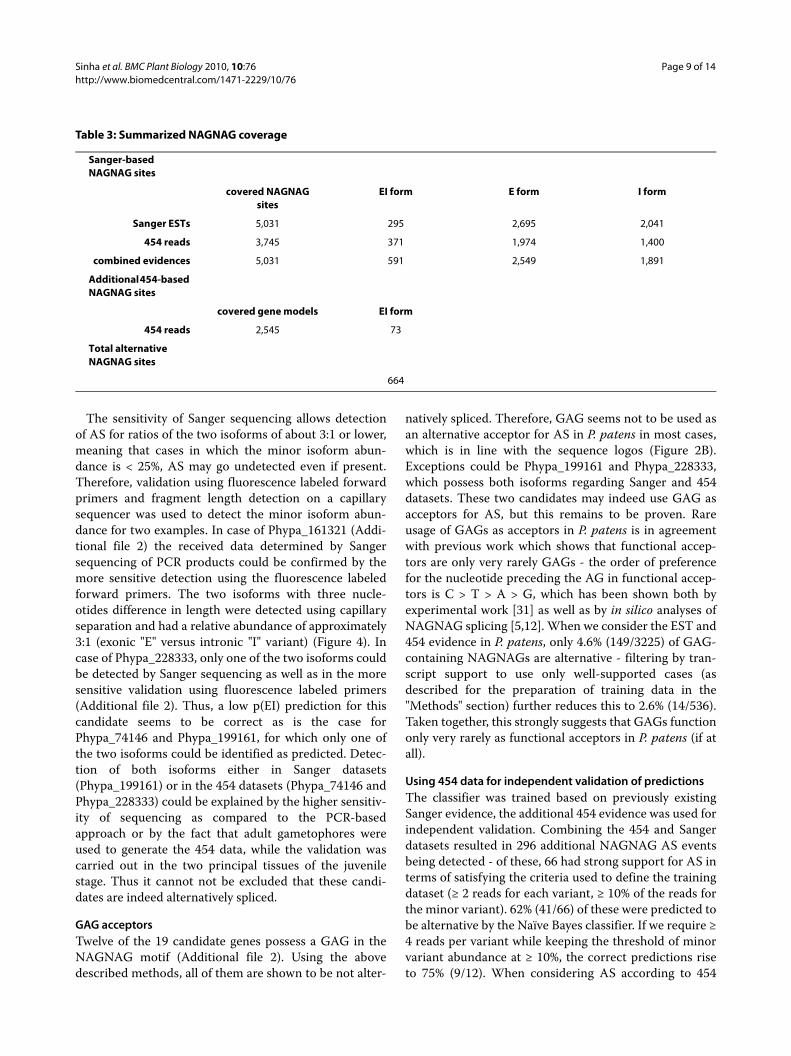
ESTs. There are 42 NAGNAGs which have support foronly one acceptor in the Sanger data, and for only theother acceptor in 454 data. Combining the results fromSanger and 454 data, P. patens has 664 alternative NAG-NAGs. Again these results show that NAGNAG AS is aswidespread in the moss P. patens as it is in the seed plantsA. thaliana and O. sativa. An overview of the NAGNAGsites covered by the different transcript evidence pools ispresented in Table 3.
Experimental confirmation of the NAGNAG ASExperiments were performed on 19 candidate NAG-NAGs, 14 as controls (seven with AS according to tran-script data, and seven without AS) to see whether thesplicing outcomes according to Sanger and 454 readscould be confirmed by a PCR based approach, and five onthe basis of an orthologous alternative NAGNAG intronin A. thaliana (see above). Of the seven candidates withsupport for AS from Sanger or 454 datasets, three werepredicted to be alternative spliced with p(EI) values > 0.9(Additional file 2). Using Sanger sequencing of cDNA
based PCR products, all three candidates were indeedverified as being alternatively spliced in P. patens pro-tonema and gametophore tissue, respectively. Eight can-didate genes were used as potential negative controls, astheir p(EI) predictions were 0.365 and lower. All candi-dates showed support for the single predicted isoform bymeans of available transcript evidence and consequentlyonly this single isoform could be detected during experi-mental validation (Additional file 2). Having support forboth variants from either the Sanger or the 454 datasets,but a p(EI) < 0.9, four more candidates were chosen to bevalidated. NAGNAG AS could be confirmed for the geneproduct Phypa_161321 by Sanger sequencing of cDNAPCR products, although it has a low p(EI) of 0.181 (Addi-tional file 2). The experimental validation is supported bythe Sanger dataset, where 13 "E" variants as well as 27 "I"variants could be identified. This is the only case whereprediction from the Naïve Bayes Classifier does not agreewith the experimental results. In case of Phypa_74146and Phypa_199161, only one of the two isoforms could bedetected, reflecting the low p(EI) values.
Figure 3 ROC plot. Depicting the in silico performance on the 3-class [I-class (red), E-class (green), and EI-class (blue)] classification problem. The EI-class, or AS, harder to predict (AUC = 0.96) than the two constitutive variants, E and I (AUC = 0.99 for both).
Sinha et al. BMC Plant Biology 2010, 10:76http://www.biomedcentral.com/1471-2229/10/76
Page 9 of 14
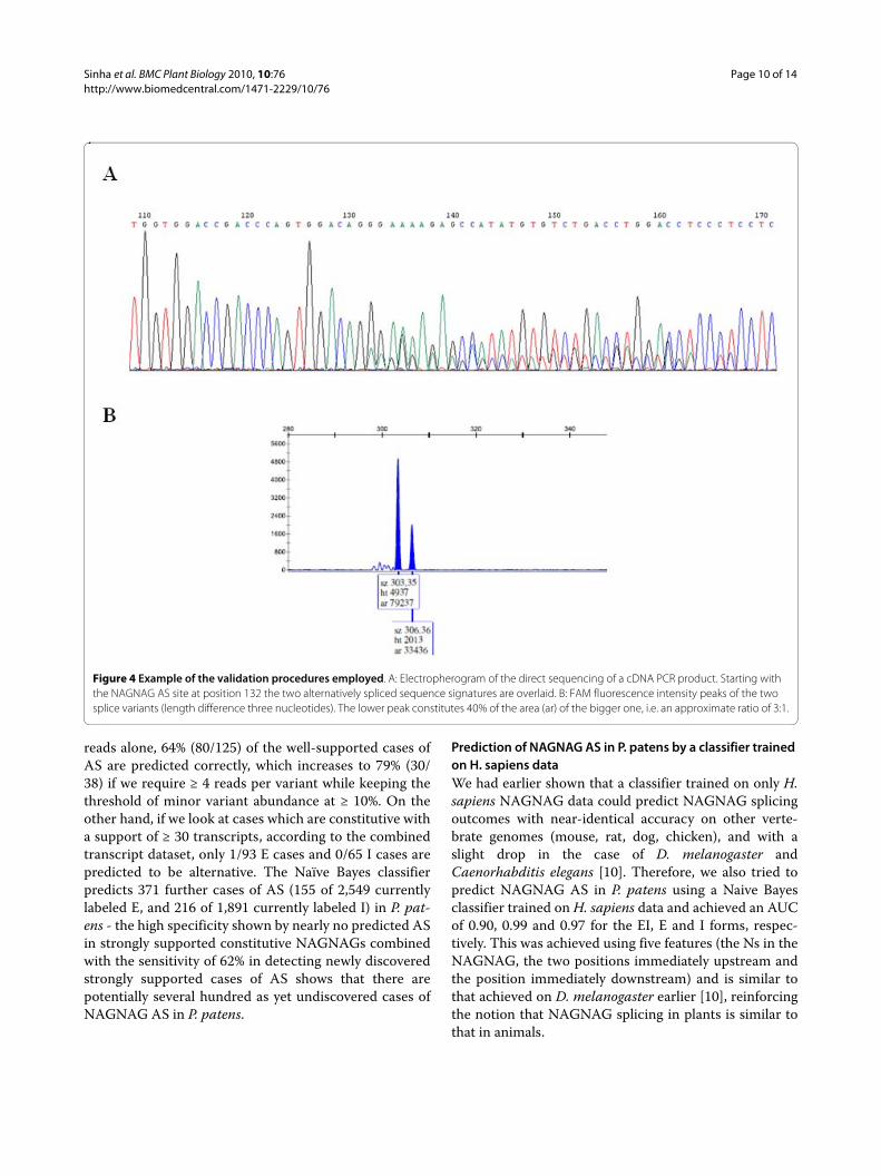
The sensitivity of Sanger sequencing allows detectionof AS for ratios of the two isoforms of about 3:1 or lower,meaning that cases in which the minor isoform abun-dance is < 25%, AS may go undetected even if present.Therefore, validation using fluorescence labeled forwardprimers and fragment length detection on a capillarysequencer was used to detect the minor isoform abun-dance for two examples. In case of Phypa_161321 (Addi-tional file 2) the received data determined by Sangersequencing of PCR products could be confirmed by themore sensitive detection using the fluorescence labeledforward primers. The two isoforms with three nucle-otides difference in length were detected using capillaryseparation and had a relative abundance of approximately3:1 (exonic "E" versus intronic "I" variant) (Figure 4). Incase of Phypa_228333, only one of the two isoforms couldbe detected by Sanger sequencing as well as in the moresensitive validation using fluorescence labeled primers(Additional file 2). Thus, a low p(EI) prediction for thiscandidate seems to be correct as is the case forPhypa_74146 and Phypa_199161, for which only one ofthe two isoforms could be identified as predicted. Detec-tion of both isoforms either in Sanger datasets(Phypa_199161) or in the 454 datasets (Phypa_74146 andPhypa_228333) could be explained by the higher sensitiv-ity of sequencing as compared to the PCR-basedapproach or by the fact that adult gametophores wereused to generate the 454 data, while the validation wascarried out in the two principal tissues of the juvenilestage. Thus it cannot not be excluded that these candi-dates are indeed alternatively spliced.
GAG acceptorsTwelve of the 19 candidate genes possess a GAG in theNAGNAG motif (Additional file 2). Using the abovedescribed methods, all of them are shown to be not alter-
natively spliced. Therefore, GAG seems not to be used asan alternative acceptor for AS in P. patens in most cases,which is in line with the sequence logos (Figure 2B).Exceptions could be Phypa_199161 and Phypa_228333,which possess both isoforms regarding Sanger and 454datasets. These two candidates may indeed use GAG asacceptors for AS, but this remains to be proven. Rareusage of GAGs as acceptors in P. patens is in agreementwith previous work which shows that functional accep-tors are only very rarely GAGs - the order of preferencefor the nucleotide preceding the AG in functional accep-tors is C > T > A > G, which has been shown both byexperimental work [31] as well as by in silico analyses ofNAGNAG splicing [5,12]. When we consider the EST and454 evidence in P. patens, only 4.6% (149/3225) of GAG-containing NAGNAGs are alternative - filtering by tran-script support to use only well-supported cases (asdescribed for the preparation of training data in the"Methods" section) further reduces this to 2.6% (14/536).Taken together, this strongly suggests that GAGs functiononly very rarely as functional acceptors in P. patens (if atall).
Using 454 data for independent validation of predictionsThe classifier was trained based on previously existingSanger evidence, the additional 454 evidence was used forindependent validation. Combining the 454 and Sangerdatasets resulted in 296 additional NAGNAG AS eventsbeing detected - of these, 66 had strong support for AS interms of satisfying the criteria used to define the trainingdataset (≥ 2 reads for each variant, ≥ 10% of the reads forthe minor variant). 62% (41/66) of these were predicted tobe alternative by the Naïve Bayes classifier. If we require ≥4 reads per variant while keeping the threshold of minorvariant abundance at ≥ 10%, the correct predictions riseto 75% (9/12). When considering AS according to 454
Table 3: Summarized NAGNAG coverage
Sanger-based NAGNAG sites
covered NAGNAG sites
EI form E form I form
Sanger ESTs 5,031 295 2,695 2,041
454 reads 3,745 371 1,974 1,400
combined evidences 5,031 591 2,549 1,891
Additional 454-based NAGNAG sites
covered gene models EI form
454 reads 2,545 73
Total alternative NAGNAG sites
664
Sinha et al. BMC Plant Biology 2010, 10:76http://www.biomedcentral.com/1471-2229/10/76
Page 10 of 14
reads alone, 64% (80/125) of the well-supported cases ofAS are predicted correctly, which increases to 79% (30/38) if we require ≥ 4 reads per variant while keeping thethreshold of minor variant abundance at ≥ 10%. On theother hand, if we look at cases which are constitutive witha support of ≥ 30 transcripts, according to the combinedtranscript dataset, only 1/93 E cases and 0/65 I cases arepredicted to be alternative. The Naïve Bayes classifierpredicts 371 further cases of AS (155 of 2,549 currentlylabeled E, and 216 of 1,891 currently labeled I) in P. pat-ens - the high specificity shown by nearly no predicted ASin strongly supported constitutive NAGNAGs combinedwith the sensitivity of 62% in detecting newly discoveredstrongly supported cases of AS shows that there arepotentially several hundred as yet undiscovered cases ofNAGNAG AS in P. patens.
Prediction of NAGNAG AS in P. patens by a classifier trained on H. sapiens dataWe had earlier shown that a classifier trained on only H.sapiens NAGNAG data could predict NAGNAG splicingoutcomes with near-identical accuracy on other verte-brate genomes (mouse, rat, dog, chicken), and with aslight drop in the case of D. melanogaster andCaenorhabditis elegans [10]. Therefore, we also tried topredict NAGNAG AS in P. patens using a Naive Bayesclassifier trained on H. sapiens data and achieved an AUCof 0.90, 0.99 and 0.97 for the EI, E and I forms, respec-tively. This was achieved using five features (the Ns in theNAGNAG, the two positions immediately upstream andthe position immediately downstream) and is similar tothat achieved on D. melanogaster earlier [10], reinforcingthe notion that NAGNAG splicing in plants is similar tothat in animals.
Figure 4 Example of the validation procedures employed. A: Electropherogram of the direct sequencing of a cDNA PCR product. Starting with the NAGNAG AS site at position 132 the two alternatively spliced sequence signatures are overlaid. B: FAM fluorescence intensity peaks of the two splice variants (length difference three nucleotides). The lower peak constitutes 40% of the area (ar) of the bigger one, i.e. an approximate ratio of 3:1.

Sinha et al. BMC Plant Biology 2010, 10:76http://www.biomedcentral.com/1471-2229/10/76
Page 11 of 14
ConclusionsHere we describe the first computational prediction ofalternative splicing (AS) in a non-seed plant and find thatNAGNAG AS in P. patens, a moss, can be predicted withhigh accuracy. Since the extent of NAGNAGs in P. patenshad not yet been reported, this work involved both char-acterization as well prediction of NAGNAG splicing in P.patens. Using ESTs, we found that NAGNAG AS is aswidespread in the bryophyte P. patens as it is in the seedplants A. thaliana and O. sativa. Thus, NAGNAG AS islikely to be a common feature of AS in all land plants, justas it is in animals. Although we detected homologs withNAGNAG events among the two land plants P. patensand A. thaliana, NAGNAG splicing seems not to be con-served at the intron level.
Using carefully constructed training and test datasets,an in silico performance of AUC = 0.96, 0.99 and 0.98 wasachieved for the EI, E and I forms, respectively. The mostinformative features (according to information gain [32])were the nucleotides in the NAGNAG and its immediatevicinity, and even a relatively simple classifier like theNaïve Bayes classifier could match the more sophisticatedBayesian network and Support vector machine. The per-formance achieved by a Naïve Bayes classifier trained onH. sapiens data (AUC = 0.90, 0.99 and 0.97 for the EI, Eand I forms, respectively) was similar to that achieved onD. melanogaster earlier [10]. This indicates that, as in ani-mals, the mechanism behind NAGNAG AS in plants issimple in nature and mostly dependent on the splice siteneighborhood. Independent validation of the predictionsof the classifier (trained on Sanger EST data alone) using454 data showed that 64% (80/125) of the well-supportedcases of NAGNAG AS could be predicted correctly.
In total, seven candidates were chosen for independentexperimental confirmation of the Sanger and 454 evi-dence of NAGNAG splicing. The experimental confirma-tion depends on detection of isoforms using sequenceelectropherograms and is less sensitive than size poly-morphism detection using fluorescence-labeled primers.The latter method was used on two of the seven examplesand confirmed the results of the previous method. Whilethere is transcript support for alternative use of GAGacceptors this could not be proven in our experimentalvalidation. In addition, a further 12 experiments wereperformed - six as negative controls, all of which agreedwith the predictions, and five to check for possible con-served NAGNAG AS with A. thaliana, which could notbe detected.
When additional 454 transcript evidence was used tosupplement the Sanger EST data, a total of 664 alternativeNAGNAGs were found in P. patens. Since the averagecoverage per constitutive NAGNAG was still onlyapproximately ten ESTs, this number shall likely continueto rise with deeper coverage of the transcriptome. Never-
theless, the results provide the first evidence that NAG-NAG AS is widespread in P. patens. Our findings are inagreement with a recent study which showed that NAG-NAG AS shares common properties in A. thaliana and O.sativa and animals [24]. This indicates that the mecha-nism behind NAGNAG AS in land plants is similar tothat in animals. The pervasiveness of NAGNAG AS sug-gests that it may be a general feature of splicing in animalsand plants, and possibly in all eukaryotes.
MethodsIdentification of alternative splicing at NAGNAG acceptors using ESTs346,871 P. patens Sanger EST reads (available at http://www.cosmoss.org) from various developmental stagesand tissue types (predominantly protonema and juvenilegametophores) were aligned using GenomeThreader[33]. EST alignments (max. intron length 20,000) withless than 95% identity and 90% EST length coverage wereexcluded from further analyses to obtain only reliablealternative acceptors. In addition, EST alignments match-ing a single exon as well as alignments ending at an exonboundary supporting either the E or I site were discarded.The sequence regions used for feature extraction (Figure5) and EST evidence counts were created using the BioP-erl [34] module Bio::DB::SeqFeature::Store.
Sequence logosSequence logos were created using the WebLogo soft-ware http://weblogo.berkeley.edu/logo.cgi[35] with thesequence regions shown in Figure 5.
Feature design and extraction; classifiersFeature extraction was done based on annotated datausing a Perl script (Additional file 3; see Additional file 4for example input data. The script produces outputwhich, together with Additional file 5, can be used withstandard classifiers). The region used for analysis can beseen in Figure 5. Since the composition of the splice siteneighborhood influences splicing in general, the basepairs at positions -20 to +3 with respect to the NAGNAGwere each used as a single feature, as were the two Ns inthe NAGNAG motif. The last three positions of the
Figure 5 Nomenclature of features used in this study. Nomencla-ture of sequence features used to analyze NAGNAG splicing. The re-gion used to derive all 31 features is shown, along with the names given to the positional features.
Sinha et al. BMC Plant Biology 2010, 10:76http://www.biomedcentral.com/1471-2229/10/76
Page 12 of 14
upstream exon were also included, since they can influ-ence both the process of splicing, as well as reflect influ-ence of codon usage near the exon boundary. Thus, wehad a total of 28 features which each represented a nucle-otide, and thus had four possible values (A, C, G, T). Aweak polypyrimidine tract (PPT) can contribute to AS,and the number of pyrimidines in the 3' region of theintron is a measure of PPT strength. Therefore, wedesigned a feature called "Y-content", which refers to thenumber of pyrimidines in the 20 bp upstream of theNAGNAG. Splice site strength, being one of the mostimportant determinants of splicing outcome, was alsoincluded as a feature - the strength of the two possiblesplice sites for each NAGNAG exon, as computed usingSpliceMachine [36], contributed two more features. Intotal, 31 features were used. We used the WEKA packageand Bayesian Networks, Naive Bayes classifiers, and Sup-port vector machines [32]. For feature selection withinWEKA, we used the method "CfsSubsetEval". In addition,we also used manual inclusion and exclusion of features.
Information gainInformation gain is defined as the reduction in theentropy of the class variable, given the feature [32]. Theformula for information gain is:
where H(Class) is the entropy of the class variable, andH(Class|Feature) is the conditional entropy of the classvariable, given the feature. Information gain is a wellestablished measure for feature selection in MachineLearning. We used the WEKA package for computinginformation gain, in order to rank the features accordingto how informative they were. We also used it for predic-tion based on SVMs, as implemented in the SMO option,and for prediction using Naïve Bayes classifiers.
Functional annotation and GO enrichment analysisFor every (potential) NAGNAG splicing region an over-lapping P. patens gene model was assigned using the startand stop coordinates on the genomic scaffolds. The cor-responding predicted protein sequences were subjectedto BLAST2GO [37] GO term annotation which wasextended by various subcellular target prediction andhomology-based methods (see http://www.cosmoss.org/annotation/references?cosmoss_ref=1 for details). Theresulting GO annotation was mapped to GO slim termsusing the Blast2GO internal mapping function using the"goslim_plant.obo" ontology subset. GO enrichmentanalysis was performed against the complete P. patenswith the BLAST2GO internal Fisher's exact test/GOSSIP[38] using the two-tailed test, with false discovery rate(FDR) correction and a q-value cut-off < 0.05. The A.
thaliana alternative NAGNAG splicing gene set was con-structed using the alternative NAGNAG acceptor casesidentified within the A. thaliana genome from [26] and[24]. The resulting alternative NAGNAG acceptor setcontains 290 A. thaliana proteins. These proteins weresubjected to a GO enrichment analysis as describedabove for P. patens. The A. thaliana GOA was down-loaded from ftp://ftp.arabidopsis.org/home/tair/Ontolo-gies/Gene_Ontology/ATH_GO_GOSLIM.txt(17.11.2009) and mapped to GO slim (goslim_plant.obo)with BLAST2GO.
Candidate selection for evolutionary conserved NAGNAG acceptorsP. patens cosmoss v1.2 and A. thaliana TAIR 8 proteinswere subjected to a BLAST based single linkage cluster-ing using BLASTCLUST [39]. The parameters were set to70% length coverage and 70% alignment identity toobtain only highly conserved homologs. In total 1,088clusters with at least one P. patens, respectively A. thali-ana, protein were found. Five candidates out of seven P.patens genes, each sharing a cluster with A. thalianaalternative NAGNAG acceptor containing genes [24,26],were selected for experimental validation. In addition,these P. patens candidate genes contain a potential NAG-NAG acceptor in the same intron as the corresponding A.thaliana homolog.
Experimental confirmation of splice variantsP. patens total RNA was isolated from protonema andgametophore tissue using the RNeasy Plant Mini Kit(Qiagen, Hilden, Germany). cDNA synthesis was carriedout with 250 ng total RNA using Superscript III ReverseTranscriptase (Invitrogen, Karlsruhe, Germany) accord-ing to the manufacturers' instructions. For validation ofdifferent splice variants, PCR was performed from pro-tonema and gametophore RNA, respectively, using nativePfu-Polymerase (Fermentas, St. Leon-Rot, Germany).PCR primers were obtained from Sigma (München, Ger-many). PCR reactions were carried out using 12 ng cDNAas template. Products were extracted using the QIAquickPCR purification Kit (Qiagen, Hilden, Germany) anddirectly sequenced (GATC, Konstanz, Germany).Sequences and chromatograms were analysed with Chro-masPro Version 1.34. Alternatively, PCR products ampli-fied with carboxyfluorescein (FAM) labeled forwardprimers were analysed by capillary electrophoresis, whereAS was detected as a size difference of three nucleotidesin length. PCR products were diluted as appropriate andsubjected to capillary electrophoresis for separation anddetection. For this purpose, 10 μL HiDi formamide(Applied Biosystems) and 0.5 μL HD400 GS internal sizestandard were added to each well, and the plate wasmounted on a 3100 Genetic Analyzer with Foundation
IG Class Feature H Class H Class Feature( | ) ( ) ( | )= −

Sinha et al. BMC Plant Biology 2010, 10:76http://www.biomedcentral.com/1471-2229/10/76
Page 13 of 14
Data Collection software v. 2.0 and Gene Mapper ID soft-ware v. 3.2 (Applied Biosystems, Darmstadt, Germany).
Tissue culture and generation of additional transcript evidencePhyscomitrella patens strain Gransden 2004 [23] was cul-tivated on solidified (1% w/v agar) mineral medium [250mg L-1 KH2PO4, 250 mg L-1 MgSO4 × 7-H2O, 250 mg L-1
KCl, 1000 mg L-1 Ca(NO3)2 × 4H2O, 12.5 mg L-1 FeSO4 ×7H2O, pH 5.8 with KOH] on 9 cm petri dishes enclosedby laboratory film in a Percival cultivation chamber (CLF,Germany) at 22°C with a 16 h light, 8 h dark regime under70 μmol*s-1*m-2 white light (long day conditions). Game-tophore colonies were grown from single gametophorestransferred to the dishes from precultured colonies.Induction of gametangia was performed by placing thedishes under inductive conditions [40], i.e. 20 μmol *s-
1*m-2 white light and 15°C with a 8 h light, 16 h darkregime until development of gametangia. After harvest-ing and freezing, the material was ground under liquidnitrogen and total RNA isolated using the Ambion mir-Vana miRNA isolation kit (Applied Biosystems, Darm-stadt, Germany). RNA isolation and subsequentsequencing pool creation steps were carried out by VertisBiotechnologie (Freising, Germany). Poly(A)+ RNA wasprepared by oligo(dT) chromatography and cDNA wassynthesized using a N6 randomized primer. Afterwards,454 adapters A (CCATCTCATCCCTGCGTGTCTC-CGACTCAG) and B (CTGAGACTGCCAAGGCACA-CAGGGGATAGG) were ligated to the 5' and 3' ends ofthe cDNA. The resulting N0 cDNA was amplified usingPCR (16 cycles) with a proof reading enzyme. Normaliza-tion was carried out by one cycle of denaturation andreassociation of the cDNA, resulting in N1-cDNA. Reas-sociated ds-cDNA was separated from the remaining ss-cDNA (normalized cDNA) by passing the mixture over ahydroxylapatite column. After hydroxylapatite chroma-tography, the ss-cDNA was amplified with 9 PCR cycles.Finally, the cDNA in the size range of 500-700 bp waseluted from a preparative agarose gel and subjected to GSFLX Titanium sequencing (GATC, Konstanz, Germany),resulting in 631,313 raw reads. After low quality andadapter clipping using LUCY [41] and SeqClean http://compbio.dfci.harvard.edu/tgi/software/, and polyA-tailremoval with trimmest [42], 589,283 reads with a meanlength of 343 nucleotides remained. The 454 reads (Addi-tional file 1) were mapped against the genome asdescribed above for the P. patens Sanger ESTs and areavailable at http://www.cosmoss.org for download in agenome browser track "454 reads sexual gametophores(normalized library)" http://www.cosmoss.org/cgi/gbrowse/physcome/
Additional material
Authors' contributionsRS analyzed the transcript evidences for NAGNAGs, designed and extractedfeatures, used WEKA to perform the classification and analyses, computed thegenome-wide prediction, and drafted the manuscript. ADZ obtained the tran-script evidences for all genomic NAGNAGs in P. patens, helped in featureextraction, performed the GO-analysis, and contributed to writing the manu-script. KB performed the experimental validations and contributed to writingthe manuscript. DL participated in the transcript evidence analyses, andhelped with the GO-analysis. RR and MP supervised part of the work. RB andSAR contributed to writing the manuscript, conceived of and supervised thestudy. All authors have read and approved the final manuscript.
AcknowledgementsThis work was supported by the German Research Foundation (DFG grant Re 837/10-2 to R.R. and S.A.R.), by the Federal Ministry of Education and Research (BMBF grant FRISYS 0313921 to R.B., R.R. and S.A.R.) and by the Excellence Initia-tive of the German Federal and State Governments (EXC 294 to R.B., R.R. and S.A.R.). We are grateful to S. Richardt and E. Heupel for skillful technical assis-tance and to M. Heinrich for analysing the FAM-labeled PCR products.
Author Details1Bioinformatics group, University of Freiburg, Georges-Koehler-Allee 106, 79110 Freiburg, Germany, 2Faculty of Biology, University of Freiburg, Hauptstrasse 1, 79104 Freiburg, Germany, 3Plant Biotechnology, Faculty of Biology, University of Freiburg, Schaenzlestrasse 1, 79104 Freiburg, Germany, 4Freiburg Initiative for Systems Biology (FRISYS), University of Freiburg, Schaenzlestrasse 1, 79104 Freiburg, Germany, 5Centre for Biological Signalling Studies (bioss), University of Freiburg, Albertstr. 19, 79104 Freiburg, Germany, 6Genome Analysis, Leibniz Institute for Age Research - Fritz Lipmann Institute, Beutenbergstr. 11, 07745 Jena, Germany and 7Philipps-Universität Marburg, Laboratorium für Zellbiologie, Karl-von-Frisch Str., 35032 Marburg, Germany
References1. Graveley BR: Alternative splicing: increasing diversity in the proteomic
world. Trends in Genetics 2001, 17(2):100-107.2. Hughes TA: Regulation of gene expression by alternative untranslated
regions. Trends in Genetics 2006, 22(3):119-122.3. Stalder L, Mühlemann O: The meaning of nonsense. Trends in Cell Biology
2008, 18(7):315-321.4. Wang ET, Sandberg R, Luo S, Khrebtukova I, Zhang L, Mayr C, Kingsmore
SF, Schroth GP, Burge CB: Alternative isoform regulation in human tissue transcriptomes. Nature 2008, 456(7221):470-476.
5. Hiller M, Huse K, Szafranski K, Jahn N, Hampe J, Schreiber S, Backofen R, Platzer M: Widespread occurrence of alternative splicing at NAGNAG acceptors contributes to proteome plasticity. Nat Genet 2004, 36(12):1255-1257.
6. Zavolan M, Kondo S, Schonbach C, Adachi J, Hume DA, Group RG, Members GSL, Hayashizaki Y, Gaasterland T: Impact of Alternative Initiation, Splicing, and Termination on the Diversity of the mRNA Transcripts Encoded by the Mouse Transcriptome. Genome Res 2003, 13(6b):1290-1300.
Additional file 1 All NAGNAGs in Physcomitrella patens. Information on all 5,031 NAGNAGs in Physcomitrella patens, including gene name, EST name, genomic location, transcript support, and the predictions of the Naïve Bayes classifier.Additional file 2 Summarized experimental results.Additional file 3 Perl script. The Perl script used for feature extraction.Additional file 4 Transcript support data. The transcript support data derived from the http://www.cosmoss.org database.Additional file 5 Names file. Names file for combination with the script output.
Received: 15 December 2009 Accepted: 28 April 2010 Published: 28 April 2010This article is available from: http://www.biomedcentral.com/1471-2229/10/76© 2010 Sinha et al; licensee BioMed Central Ltd. This is an Open Access article distributed under the terms of the Creative Commons Attribution License (http://creativecommons.org/licenses/by/2.0), which permits unrestricted use, distribution, and reproduction in any medium, provided the original work is properly cited.BMC Plant Biology 2010, 10:76
Sinha et al. BMC Plant Biology 2010, 10:76http://www.biomedcentral.com/1471-2229/10/76
Page 14 of 14
7. Dou Y, Fox-Walsh KL, Baldi PF, Hertel KJ: Genomic splice-site analysis reveals frequent alternative splicing close to the dominant splice site. RNA 2006, 12(12):2047-2056.
8. Ermakova EO, Nurtdinov RN, Gelfand MS: Overlapping alternative donor splice sites in the human genome. Journal of Bioinformatics and Computational Biology 2007:991-1004.
9. Sugnet CW, Kent WJ, Jr AM, Haussler D: Transcriptome and Genome Conservation of Alternative Splicing Events in Humans and Mice. Pacific Symposium on Biocomputing 2004, 9:66-77.
10. Sinha R, Nikolajewa S, Szafranski K, Hiller M, Jahn N, Huse K, Platzer M, Backofen R: Accurate prediction of NAGNAG alternative splicing. Nucl Acids Res 2009, 37(11):3569-3579.
11. Chern T-M, van Nimwegen E, Kai C, Kawai J, Carninci P, Hayashizaki Y, Zavolan M: A Simple Physical Model Predicts Small Exon Length Variations. PLoS Genetics 2006, 2(4):e45.
12. Akerman M, Mandel-Gutfreund Y: Alternative splicing regulation at tandem 3' splice sites. Nucl Acids Res 2006, 34(1):23-31.
13. Barbazuk WB, Fu Y, McGinnis KM: Genome-wide analyses of alternative splicing in plants: Opportunities and challenges. Genome Research 2008, 18(9):1381-1392.
14. Wang B-B, Brendel V: Genomewide comparative analysis of alternative splicing in plants. PNAS 2006, 103(18):7175-7180.
15. Wang B-B, O'Toole M, Brendel V, Young N: Cross-species EST alignments reveal novel and conserved alternative splicing events in legumes. BMC Plant Biology 2008, 8(1):17.
16. Campbell M, Haas B, Hamilton J, Mount S, Buell CR: Comprehensive analysis of alternative splicing in rice and comparative analyses with Arabidopsis. BMC Genomics 2006, 7(1):327.
17. Ner-Gaon H, Leviatan N, Rubin E, Fluhr R: Comparative Cross-Species Alternative Splicing in Plants. Plant Physiol 2007, 144(3):1632-1641.
18. Filichkin SA, Priest HD, Givan SA, Shen R, Bryant DW, Fox SE, Wong W-K, Mockler TC: Genome-wide mapping of alternative splicing in Arabidopsis thaliana. Genome Research 2009, 20:45-58.
19. Kim E, Magen A, Ast G: Different levels of alternative splicing among eukaryotes. Nucl Acids Res 2007, 35(1):125-131.
20. Berget SM: Exon recognition in vertebrate splicing. J Biol Chem 1995, 270:2411-2414.
21. Lorkovic ZJ, Kirk DAW, Lambermon MHL, Filipowicz W: Pre-mRNA splicing in higher plants. Trends in Plant Science 2000, 5(4):160-167.
22. Lim LP, Burge CB: A computational analysis of sequence features involved in recognition of short introns. Proceedings of the National Academy of Sciences of the United States of America 2001, 98(20):11193-11198.
23. Rensing SA, Lang D, Zimmer AD, Terry A, Salamov A, Shapiro H, Nishiyama T, Perroud P-F, Lindquist EA, Kamisugi Y, et al.: The Physcomitrella Genome Reveals Evolutionary Insights into the Conquest of Land by Plants. Science 2008, 319(5859):64-69.
24. Iida K, Shionyu M, Suso Y: Alternative Splicing at NAGNAG Acceptor Sites Shares Common Properties in Land Plants and Mammals. Mol Biol Evol 2008, 25(4):709-718.
25. Bluthgen N, Brand K, Cajavec B, Swat M, Herzel H, Beule D: Biological profiling of gene groups utilizing Gene Ontology. Genome Inform 2005, 16(1):106-115.
26. Schindler S, Szafranski K, Hiller M, Ali G, Palusa S, Backofen R, Platzer M, Reddy A: Alternative splicing at NAGNAG acceptors in Arabidopsis thaliana SR and SR-related protein-coding genes. BMC Genomics 2008, 9(1):159.
27. Hiller M, Szafranski K, Sinha R, Huse K, Nikolajewa S, Rosenstiel P, Schreiber S, Backofen R, Platzer M: Assessing the fraction of short-distance tandem splice sites under purifying selection. Rna 2008, 14(4):616-629.
28. Lang D, Zimmer AD, Rensing SA, Reski R: Exploring plant biodiversity: the Physcomitrella genome and beyond. Trends in Plant Science 2008, 13(10):542-549.
29. Ling C, Huang J, Zhang H: AUC: a better measure than accuracy in comparing learning algorithms. Canadian Artificial Intelligence Conference 2003 2003:329-341.
30. Reski R: Development, genetics and molecular biology of mosses. Botanica Acta 1998, 111:1-15.
31. Hollins C, Zorio DAR, Macmorris M, Blumenthal T: U2AF binding selects for the high conservation of the C. elegans 3' splice site. RNA 2005, 11(3):248-253.
32. Witten IH, Frank E: Data Mining: Practical machine learning tools and techniques. Second edition. Morgan Kaufmann, San Francisco; 2005.
33. Gremme G, Brendel V, Sparks ME, Kurtz S: Engineering a software tool for gene structure prediction in higher organisms. Information and Software Technology 2005, 47(15):965-978.
34. Stajich JE, Block D, Boulez K, Brenner SE, Chervitz SA, Dagdigian C, Fuellen G, Gilbert JGR, Korf I, Lapp H, et al.: The bioperl toolkit: Perl modules for the life sciences. Genome Research 2002, 12(10):1611-1618.
35. Crooks GE, Hon G, Chandonia J-M, Brenner SE: WebLogo: A Sequence Logo Generator. Genome Res 2004, 14(6):1188-1190.
36. Degroeve S, Saeys Y, De Baets B, Rouze P, Peer Y Van de: SpliceMachine: predicting splice sites from high-dimensional local context representations. Bioinformatics 2005, 21(8):1332-1338.
37. Conesa A, Gotz S, Garcia-Gomez JM, Terol J, Talon M, Robles M: Blast2GO: a universal tool for annotation, visualization and analysis in functional genomics research. Bioinformatics 2005, 21(18):3674-3676.
38. Bluethgen N, Brand K, Cajavec B, Swat M, Herzel H, Beule D: Biological profiling of gene groups utilizing Gene Ontology. Genome Inform 2005, 16(1):106-115.
39. Altschul SF, Gish W, Miller W, Myers EW, Lipman DJ: Basic Local Alignment Search Tool. Journal of Molecular Biology 1990, 215(3):403-410.
40. Hohe A, Rensing SA, Mildner M, Lang D, Reski R: Day length and temperature strongly influence sexual reproduction and expression of a novel MADS-box gene in the moss Physcomitrella patens. Plant Biology 2002, 4(5):595-602.
41. Chou H-H, Holmes MH: DNA sequence quality trimming and vector removal. Bioinformatics 2001, 17(12):1093-1104.
42. Rice P, Longden I, Bleasby A: EMBOSS: The European Molecular Biology Open Software Suite. Trends in Genetics 2000, 16(6):276-277.
doi: 10.1186/1471-2229-10-76Cite this article as: Sinha et al., Identification and characterization of NAG-NAG alternative splicing in the moss Physcomitrella patens BMC Plant Biology 2010, 10:76