identification and qualitative analysis of renal...
TRANSCRIPT
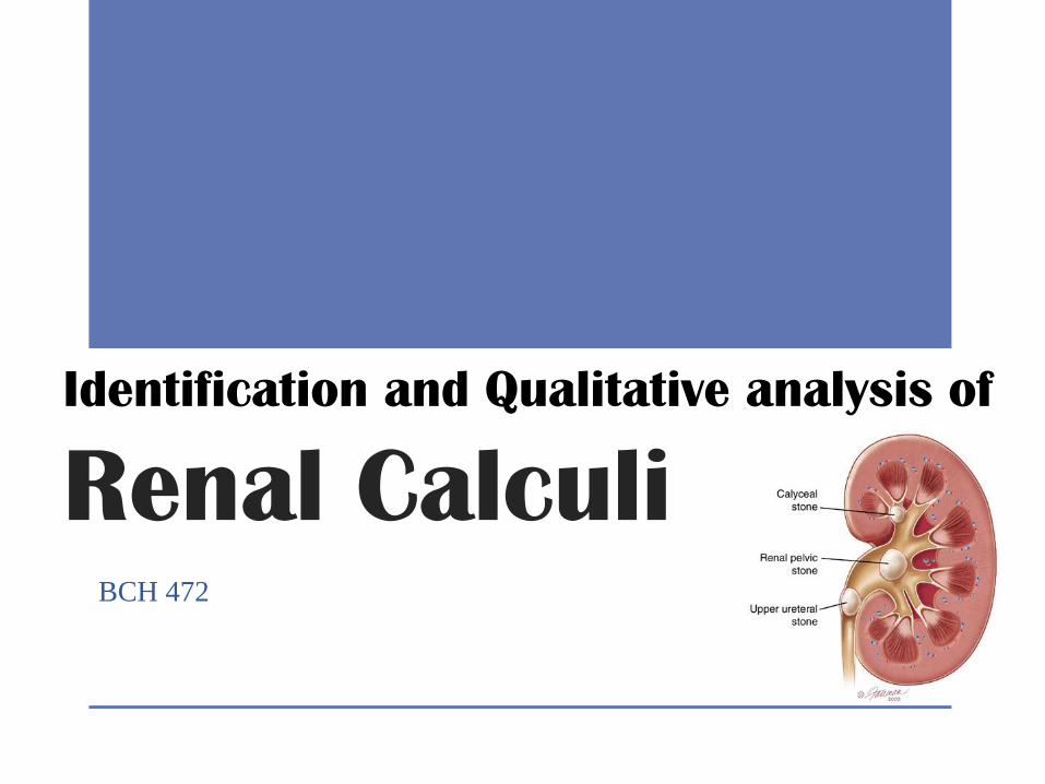
Identification and Qualitative analysis of
Renal Calculi BCH 472
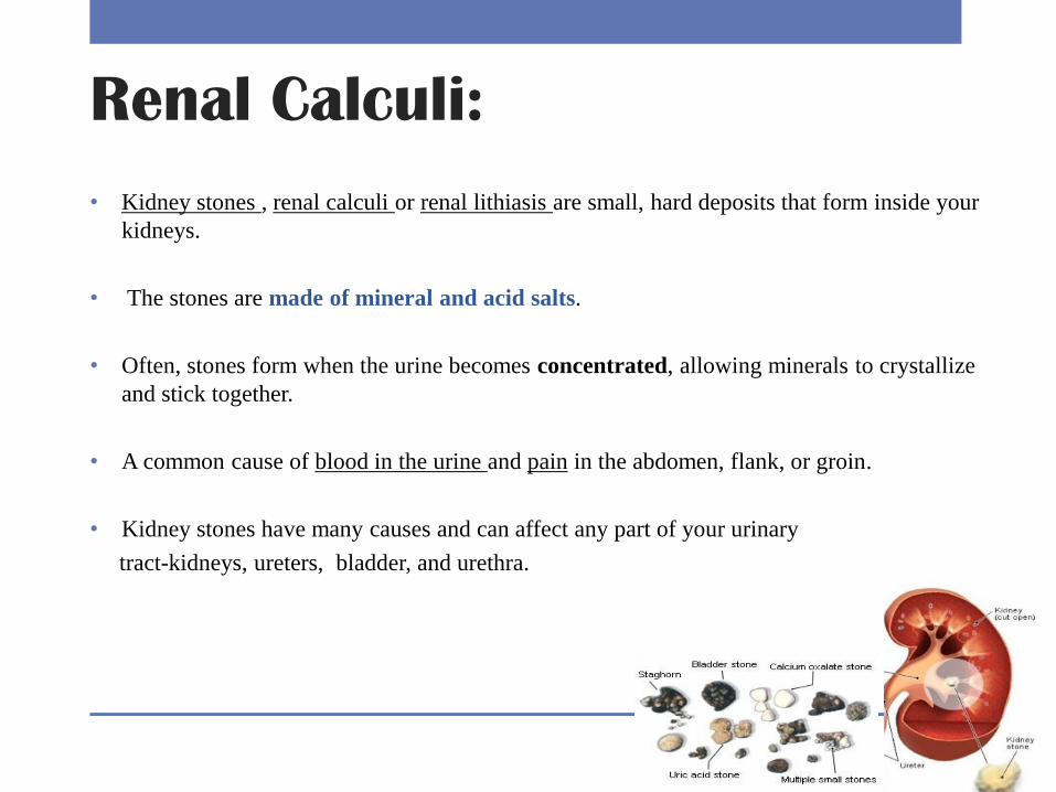
Renal Calculi:
• Kidney stones , renal calculi or renal lithiasis are small, hard deposits that form inside your
kidneys.
• The stones are made of mineral and acid salts.
• Often, stones form when the urine becomes concentrated, allowing minerals to crystallize
and stick together.
• A common cause of blood in the urine and pain in the abdomen, flank, or groin.
• Kidney stones have many causes and can affect any part of your urinary
tract-kidneys, ureters, bladder, and urethra.
Pathogenesis of renal stones :
• There are two basic aspects in the pathogenesis of renal stones:
• Increased urinary excretion of stone forming elements like calcium, phosphorus,
uric acid, oxalate, and cystine
• Low fluid intake (decreased urine volume). A low fluid intake results in the
production of concentrated urine, causing supersaturation and crystallisation of
stone-forming compounds.
• In addition, low urine flow rates favor crystal deposition on the urothelium.
• Physio-chemical changes which influence stone formation like: pH of urine, stone
matrix, and protective substances in the urine.
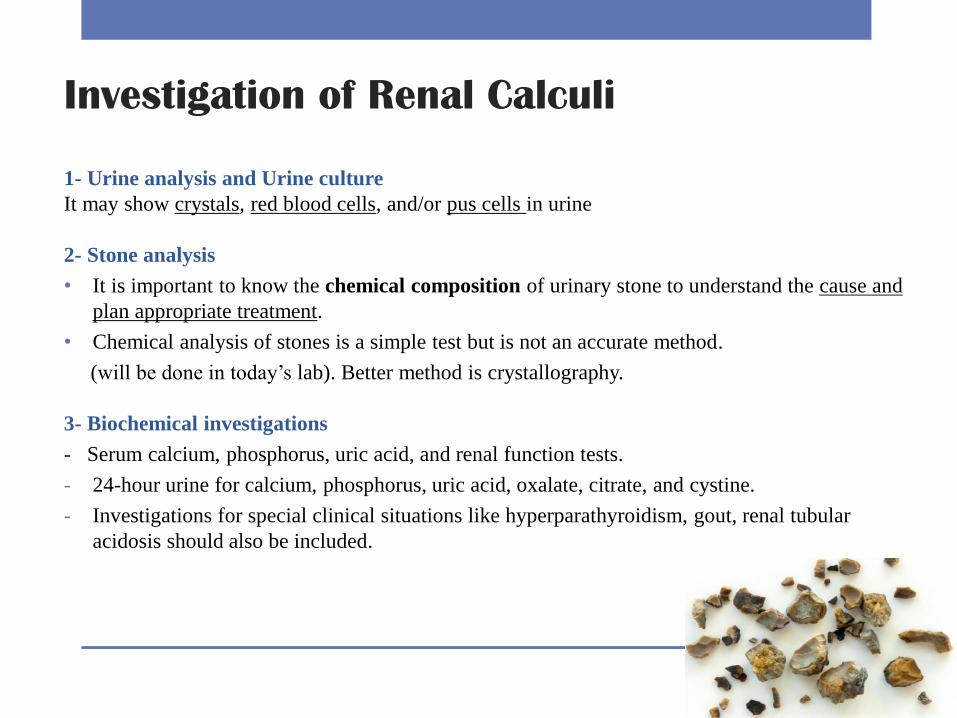
Investigation of Renal Calculi
1- Urine analysis and Urine culture
It may show crystals, red blood cells, and/or pus cells in urine
2- Stone analysis
• It is important to know the chemical composition of urinary stone to understand the cause and
plan appropriate treatment.
• Chemical analysis of stones is a simple test but is not an accurate method.
(will be done in today’s lab). Better method is crystallography.
3- Biochemical investigations
- Serum calcium, phosphorus, uric acid, and renal function tests.
- 24-hour urine for calcium, phosphorus, uric acid, oxalate, citrate, and cystine.
- Investigations for special clinical situations like hyperparathyroidism, gout, renal tubular
acidosis should also be included.
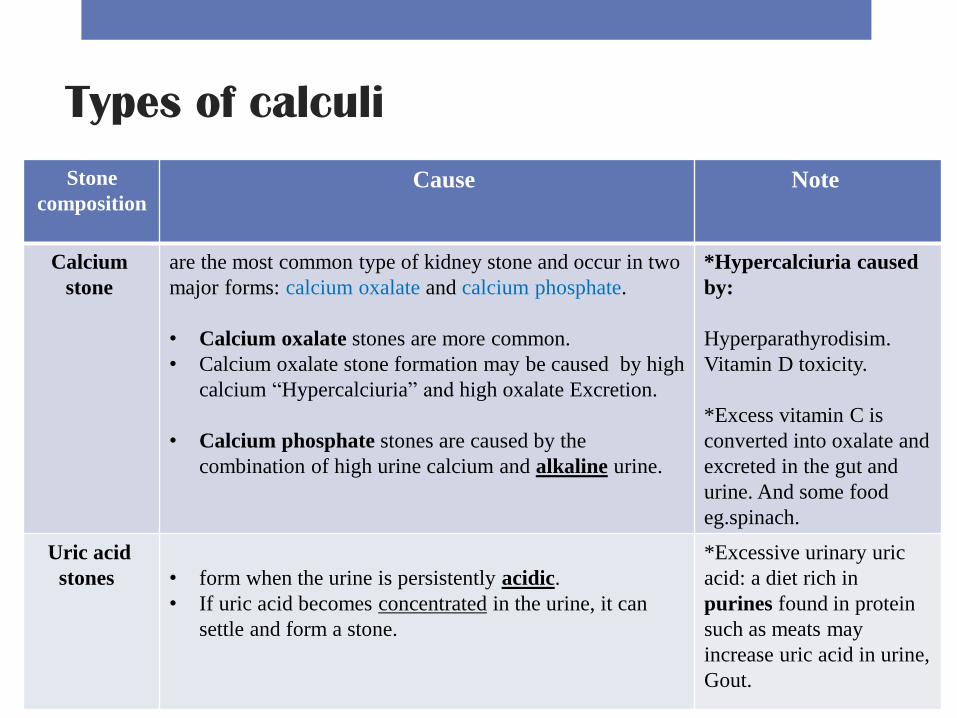
Types of calculi
Stone
composition Cause
Note
Calcium
stone
are the most common type of kidney stone and occur in two
major forms: calcium oxalate and calcium phosphate.
• Calcium oxalate stones are more common.
• Calcium oxalate stone formation may be caused by high
calcium “Hypercalciuria” and high oxalate Excretion.
• Calcium phosphate stones are caused by the
combination of high urine calcium and alkaline urine.
*Hypercalciuria caused
by:
Hyperparathyrodisim.
Vitamin D toxicity.
*Excess vitamin C is
converted into oxalate and
excreted in the gut and
urine. And some food
eg.spinach.
Uric acid
stones
• form when the urine is persistently acidic.
• If uric acid becomes concentrated in the urine, it can
settle and form a stone.
*Excessive urinary uric
acid: a diet rich in
purines found in protein
such as meats may
increase uric acid in urine,
Gout.
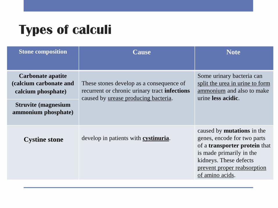
Types of calculi
Stone composition Cause
Note
Carbonate apatite
(calcium carbonate and
calcium phosphate)
These stones develop as a consequence of
recurrent or chronic urinary tract infections
caused by urease producing bacteria.
Some urinary bacteria can
split the urea in urine to form
ammonium and also to make
urine less acidic.
Struvite (magnesium
ammonium phosphate)
Cystine stone
develop in patients with cystinuria.
caused by mutations in the
genes, encode for two parts
of a transporter protein that
is made primarily in the
kidneys. These defects
prevent proper reabsorption
of amino acids.
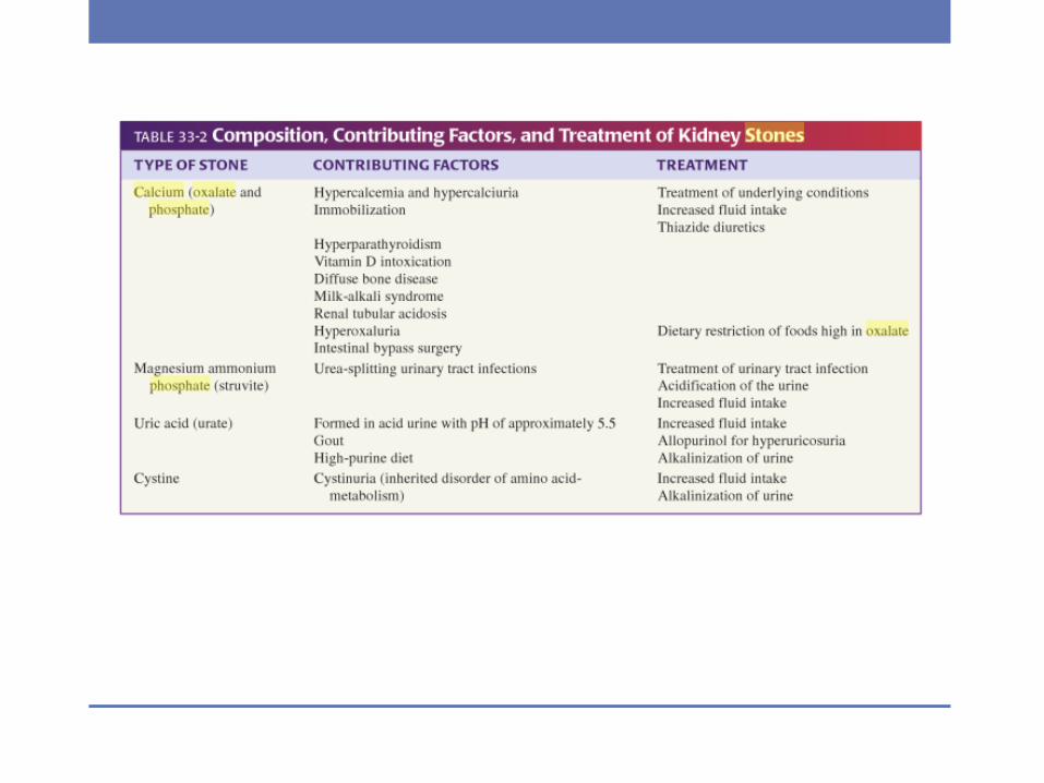
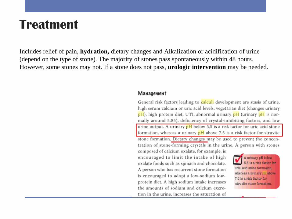
Treatment
Includes relief of pain, hydration, dietary changes and Alkalization or acidification of urine
(depend on the type of stone). The majority of stones pass spontaneously within 48 hours.
However, some stones may not. If a stone does not pass, urologic intervention may be needed.
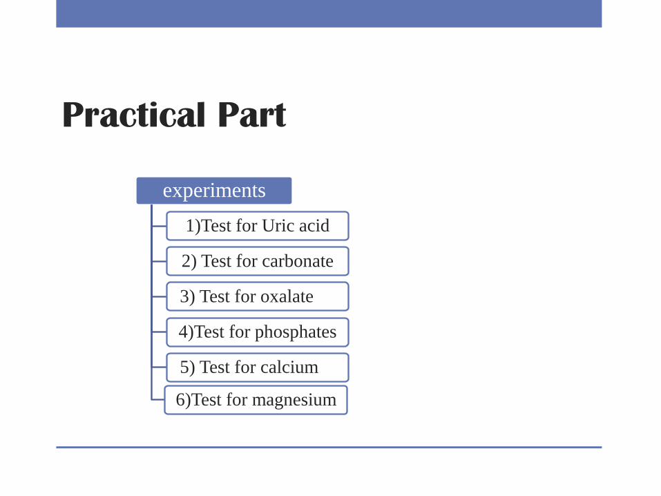
Practical Part
experiments
1)Test for Uric acid
2) Test for carbonate
3) Test for oxalate
4)Test for phosphates
5) Test for calcium
6)Test for magnesium
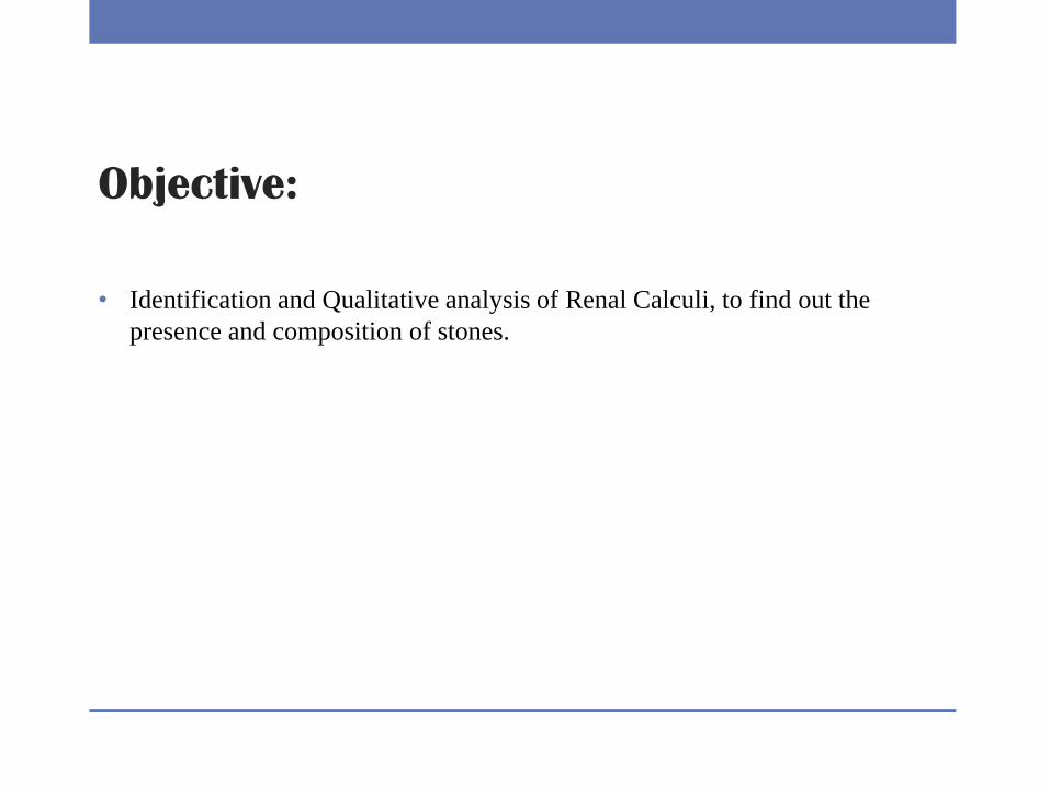
Objective:
• Identification and Qualitative analysis of Renal Calculi, to find out the
presence and composition of stones.

1)Test for Uric acid Principle: Uric acid undergoes oxidation when treated with HNO3.
Method:
1-Put a small amount of the sample1.
2-Add 5-7 drops of concentrated nitric acid.
3-Heat in a water bath.
yellow to orange color on the inner surface of the test tube.
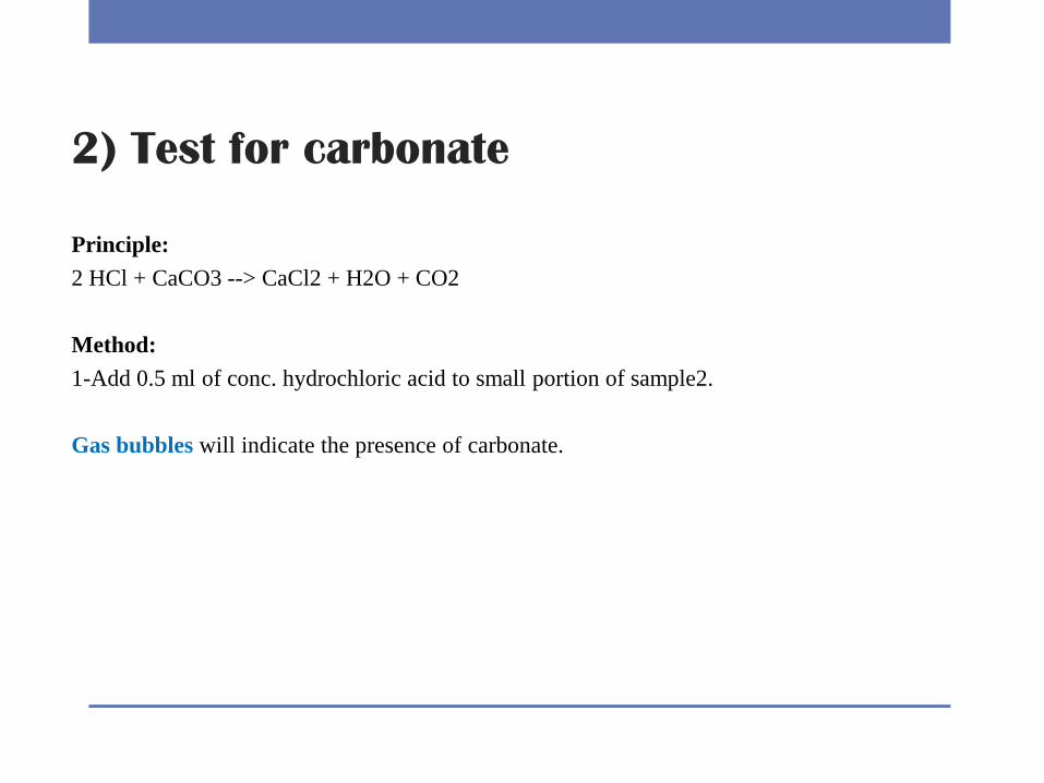
2) Test for carbonate
Principle:
2 HCl + CaCO3 --> CaCl2 + H2O + CO2
Method:
1-Add 0.5 ml of conc. hydrochloric acid to small portion of sample2.
Gas bubbles will indicate the presence of carbonate.
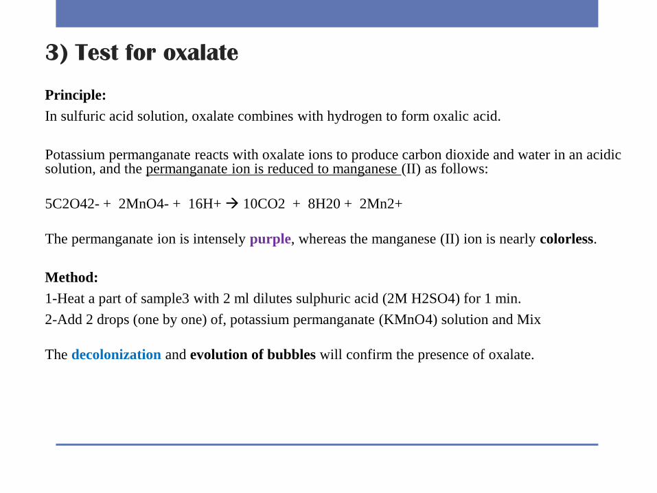
3) Test for oxalate
Principle:
In sulfuric acid solution, oxalate combines with hydrogen to form oxalic acid.
Potassium permanganate reacts with oxalate ions to produce carbon dioxide and water in an acidic (II) as follows:permanganate ion is reduced to manganese solution, and the
5C2O42- + 2MnO4- + 16H+ 10CO2 + 8H20 + 2Mn2+
The permanganate ion is intensely purple, whereas the manganese (II) ion is nearly colorless.
Method:
1-Heat a part of sample3 with 2 ml dilutes sulphuric acid (2M H2SO4) for 1 min.
2-Add 2 drops (one by one) of, potassium permanganate (KMnO4) solution and Mix
The decolonization and evolution of bubbles will confirm the presence of oxalate.
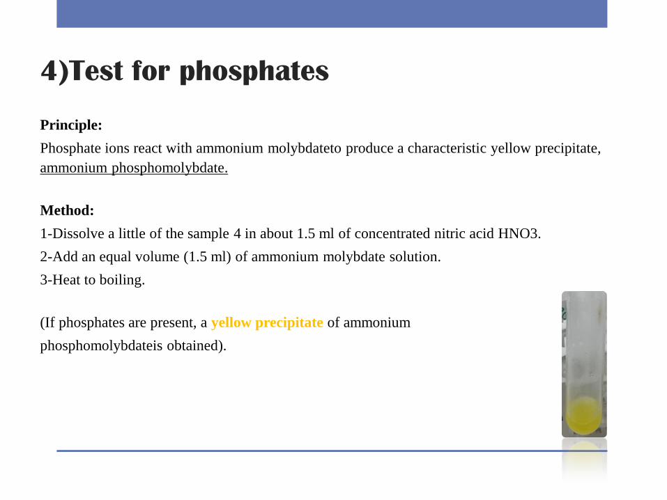
4)Test for phosphates
Principle:
Phosphate ions react with ammonium molybdateto produce a characteristic yellow precipitate,
ammonium phosphomolybdate.
Method:
1-Dissolve a little of the sample 4 in about 1.5 ml of concentrated nitric acid HNO3.
2-Add an equal volume (1.5 ml) of ammonium molybdate solution.
3-Heat to boiling.
(If phosphates are present, a yellow precipitate of ammonium
phosphomolybdateis obtained).
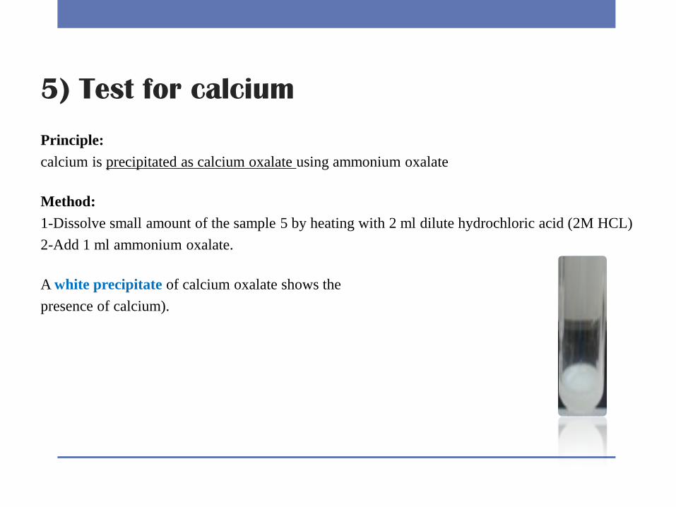
5) Test for calcium
Principle:
calcium is precipitated as calcium oxalate using ammonium oxalate
Method:
1-Dissolve small amount of the sample 5 by heating with 2 ml dilute hydrochloric acid (2M HCL)
2-Add 1 ml ammonium oxalate.
A white precipitate of calcium oxalate shows the
presence of calcium).
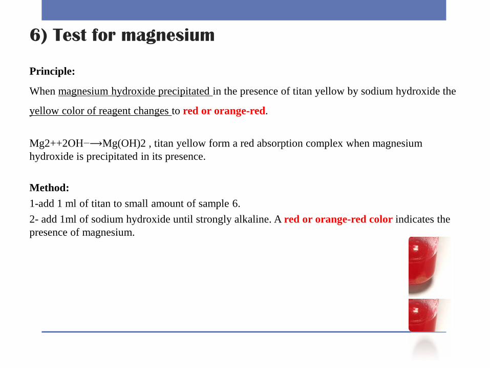
6) Test for magnesium
Principle:
When magnesium hydroxide precipitated in the presence of titan yellow by sodium hydroxide the
yellow color of reagent changes to red or orange-red.
Mg2++2OH−⟶Mg(OH)2 , titan yellow form a red absorption complex when magnesium
hydroxide is precipitated in its presence.
Method:
1-add 1 ml of titan to small amount of sample 6.
2- add 1ml of sodium hydroxide until strongly alkaline. A red or orange-red color indicates the
presence of magnesium.
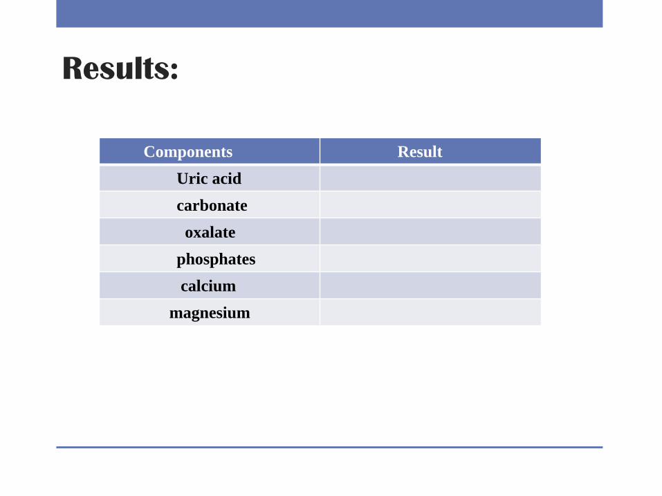
Results:
Result Components
Uric acid
carbonate
oxalate
phosphates
calcium
magnesium
Discussion:
Comment in each results you obtained and mention whether the sample contains
these component or not? And the disease that cause each type of stone.
Questions :
How change in urine pH can influence the type of stone formed?
Why Hyperparathyrodisim increases chance of calcium stone formation?