identification of a new regulatory pathway for the k-state
TRANSCRIPT

HAL Id: tel-01869028https://tel.archives-ouvertes.fr/tel-01869028
Submitted on 6 Sep 2018
HAL is a multi-disciplinary open accessarchive for the deposit and dissemination of sci-entific research documents, whether they are pub-lished or not. The documents may come fromteaching and research institutions in France orabroad, or from public or private research centers.
L’archive ouverte pluridisciplinaire HAL, estdestinée au dépôt et à la diffusion de documentsscientifiques de niveau recherche, publiés ou non,émanant des établissements d’enseignement et derecherche français ou étrangers, des laboratoirespublics ou privés.
Identification of a new regulatory pathway for theK-State in bacillus subtilis
Mathieu Miras
To cite this version:Mathieu Miras. Identification of a new regulatory pathway for the K-State in bacillus subtilis. Humangenetics. Université Paul Sabatier - Toulouse III, 2017. English. �NNT : 2017TOU30082�. �tel-01869028�

tre :
Université Toulouse 3 Paul Sabatier (UT3 Paul Sabatier)
ED BSB : Microbiologie
Mathieu MIRAS 28 Avril 2017
IDENTIFICATON OF A NEW REGULATORY PATHWAY FOR THE
K-STATE IN BACILLUS SUBTILIS
Laboratoire de Microbiologie et Génétique Moléculaires - UMR 5100
Patrice POLARD
Melanie BLOKESCH Associate Professor EPFL RapporteurMireille ANSALDI Directeur de recherche CNRS Rapporteur
Patrice POLARD Directeur de recherche CNRS Directeur de thèseClaude GUTIERRez Professeur UT3 Paul Sabatier Président du juryDavid DUBNAU Principal Investigator PHRI Examinateur

!

1
TABLE OF CONTENTS

2
TABLE OF CONTENTS ............................................................................................ 1!
RESUME ...................................................................................................................... 5!
ABSTRACT .................................................................................................................. 8!
ACKNOWLEDGEMENTS ...................................................................................... 11!
INTRODUCTION ...................................................................................................... 15!
A brief history of DNA ............................................................................................ 16!
DNA acquisition in prokaryotes .............................................................................. 18!
Conjugation .......................................................................................................... 18!
Transduction ........................................................................................................ 20!
Transformation ..................................................................................................... 22!
Transformation is a conserved phenomenon among bacteria .............................. 22!
Bacillus subtilis ........................................................................................................ 24!
General introduction ............................................................................................ 24!
A short history of Bacillus subtilis ....................................................................... 25!
Cell types in Bacillus subtilis ............................................................................... 26!
Competence for transformation and the K-state ...................................................... 28!
Foreword .............................................................................................................. 28!
Overview of the transformation process in B. subtilis ......................................... 29!
K-state regulation in B. subtilis ............................................................................ 31!
Regulation of comK ......................................................................................... 32!
Regulation of comK expression by quorum sensing ........................................ 34!
Regulation of comK basal expression: “the uptick” ........................................ 35!
The escape from the K-state ............................................................................ 37!
Two component systems in bacteria ........................................................................ 38!
General introduction ............................................................................................ 38!

3
The phosphorelay ................................................................................................. 41!
The DegS-DegU Two Component System in B. subtilis ..................................... 41!
Biofilms .................................................................................................................... 45!
Introduction to biofilms ....................................................................................... 45!
Description of B. subtilis biofilms ....................................................................... 45!
Biofilm life cycle ................................................................................................. 46!
The Spo0A pathway ............................................................................................. 48!
The SinR-SlrR regulation switch ......................................................................... 49!
Biofilms and the DegS-DegU pathway ................................................................ 50!
Quorum Sensing in biofilms ................................................................................ 51!
Making a biofilm, a hallmark of undomesticated B. subtilis strains .................... 52!
THESIS PURPOSE ................................................................................................... 53!
CHAPTER I ............................................................................................................... 57!
Genome sequence of the Bacillus subtilis biofilm-forming transformable strain
PS216 ........................................................................................................................... 57!
ABSTRACT ............................................................................................................. 58!
RESULTS ................................................................................................................ 59!
MATERIALS AND METHODS ............................................................................. 59!
NUCLEOTIDE SEQUENCE ACQUISITION NUMBER. .................................... 60!
REFERENCES ........................................................................................................ 61!
CHAPTER II .............................................................................................................. 63!
Domesticated and undomesticated strains of Bacillus subtilis share the same
basic network for competence development ............................................................ 63!
The firefly luciferase: a powerful tool for gene expression studies ......................... 64!
Effect of different competence regulators KO on K-state development .................. 65!

4
Strains Table ............................................................................................................ 70!
CHAPTER III ............................................................................................................ 71!
A DegU-P and DegQ-dependent regulatory pathway for the K-state in Bacillus
subtilis .......................................................................................................................... 71!
ABSTRACT ............................................................................................................. 72!
INTRODUCTION ................................................................................................... 73!
MATERIALS AND METHODS ............................................................................. 76!
RESULTS ................................................................................................................ 80!
DISCUSSION .......................................................................................................... 94!
REFERENCES ...................................................................................................... 100!
SUPPLEMENTAL DATA .................................................................................... 110!
THESIS SUMMARY ............................................................................................... 115!
CONCLUSION ........................................................................................................ 115!
REFERENCES ......................................................................................................... 122!

5
RESUME
6
Bacillus subtilis, une bactérie Gram-positive présente dans le sol, peut lorsque
les nutriments sont en concentrations limitantes, sporuler, former des biofilms ou
devenir compétente. La compétence est, chez B. subtilis, caractérisée par un arrêt de
la division cellulaire, une tolérance aux antibiotiques et l’expression de plus d’une
centaine de gènes. L’expression de la compétence, aussi désignée sous le nom de « K-
state », est dépendante de la synthèse du facteur de transcription ComK et se fait de
façon stochastique résultant en la formation de deux sous-populations bactériennes,
non-compétentes et compétentes. L’émergence, à partir de cellules génétiquement
identiques, de deux sous-populations distinctes est une stratégie de survie très
répandue chez les procaryotes, connue sous le nom de « bet-hedging ».
Bien que les mécanismes de régulation du développement de la compétence
ont, chez B. subtilis, largement été étudiés au cours des dernières années, la raison
pour laquelle les souches non-domestiques sont très peu transformables (1-2% de la
population) comparé aux souches domestiques (~15%) reste méconnue. Nous
démontrons ici que c’est essentiellement dû à une mutation de transition dans le
promoteur du gène degQ. Cette mutation diminue la synthèse de DegQ, protéine
impliquée dans la régulation de la formation de biofilms, de la synthèse
d’exoprotéases et de la transformation génétique. DegQ est une protéine impliquée
dans le transfert d’un groupe phosphoryl entre la kinase DegS et son substrat DegU.
Une faible quantité de DegQ diminue la concentration en DegU~P ce qui a pour
conséquence la désinhibition de l’opéron srfA entrainant une accumulation de ComK
et l’expression de la compétence. C’est ainsi que, dans les souches domestiques de B.
subtilis, un plus grand nombre de bactéries atteignent le niveau nécessaire en ComK
pour activer une boucle d’auto-activation transcriptionnelle de comK. Nous

7
démontrons aussi que l’activation transcriptionnelle de srfA est, dans les souches non-
domestiques, transitoire alors que la population bactérienne entre en phase
stationnaire de croissance. Ces données indiquent que le développement de la
compétence est moins fréquent et plus transitoire dans les souches non-domestiques
de B. subtilis. De plus, cette limitation du K-state dans les souches non-domestiques
est plus importante que précédemment « pensé » probablement dû à la domestication
de B. subtilis au cours de ces 50 dernières années.
Ce travail reflète non seulement, l’importance de l’utilisation de souches non-
domestiques dans la caractérisation des voies de régulation de la compétence chez B.
subtilis, mais aussi la portée du choix de modèle biologique dans l’étude de
phénomènes biologiques complexes.

8
ABSTRACT
9
Bacillus subtilis, a Gram-positive soil bacterium, can enter into several
developmental pathways such as sporulation, biofilm formation and competence
development for DNA transformation when it becomes limited for essential nutrients.
During competence, cells do not divide, are tolerant to antibiotics and competent cells
express more than a 100 genes. The competent state has been named the K-state after
its master regulator ComK.
In B. subtilis, the entry into the K-state is stochastically determined by the
activation of the transcription factor ComK and occurs, in the domesticated strains of
B. subtilis, in approximately 15% of the population. The emergence from genetically
identical cells of two distinct subpopulations (competent cells and non-competent
cells) is known to be a classic survival strategy for bacteria, known as bet-hedging.
Regulation of entry into the K-state has been intensively studied and is well
understood; however, the reasons why undomesticated isolates of B. subtilis are
poorly transformable compared to the domesticated strains remained unexplained. We
show here that fewer cells enter the K-state, suggesting that some regulatory pathway
limiting its expression has been lost in the domesticated backgrounds. We
demonstrate that this is largely due to an inactivating point mutation in the degQ
promoter region resulting in a decrease of the amount of DegQ. DegQ is known to
stimulate phosphate transfer from the DegS autokinase to its cognate response
regulator DegU. A low level of DegQ thus decreases the concentration of the
phosphorylated form of DegU, leading to the de-repression of the srfA operon, which
increases the amount of ComS leading to the stabilization of ComK. Thus, in
domesticated strains of B. subtilis, more cells reach the concentration threshold of
ComK needed to activate the positive auto-regulatory loop of ComK acting on its own

10
promoter. We also show that the activation of srfA transcription in undomesticated
strains is transient, as it is turned off when cells enter the stationary phase.
Taken together, these data indicate that the K-state and transformability are
less frequent and more transient in the undomesticated strains of B. subtilis.
Consideration of the regulatory mechanisms and the fitness advantages and costs of
the K-state must from now on take these features into consideration. These results
underscore that our understanding of real-life biology requires the use of wild isolates.

11
ACKNOWLEDGEMENTS

12
It has been five years since I began working towards my PhD. During these
years, I have received support from colleagues, friends and family and I would like to
dedicate this work to them.
To my mentor, David Dubnau, you invited me to be a part of your laboratory
and I could not imagine doing my thesis work with anyone else. Thank-you for
guiding me through the PhD’s rough waters, for giving me the freedom and
encouragements. Thank-you for your wisdom and your contagious (but precious)
optimism: I think I will never forget the sentence “This is gonna work!!”. There is no
doubt you helped me become a better scientist. Thank-you!
To my thesis committee. Claude Gutierrez, Mireille Ansaldi and Melanie
Blokesch, thank-you for being a member of this committee, for coming to Toulouse
and for reading this work. Thank-you to Patrice Polard for allowing me to go to
Dave’s lab by giving me those precious signatures and for guiding me during the last
few months of my thesis.
To the “Daffettes”. Hedia, Ségolène, Nawel, Fabienne and Annaïk, thank-you
for training me as a student and for coming to my defense after all these years. You
showed me that good science could be done in a happy and friendly environment and
that is something I have been trying to reproduce every day since then. Annaïk, thank-
you for recommending me to Dave. I would definitely not be “here” without you!

13
This work would not have been possible without the excellent undergraduate
education I received at the Université Paul Sabatier. I thank the professors and
teachers in Departments of Biology for their instruction.
To the Dubnau Lab, past and present: During these five years, you guys have become
like family. Thank-you for “enjoying” my eclectic music playlists, for many fun and
encouraging conversations during lunch over these years, for enjoying all these sports
events (mainly soccer) from the lab and of course, for sharing your stock solutions so
kindly #TeamSpirit. Jeanie, thank-you for opening the doors of your house on several
occasions, for always sharing your political and scientific opinions with passion…
Please, don’t give up the fight, I am sure we can “Make this world and our planet
great again”. Jeanette, thank-you for being my lab “mom”, and for making fun of my
accent. I really have enjoyed working by your side and have appreciated our
conversations and your advices. Val, thank-you for always being helpful, for your
insights about this manuscript, and for taking me to my first hockey game. Christine
and Micaela, thank-you for bringing new accents in the lab, for these treats brought
back from Germany and Italy, for your laughs and your friendship. Miguel and Nico,
thank-you for helping me during my first months in America, not only as a PhD
student, or for these passionate soccer conversations but also for showing me how to
fill a tax return form…
To the PHRI/Rutgers kitchen staff: for making all these plates so I could dedicate my
time showing (again and again) Jeanette how to use the Geneious software…
To all, I will miss having you around, and wish you all the best.

14
To my friends. Manu, Marina, Beun, Marion, Pierre, JF, Paula, Ced, Theresa,
Anne-So, Fabe, Romain, Mathieu, Pablo, thank-you for always being around despite
the distance and for making me feel like I never left when I got the chance to see you.
To my college mates: Cindy, Saad, Thomas and Thibault. I will not forget all these
times we could not stop laughing and of course ALL these “Belote games’ we played
anytime we got a chance. To friends from the New World: Thomas, JL, JP, Stéphane,
Benoit, Iuri, Julien, Romain, Jessie, Charles, Gaétan, Soufiane it’s been a great
pleasure meeting you, playing soccer and spending time with you. You guys made
this American adventure unforgettable.
To my family, and more particularly to my parents. To become a scientist, I
think you need to be, over all, curious. And since I am young, you have always tried
to open my eyes and mind to people, to new things, to new languages and cultures. I
think I would have never become a scientist without all these travels and trivial cards
games. Thank-you for always supporting me, for taking time to skype me when it was
late at night and for sending me these precious packages filled with surprises, books
and treats, especially dark chocolate without which I could have not survived to these
cold winters.

15
INTRODUCTION

16
A brief history of DNA
The DNA story began in the mid-nineteenth century
with the work of a young Swiss physician, Friedrich
Miescher. After graduation, he went to Germany to work in
one of the first laboratories focusing on the composition of
lymphoid cells. As it was difficult to extract material from
the lymph glands, Miescher had the idea to accumulate
bandages from a nearby clinic to collect the pus. During his experiments with this
material, he identified a substance with unexpected properties that did not match those
of proteins; this substance was made of oxygen, hydrogen, nitrogen and phosphorus
but there was a unique ratio of phosphorus to nitrogen. Since it had been purified
from the nucleus of the cells, Miescher named this substance “nuclein,” which ended
up being partially purified deoxyribonucleic acid (DNA) (Dahm, 2008).
Born in 1869 in Lithuania, Phoebus Levene migrated
to the U.S. to practice medicine in the Lower East Side of
Manhattan a few years before World War II. As he was
interested in the chemical structure of sugars, Levene further
characterized Miescher’s “nuclein” by showing it was made
of a nitrogenous base, a sugar, and a phosphate group and
that the different units were connected to each other via a phosphate group (Levene,
1919).

17
In 1928, the British bacteriologist Frederick Griffith was
working on the epidemiology and pathology of Streptococcus
pneumoniae, the pathogen responsible for pneumonia, and
showed in what is now known as “Griffith’s Experiment” the
first evidence of bacterial transformation. Indeed, he realized he
could transfer the virulence trait from a heat-inactivated (dead)
virulent strain of S. pneumoniae to a live but non-virulent strain of S. pneumoniae,
simply by mixing both live and dead strains. Unaware of the precise substance
involved in the transfer of this new virulence phenotype acquired by the non-
pathogenic strain, Griffith named this mysterious agent the “transforming principle”
(Griffith, 1928) (Figure 1).
Figure 1: The Griffith Experiment 1- The mice infected by a virulent “S” strain of S. pneumoniae are killed. 2 and 3- The mice infected by either non-virulent “R” strain or heat-killed virulent “S” strain of S. pneumoniae survive. 4- After co-infection by “R” and “S” killed by heat, the mice die. Griffith was then able to isolate both live “R” and live “S” strains of S. pneumoniae from the blood of these dead mice. He concluded that the “R” strain had been "transformed" into the lethal “S” strain by a "transforming principle”.
Figure1: Griffith’s Experiment Mice infected by virulent “S” strain of S. pneumoniae are killed. Mice infected by not virulent “R” strain or heat-killed virulent “S” strain of S.
pneumoniae are not killed. After co-infection by “R” and “S” killed by heat, mice die. Griffith was then able to isolate both live “R” and live “S”
strains of S. pneumoniae from the blood of these dead mice. He concluded that the “R” strain had been "transformed" into the lethal “S” strain by
a "transforming principle" that was somehow part of the dead “S” strain bacteria.
!"#$%&'&()*+$)+&',-*.$/0&1$2++3*$&/$.*,.$
1&3+*$
4)5)(6$!"#$%*''+$ 4)5)(6$!7#$%*''+$
!7#$%&'&()*+$)+&',-*.$/0&1$2++3*$&/$
8*,'-89$1&3+*$
:&$%&'&()*+$)+&',-*.$/0&1$2++3*$&/$
8*,'-89$1&3+*$
!7#$,(.$!"#$%&'&()*+$)+&',-*.$/0&1$2++3*$&/$.*,.$1&3+*$
;*,-$<)''*.$!"#$%*''+$ 4)5)(6$!7#$%*''+$=$;*,-$<)''*.$!"#$%*''+$
!" #" $" %"

18
In the mid-nineties, the scientific community wondered
about a big mystery: in which molecule is the genetic
information hidden? Son of a Baptist minister at the Mariners’
Temple In New York’s Lower East Side, Oswald Theodore
Avery, a Canadian doctor, repeated Griffith’s original
experiment using purified DNA from a virulent strain of S. pneumoniae and showed
that it was sufficient to transfer the virulence trait to a non-virulent strain (Avery,
Macleod, & McCarty, 1944) (Hershey, 1952). Avery was the first to propose that
Griffith’s transforming principle was actually DNA.
DNA acquisition in prokaryotes
Bacteria can acquire new traits either by mutation or by the acquisition of
external genetic material. This external acquisition of DNA is termed horizontal gene
transfer (HGT) (de la Cruz & Davies, 2000) (Ochman, Lawrence, & Groisman, 2000),
referring to the transfer of genes between organisms in a manner that contrasts with
vertical transfer, the inheritance of genes from the parental generation to offspring via
sexual or asexual reproduction. HGT has been shown to be an important factor in the
evolution of many organisms and the primary reason for the spread of bacterial
antibiotic resistance (Koonin, Makarova, & Aravind, 2001). HGT occurs through
three mechanisms: conjugation, transduction and transformation (Koonin et al., 2001).
Conjugation
Conjugation is the transfer of DNA from a donor to a recipient by direct
physical contact between two cells (Clark & Adelberg, 1962). Many but not all
species of bacteria can conjugate and conjugation is possible between cells of the

19
same species or even between cells of two different species (Trieu-Cuot, Gerbaud,
Lambert, & Courvalin, 1985) (Penalva, Moya, Dopazo, & Ramon, 1990) (Kuhsel,
Strickland, & Palmer, 1990). A plasmid called the F factor (Fertility factor) is
required for conjugation (Lederberg, Cavalli, & Lederberg, 1952). As it carries its
own origin of replication and an origin of tranfert, the F plasmid is an episome. It also
carries the tra locus that incodes for pilin proteins. Only one copy of the F plasmid
can be found in a given bacterium. In bacteria there are two “mating types,” a donor
(or F+) and a recipient (or F-) and the direction of transfer of genetic material is
unidirectional (Lederberg et al., 1952). After the pilus of the donor cell recognizes
and binds to specific receptors sites on the cell wall of the recipient cell, a single
stranded DNA molecule from the mobile plasmid is transferred from the donor to the
recipient and is later converted to double stranded DNA (Wozniak & Waldor, 2010)
(Figure 2).

20
Figure 2: Schematic drawing of bacterial conjugation 1- The donor cell produces a pilus, which is encoded on the mobile plasmid. 2- The pilus attaches to the recipient cell and brings the two cells in close proximity. 3- The plasmid is nicked and a single strand of DNA is then transferred to the recipient cell. 4- After genetic transfer, both cells synthesize the complementary strands to produce a double stranded circular plasmid. Both cells are now donors.
Transduction
Transduction is the transfer of genetic information from a donor to a recipient
by means of a bacteriophage and can happen through either the lytic cycle or the
lysogenic cycle (Zinder & Lederberg, 1952) (Kresge, Simoni, & Hill, 2011). In the
lysogenic cycle, the bacteriophage’s genome is integrated in the host genome to form
the prophage and will not be expressed. The prophage is then transmitted as well as
the rest of the genetic information to the daughter cells (called lysogens) as the
bacterium divides. The switch to the lytic cycle can be induced at any time and the
prophage DNA is excised from the bacterial chromosome, transcribed, and translated
to produce phage elements. The prophage DNA will then be incorporated in the phage
particles, which are released by lysis of the host. Because the packaging of prophage
DNA in the phage heads is a low fidelity process, it happens that small pieces of the
!"#$%$&$%'()*+,) -$./(0)1('&%/2)
!"#"$% &'()*)'#+%
1/(3&)
,-.%!"#"$%/'0%!"#"$%
1%
2%
3%
4%

21
host chromosomal DNA may be incorporated as well. The phage coat protects its
genome in the environment so that transduction, unlike transformation, is not affected
by extracellular nucleases. We can assume that, because this mode of gene transfer
does not depend on many dedicated bacterial genes, phages played an important role
in the ecology and evolution of bacteria. Indeed, by moving pieces of bacterial DNA
among themselves, phages probably contributed in making bacteria what bacteria are
nowadays (Spizizen, Reilly, & Evans, 1966) (Figure 3).
Figure 3: Schematic drawing of bacterial transduction. 1- Phage infects a susceptible bacterial cell. 2- The host bacterial DNA is hydrolyzed while phage DNA and proteins are produced. 3- New viral particles are synthesized and will occasionally incorporate phage DNA into the mature virion, instead of phage DNA. The cell lyses and releases the new bacteriophages. 4- Transducing phages infect new cell (recipient host) and transfer bacterial DNA, but are defective as lytic phages. 5- New DNA is incorporated into recipient’s genome by recombination.
!"#$%$&$%'()*+,)
!"#"$%&'((%
-"'./)
)%-"'./)*+,)0#'.%/12&)*%
+'&,-,'#.%/"0.%&'((%
3#'1&45671.)8"'./)1%
-"'./)972")4$1$#)*+,)
2%
3$4#056&'5%&'((%
7%
*$1$#)6/(())*+,)0#'.%/12&)

22
Transformation
Transformation is the gene transfer resulting from the uptake of exogenous
(environmental) DNA. Certain bacteria species (e.g. Bacillus, Haemophilus,
Neisseria, Pneumococcus) can naturally incorporate DNA into their genomes. Like
conjugation systems, transformation systems depend on specialized operons that
encode DNA uptake machinery. Proteins encoded by these genes include those
necessary for DNA binding, internalization, and possible recombination of DNA with
the genome. This process will be discussed in great detail (see “Competence for
transformation and the K-state”).
Transformation is a conserved phenomenon among bacteria
Cells expressing genes needed for acquiring DNA from the environment are
said to be in a state of competence. Since this state is mediated by genes present in the
bacterial genome, and is not induced artificially, this form of competence is referred
to as genetically-programmed (Erickson, 1970) (Chen, Christie, & Dubnau, 2005). In
contrast, other bacteria such as Escherichia coli are not able to take up DNA
naturally, but the competent state can be induced through the addition of either
chemical or physical agents (electroshock), which permit DNA to pass through the
cell wall. Most naturally competent bacteria only take up DNA at a specific time
during growth. It has been argued that the benefits of genetic competence are that the
acquired DNA can be used as a source of nutrition, to repair existing genes, or for the
acquisition of new genetic material (Redfield, 1993). Examples of natural bacterial
competence are represented in both Gram-negative (i.e. Neisseria gonorrhoeae,
Haemophilus influenzae, Helicobacter pylori) and Gram-positive (i.e. Streptococcus
pneumonia, Bacillus subtilis) species (Chen et al., 2005). While in Gram-positive
bacteria, DNA must go through the thick peptidoglycan layer of the cell wall and the

23
cytoplasmic membrane; it must also traverse the highly impermeable outer membrane
of Gram-negative bacteria. Therefore, additional steps are involved in the Gram-
negative transformation systems, and the initial interaction of DNA with the cell
envelope is also different in the two types of bacteria. Despite these differences, the
protein machinery required to transport DNA in Gram-positive and Gram-negative
bacteria is conserved, and interestingly, is also closely related to protein machines
used for molecular secretion (Burton & Dubnau, 2010).

24
Bacillus subtilis
General introduction
Bacillus subtilis (B. subtilis) is a bacterium naturally found in soil and
vegetation that belongs to the Firmicute phylum of bacteria, which includes Gram-
positive bacteria with a low G+C content. Firmicutes have been differentiated from
one another based on different characteristics such as the nature of their cell envelope,
their aerotolerance (how well they live and grow in oxygen) and their ability to form
endospores. Consequently there are seven classes of Firmicutes: the Erysipelotrichia,
the Negativicutes, the Limnochordia, the Tissierellia, the Thermolithobacteria, the
Clostridia and the Bacilli. Because stress and starvation are common in its
environment, B. subtilis has evolved a set of strategies that allow its survival under
these harsh conditions: formation of stress resistant endospores or uptake of external
DNA, which allows the bacteria to adapt by recombination. And partly because B.
subtilis is also a non-pathogenic bacterium for humans it has been used as a model
organism to further understand pathogenic microorganisms belonging to the same
phylum such as Streptococcus pneumoniae (a major cause of pneumonia), Bacillus
anthracis (agent of anthrax) or Listeria monocytogenes (food infection). B. subtilis is
used in industry to produce antibiotics (subtilin, bacitracin) and secrete several
commercial enzymes used in the food industry (amylases for bread production) or the
detergent industry (proteases). Also, a strain of B. subtilis formerly known as Bacillus
natto is typically used in Japan to produce a treat called “natto”. This traditional
course is made of soybeans fermented with Bacillus natto and can be eaten for
breakfast.

25
A short history of Bacillus subtilis
B. subtilis was originally discovered more than a hundred and fifty years ago
and the domesticated laboratory strains have now been used for more than half a
century (Zeigler et al., 2008). B. subtilis laboratory strains derive from a single
tryptophan-requiring auxotrophic strain, strain 168. In the mid 1900s, two Yale
University botanists, Paul Burkholder and Norman Giles, isolated strain 168 after the
B. subtilis Marburg strain was mutagenized with X-rays (Burkholder & Giles, 1947).
Unfortunately the Yale group abandoned its B. subtilis experiments to focus on other
research interests, and most of its B. subtilis collection has been lost. At least five
mutants (auxotrophs requiring threonine (strain 23), nicotinic acid (strain 122), or
tryptophan (strains 160, 166 and 168)) were preserved from those original
experiments, and were transferred to Charles Yanofsky and John Spizizen, who
showed that three of them (strains 122, 166 and 168) could be transformed to
prototrophy when exposed to DNA from strain 23 (Spizizen, 1958). The highly
transformable strain 168 became the subject of many other studies and was therefore
disseminated around the world. By the mid 1970s, so many mutants had been
developed from strain 168 that a centralized repository, the Bacillus Genetic Stock
Center (BGSC), was established in Ohio to maintain them. Interestingly, X-irradiation
and domestication brought many changes in B. subtilis strain 168 behaviors: a
dramatic reduction in its ability to form complex, structured and matrix-adhered
colonies known as biofilms, an inability to swarm on solid surfaces and an increased
competence for genetic transformation (Branda, Gonzalez-Pastor, Ben-Yehuda,
Losick, & Kolter, 2001) compared to its probable ancestor NCIB3610 (hereafter
referred to as 3610). One major change was the loss of a plasmid, called pBS32,
which encodes a cassette of phage genes (Konkol, Blair, & Kearns, 2013). While
26
pLS32 is not present in the closest relatives of strain B. subtilis 3610, it is found in
more distantly related strains (Tanaka & Ogura, 1998).
Cell types in Bacillus subtilis
Soil is a variable environment and accordingly B. subtilis can adapt by
differentiating into distinct subpopulations of specialized cells, which express
different traits that may confer a survival advantage in adverse conditions (Lopez,
Fischbach, Chu, Losick, & Kolter, 2009) (Lopez, Vlamakis, & Kolter, 2009). Thus, B.
subtilis is a good model organism for the study of alternative lifestyles. When
nutrients are abundant, cells can grow in a free-floating planktonic form. At the onset
of stationary phase, or when B. subtilis is shifted to poor nutrient conditions, some B.
subtilis cells adapt by becoming naturally transformable (Chen et al., 2005). Other
cells secrete toxins, which kill siblings by inducing cell lysis and the release of
cellular components for scavenging (Lopez, Fischbach, et al., 2009). Also, during
growth, cells may switch from a sessile to a motile state or vice versa. The sessile
lifestyle choice leads to the formation of a multi-cellular, community known as a
biofilm. Production of the extracellular matrix, which is essential for biofilm
formation, is carried out by a subpopulation of specialized cells in B. subtilis (Chai,
Chu, Kolter, & Losick, 2008). However, the whole community benefits from the
presence of the extracellular matrix, because all of the cells are encased within the
matrix in mature biofilms, and thus protected from environmental insults such as
antibiotics or phage (Vlamakis, Aguilar, Losick, & Kolter, 2008). As an extreme
survival mechanism, the population will produce spores, and the mother cells lyse
during the process. These spores are metabolically inactive, resistant to heat, radiation
and toxic chemicals, and able to persist over long periods of time without nutrients. If
optimal conditions arise and nutrients become available, spores germinate and resume

27
growth. The development of each of these specialized states is triggered by specific
signals. For example, sporulation is proposed to be activated when the intracellular
concentrations of essential metabolic molecules, like GTP, decrease (Lopez, Gontang,
& Kolter, 2010) but the signal itself remains unknown. Transformation is regulated in
part by quorum-sensing, a form of cell-cell communication in which cells secrete
signaling molecules that accumulate in the medium as they grow and typically elicit a
coordinated response from the population (Hahn & Dubnau, 1991) (Lazazzera, 2000).
Overall, although the cells of a B. subtilis culture are genetically identical, different
triggers activate specific changes in gene expression that often result in distinct
subpopulations of cells (Lazazzera, 2000) (Lopez, Vlamakis, & Kolter, 2009).

28
Competence for transformation and the K-state
Foreword
The timing for competence expression is different among bacteria. S.
pneumoniae, for example, expresses competence only during early exponential
growth (Pakula & Walczak, 1963) while Neisseria gonorrhoeae is competent during
the entire exponential phase (Sparling, 1966). B. subtilis develops competence at the
onset of the stationary phase. The percentage of competent cells is also different
between bacteria: only 10 to 20% of the cells express competence in domesticated B.
subtilis while the entire population becomes competent in S. pneumoniae. Also,
factors triggering the expression of competence are different; as the chemical
composition of the media is important for S. pneumoniae, whereas nutrient limitation
is important for B. subtilis, although this has not been studied in any detail (Tomasz,
1966) (Morrison & Baker, 1979).
Competence provides the cell population with an alternative mechanism of
survival under environmentally challenging conditions. Indeed, different theories
have been proposed on the benefits of genetic transformation, such as the use of DNA
to repair damaged genes, the use of DNA as a source of nutrition (carbon, nitrogen
and phosphorous) or the use of this exogenous DNA to allow genetic diversity in the
population (Finkel & Kolter, 2001). A unique feature of competence in B. subtilis is
that cells expressing competence do not divide. It has also recently been shown that
competent cells are tolerant of antibiotics, a form of persistence (Hahn, Tanner,
Carabetta, Cristea, & Dubnau, 2015). Because more than 100 genes are expressed
when cells are competent (Hamoen, Smits, de Jong, Holsappel, & Kuipers, 2002)
(Berka et al., 2002) (Ogura et al., 2002), and because most of these genes are not
needed for transformation, the competent state has been called the K-state, named for

29
the master regulator ComK (Maamar & Dubnau, 2005). The emergence from
genetically identical cells of two distinct subpopulations (competent cells and non-
competent cells) is a classical survival strategy for bacteria, known as bet-hedging
(Suel, Kulkarni, Dworkin, Garcia-Ojalvo, & Elowitz, 2007) (Veening, Smits, &
Kuipers, 2008).
Overview of the transformation process in B. subtilis
The proteins that are essential for DNA-uptake are encoded by three different
operons, comE (Albano, Hahn, & Dubnau, 1987) (Hahn, Inamine, Kozlov, & Dubnau,
1993) (Inamine & Dubnau, 1995), comF (Londono-Vallejo & Dubnau, 1993)
(Londono-Vallejo & Dubnau, 1994) and comG (Albano, Breitling, & Dubnau, 1989)
(Albano & Dubnau, 1989) (Briley et al., 2011) as well as the gene product of comC
(Mohan, Aghion, Guillen, & Dubnau, 1989) (Chung & Dubnau, 1995). Other genes
are needed for the processing and integration of DNA following uptake. The process
of transformation can be subdivided in four distinct steps. First, double-stranded DNA
(dsDNA) from the environment is bound to competent cells. Previous studies showed
that there is no sequence preference and that there are approximately 50 binding sites
per cell (Dubnau, 1991). It was proposed that a “competence pseudopilus,” made up
of pilin subunits encoded by the comG operon which are processed by the protease
ComC, traverses the cell wall and makes contact with the exogenous dsDNA.
However, while the competence pseudopilus is required for DNA binding, none of its
components have been shown to have DNA binding properties (Chen & Dubnau,
2004), suggesting that some unknown DNA binding protein exists. Following the
association of DNA with the cell, the ds-DNA is non-specifically cleaved by the
membrane-bound endonuclease NucA, where the average size of the fragments is 11-
18kb (Provvedi, Chen, & Dubnau, 2001) (Figure 4). Next, the dsDNA is transported

30
through the plasma membrane via the aqueous ComEC channel and one strand is
degraded by an unknown nuclease. ComFA was proposed to be a DNA translocase
and/or helicase, which may provide energy for the movement of the DNA through the
ComEC channel (Londono-Vallejo & Dubnau, 1993) (Londono-Vallejo & Dubnau,
1994) (Dubnau & Cirigliano, 1972) (Lacks, Greenberg, & Neuberger, 1975). In B.
subtilis there is no evidence that the complementary strand is degraded while the other
strand is transported through the channel, as has been shown in S. pneumoniae
(Mejean & Claverys, 1993). Also, an ortholog of the endonuclease that degrades the
non-transforming strand in S. pneumoniae is lacking in B. subtilis. Finally, once in the
cytoplasm, the single stranded DNA (ssDNA) is integrated into the genome by
homologous recombination (Fernandez, Ayora, & Alonso, 2000). This final step
requires the involvement of competence-induced proteins DprA, RecA and SSB.
Indeed, DprA (DNA processing protein A) presumably binds the ssDNA to protect it
from degradation by nucleases, and also recruits the recombinase RecA (Yadav et al.,
2013) (Yadav, Carrasco, Serrano, & Alonso, 2014) (Lenhart, Schroeder, Walsh, &
Simmons, 2012). RecA polymerizes on ssDNA and promotes a homology search
along chromosomal DNA (Yadav et al., 2013) (Yadav et al., 2014) to form a
recombination heteroduplex intermediate with the host chromosome (Figure 4).

31
Figure 4: DNA uptake machinery in B. subtilis ComG pre-pilin subunits are first translated as integral membrane proteins with a cytoplasmic leader peptide. They are then processed by the pre-pilin peptidase ComC before being translocated out of the membrane where they oligomerize into a pilus. The energy necessary for the movement of the pilus through the conserved membrane protein ComGB is provided by the associated ATPase ComGA. When the double stranded DNA (dsDNA) makes contact with the pilus, it is believed that ComEA delivers the DNA to the membrane channel ComEC where one strand of DNA is likely degraded by an unknown nuclease. This single-stranded DNA (ssDNA) internalization is driven by the ATP-dependent translocase ComFA. Cytoplasmic ssDNA is bound by DprA, which recruits RecA to allow homologous recombination with the host DNA.
K-state regulation in B. subtilis
Competence development in B. subtilis has become an important model for
bistable gene expression where the expression of a key regulatory protein (ComK) is
tightly regulated to prevent the entire population from becoming competent. In B.
subtilis, competence development is the result of a dramatic increase in the cellular
concentration of the transcriptional regulator, ComK (Hamoen, Van Werkhoven,
Bijlsma, Dubnau, & Venema, 1998; van Sinderen et al., 1995; van Sinderen &
Venema, 1994). ComK is referred to as the “master regulator of competence,”
because it is both necessary and sufficient for the transcriptional activation of all of
!"#$%$&'
()*+,'
()-./,0)'
!$-12'
!$-13'
!$-!'
4)56*$7&"8,0'
!$-9!'
!$-:2'
!$-92'
;5/2'
<)82'
*%=;>2'
%%=;>2'
?$-$&$7"'%),/8@' <)8$-.+0,6$0'
?$%#';>2'
2A4'2;4'B'4+' 2A4'
2;4'B'4+'
4+&C%'
4/)=5+&+0'

32
the K-state genes. ComK is a 22kDa transcription factor that binds as a tetramer or
dimer of dimers to A/T rich regions within its targets promoter genes that contain
“ComK boxes” (van Sinderen et al., 1995) (Hamoen et al., 1998). But, in addition to
competence regulation, ComK also activates the transcription of genes involved in
many other different processes such as cell shape determination, cell division,
transcriptional regulation, transport, protein synthesis and stress responses (Ogura et
al., 2002) (Berka et al., 2002). As noted already, ComK regulates more than genes
required for transformation, and the “K-state,” which can be broadly described as a
global, persistence-like, bet-hedging, expression state (Berka et al., 2002) (Hahn et
al., 2015). Thus, the K-state enhances fitness by a means distinct from
transformability (Maamar & Dubnau, 2005).
Regulation of comK
As mentioned previously, cells expressing ComK are fully dedicated to enter
the K-state and stop DNA replication and cellular division. This represents a risk for
the cell population. To avoid such a situation, the expression of ComK is tightly
regulated during growth. During exponential growth, the transcription of comK is
inhibited via the direct binding of the transcriptional repressors AbrB (Hamoen et al.,
2003), CodY (Serror & Sonenshein, 1996) and Rok (Hoa, Tortosa, Albano, &
Dubnau, 2002) (Figure 4). In addition to this transcriptional repression, ComK
stability is also regulated. Any ComK that is made will be degraded by a second
regulation system, ClpC/ClpP mediated proteolysis (Turgay, Hahn, Burghoorn, &
Dubnau, 1998). The adaptor protein MecA directly interacts with ComK and delivers
it to the ATP-dependent chaperone ClpC, where it is unfolded and then completely
degraded by ClpP (Figure 5) (Turgay, Hamoen, Venema, & Dubnau, 1997) (Turgay

33
et al., 1998). These negatively acting mechanisms are relieved as the cells approach
stationary phase, which is when the small peptide ComS is produced. ComS binds
directly to the ComK-binding site of MecA releasing ComK from degradation
(Turgay et al., 1997) (Prepiak & Dubnau, 2007). The expression of comS is a
consequence of quorum-sensing mechanisms taking place in late exponential phase.
Once released from degradation, the basal level of ComK exceeds a threshold in some
cells and comK expression will then be amplified by a positive feedback loop in
which ComK acts on its own promoter (PcomK). The resulting burst of ComK
synthesis triggers the expression of the transformation apparatus genes and the other
K-state genes. (Figure 5).
Figure 5: Overview of the competence regulation in B. subtilis A represention of the relevant effectors involved in the expression of competence for DNA uptake. On the left is the phosphorelay composed of five histidine kinases KinA-E, Spo0F (0F), Spo0B (0B) and Spo0A (0A) ultimately leading to the phosphorylation of the master regulator Spo0A. Depending on its concentration in the cells, Spo0A-P will activate and repress directly or indirectly the comK promoter. To the right, the synthesis and processing of the two qurorum sensing pheromones ComX and CSF are shown, which leads to the stabilization of ComK through the activation of the transcription of the srfA operon and the synthesis of ComS peptide. ComS, in turn, blocks ComK degradation by the MecA-ClpC-ClpP complex. Once ComK is stabilized, it autoactivates its own transcription, and triggers the expression of the com operons, which encode the proteins to assemble the DNA uptake apparatus.
!"#$%$ &$
%$&$
%$&$ 0F
0B
0A %$&$
#"!$'()*#*+$
,-."/$,-012/3-$
#"!$ !"#$
#"!$ !"#$
#"!$%$&$ !"#$%$ &$
KinB KinC
KinD
KinE
#"!$ !"#$
KinA
!"#$
!"#$%
#"!$
ComP
!"#$%&'()
!"#$%&'()
'*04$ &$%$'*04$
&'())%
5/6'$
'78$ &92'$
'78$
&'()*%!"#+%
Spo0K
,-'*%
5*:$
!"#.)% !"#.*% !"#./%
!"#0)% !"#0*% !"#0/%
!"#1)% 1*% 1/%12%1.% 10%11%'*.;$
'*07$
,-<4$'+6'$'+6&$

34
Regulation of comK expression by quorum sensing
In strain 168 derivatives the maximum of K-state expression is reached two
hours after entering in the stationary phase because of the accumulation of quorum
sensing pheromones. Quorum-sensing is a cell-cell communication mechanism that
monitors cell density. Secreted molecules accumulate in the medium and activate a
cognate receptor at high concentration, triggering an intracellular response
(Lazazzera, 2000). In B. subtilis, two quorum-sensing peptides are known to regulate
the expression of comS: ComX (Magnuson, Solomon et al. 1994) and competence
stimulating factor, CSF (Solomon, Magnuson, Srivastava, & Grossman, 1995).
ComX is a signaling peptide that regulates competence through the ComP-
ComA two-component regulatory system (Hahn & Dubnau, 1991) (Solomon et al.,
1995). Synthesized as a 55 amino acid precursor, pre-ComX is cleaved and modified
by ComQ, to generate its mature form consisting in a 10 amino acid peptide with a
hydrophobic isoprenoid modification on a tryptophan and a cyclization event
(Weinrauch, Msadek, Kunst, & Dubnau, 1991) (Ansaldi, Marolt, Stebe, Mandic-
Mulec, & Dubnau, 2002). The mature ComX is secreted and when present in high
enough concentrations, binds to ComP, a membrane histidine kinase. Binding
activates the autophosphorylation of a conserved histidine residue in the cytoplasmic
domain of ComP (Piazza, Tortosa, & Dubnau, 1999). The phosphoryl group is
transferred to the N-terminal regulatory domain of its cognate response regulator
ComA (Weinrauch et al., 1991) allowing its binding to DNA and the activation of the
srfA operon that encodes ComS (Figure 5) (Nakano, Xia, & Zuber, 1991) (Nakano &
Zuber, 1991) (Roggiani & Dubnau, 1993).

35
CSF (Competence and Sporulation Factor) is the second signaling peptide that
regulates competence through quorum-sensing. CSF comes from the last five codons
of the gene product of phrC (Solomon, Lazazzera, & Grossman, 1996) and is
exported via an unknown mechanism after cleavage to produce a pentapeptide
(ERGMT) without any post-translational modifications. Secreted CSF is internalized
back into the cell through the oligopeptide permease Spo0K (Rudner, LeDeaux,
Ireton, & Grossman, 1991) (Solomon et al., 1995) (Lazazzera, Solomon, &
Grossman, 1997). This peptide then prevents the dephosphorylation of ComA-P by
inhibiting the phosphatase RapC (Figure 5) (Solomon et al., 1995).
Regulation of comK basal expression: “the uptick”
Another cause of temporal regulation comes from the tight regulation of comK
basal expression. The basal expression of comK, measured in the absence of ComK
autoregulation, increases gradually with growth, reaching a maximum as cells enter
the stationary phase, and then declines (Leisner, Stingl, Radler, & Maier, 2007)
(Mirouze, Desai, Raj, & Dubnau, 2012). This uptick in basal expression is the result
of an increasing concentration of the phosphorylated form of the master regulator
Spo0A. Spo0A-P levels are controlled by a multi-component phosphorelay (further
described below). Spo0A-P affects the comK promoter both directly and indirectly.
Spo0A-P represses the expression of abrB thus relieving the repression of AbrB on
the comK promoter (Figure 6A) (Strauch, Webb, Spiegelman, & Hoch, 1990).
Secondly, early on when Spo0A-P levels are low, it binds to three high affinity sites
within the comK promoter and activates comK transcription by antagonizing the
repressive effect of Rok (Mirouze et al., 2012). As Spo0A-P levels accumulate in the
cells, it binds to two lower affinity sites within the comK promoter, shutting off the

36
transcription of comK. Thus, Spo0A-P establishes a temporal gate referred as to a
“window of opportunity” for cells to enter the K-state, by mediating a transient uptick
in comK expression (Figure 6A) (Mirouze et al., 2012).
Figure 6: comK uptick regulation in B. subtilis A- Low levels of Spo0A-P bind to three low affinity sites (A1-A3) and activate basal comK transcription by antagonizing the repressor Rok. At higher concentrations, Spo0A-P binds to repressor sites (R1-2) that are downstream of the transcription start site, and block gene expression. B- Schematic representation of the window of opportunity for competence development in B. subtilis defined by the levels of Spo0A-P and their effects on the comK promoter.
In a small percentage of cells the basal level of ComK is high enough to allow
expression of the DNA uptake apparatus and other K-state genes, because the
expression is above a threshold and ComK has been further stabilized through the
quorum-sensing produced ComS. ComS is made in all cells in a population, whereas
noise in the basal expression allows only some cells to exceed the threshold for
positive autoregulation. Thus the temporal gate provided by the uptick results in a
“window of opportunity” for K-state expression explaining both the timing of
expression and its bistable nature (Mirouze et al., 2012) (Smits et al., 2005). The
!!"#$"
!"# !$# !%# &"# &$#
!!"#$"
!"# !$# !%# &"# &$#
!!"#$"
!"# !$# !%# &"# &$#
#$%&'(!"
'()*!+,#
'()*!+,#
'()*!+,#
#$%&'(!"!!"#
$"')*+,-.
"
/01230%45"
/26$%174"87-2"
!# -#

37
accumulation of ComX contributes to the timing by ensuring that the K-state can only
occur when the cell density is high (Figure 6B).
The escape from the K-state
When the cell population is facing favorable environmental conditions the
non-competent cells resume growth, whereas competent cells are delayed at least 90
minutes before they start growing again. This is because growth is inhibited by
ComGA, Maf and MreB, all of which are expressed in the K-state under ComK
control. The late competence protein ComGA is known to inhibit cell elongation by
preventing the formation of FtsZ rings but this regulation mechanism remains
unknown. ComGA prevents the degradation of ppGpp by binding to RelA (Hahn et
al., 2015) and the accumulation of this small molecule inhibits cell elongation. It is
possible that this is the mechanism of inhibition of FtsZ rings because these structures
do not form if cell-size remains small. The highly conserved protein Maf is also
involved in the regulation of cell division in competent cells where Maf acts
downstream of ComGA (Briley et al., 2011). comGA null mutant cells remain blocked
in division in a later stage (Haijema, Hahn, Haynes, & Dubnau, 2001). When the cells
become competent ComGA accumulates at the cell poles where it co-localizes with
other proteins to form the DNA uptake machinery (Hahn, Maier, Haijema, Sheetz, &
Dubnau, 2005). The actin-like protein MreB also plays a role in the delay in growth
observed for competent cells (Mirouze, Ferret, Yao, Chastanet, & Carballido-Lopez,
2015). During vegetative growth, MreB localizes along the sidewalls and promotes
cell elongation whereas, in competent cells, MreB relocalizes at the cell poles with
ComGA. After 120 minutes, MreB and ComGA co-localization is lost, allowing
MreB to relocalize along the sidewalls to reinitiate elongation suggesting that

38
ComGA sequesters MreB in competent cells to prevent cell elongation and therefore
escape from competence. Thus ComGA may work in two ways to delay the
resumption of growth. This delay in growth for the competent cells may be a “check-
point” allowing the cells to repair the chromosome after recombination and before
resuming growth. Also, it is likely that the delay has been selected because it confers
antibiotic tolerance (persistence) (Nester & Stocker, 1963) (Haijema et al., 2001;
Johnsen, Dubnau, & Levin, 2009) (Briley et al., 2011; Hahn et al., 2015) (Yuksel,
Power, Ribbe, Volkmann, & Maier, 2016).
Two component systems in bacteria
General introduction
Free-living organisms modulate gene expression in response to environmental
changes. To do so, they require sensors to detect physical and/or chemical signals as
well as regulators to change the levels of gene products. Many of these signaling
systems have been described as two-component systems, since they depend upon the
interaction of two regulatory proteins: a sensor kinase and a response regulator
(Henner, Ferrari, Perego, & Hoch, 1988) (Giraldo, Andreu, & Diaz-Orejas, 1998;
Jiang, Shao, Perego, & Hoch, 2000).
Sensor kinases typically have two functional domains: an N-terminal stimulus
detection domain (or input domain, which is often transmembrane) and a C-terminal
autokinase domain. The autokinase domain contains a phosphotransferase subdomain
(with a histidine that becomes phosphorylated) as well as an ATP-binding subdomain
(Figure 7A). Because of the variety of signals, input domains are very heterogeneous
in amino acid sequence and size compared to autokinase domains, which are of

39
similar length and show many conserved amino acids suggesting a common
evolutionary origin (Mitrophanov & Groisman, 2008). When the input domain of the
kinase is activated, it performs an autophosphorylation reaction, transferring a
phosphoryl group from ATP to a specific histidine residue within the
phosphotransferase sub-domain. The phosphoryl group is then transferred from the
kinase to an aspartate residue on the response regulator’s receiver domain (Figure
7A). This typically triggers a conformational change that activates the response
regulator’s effector domain, which in turn produces the cellular response to the signal,
usually by activating (or repressing) expression of target genes (Mitrophanov &
Groisman, 2008) (Capra & Laub, 2012). While the majority of effector domains have
DNA-binding activity to regulate the transcription of specific genes, some have an
enzymatic domain or no C-terminal domain at all, i.e. in the chemotaxis system and in
the Spo0A phosphorelay (Stock, Robinson, & Goudreau, 2000).

40
Figure 7: Two component systems in bacteria A- Two component system arecomposed of a histidine kinase and its cognate response regulator. After activation by signal binding to the input domain of the kinase, a histidine residue is phosphorylated within the autokinase domain. The phosphoryl group is then transferred from the histidine to an aspartate residue on the response regulator’s receiver domain, which triggers a conformational change that activates the response regulator’s effector domain. B- Phosphorelays are evolved two component systems. Shown here is the phosphorelay in B. subtilis leading to the phosphorylation of Spo0A (here 0A). Input signals can be sensed by a pool of five kinases (KinA, B, C, D and E) leading to the phosphorylation cascade where Spo0F and Spo0B (0F and 0B respectively) are used as intermediates between these kinases and their cognate response regulator Spo0A.
The number of two-component systems present in a bacterial genome is highly
correlated with genome size as well as ecological niche; bacteria that occupy niches
with frequent environmental fluctuations indeed have more histidine kinases and
response regulators (Capra & Laub, 2012). Furthermore it has been shown that
bacteria can acquire new two-component systems through gene duplication or by
horizontal gene transfer (Alm, Huang, & Arkin, 2006). In most cases, response
regulator genes are located in the same operon as their cognate histidine kinase.
!"#$%&'(!()$
*+,"-$*+./0-1+$
!"#$ #"!$
!"#$ #"!$
!"#$2$3$ #"!$2$ 3$
KinB KinC
KinD
KinE
!"#$ #"!$
KinA
#"!$!"#$2$3$ #"!$2$ 3$
453$463$
#"!7,"1+$8"1-!+$
463$
4!9$2$3$:+!9(1!+$0+;<)-'(0$
4$ =$
6>4$
2$3$
2$3$ 0F
0B
0A 2$3$

41
The phosphorelay
To respond to multiple signal inputs, bacteria may use more complex types of
two-component-based systems called phosphorelays. In these systems, the response
regulator is not directly phosphorylated by the sensor kinase but instead additional
regulatory and phosphotransferase domains may be involved. For example, the sensor
kinase may transfer the phosphoryl group to a single domain response regulator that
will subsequently transfer it to a second phosphotransferase domain, which then
serves as the primary phosphoryl donor to the response regulator. In B. subtilis,the
“sporulation phosphorelay” is one such example, where the sensor kinases KinA-E
transfer a phosphoryl group to the transcription factor Spo0A through Spo0F and
Spo0B (Burbulys, Trach, & Hoch, 1991) (Figure 7B). In this case, the different
domains are on different proteins, but for other phosphorelays, the sensor kinase and
the additional domains can form a multi-domain protein, as in the BvgS sensor kinase
of Bordetella pertussis (Uhl & Miller, 1996).
The DegS-DegU Two Component System in B. subtilis
The DegS-DegU two-component system in B. subtilis is involved in the
control of many cellular processes, including exoprotease production and the K-state
(Dahl, Msadek, Kunst, & Rapoport, 1992) (Msadek, Kunst, Klier, & Rapoport, 1991).
DegS is a 44 kDa protein with a histidine kinase domain, but without a
transmembrane segment, suggesting that DegS is either cytosolic or associated to the
membrane through another protein. The signal that activates DegS has not yet been
identified, but recently (Hsueh et al., 2011) (Cairns, Marlow, Bissett, Ostrowski, &
Stanley-Wall, 2013) it has been proposed that flagellar rotation or basal body
assembly may serve as signals that inhibit DegS phosphorylation. The degU and degS

42
genes constitute an operon that contains three promoters, which will be discussed in
more detail later. (Figure 8A).
Figure 8: The DegS-DegU two component system in B.subtilis. A- Schematic representation of the regulation of the response regulator DegU by its cognate kinase DegS. DegQ facilitates the phosphotransfer from DegS to DegU by an unknown mechanism. The cell regulates the level of DegU-P by interrupting the positive feedback loop of DegU-P on the P3 promoter of degU. DegU DNA binding is also regulated by the phosphatase RapG which is inhibited when PhrG accumulates in the cell. B- Representation of the different effects of DegU-P depending on its concentration in the cell. Unphosphorylated DegU activates competence, while a low and intermediate level of DegU-P triggers swarming and biofilm formation. Instead, a high concentration of DegU-P in the cell activates the synthesis of exoproteases and inhibits competence, swarming motility and biofilm formation.
DegU, a 25 kDa protein that belongs to the LuxR-FixJ family, has a helix-
turn-helix DNA domain at its C-terminus that recognizes AT-rich octamers. DegU is
a response regulator activated by phosphorylation of a conserved aspartate residue
within N-terminal domain by its cognate kinase DegS (Ogura, Shimane, Asai,
Ogasawara, & Tanaka, 2003). Importantly, the small protein DegQ stimulates
!"#$%$&'
()*+,'()-./,0)'
1/)213/4'
Spo0K
13/4'
5,64'
13/4'
!"#$% !"#&%
!"# !$# !%#
7)89'7)89'1':'
7)8;'7)8;'1':'
7)8<'
='
>'7?='
!&6!'!&61'
DegU-noP DegU~P DegU~P DegU~P
K-state Smarming Biofilm Proteases

43
phosphate transfer from phosphorylated DegS (DegS~P) to DegU (Figure 8A)
(Kobayashi, 2007b).
DegU is also regulated direct interaction by RapG, which inhibits DegU’s
DNA binding. This inhibition is relieved by the accumulation of the small peptide
PhrG, that prevents RapG binding (Ogura et al., 2003) (Figure 8A). When
unphosphorylated, DegU is known to regulate the comK promoter by stimulating
ComK binding to its own promoter when concentrations are low (Hamoen, Van
Werkhoven, Venema, & Dubnau, 2000). No other targets of unphosphorylated DegU
are known, except possibly the fla-che promoter (Tsukahara & Ogura, 2008) (Mordini
et al., 2013), whereas phosphorylated DegU is known to trigger the expression of
many genes, including aprE, nprE (degradation of proteins), sacB, sacX (sucrose
metabolism) and degQ, and repress that of wapA (WapA carries a C-terminal toxin
domain which is deployed to inhibit the growth of neighboring cells) (Msadek et al.,
1991) (Crutz & Steinmetz, 1992) (Dartois, Debarbouille, Kunst, & Rapoport, 1998).
For example, when levels of DegU-P are low, it activates the fla-che operon, which is
critical for motility (Hamoen et al., 2000) (Tsukahara & Ogura, 2008). Low levels of
DegU~P also promotes complex colony architecture during biofilm formation, but
higher DegU~P concentrations inhibits it (Verhamme, Kiley, & Stanley-Wall, 2007).
When levels of DegU-P are high, degU transcription is activated by an autoregulatory
loop (Kobayashi, 2007b) (Ogura & Tsukahara, 2010), but the AAA+ (ATPase
associated with diverse cellular activities) protease ClpCP may specifically degrade
the DegU-P, leading to modulation of DegU autoactivation (Ogura & Tsukahara,
2010). The hyperphosphorylation of DegU results in overproduction of degradative
enzymes and prevents K-state development (Hahn, Luttinger, & Dubnau, 1996). The
genetic context of the DegS-DegU two-component system and its role in the

44
regulation of competence development in B. subtilis will be discussed later in this
manuscript (Figure 8B).

45
Biofilms
Introduction to biofilms
Biofilm formation is a universal trait among bacteria, and biofilms can be
found on many diverse natural or artificial surfaces (Hall-Stoodley, Costerton, &
Stoodley, 2004) (Stewart & Franklin, 2008). Since biofilms are problematic in many
man-made settings, they have been studied intensively during the past decade.
Biofilms confer resistance to antimicrobial and antibacterial agents (Mah & O'Toole,
2001) (Stewart, 2002) (Davies, 2003). There is also an industrial interest in
characterizing biofilms to possibly exploit them for bioremediation, which is a waste-
management technique that involves the use of organisms to remove or neutralize
pollutants from a contaminated site, or as a potential source of energy in the form of
microbial fuel cells (Singh, Paul, & Jain, 2006) (Logan, 2009). Natural biofilms likely
contain mixtures of different microbial species, but much work has focused on single
species biofilms.
Description of B. subtilis biofilms
Biofilms are communities of surface-associated microorganisms encased in a
self-produced extracellular matrix. The B. subtilis matrix is primarily composed of
exopolysaccharide (EPS) and proteins. The major EPS component of all B. subtilis
biofilms is synthesized by the products of the epsABCDEFGHIJKLMNO operon
(Branda, Vik, Friedman, & Kolter, 2005) (Kearns, Chu, Branda, Kolter, & Losick,
2005). Another extracellular polymer, !-poly-DL-glutamic acid (PGA), is produced in
large amounts by some strains and can enhance formation of submerged biofilms, but

46
PGA is not required for wrinkled-colony morphology or for pellicle formation
(Branda, Chu, Kearns, Losick, & Kolter, 2006). In addition to EPS, many other
proteins are found in biofilms, and two important structural proteins have been so far
described: the translocation-dependant antimicrobial spore component (TasA) and the
biofilm surface layer protein BslA (Kobayashi & Iwano, 2012). So far, TasA has been
described to form long fibers attached to the cell through TapA (TasA anchoring and
assembly protein) but this theory has recently been challenged (not yet published).
These two proteins are encoded by the tapA-sipW-tasA, sipW encoding for the type I
signal peptidase W which processes both TasA and TapA (Stover & Driks, 1999).
B. subtilis biofilms have been studied as colonies at an air-agar interface,
floating biofilms that form at the air-liquid interface (also termed pellicles) and, in the
case of certain domesticated strains, submerged, surface-adhered biofilms that form at
the liquid-solid interface. The capacity to form robust biofilms has largely been lost in
the descendants of the laboratory strain 168, and is best studied on natural isolates,
such as 3610, the probable parent of 168 (McLoon, Guttenplan, Kearns, Kolter, &
Losick, 2011). In the laboratory, matrix production and biofilm formation are
promoted when cells are grown in a defined media (MsGG).
Biofilm life cycle
It has been shown that biofilms contain different cell types, in that cells
producing matrix, expressing motility or sporulating are all found in a same biofilm in
B. subtilis (Vlamakis et al., 2008). Using time-lapse microscopy, it was showed that at
the early stage of biofilm formation on a solid surface, most cells produce flagella and
were motile (Vlamakis et al., 2008). Later, the number of motility-expressing cells
decreases, and the few remaining motile cells are located at the edge and the base of
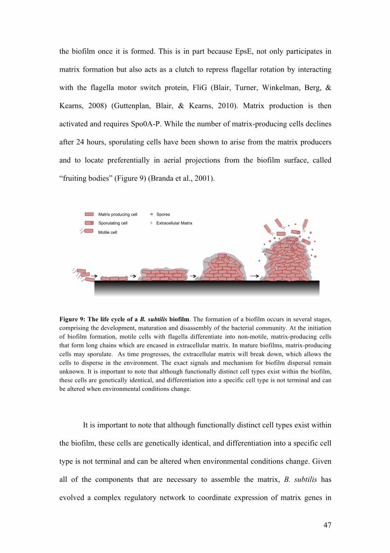
47
the biofilm once it is formed. This is in part because EpsE, not only participates in
matrix formation but also acts as a clutch to repress flagellar rotation by interacting
with the flagella motor switch protein, FliG (Blair, Turner, Winkelman, Berg, &
Kearns, 2008) (Guttenplan, Blair, & Kearns, 2010). Matrix production is then
activated and requires Spo0A-P. While the number of matrix-producing cells declines
after 24 hours, sporulating cells have been shown to arise from the matrix producers
and to locate preferentially in aerial projections from the biofilm surface, called
“fruiting bodies” (Figure 9) (Branda et al., 2001).
Figure 9: The life cycle of a B. subtilis biofilm. The formation of a biofilm occurs in several stages, comprising the development, maturation and disassembly of the bacterial community. At the initiation of biofilm formation, motile cells with flagella differentiate into non-motile, matrix-producing cells that form long chains which are encased in extracellular matrix. In mature biofilms, matrix-producing cells may sporulate. As time progresses, the extracellular matrix will break down, which allows the cells to disperse in the environment. The exact signals and mechanism for biofilm dispersal remain unknown. It is important to note that although functionally distinct cell types exist within the biofilm, these cells are genetically identical, and differentiation into a specific cell type is not terminal and can be altered when environmental conditions change.
It is important to note that although functionally distinct cell types exist within
the biofilm, these cells are genetically identical, and differentiation into a specific cell
type is not terminal and can be altered when environmental conditions change. Given
all of the components that are necessary to assemble the matrix, B. subtilis has
evolved a complex regulatory network to coordinate expression of matrix genes in
Spores Matrix producing cell
Sporulating cell
Motile cell
Extracellular Matrix

48
response to the shifting environmental conditions that it encounters in its natural
environment.
The Spo0A pathway
As its name suggests, Spo0A was first discovered as a gene required for the
sporulation pathway. Further studies revealed that Spo0A is an essential
transcriptional factor for all the adaptation pathways in B. subtilis by controlling the
expression of more than a hundred promoters, which differ in their Spo0A-P binding
affinities. Spo0A-P induces biofilm formation when present at intermediate levels, but
as the biofilm matures and the concentration of Spo0A-P increases in a subpopulation
of cells, sporulation will be triggered (Fujita, Gonzalez-Pastor, & Losick, 2005). As
previously described, Spo0A-P also regulates the K-state.
Spo0A-P promotes biofilm formation by inhibiting the action of two major
repressors of the epsA-O and the tapA operons, SinR and AbrB (Figure 10). The
derepression of SinR via Spo0A-P is indirect,. Spo0A-P directly increases the
transcription of sinI. SinI is an antagonist of the matrix gene repressor SinR, via
protein-protein interactions that inhibit SinR DNA binding. SinI is a SinR paralog,
that lacks the N-terminal DNA binding domain but contains a C-terminal
oligomerization domain similar to that of SinR. Furthermore, SinR activity is
regulated by SlrR. SlrR is another SinR paralog, but contains both domains found in
SinR. Its transcription is repressed by SinR (Chu et al., 2008). When slrR expression
is derepressed by SinR inactivation through SinI, induced SlrR binds to SinR and
reprograms SinR to repress expression of motility-promoting genes (see below)
(Vlamakis et al., 2008) (Chai et al., 2008). The second repressor of matrix production
AbrB is directly repressed by very low concentrations of Spo0A-P. In addition to its

49
inhibitory effect on the epsA-O and the tapA operons, AbrB has been shown to repress
the expression of the regulatory protein SlrR and the matrix protein BslA (Chu et al.,
2008) (Chai, Kolter, & Losick, 2009). The presence of the two matrix production
repressors SinR and AbrB with overlapping targets indicates that the regulation of
biofilm formation is tightly regulated in order to coordinate expression of the matrix
genes.
The SinR-SlrR regulation switch
The SinR-SlrR complex serves as a switch between biofilm formation and
motility, which is why, as previously mentioned, both SinR and AbrB control slrR
gene expression. At low concentrations of SlrR, lytABC and lytF (genes encoding for
autolysins, which are proteins involved in the separation cell chains), and hag (the
gene encoding flagellin) expression are not repressed (Figure 10). But when the level
of SlrR is high enough, the activity of SinR decreases in the cell, allowing the
expression of matrix coding genes and the development of biofilms. Additionally, the
SinR-SlrR complex represses the expression of hag, lytABC and lytF, as cell chaining
is essential for the onset of biofilm formation (Chai, Kolter, & Losick, 2010) (Chai,
Norman, Kolter, & Losick, 2010). The switch between the low level to the high level
of SlrR is dependent on SinI-mediated inhibition of SinR, which is directly controlled
by Spo0A-P (Figure 10). The SinR-SlrR switch remarkably leads to epigenetically
heritable changes (Vlamakis, Chai, Beauregard, Losick, & Kolter, 2013).

50
Figure 10: The SinI-SinR-SlrR switch between motile and sessile life A- When the concentration of Spo0A-P is low, motility genes and autolysin are expressed while biofilm formation is inhibited as the tasA-sipW-tapA and epsA-O operons are repressed. Without SinI, the repression of SinR low, and therefore SlrR is not present in highe enough concentrations to inhibit SinR. So, SinR represses biofilm formation, and cells are motile B- The concentration of Spo0A-P increases to some threshold level, and the SinR/SinR-SlrR ratio is reversed, by lowering the amount of free SinR in the cells. The repression of matrix production is relieved, while the synthesis of autolysin and motility is inhibited allowing the development of biofilm formation.
Biofilms and the DegS-DegU pathway
As previously described, DegU is a major regulator that is involved in the
regulation of many cellular adaptations such as the K-state, motility and secretion of
degradative enzymes. Furthermore, a degU mutant cannot form normal biofilms
because of the loss of the surface hydrophobicity protein BslA (Kobayashi & Iwano,
2012) (Kobayashi, 2007a) (Verhamme, Murray, & Stanley-Wall, 2009). BslA is a
small extracellular protein that, with TasA and exopolysaccharide, facilitates the
assembly of the matrix within a biofilm (Ostrowski, Mehert, Prescott, Kiley, &
Stanley-Wall, 2011). BslA forms a hydrophobic layer on the surface of the B. subtilis
0A !"#"
$%&'"
$%&("
!"#$%$)*("
$%&("$%&("$%&("$%&("$)*("+"
0A !"#" 0A !"#" 0A !"#"
$%&'"$%&'"
$%&'"
$%&("
!"#$%$)*("
$%&("$)*("
$%&("$)*("
$%&("$)*("
,"$%&("
-.*/"
/%01)2"
&'()% !*(+% &'!)%
,(!)-./0123456789:%
304)%56";'<%
-750)68%&"
"=&)% "=&-% "=&.%
"=&1%
-" /"

51
biofilm (Kobayashi & Iwano, 2012) (Hobley et al., 2013). Transcription of bslA has
been shown to be indirectly activated by the transcription factor Rok (Kovacs &
Kuipers, 2011), is directly inhibited by AbrB (Verhamme et al., 2009) and by an
intermediate level of DegU-P during biofilm formation. Additionaly, a degU null
mutant cannot synthesize the PGA polymer encoded by the pgs operon (Stanley &
Lazazzera, 2005), which in some bacilli is a prominent feature of biofilms. DegU-P,
in addition to its activator function at an intermediate level, can also inhibit biofilm
formation when its concentration is high (above some threshold) (Verhamme et al.,
2007) (Figure 8B).
Quorum Sensing in biofilms
One of the first molecules identified as an inducer of matrix gene expression
was surfactin. In addition to its surfactant and anti-microbial activities, surfactin
triggers the phosphorylation of Spo0A through the membrane-localized kinase KinC.
Here, instead of responding to direct binding of surfactin, KinC is activated by the
function of the molecule. Surfactin is a lipopeptide that inserts into the membrane and
results in potassium leakage, which activates KinC by an unknown mechanism and
leads to the expression of matrix genes (Lopez et al., 2010). Surprisingly, surfactin is
only produced by a sub-population of cells, and the cells that produce surfactin do not
respond to the molecule. Since the surfactin producers do not respond to the signal
they make and the signal is unidirectional, the surfactin production in B. subtilis can
be described as a paracrine signaling system (Lopez, Vlamakis, Losick, & Kolter,
2009). It contrasts with previously described quorum-sensing systems where every
cell in a population is thought to produce and respond to the signaling molecule.

52
Making a biofilm, a hallmark of undomesticated B. subtilis strains
As many laboratories work with the domesticated strain 168 or its derivatives,
the ability of B. subtilis to form robust biofilms has only been studied and
characterized at the beginning of this century (Branda et al., 2006). This work has
been carried out in the “wild” strain 3610, a strain believed to be the closest relative
of the laboratory strain 168 as these two strains share a great similarity in genome
sequence. A few mutations that are responsible for the phenotypic differences
between these two strains have been identified. Interestingly, single point mutations in
four genes (sfp, epsC, swrA and degQ) and the lack of the rapP gene are responsible
for the defect in matrix production in the domesticated strain. The sfp gene encodes a
broad-substrate-specificity phosphopantetheinyl transferase, which is involved in the
production of surfactin and a point mutation in strain 168 impairs its function. The
second point mutation is in epsC, which results in a decrease of exopolysaccharide
synthesis. The third mutation contributing to the impairment of biofilm formation is
located within swrA, which is known to be essential for swarming motility and poly-
!-polyglutamic acid synthesis (Stanley & Lazazzera, 2005). The last point mutation is
in the promoter sequence of degQ, a protein involved in the regulation of the DegS-
DegU pathway that is important for the regulation of biofilm formation (Verhamme et
al., 2009) (Marlow et al., 2014). As noted above, DegQ facilitates the transfer of a
phosphoryl group from DegS-P to DegU. The regulatory gene rapP, encoded on the
plasmid pLS32 is no longer present in strain 168. RapP has also recently been shown
to be involved in the regulation of biofilm formation (Omer Bendori, Pollak, Hizi, &
Eldar, 2015) (Parashar, Konkol, Kearns, & Neiditch, 2013) is unclear.

53
THESIS PURPOSE

54
It is well known that all the developmental adaptations (competence,
sporulation, biofilm formation) in B. subtilis share regulatory proteins (Spo0A,
DegS/DegU, SinI/SinR...). These adaptations have so far been studied in optimal
media with selected B. subtilis strains such as the laboratory strain 168 for the study
of competence, and the undomesticated 3610 strain for the study of biofilm formation.
In a given environment, while all the cells have the same genome, some cells will
induce one adaptation while others will develop another. Because biofilms contain
sporulating and non-sporulating cells, and presumably competent cells as well, they
appear to be the perfect context in which to study the relationships between all the
environment adaptations in B. subtilis. It is now clear that strain 168 has been selected
over decades for its high expression of competence for transformation, loosing at the
same time its ability to form biofilms. To study competence development in a biofilm
context it is therefore important to first understand competence regulation in
undomesticated isolates of B. subtilis.
In what follows I will refer to the state of competence for transformation as
the K-state because it is induced by the transcription factor ComK. The use of this
term serves as a reminder that this state of gene expression involves more than just
transformation and is a state of dormancy distinct from spores that confers tolerance
to antibiotics.
I began this scientific exploration by observing that most of the
undomesticated, biofilm forming strains of Bacillus subtilis are poorly transformable:
(i) the number of cells entering into the K-state is lower in undomesticated strains
compared to the laboratory strain 168; (ii) the ensemble rate of transcription of
ComK, the master regulator of competence, is significantly lower in wild isolates
(PS216 and 3610) than in the reference strain 168 while the uptick (explained below)

55
is the same in these three backgrounds. This difference suggests that an important
aspect of regulation has been lost by domestication and that our understanding of K-
state control is therefore incomplete. This work is devoted to correcting this
deficiency.
The main results of this study are presented here in three different chapters.
The first chapter introduces the publication of the work we have done on the
characterization of a genomic sequence of a new strain of B. subtilis: the PS216 strain
is indeed another undomesticated strain of B. subtilis that has been isolated in
Slovenian soil that form robust and structured biofilms.
The second chapter shows the work done on the characterization of the basic
network for competence regulation in the domesticated strain 168 and two
undomesticated strains of B. subtilis: the ancestral strain 3610 and the neo-
characterized PS216 strain. By comparing the genome sequences of these three
different strains and studying the effect of competence gene knock outs on the
expression of transformability and the K-state, we concluded that the core circuitry
for competence development is identical in domesticated and undomesticated strains.
We also confirmed that, in comparison to PS216 and 3610, the laboratory strain 168
carries in its genome a previously identified point mutation in the promoter of degQ
that will be of great interest in this study.
In the third chapter we show that this point mutation is involved in the
increased level of ComK in the undomesticated strain 168 and that its effect is
mediated by regulation of the phosphorylation state of DegU-P, a response regulator
known to be involved in the control of motility and biofilm formation. We discovered
that because of this point mutation in the promoter region of degQ, ComK is

56
stabilized in the laboratory strain compared to the undomesticated strain 3610. We
finally showed that, after screening other candidates, rapP, a gene encoded by a
plasmid in strain 3610 is also involved in the regulation of competence development
in this isolate, as it turns down the expression of the srfA operon and therefore limits
the percentage of cells entering into the K-state.

57
CHAPTER I
Genome sequence of the
Bacillus subtilis biofilm-
forming transformable
strain PS216

58
Genome sequence of the Bacillus subtilis biofilm-forming
transformable strain PS216
Russell Durrett1, Mathieu Miras2,3, Nicolas Mirouze2,4, Apurva Narechania5, Ines
Mandic-Mulec6 and David Dubnau*2
1 Institute for Computational Biomedicine, Weill Medical College of Cornell
University, New York, NY 10021, USA
2Public Health Research Institute, New Jersey Medical School, Newark, NJ 07103,
USA
3Université de Toulouse (Toulouse III), 118 route de Narbonne, 31062 Toulouse,
France
4 UMR1319 Micalis, Bat. Biotechnologie (440), I.N.R.A., Domaine de Vilvert, 78352
Jouy-en-Josas Cedex, France
5Sackler Institute for Comparative Genomics, American Museum of Natural History,
New York, New York, USA 10024
6Biotechnical Faculty, University of Ljubljana, 1000 Ljubljana, Slovenia
*To whom correspondence should be addressed,
ABSTRACT
PS216, a strain of Bacillus subtilis isolated in Slovenia has been sequenced.
PS216 is transformable and forms robust biofilms, making it useful for the study of
competence regulation in an undomesticated bacterium.

59
RESULTS
Bacillus subtilis is the most studied Gram-positive model organism
(Sonenshein, 2002 #4258). It has become apparent that the standard reference strain
168 has been modified by decades of inadvertent selection in the laboratory, thereby
acquiring a high frequency of transformability and losing the ability to form biofilms
(McLoon, 2011 #4080). Recently, “undomesticated” strains, notably NCIB3610, have
been investigated intensively because of their ability to form robust biofilms
(McLoon, #4080). However, NCIB3610 is poorly transformable, limiting its
usefulness for the study of genetic competence and compromising its ability to be
manipulated genetically. B. subtilis PS216, which was isolated in Slovenia from
sandy soil, forms robust biofilms and is more transformable than NCIB3610. Based
on phylogenetic analysis of three concatenated protein coding genes (dnaJ, gyrA and
rpoB), PS216 is most closely related to B. subtilis ssp. subtilis and belongs to a clade
demarcated as the putative ecotype 10 (Stefanic, #4727). Strain PS216 resides in the
same quorum-sensing pherotype group as 168 (Tortosa, 2001 #1706;Stefanic, 2009
#4201).
MATERIALS AND METHODS
The genome sequence of B. subtilis PS216 was generated as described in
(Koren, #4728). Briefly, 274 Mb of PacBio long-read data was error-corrected with
150 bp MiSeq data using the pacBioToCA pipeline, resulting in approximately 71 Mb
of corrected long reads that were then assembled by the Celera assembler. This
assembly contained 146 contigs, 90% of the assembly in 26 contigs larger than 42Kb.

60
The initial assembly yielded a total of 112 single nucleotide changes compared
to the reference strain 168 (NC_000964) and 140 SNPs compared to strain NCIB3610
(NZ_CM000488). SNPs were identified in SAMtools (Li, 2009 #4726) using short
read alignments generated by BWA (Li, 2009 #4725). Sequencing of PCR products
confirmed four of the nucleotide changes in genes of interest (oppD, comP, degQ,
sigH). Of the 112 nucleotides that differed between strains 168 and PS216, 27 were
identical in sequence between PS216 and NCIB3610. These include the confirmed
nucleotide changes in degQ, oppD and sigH.
Notably, no large plasmids were detected in PS216 such as the one present in
NCIB3610 (McLoon, 2011 #4080). We used NUCmer (Kurtz, 2004 #4721) and
ABACAS (Assefa, 2009 #4723) to order and orient the contigs with respect to the
reference, an analysis which revealed that both the 20,521 bp ICEBs1 element (Lee,
#4701) and the 134,385 bp SPß temperate bacteriophage present in 168 (Lazarevic,
1999 #4719) were missing from PS216. The absence of the latter two elements was
verified by sequencing a PCR product that crossed the two insertion sites.
We anticipate that this sequence information for PS216 will facilitate
comparative studies of development and physiology in Bacillus.
NUCLEOTIDE SEQUENCE ACQUISITION NUMBER.
This Whole Genome Shotgun project has been deposited at
DDBJ/EMBL/GenBank under the accession AQGR00000000. The version described
in this paper is the first version, AQGR01000000.

61
ACKNOWLEDGEMENTS
We appreciate discussions with Barry Kreiswirth. This work was supported by
NIH grant 5R01GM057720-43 awarded to DD and by Slovenian ARRS grants J4-
3631 and JP4-116, awarded to IMM.
REFERENCES
1. Sonenshein AL, Hoch JA, Losick R. 2002. Bacillus subtilis and its closest
relatives. From genes to cells. ASM Press, Washington, DC.
2. McLoon AL, Guttenplan SB, Kearns DB, Kolter R, Losick R. 2011. Tracing the
domestication of a biofilm-forming bacterium. J. Bacteriol. 193:2027–2034.
3. Stefanic P, Decorosi F, Viti C, Petito J, Cohan FM, Mandic-Mulec I. 2012. The
quorum sensing diversity within and between ecotypes of Bacillus subtilis. Environ.
Microbiol. 14:1378–1389.
4. Tortosa P, Logsdon L, Kraigher B, Itoh Y, Mandic-Mulec I, Dubnau D. 2001.
Specificity and genetic polymorphism of the Bacillus competence quorum-sensing
system. J. Bacteriol. 183:451–460.
5. Stefanic P, Mandic-Mulec I. 2009. Social interactions and distribution of Bacillus
subtilis pherotypes at microscale. J. Bacteriol. 191:1756–1764.
6. Koren S, Schatz MC, Walenz BP, Martin J, Howard JT, Ganapathy G, Wang
Z, Rasko DA, McCombie WR, Jarvis ED, Phillippy AM. 2012. Hybrid error
correction and de novo assembly of single-molecule sequencing reads. Nat.
Biotechnol. 30:693–700.
7. Li H, Handsaker B, Wysoker A, Fennell T, Ruan J, Homer N, Marth G,
Abecasis G, Durbin R, 1000 Genome Project Data Processing Subgroup. 2009.
The sequence alignment/map format and SAMtools. Bioinformatics 25:2078–2079.

62
8. Li H, Durbin R. 2009. Fast and accurate short read alignment with Burrows-
Wheeler transform. Bioinformatics 25:1754–1760.
9. Kurtz S, Phillippy A, Delcher AL, Smoot M, Shumway M, Antonescu C,
Salzberg SL. 2004. Versatile and open software for comparing large genomes.
Genome Biol. 5:R12.
10. Assefa S, Keane TM, Otto TD, Newbold C, Berriman M. 2009. ABACAS:
algorithm-based automatic contiguation of assembled sequences. Bioinformatics
25:1968–1969.
11. Lee CA, Thomas J, Grossman AD. 2012. The Bacillus subtilis conjugative
transposon ICEBs1 mobilizes plasmids lacking dedicated mobilization functions. J.
Bacteriol. 194:3165–3172.
12. Lazarevic V, DüsterhoÅNft A, Soldo B, Hilbert H, Mauël C, Karamata D.
1999. Nucleotide sequence of the Bacillus subtilis temperate bacteriophage SPbetac2.
Microbiology 145(Pt 5):1055–1067.

63
CHAPTER II
Domesticated and
undomesticated strains of
Bacillus subtilis share the
same basic network for
competence development

64
The firefly luciferase: a powerful tool for gene expression
studies
To further investigate if the core circuitry for competence regulation was the
same in the undomesticated backgrounds compared to the reference strain 168, we
looked at the effects on comK or comGA expression of knock outs (KO) of the main
regulators of competence determined in the reference strain. To follow the
transcriptional activity of these two genes in B. subtilis, we chose the luciferase gene
as a reporter. In the luciferase reaction, light is emitted when the luciferase acts on the
luciferin substrate. Photon emission can be detected by a properly equipped light
sensitive apparatus such as a plate reader. Possibly because the luciferase half life is
low, this is a powerful tool to study transcription rates in vivo compared to reporters
like ß-galactosidase that measure he accumulation of gene product (Figure 1A)
(Gould & Subramani, 1988).
Figure 1: The luciferase as a reporter gene to measure genes rate of transcription (A) Enzymatic reaction. (B) Genetic organization of the gene reporter.
comK
!"#$%&'()*++,*-&)'
PcomK luciferase
pUC18cm
PcomK
PcomK PcomK comK luciferase
pUC18cm
B. subtilis Chromosome
Luciferin + ATP
Luciferyl adenylate + O2
Luciferyl adenylate + PPi
Oxyluciferin + AMP + Light
A
B

65
To study the transcriptional activity of comK (and comGA), the firefly
luciferase coding sequence has been cloned downstream of the comK and comGA
promoters and inserted in the chromosome by homologous recombination (Figure
1B). This was done in two natural isolates, PS216 and NCIB3610.
Effect of different competence regulators KO on K-state
development
Because of the essential role of Spo0A in the regulation of competence
development via the regulation of the uptick (Mirouze et al., 2012), we first examined
the effect of a spo0A KO on the expression of comK and comGA. Figures 2A and 2B
show that the rate of transcription of both comK and comGA is greatly reduced in the
spo0A null mutants compared to the wild type, which is consistent with the known
role of Spo0A in the domesticated strain.
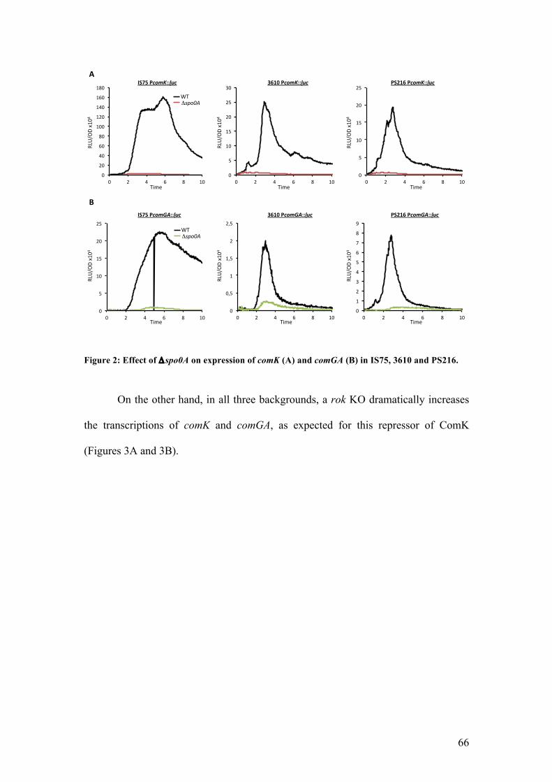
66
Figure 2: Effect of !spo0A on expression of comK (A) and comGA (B) in IS75, 3610 and PS216.
On the other hand, in all three backgrounds, a rok KO dramatically increases
the transcriptions of comK and comGA, as expected for this repressor of ComK
(Figures 3A and 3B).
!"
#!"
$!"
%!"
&!"
'!!"
'#!"
'$!"
'%!"
'&!"
!" #" $" %" &" '!"!"
("
'!"
'("
#!"
#("
)!"
!" #" $" %" &" '!"!"
("
'!"
'("
#!"
#("
!" #" $" %" &" '!"
!"
("
'!"
'("
#!"
#("
!" #" $" %" &" '!"!"
!*("
'"
'*("
#"
#*("
!" #" $" %" &" '!"!"
'"
#"
)"
$"
("
%"
+"
&"
,"
!" #" $" %" &" '!"
-./01
2"3'!$"
4567"
84"!!"#$%"
!"#$%&!"#$''%&!%(%
-./01
2"3'!$"
4567"
)*+,%&!"#$''%&!%
-./01
2"3'!$"
4567"
&"-+*%&!"#$''%&!%-./01
2"3'!$"
4567"
84"!!"#$%"
!"#$%&!"#'(''%&!%
.%
-./01
2"3'!$"
4567"
)*+,%&!"#'(''%&!%
-./01
2"3'!$"
4567"
&"-+*%&!"#'(''%&!%

67
Figure 3: Effect of !rok on expression of comK (A) and comGA (B) in IS75, 3610 and PS216.
We also investigated the effect of early competence gene KOs on the
expression of comK and comGA. For example a comS KO has a severe effect on the
expression of comGA in the three backgrounds; in the comS null mutants, ComS
cannot prevent ComK degradation via the MecA/ClpC/ClpP system (Figure 4B). We
observed the same phenomenon when looking at the expression of comK: the rate of
transcription of comK is lowered in the comS mutant compared to the wild type in the
three strains because ComK is not available to activate its own promoter (Figure 4A).
However we can distinguish some residual expression that coincides with the uptick:
the comS KO disrupts the autoregulatory loop of ComK on PcomK as ComK is
degraded but doesn’t have any effect on the uptick regulation of PcomK.
!"
#!!"
$!!"
%!!"
&!!"
'!!"
(!!"
)!!"
*!!"
+!!"
#!!!"
!" $" &" (" *" #!"!"
'!"
#!!"
#'!"
$!!"
$'!"
%!!"
%'!"
!" $" &" (" *" #!"!"
'!"
#!!"
#'!"
$!!"
$'!"
!" $" &" (" *" #!"
!"
'"
#!"
#'"
$!"
$'"
%!"
%'"
&!"
!" $" &" (" *" #!"!"
'"
#!"
#'"
$!"
$'"
%!"
%'"
&!"
!" $" &" (" *" #!"!"
'"
#!"
#'"
$!"
$'"
%!"
%'"
&!"
!" $" &" (" *" #!"
,-./0
1"2#!&"
3456"
73"!!"#"
!"#$%&!"#$''%&!%(%
,-./0
1"2#!&"
3456"
)*+,%&!"#$''%&!%
,-./0
1"2#!&"
3456"
&"-+*%&!"#$''%&!%,-./0
1"2#!&"
3456"
73"!!"#"
!"#$%&!"#'(''%&!%
.%,-./0
1"2#!&"
3456"
)*+,%&!"#'(''%&!%
,-./0
1"2#!&"
3456"
&"-+*%&!"#'(''%&!%

68
Figure 4: Effect of !comS on expression of comK (A) and comGA (B) in IS75, 3610 and PS216.
Contrary to the undomesticated strains, the laboratory strain 168 does not
produce surfactin because of a mutation in sfp (a gene involved in surfactin
production). To determine if the production of surfactin explains the lower
transformability of the wild isolates compared to strain 168 we monitored the
expression of comK and comGA in a surfactin null mutant. Eliminating the expression
of surfactin has no effect on the expression of either comK or comGA in the
undomesticated strains: the rate of transcription of our two reporter genes is
comparable in the mutants and the wild types in 3610 and PS216 (Figure 5A and 5B).
!"
#"
$!"
$#"
%!"
%#"
!" %" &" '" (" $!"!"
!)%"
!)&"
!)'"
!)("
$"
$)%"
$)&"
!" %" &" '" (" $!"!"
$"
%"
*"
&"
#"
'"
+"
("
,"
$!"
!" %" &" '" (" $!"
!"
#!"
$!!"
$#!"
%!!"
%#!"
*!!"
*#!"
!" %" &" '" (" $!"!"
#"
$!"
$#"
%!"
%#"
*!"
*#"
!" %" &" '" (" $!"!"
#"
$!"
$#"
%!"
%#"
*!"
*#"
&!"
&#"
!" %" &" '" (" $!"
-./01
2"3$!&"
-./01
2"3$!&"
-./01
2"3$!&"
4567" 4567" 4567"
-./01
2"3$!&"
4567"
-./01
2"3$!&"
4567"
-./01
2"3$!&"
4567"
84"!!"#$"
!"#$%&!"#$''%&!% ()*+%&!"#$''%&!% &",*)%&!"#$''%&!%
84"!!"#$"
!"#$%&!"#'(''%&!% ()*+%&!"#'(''%&!% &",*)%&!"#'(''%&!%
-%
.%

69
Figure 4: Effect of !srfA on expression of comK (A) and comGA (B) in IS75, 3610 and PS216.
All together, these results suggest that the basic circuitry for competence
development is the same in the laboratory strain 168 and the two undomesticated
strains tested, 3610 and PS216.
!"
#"
$!"
$#"
%!"
%#"
!" %" &" '" (" $!"!"
!)%"
!)&"
!)'"
!)("
$"
$)%"
!" %" &" '" (" $!"!"
!)%"
!)&"
!)'"
!)("
$"
$)%"
$)&"
$)'"
$)("
%"
!" %" &" '" (" $!"
!"
%!"
&!"
'!"
(!"
$!!"
$%!"
$&!"
!" %" &" '" (" $!"!"
#"
$!"
$#"
%!"
%#"
!" %" &" '" (" $!"!"
#"
$!"
$#"
%!"
%#"
*!"
*#"
&!"
&#"
!" %" &" '" (" $!"
+,-./
0"1$!&"
2345"
62"!!"#$"
!"#$%&!"#$''%&!%(%
+,-./
0"1$!&"
2345"
)*+,%&!"#$''%&!%
+,-./
0"1$!&"
2345"
&"-+*%&!"#$''%&!%+,-./
0"1$!&"
2345"
62"!!"#$"
!"#$%&!"#'(''%&!%
.%+,-./
0"1$!&"
2345"
)*+,%&!"#'(''%&!%
+,-./
0"1$!&"
2345"
&"-+*%&!"#'(''%&!%

70
Strains Table
Strain Number
Backgrounda Descriptionb
IS75 168 derivative his leu met BD6655 B. subtilis (IS75) PcomK::luc (Cm), "spo0A (Kan) BD6656 B. subtilis (3610) PcomK::luc (Cm), "spo0A (Kan) BD6657 B. subtilis (PS216) PcomK::luc (Cm), "spo0A (Kan) BD6667 B. subtilis (IS75) PcomGA::luc (Cm), "spo0A (Kan) BD6668 B. subtilis (3610) PcomGA::luc (Cm), "spo0A (Kan) BD6669 B. subtilis (PS216) PcomGA::luc (Cm), "spo0A (Kan) BD6460 B. subtilis (IS75) PcomK::luc (Cm), "rok (Kan) BD6464 B. subtilis (3610) PcomK::luc (Cm), "rok (Kan) BD6462 B. subtilis (PS216) PcomK::luc (Cm), "rok (Kan) BD6658 B. subtilis (IS75) PcomGA::luc (Cm), "rok (Kan) BD6659 B. subtilis (3610) PcomGA::luc (Cm), "rok (Kan) BD6660 B. subtilis (PS216) PcomGA::luc (Cm), "rok (Kan) BD6604 B. subtilis (IS75) PcomK::luc (Cm), "comS (Tet) BD6605 B. subtilis (3610) PcomK::luc (Cm), "comS (Tet) BD6606 B. subtilis (PS216) PcomK::luc (Cm), "comS (Tet) BD6607 B. subtilis (IS75) PcomGA::luc (Cm), "comS (Tet) BD6608 B. subtilis (3610) PcomGA::luc (Cm), "comS (Tet) BD6609 B. subtilis (PS216) PcomGA::luc (Cm), "comS (Tet) BD6652 B. subtilis (IS75) PcomK::luc (Cm), "srfA (Kan) BD6653 B. subtilis (3610) PcomK::luc (Cm), "srfA (Kan) BD6654 B. subtilis (PS216) PcomK::luc (Cm), "srfA (Kan) BD6664 B. subtilis (IS75) PcomGA::luc (Cm), "srfA (Kan) BD6665 B. subtilis (3610) PcomGA::luc (Cm), "srfA (Kan)
BD6666 B. subtilis (PS216) PcomGA::luc (Cm), "srfA (Kan) aThe IS75 derivatives are all his leu met auxotrophs. bAll of the fusion constructs are inserted by single crossover at the native loci.

71
CHAPTER III
A DegU-P and DegQ-
dependent regulatory
pathway for the K-state in
Bacillus subtilis

72
A DegU-P and DegQ-Dependent Regulatory Pathway for the K-state
in Bacillus subtilis
Mathieu Miras1,2 and David Dubnau1*
1Public Health Research Institute Center, New Jersey Medical School, Rutgers
University, Newark, NJ, USA,
2Laboratoire de Microbiologie et Génétique Moléculaires, Université de Toulouse,
Toulouse, France
ABSTRACT
The K-state in the model bacterium Bacillus subtilis is associated with
transformability (competence) as well as with growth arrest and tolerance for
antibiotics. Entry into the K-state is determined by the stochastic activation of the
transcription factor ComK and occurs in about ∼15% of the population in
domesticated strains. Although the upstream mechanisms that regulate the K-state
have been intensively studied and are well understood, it has remained unexplained
why undomesticated isolates of B. subtilis are poorly transformable compared to their
domesticated counterparts. We show here that this is because fewer cells enter the K-
state, suggesting that a regulatory pathway limiting entry to the K-state is missing in
domesticated strains. We find that loss of this limitation is largely due to an
inactivating point mutation in the promoter of degQ. The resulting low level of DegQ
decreases the concentration of phosphorylated DegU, which leads to the de-repression

73
of the srfA operon and ultimately to the stabilization of ComK. As a result, more cells
reach the threshold concentration of ComK needed to activate the auto-regulatory
loop at the comK promoter. In addition, we demonstrate that the activation of srfA
transcription in undomesticated strains is transient, turning off abruptly as cells enter
the stationary phase. Thus, the K-state and transformability are more transient and
less frequently expressed in the undomesticated strains. This limitation is more
extreme than appreciated from studies of domesticated strains. Selection has
apparently limited both the frequency and the duration of the bistably expressed K-
state in wild strains, likely because of the high cost of growth arrest associated with
the K-state. Future modeling of K-state regulation and of the fitness advantages and
costs of the K-state must take these features into account.
INTRODUCTION
The transcription factor ComK (van Sinderen et al., 1995) directly activates
more than 100 genes (Berka et al., 2002; Hamoen et al., 2002; Ogura et al., 2002).
While about 20 of these mediate the uptake, processing and integration of exogenous
DNA resulting in transformation (Burton and Dubnau, 2010), the roles of the
remaining ∼80 genes are poorly understood. Because these genes are not needed for
transformation (J. Hahn, unpublished) the expression of ComK must result in
phenotypes beyond competence that presumably enhance fitness. In fact, ComK also
induces a period of growth arrest during which the cells that express ComK exhibit
antibiotic tolerance (Nester and Stocker, 1963; Haijema et al., 2001; Johnsen et al.,
2009; Briley et al., 2011; Hahn et al., 2015; Yuksel et al., 2016). This persistent state
has been called the K-state, to emphasize that ComK regulates more than competence
for transformation (Berka et al., 2002).

74
A long recognized and remarkable feature of the K-state is that it is expressed
in a minor fraction of a clonal population, in a more or less all or nothing fashion
(Nester and Stocker, 1963; Singh and Pitale, 1967; Hadden and Nester, 1968;
Haseltine- Cahn and Fox, 1968; Maamar and Dubnau, 2005; Smits et al., 2005). Entry
into the K-state is determined stochastically and studies in domesticated strains
derived from the indole-requiring auxotrophic strain 168 (Spizizen, 1958) have
ascribed this stochastic determination to noise in the basal expression of the comK
promoter (Maamar et al., 2007; Süel et al., 2007). When the noisy expression of comK
causes a subpopulation of cells to exceed a threshold level of ComK, two dimers of
this protein bind cooperatively to the comK promoter (van Sinderen and Venema,
1994; Hamoen et al., 1998), activating a positive feedback loop and the rapid
transition of these cells into the K-state, where ComK activates downstream genes.
The frequency of these activation events is extremely low during exponential
growth and then rises as a culture approaches the stationary phase of growth. This
temporal control has two principal causes. First, growing cultures secrete ComX, a
quorum sensing pheromone, which accumulates and ultimately causes the
phosphorylation of the response-regulator protein ComA (Magnuson et al., 1994).
ComA-P then binds to and activates the promoter of the srfA operon, which encodes
the small protein ComS (Nakano and Zuber, 1991; Nakano et al., 1991; Roggiani and
Dubnau, 1993; Hamoen et al., 1995). ComS in turn competes with ComK for binding
to the MecA-ClpC-ClpP protease, which rapidly degrades ComK during growth
(Turgay et al., 1997, 1998; Prepiak and Dubnau, 2007). Stabilization occurs toward
the end of exponential growth when high cell densities have produced sufficient
ComX levels. A second cause of temporal regulation derives from the exquisitely
controlled kinetics of comK basal expression (Leisner et al., 2007; Mirouze et al.,

75
2012). Ensemble measurements show that the average basal rate of comK
transcription increases gradually during growth, reaches a maximum as cells depart
from exponential growth (T0), and then declines. This uptick in basal expression is
due to a gradual increase in the phosphorylated form of the master regulator Spo0A as
cells approach stationary phase (Mirouze et al., 2012). Spo0A is phosphorylated as a
consequence of a phosphorylation cascade in which several kinases transfer
phosphoryl groups to Spo0F (Burbulys et al., 1991). These groups are then passed to
the phosphotransfer protein Spo0B and finally to Spo0A. Low levels of Spo0A-P
directly activate the basal expression of comK, while higher levels bind to repressive
operator sites so that the rate of comK expression decreases (Mirouze et al., 2012).
Thus, Spo0A- P opens and then closes a temporal gate for transitions to the K-state.
Although K-state regulation has been well characterized, there was reason to believe
that our understanding was lacking. It has been observed that the transformability of
undomesticated isolates of Bacillus subtilis and its close relatives is much lower than
that of the domesticated derivatives of 168 (Cohan et al., 1991). In fact the model
undomesticated isolate NCIB3610 (hereafter 3610), is poorly transformable, although
it is very closely related to the wild parent of 168. The poor transformability is due in
part to comI, a gene that is absent in 168-derivatives. Interestingly, the ComI protein
appears to decrease the uptake of DNA, without affecting K-state expression (Konkol
et al., 2013). Together, these observations suggest that some regulatory feature has
been lost in the domesticated strains and that our appreciation of K-state regulation is
consequently incomplete.
In the present study, we have shown that a known promoter mutation in degQ
in domesticated strains (Yang et al., 1986; McLoon et al., 2011) is primarily
responsible for this difference in transformability. It is of interest that this mutation

76
also contributes to the failure of strain 168 derivatives to form robust biofilms
(McLoon et al., 2011). A consequence of this mutation is that the response regulator
DegU is poorly phosphorylated in domesticated strains (Stanley and Lazazzera, 2005;
Kobayashi, 2007). This deficit in DegU-P derepresses PsrfA. This causes ComK to be
stabilized, allowing more cells to pass the threshold for comK auto-activation, thereby
increasing the fraction of K-state cells. It is known that unphosphorylated DegU is
required for K-state expression because it helps ComK bind to its own promoter, thus
acting as a priming protein when the ComK concentration is low (Hamoen et al.,
2000). Thus, our present results show that the regulation of the K-state in
undomesticated strains requires the proper ratio of phosphorylated and
unphosphorylated DegU, in accordance with the view that this protein acts as a
rheostat for development (Verhamme et al., 2007).
MATERIALS AND METHODS
Microbiological Methods
Bacterial strains are listed in Supplementary Table S1. The backgrounds used
for all experiments were either IS75, a derivative of strain 168, PS216 (an
undomesticated strain of B. subtilis isolated in Slovenia and kindly provided by Inés
Mandic-Mulec) (Durrett et al., 2013), 3610!comI or 3610 comIQ12L (both gifts
from Dan Kearns). The comIQ12L mutation abolishes ComI activity (Konkol et al.,
2013), removing a block in DNA uptake. Constructs were introduced into IS75 by
transformation (Albano et al., 1987) and into 3610!comI and PS216 by transduction
using bacteriophage SPP1 (Cozy and Kearns, 2010). An exception was for the
swapping of the degQ alleles, which was carried out by transformation, as described
below. Bacterial growth was at 37#C in competence medium (Albano et al., 1987)

77
unless otherwise specified. Antibiotic selections were carried out on Lysogeny Broth
(LB) agar plates (Cozy and Kearns, 2010) containing ampicillin (100 µg ml$1),
spectinomycin (100 µg ml$1), erythromycin (5 µg ml$1), kanamycin (Kan) (5 µg
ml$1) or chloramphenicol (5 µg ml$1). In some cases selection was for erythromycin
(Ery) (1 µg ml$1) plus lincomycin (20 µg ml$1). Solid media were solidified by the
addition of 1.5% agar. Transformation frequencies were determined using genomic
DNA isolated from a leucine prototroph.
Luciferase Assays
Light output from luciferase reporter constructs was measured as previously
described (Mirouze et al., 2011). Briefly, strains were first grown in LB medium for 2
h. Cells were then centrifuged and resuspended in fresh competence medium (Albano
et al., 1987), adjusting all the cultures to an OD600 of 2. These pre-cultures were then
diluted 20-fold in fresh competence medium and 200 µl was distributed in duplicate
in the wells of a 96-well black plate (Corning Incorporated Costar). Ten microliters of
luciferin was added to each well to reach a final concentration of 1.5 mg/ml (4.7
mM). The cultures were incubated at 37#C with agitation in a PerkinElmer Envision
2104 Multilabel Reader equipped with an enhanced sensitivity photomultiplier for
luminometry. The temperature of the clear plastic lid was maintained at 38#C to avoid
condensation. Relative Luminescence Units (RLU) and OD600 were measured at 1
min intervals after two 30-s shaking steps. The data were processed using a script
written in MATLAB, exported to Excel and plotted as RLU/OD versus time from the
beginning of growth.
SDS-PAGE and Immunoblotting

78
Cell pellets were resuspended in STM buffer (50 mM Tris pH 8.0, 25%
sucrose, 50 mM NaCl, 5 mM MgCl2) containing 300 µg ml$1 lysozyme and
incubated at 37#C for 5 min. The volume of STM was normalized to the turbidity
measurement of the culture (determined in a Klett colorimeter) when the sample was
collected. Sample buffer (final concentration of 20 mM Tris HCl pH 6.8, 10%
glycerol, 1% SDS, 0.01% bromophenol blue, 2% 2-mercaptoethanol) was added to
the samples, which were then incubated at 100#C for 5 min. Samples were separated
by electrophoresis in 12% Tris-tricine SDS polyacrylamide gels (Schägger and von
Jagow, 1987). The proteins were transferred to nitrocellulose membranes (Millipore)
using a Trans Blot Turbo semidry transfer apparatus (Bio-Rad). Primary antiserum
raised in rabbits against ComK was used at a dilution of 1:5000. Signal was detected
using a secondary dilution of 1:10,000 goat anti- rabbit antiserum conjugated to
horseradish-peroxidase (HRP) followed by visualization using Enhanced
Chemiluminescence (ECL) Prime Western Blot Detection Reagent (GE Healthcare)
according to the manufacturers’ instructions. Images were recorded by using a
Thermo Scientific MyECL Imager and band intensities were measured using ImageJ
software (Schneider et al., 2012).
degQ Allele Swapping with pMiniMAD2
To swap the degQ promoter between IS75 and 3610 at the native loci, we used the
pMiniMAD2 cloning strategy as described (Cozy and Kearns, 2010; Mukherjee et al.,
2013). A 2 kb fragment containing degQ from strain 168 or 3610 was amplified using
primers (HinDIII-degQ9:
5%GCAGCAAAGCTTCTGCGATTTCCGGATAAAAGAACATAATAATCCCAG-
3% and BamHI-degQ10: 5%-GCAGCAGGATC

79
CGCAGCCTGCTTCTTATATGCTGATCG-3%). The amplicons, which carried the
degQ wild and mutant alleles near their centers, were cloned into the HinDIII and
BamHI sites on pMiniMAD2, to produce the plasmids pED1932 and pED1931, which
carried the wild-type and mutant degQ alleles, respectively. These plasmids were used
to transform IS75 (with pED1932) and 3610 (with pED1931) where they integrated
by single crossover events. Plasmid-free strains carrying the swapped degQ promoters
were isolated (Cozy and Kearns, 2010; Mukherjee et al., 2013) to create IS75
degQ3610 (BD7454) and 3610 degQIS75 (BD7445). The presence of the swapped
promoters was confirmed by sequencing of PCR products amplified from the
chromosome, carried out by Eton Biosciences (Union, NJ, USA).
The Phyper-spank-degQ Construction
The primers degQ15 (5%-TTAGTCGACAGCTAGCCACCATAC
ACAATTCATTGATCTTTCA-3%) and degQ16 (5%-CTTGCAT
GCGGCTAGCTACTCGTTAATCCTACTGTATACAAGGA-3%) were used to
amplify a 676 bp sequence containing the degQ gene without its promoter. The
amplicon was inserted into the Phyper-spank vector pED1870 that had been cut with
NheI, using the In-Fusion HD cloning kit (Clontech, Inc.), as per the manufacturer’s
instructions. The resulting plasmid was integrated into the desired host strains by
transformation with single crossover events, by selection for Kan resistance.
pED1870 carries the Phyper-spank, lacI and lacO sequences. This and all other
constructs were verified by sequencing. The resulting plasmid creates a strain in
which degQ is under Phyper-spank at its native locus.
Microscopy

80
Cells were harvested, diluted into PBS (81 mM Na2HPO4 + 24.6 mM
Na2HPO4 + 100 mM NaCl) and 1 µl of each culture was placed on a pad of 1%
agarose made up in 0.5X TAE buffer. Images were collected using a Nikon Eclipse Ti
inverted microscope equipped with an Orca Flash 4.0 digital camera (Hamamatsu),
with a Nikon TIRF 1.45 NA Plan Neoflur 100 oil immersion objective. NIS-Elements
AR (v 4.40, Nikon) software was used to collect and analyze images, which were then
imported into Photoshop to configure the images for publication. Fluorescence
intensities were determined using the automated General Analysis tool of NIS-
Elements.
RESULTS
Undomesticated Bacillus subtilis Strains Express the K-state in Few Cells
It has been observed that the transformability of undomesticated B. subtilis
strains is generally lower than that of the domesticated strain 168 and its derivatives
(Cohan et al., 1991). Because the K-state is expressed bistably, this difference may
reflect differing efficiencies of transformation per cell or differing frequencies of K-
state cells in the population. We therefore determined the percentage of competence-
expressing cells during growth in five randomly chosen natural isolates: the B. subtilis
subsp. subtilis RO-OO-2 and RO-FF-1 strains, the B. subtilis subsp. spizizenii RO-E-
2 [all three isolated in the Mojave desert (Cohan et al., 1991)], the commonly used
model strain 3610 (Branda et al., 2006; McLoon et al., 2011) and B. subtilis PS216,
isolated in Slovenia (Durrett et al., 2013). Throughout this study we have used comI
mutants of 3610. ComI does not affect the expression of comK but does inhibit the
uptake of transforming DNA (Konkol et al., 2013). In each case, after verifying that
the regulatory sequences upstream of comK were the same in all the strains (not

81
shown), comK promoter (PcomK) fusions to the genes encoding Cyan Fluorescent
Protein (CFP) or Green Fluorescent Protein (GFP) were integrated by single
reciprocal recombination and the percentages of comK-expressing cells were
enumerated microscopically and compared to that of the 168- derivative IS75, which
is the reference domesticated strain used throughout this study. We determined that
the time of maximal transformability for PS216 and 3610 was attained after 240 min
of growth and after 315 min for IS75 (Supplementary Figure S1). The results in
Figure 1 were therefore obtained with samples taken at those times, which correspond
to about T0 and T2 for the undomesticated and domesticated strains, respectively. The
growth curves for all the strains under these conditions were similar (not shown). The
results summarized in Figure 1 show that the samples from the undomesticated
isolates contain significantly fewer competence-expressing cells (0.2–5%) than the
domesticated strain sample (15.4%).
!"#""#$% !"#&&#'% !"#(#$%
)*#$'+% ,+'-% .*/0%
-1$2% -102% 02%
-132%,1+2% '0132%

82
Figure 1: Undomesticated strains express the K-state with low frequency. Shown are representative images of the indicated strains expressing PcomK fusions to the genes encoding GFP (top row) or CFP (bottom row). The fields were selected to show at least one expressing cell. The measured frequencies of K-state cells are indicated in the lower right of each panel. As explained in the text, the domesticated strain (IS75) was imaged at T2 and the undomesticated strains at T0. Strain numbers are presented in Supplementary Table S1.
To compare the transcription rates from PcomK in strains 3610 and PS216 to
that of the domesticated strain, we utilized promoter fusions to firefly luciferase,
which reports transcription rates rather than the accumulation of a gene product
(Mirouze et al., 2011). The fusions were integrated by single-reciprocal
recombination placing the reporter gene under control of the normal comK regulatory
sequences. The peak rates of transcription from PcomK in the domesticated strain and
in both 3610 and PS216 approximately reflect the frequencies of K-state cells in each
population (Figure 2). The peak transcription rates in the undomesticated strains are
about 9.6- and 5.7-fold lower than in the IS75 background, while the fractions of
competence- expressing cells differ by 38- and 4.2-fold, respectively. We do not
expect an exact correspondence between the peak rates of transcription and
transformation frequencies; light output from luciferase does not inform us about the
amount of ComK synthesized, the activation of the downstream genes needed for
transformation and the assembly of the transformation machinery. In the domesticated
strain, the transcription rate remains elevated even as the cultures enter stationary
phase (Figure 2A). In 3610 and PS216, the rates decline rapidly from a maximum
reached at about T0 (Figures 2B,C). These data demonstrate that unknown
mechanisms limit the probability of transitions to the K-state and cause the expression
of comK to cease abruptly. Interestingly, these mechanisms have been lost in the
domesticated strains.

83
Figure 2: Expression from PcomK is higher in IS75 (A) than in the undomesticated strains 3610 (B) and PS216 (C). (D) The three curves plotted on the same scale. The vertical arrows on panels (A–C) point to T0. Strain numbers are presented in Supplementary Table S1.
A degQ Mutation in the Domesticated Strains Causes Increased comK
Expression
To identify gene(s) that limit transitions to the K-state in undomesticated
strains, we transformed PS216 with DNA from a domesticated strain that carries a
fusion of the comG promoter to lacZ, linked to a kanamycin (Kan) resistance
determinant. It was reasoned that selection for this marker on plates containing Kan
and 5-bromo-4-chloro-3-indolyl-&-D-galactopyranoside (X-gal) would select for
transformable cells and might transfer an unlinked mutation from the domesticated
strain, capable of conferring increased expression from PcomG. Indeed, dark blue
colonies were observed, representing about 1% of the total number of KanR colonies,
a frequency consistent with “congression,” the simultaneous transformation of B.
!"#$%&
'()*+
,&-./0
&
.12&3!"#$44%&!&&51./&3!"#$44%&!&367.1&3!"#$44%&!&
/&
8/&
.//&
.8/&
7//&
78/&
5//&
/& 7& 0& 1& 2& ./&
'()*+
,&-./0
&
!"#$%&
'()*+
,&-./0
&
!"#$%&
!"#$%&
'()*+
,&-./0
&
/&
8&
./&
.8&
7/&
78&
5/&
/& 7& 0& 1& 2& ./&
51./&3!"#$44%&!&
/&8&
./&
.8&7/&78&5/&58&0/&08&8/&
/& 7& 0& 1& 2& ./&
367.1&3!"#$44%&!&&
/&
8/&
.//&
.8/&
7//&
78/&
5//&
/& 7& 0& 1& 2& ./&
.12&3!"#$44%&!&&!"
#"$"
%"

84
subtilis by unlinked markers. Although PS216 colonies are normally mucoid, all of
these blue colonies were not. Mucoidy in B. subtilis indicates the production of poly-
!-glutamic acid, due to the expression of the pgs operon and is dependent on the
presence of phosphorylated DegU and the small protein, DegQ (Stanley and
Lazazzera, 2005). DegQ increases the net transfer of a phosphoryl group from the
histidine kinase DegS to its cognate response regulator protein DegU (Kobayashi,
2007). Because derivatives of strain 168 carry a promoter mutation that markedly
decreases the expression of degQ (Yang et al., 1986; McLoon et al., 2011), we
reasoned that the blue colonies might have inherited this mutation. The degQ
promoters of several blue transformants were sequenced and indeed all had the T'C
promoter mutation at position $10 that exists in 168 derivatives.
To determine whether this mutation was responsible for the high comK
expression of domesticated strains, we swapped the wild-type and mutant degQ
alleles between the domesticated and undomesticated strains, using pMiniMAD2. For
this, and for all subsequent experiments reported here, we have used 3610, which has
been widely adopted as a model undomesticated B. subtilis, rather than the less
intensively studied PS216. Figure 3A shows the expression profiles of PsrfA, PcomK,
and PcomG luciferase fusions in the IS75 background, for strains carrying the
indigenous mutant (degQIS75) and wild-type (degQ3610) alleles.

85
Figure 3: Effect of the mutant (degQIS75) and wild-type (degQ3610) degQ alleles on expression of srfA, comK, and comG in IS75 (A) and 3610 (B). In each panel, the data from strains with indigenous and swapped alleles are indicated by solid and dashed lines, respectively. The vertical arrows in each panel point to T0. Strain numbers are presented in Supplementary Table S1.
Clearly the introduction of the wild-type degQ allele resulted in a marked
diminution of the transcription rates of each reporter so that the peak levels were
similar to those of 3610 (compare to Figure 3B). The converse was also true (Figure
3B); introduction of the degQIS75 allele into the 3610 background increased the
maximum transcription rates of all three promoters to approximately the levels
measured in IS75 (Figure 3A). A degQ knockout was also tested (Supplementary
Figure S2) using a PcomG-luc reporter in the 3610 background. As expected, a large
increase in comG transcription was noted. The effects of degQ allele swapping shown
in Figure 3 can thus be explained most economically by a repressing effect of DegU-P
on PsrfA, which would decrease the amount of ComS, destabilizing ComK. In fact it
has been shown that mutations resulting in an elevated level of DegU-P do indeed
!!"#$""%&'# !'()*""%&'# !'()+""%&'#
$#
%#
&#
'#
(#
)$#
)%#
)&#
)'#
)(#
$# %# &# '# (# )$#
*+,-.
/#0)$&
#
12345#
$#
%$#
&$#
'$#
($#
)$$#
)%$#
)&$#
)'$#
)($#
%$$#
$# %# &# '# (# )$#
*+,-.
/#0)$&
#
12345#
$#
6#
)$#
)6#
%$#
%6#
$# %# &# '# (# )$#
*+,-.
/#0)$&
#
12345#
7896,-./7896#7896,-./:')$#
!!"#$""%&'# !'()*""%&'# !'()+""%&'#
$#
%#
&#
'#
(#
)$#
)%#
)&#
)'#
)(#
$# %# &# '# (# )$#
*+,-.
/#0)$&
#
12345#
$#
%$#
&$#
'$#
($#
)$$#
)%$#
)&$#
)'$#
$# %# &# '# (# )$#
*+,-.
/#0)$&
#
12345#
$#
)#
%#
:#
&#
6#
'#
9#
(#
;#
$# %# &# '# (# )$#
*+,-.
/#0)$&
#
12345#
:')$,-./:')$#:')$,-./7896#
!"
#"
7896,-./7896#7896,-./:')$#
7896,-./7896#7896,-./:')$#
:')$,-./:')$#:')$,-./7896#
:')$,-./:')$#:')$,-./7896#

86
depress the transcription of srfA in the domesticated background (Hahn and Dubnau,
1991).
The Sharp Decrease in Transcription in 3610 Is Not Due to DegQ
Somewhat surprisingly, the characteristic sharp decreases in the transcription
rates of srfA and of comK in the 3610 background are not affected by the introduction
of the mutant degQ promoter (Figure 3). Nor are the sustained high transcription rates
from these promoters in the domesticated background obliterated when the wild-type
degQ allele is introduced. This is seen clearly when the peak values of the swapped
and un-swapped strains are normalized (Supplementary Figure S3). The comK
transcription patterns can be explained by differences in srfA transcription if we make
the simplifying assumption that ComS is unstable. Thus, when the rate of comS
transcription decreases sharply, ComK would no longer be protected from
degradation, and the transcription of comK and of comG would decrease. In accord
with this reasoning, the transcription rates of srfA, comK and comG begin to drop
after 2.3, 3.1, and 3.1 h of growth, respectively (Figure 3). The converse would apply
in the domesticated strains; sustained transcription from the srfA promoter would
provide a steady supply of ComS, stabilizing ComK. To test this idea, we measured
the transcription rates of srfA in three different backgrounds. As noted above, PS216
is intermediate between 3610 and IS75 in transformability, in comK expression and in
the percentage of cells that enter the K-state. Supplementary Figure S4 shows that
while transcription from PsrfA drops to zero in 3610, it decreases to an intermediate
level in PS216. This can be seen clearly when the three curves are normalized to the
same peak value (Supplementary Figure S4B). These comparisons are in agreement

87
with the hypothesis that the kinetics of srfA transcription underlie the strain
differences in transformability.
The DegQ Effect on K-state Expression Is Mediated by Increased DegU-P
If the presence of the mutant degQ allele affects competence expression
because of its depressing effect on DegU phosphorylation, it should be mimicked, at
least in 3610, by inactivation of degS, which encodes the DegU-cognate histidine
kinase. The results obtained in the 3610 background support a role for DegU-P as a
regulator of srfA and hence of comK and comG expression (Figure 4). The degS
knockout strain reaches a higher rate of srfA transcription than the wild-type and
presumably produces more ComS (Figure 4B). Consequently ComK is stabilized,
more comK transcription takes place and comG transcription is actually increased
(Figure 4B). In contrast with this straightforward result, Figure 4A reveals that the
inactivation of degS has no effect on srfA transcription in the IS75 background. This
is not unexpected, because in this background, which is naturally lacking in DegQ,
the amount of DegU-P is low and does not limit srfA transcription. However, the
expression from comK is affected, decreasing in peak value about twofold. This
relatively minor effect of degS inactivation can be explained as follows.
Unphosphorylated DegU is known to increase the affinity of ComK for its own
promoter (Hamoen et al., 2000). Because DegU-P activates one of the promoters that
drive degU expression (Ogura and Tsukahara, 2010), it is likely that the inactivation
of degS decreases the amount of DegU and thus compromises ComK binding to
PcomK. The expression from PcomG is also affected (Figure 4A) but less than that
from PcomK, suggesting that ComK is normally produced in excess.

88
Figure 4: Effect of degS on expression of srfA, comK, and comG in IS75 (A) and 3610 (B). The vertical arrows in each panel point to T0. Strain numbers are presented in Supplementary Table S1.
Further evidence that the depressing effect on srfA expression in 3610 is due
to phosphorylated DegU was obtained using a degUD56N allele. The DegUD56N
protein cannot be phosphorylated. Supplementary Figure S5 shows that the
DegUD56N mutant strain is de-repressed for srfA transcription similarly to the degS
inactivated strain (compare to Figure 4B). As observed when the degQ alleles were
swapped (Figure 3; Supplementary Figure S3), no effect of either the D56N or degS
knockout mutations was observed on the sharp downturn in srfA transcription in the
3610 background.
DegQ Regulates the Stability of ComK
If the effects of DegU-P on PsrfA, and hence on comS transcription underlie
the differences in K-state expression, we would expect to see this reflected in
measurements of ComK stability. Because the amount of ComK in the presence of
!"#$%
&'()*+
'
,-./0'
*'
1'
)*'
)1'
2*'
21'
*' 2' +' 3' 4' )*'
!"#$%
&'()*+
'
,-./0'
5!"#$66%&'' 5'()*66%&'' 5'()+66%&''
*'
2*'
+*'
3*'
4*'
)**'
)2*'
)+*'
)3*'
)4*'
*' 2' +' 3' 4' )*'
!"#$%
&'()*+
'
,-./0'
!"
*'
2'
+'
3'
4'
)*'
)2'
)+'
*' 2' +' 3' 4' )*'
!"#$%
&'()*+
'
,-./0'
5!"#$66%&''
*'
1'
)*'
)1'
2*'
21'
7*'
71'
*' 2' +' 3' 4' )*'
!"#$%
&'()*+
'
,-./0'
*'
2'
+'
3'
4'
)*'
)2'
)+'
)3'
*' 2' +' 3' 4' )*'
!"#$%
&'()*+
'
,-./0'
5'()*66%&'' 5'()+66%&''#"
*'
2'
+'
3'
4'
)*'
)2'
)+'
)3'
*' 2' +' 3' 4' )*'
89:1'89:1;,-./0'
89:1'89:1;,-./0'
89:1'89:1;,-./0'
73)*'73)*;,-./0'
73)*'73)*;,-./0'
73)*'73)*;,-./0'

89
elevated DegQ is quite low, we resorted to the following strategy to investigate its
stability. A strain was constructed that carries a copy of comK under control of the
xylose-inducible Pxyl promoter inserted in the ectopic amyE locus of IS75. The
native comK gene was left intact so that the effects of induction due to the addition of
xylose would be amplified by the auto- activation of comK transcription. Also present
was a copy of degQ under control of the isopropyl-&-D-thiogalactoside (IPTG)-
inducible Phyper-spank promoter, located in the chromosome at the degQ locus. This
strain was grown in the continuous presence of xylose, with and without IPTG. When
late log phase was reached, rifampicin and puromycin were added to terminate
mRNA and protein synthesis, samples were collected thereafter at regular intervals,
and Western blotting was used to monitor the decay of ComK (Figure 5).
As predicted, the stability of ComK was markedly reduced when degQ was
induced. In the uninduced culture the half-life of ComK is well in excess of 20 min;
extrapolation would suggest it to be at least 60 min. The half-life of ComK in the
presence of induced DegQ is 7– 8 min. As expected, the initial amount of ComK was
also reduced in the induced culture. These data confirm that the enhanced
transformability of the domesticated strain can be ascribed to the increased stability of
ComK, caused by the de-repression of srfA (comS).

90
Figure 5: DegQ lowers ComK stability. A strain carrying Pxyl-comK and Phyper-spank-degQ (BD8288) was grown in the presence of xylose and with and without IPTG. At T$1, puromycin and rifampicin, each at 200 µg/ml were added. At the indicated times, samples were withdrawn for Western blotting with anti-ComK antiserum. (A) The -IPTG gel was exposed for a shorter time to compensate for its higher initial intensity of the ComK band. Extracts from a comK strain were included to assist in the identification of the correct band. The ±IPTG images were from the same gel and placed one above the other (B) Bands intensities were quantified from digitized images using ImageJ software and plotted against time after the addition of the inhibitors.
Single Cell Expression in degQ-Swapped Strains
Ensemble measurements of srfA and comG transcription show that their
expression is correlated, and influenced by the levels of DegU-P. Figure 6 shows this

91
behavior on a single cell level. For these experiments, cells expressing promoter
fusions of genes encoding mCherry and GFP to PsrfA and PcomG, respectively, were
examined microscopically. Unlike firefly luciferase these reporters reflect the
accumulation of gene products. These images were collected for the wild-type IS75
and 3610 strains as well as for strains in which the degQ alleles had been swapped. It
is obvious that the GFP expressed from PcomG accumulated in more cells in the IS75
background than in 3610, as expected. Also as expected, the intensity of the mCherry
fluorescence expressed from PsrfA was much higher in IS75 than in the
undomesticated strain. When the degQIS75 allele was introduced into 3610, the
expression from PsrfA increased and the frequency of PcomG-expressing cells also
increased. Nevertheless, the mCherry fluorescence in IS75 is still more intense than
that of the 3610 degQIS75 strain, despite the fact that the maximum rates of srfA
expression are the same in both strains (Figure 3). This probably reflects the sustained
srfA transcription rate in IS75 and the sharp decrease in the 3610 background. As
expected, when degQ3610 was swapped into IS75 the mCherry fluorescence decreased
to a level lower than that of wild-type IS75 and even lower than that of 3610 degQIS75,
consistent with the fourfold higher amplitude of the PsrfA transcription rate curve in
3610 degQIS75 than in IS75 degQ3610 (Figure 3). These data demonstrate that the
results presented above for promoter transcription rates were reflected on the single-
cell level.

92
Figure 6: Single-cell expression of PsrfA-mCherry and PcomG-gfp. The indicated strains, all of which carried these two fusion constructs, were grown to the time of maximum K-state expression and samples were taken for microscopy. Representative images are shown. The 3610 and IS75 degQ3610 images were selected to include at least one K-state cell each. On the right are the percentages of K-state cells determined by examining at least 1200 cells for each strain. Strain numbers are presented in Supplementary Table S1.
We next sought to determine whether the K-state was expressed preferentially
in cells that had accumulated more mCherry, expressed from PsrfA. For this, we
measured the average pixel intensities of mCherry in K-state cells, identified by their
GFP signals, compared to the intensities of non-K- state cells. Equal numbers of K-
state and non-K-state cells were selected from each microscope field to minimize
inter-field bias. Table 1 shows that at the time of these measurements, K-state cells in
!"#$%
!"#$%!"#$%&'(%
&'()%
&'()%!"#$)*+,-
*+,-./%0,123% 4./0155234"//5-4672855#09%
41+617/8-1%9:%69;<1/17/%6122=%
>(?@A%
)?BA%
(?#A%
>B?'A%

93
both the 3610 and IS75 backgrounds do not appear to exhibit a noticeably different
mCherry signal than the non-K- state cells. This would suggest that variation in
transcription from PsrfA may not be an important determinant of K-state transitions.
However, this conclusion must be tempered by two considerations. First, the decision
to enter the K-state was made, on the average, before these measurements were made.
Second, the mCherry signal does not necessarily reflect the concentration of ComS.
RapP Influences the Sharp Downturn in srfA Expression in 3610
As noted above (Figure 3), the sharp downturn in srfA, and hence in comK and
comG transcription, is clearly not dependent on DegQ and remains unexplained. We
have considered the possibility that the differing kinetics of srfA transcription in 3610
and IS75 are influenced by the absence of a functional copy of swrA in 168-
derivatives, because SwrA has been reported to modulate the binding of DegU-P to
some promoters (Ogura and Tsukahara, 2012). To test this we used pMiniMAD2 to
swap the functional and mutant swrA alleles between the two strains. No effect on
srfA transcription was observed (not shown). Similarly, we wondered whether
surfactin, the product of the srfA operon, would exert an effect on the kinetics of srfA
transcription. Accordingly, we inactivated the srfA operon in the 3610 strain and no
effect on PsrfA transcription was observed (not shown). Another strain difference is
that 3610 carries a large plasmid, which encodes RapP, a phosphatase that acts on
Spo0F-P (Parashar et al., 2013). It has been reported that the inactivation of rapP
mitigates the sharp downturn in srfA transcription (Parashar et al., 2013; Omer
Bendori et al., 2015). This result has been verified in our hands; we have consistently
observed a shoulder in the srfA transcription rate curves in the rapP knockout strain,

94
and we have found that the transcription rate does not drop to zero as it does in the
rapP+ parent (Supplementary Figure S6A). Although the inactivation of rapP
mitigates the downturn in srfA transcription in 3610, it does not phenocopy the
sustained transcription observed in IS75 (compare Supplementary Figure S6A with
Figure 4A). Clearly other genes must be involved that differ between the
domesticated and undomesticated strains.
Given the more sustained transcription of srfA in the 3610 !rapP strain, we
would expect the transcription of comK to increase due to stabilization of ComK.
Instead, the transcription of comK was reduced (Supplementary Figure S6B). This
unexpected effect was caused by a depressing effect of rapP inactivation on the basal
level of comK transcription. This is shown by the expression of PcomK when auto-
regulation is abrogated due to the absence of a functional copy of comK
(Supplementary Figure S6C). In the comK rapP background, the basal expression of
comK first increases as in the rapP+ strain but then abruptly declines.
These data have an interesting implication; although PS216, like IS75, lacks a
rapP ortholog, the basal level expression of comK is similar in amplitude and overall
kinetics in IS75, 3610, and PS216 (Supplementary Figure S7). It appears that
selection has used both RapP-dependent and independent mechanisms to maintain the
balanced increase and decrease in basal comK expression.
DISCUSSION
The first important conclusion of this study is that the frequency of K-state
cells in the population is controlled in natural isolates by a pathway that regulates the
amount of DegU-P, providing one more illustration of the importance of using

95
undomesticated strains as a way to approximate real-life biology (McLoon et al.,
2011). This previously unrecognized pathway for K-state regulation, summarized in
Figure 7, acts by controlling the transcription of the srfA operon, which in turn affects
the stability of ComK. Although noise in the basal expression of comK selects cells
for competence, it appears that the instability of ComK in undomesticated strains
effectively decreases the fraction of cells that exceed the threshold level of ComK
needed to activate the auto-induction of comK transcription. This pathway is lost in
168-derivatives due to a promoter mutation that reduces the transcription of degQ.
Since this mutation is present in all of the sequenced 168 derivatives, it must have
been present in 168 itself, and is responsible for the choice of the indole- requiring
168 strain as a highly transformable subject for further investigation (Spizizen, 1958).
We do not know how the elevated level of DegU-P that is present in 3610 acts to
decrease the transcription from PsrfA, nor do we know whether DegU-P acts directly
on this promoter. Although degS and degU mutations that cause the accumulation of
very high levels of DegU-P were known to inhibit the K-state (Msadek et al., 1990;
Hahn and Dubnau, 1991), we show here for the first time that this mechanism is
biologically relevant; DegU plays both positive and negative roles in regulation of the
K-state in undomesticated strains. It is conceivable that the mucoidy, which can
accompany increased DegU-P may itself compromise the binding of DNA to K-state
cells, thereby contributing to the low transformability of undomesticated strains apart
from the effect of DegU-P on PsrfA.
Interestingly, PdegQ is activated by ComA-P (Msadek et al., 1991),
suggesting the following attractive mechanism. As the pheromone ComX
accumulates, both comS and degQ would be transcribed. ComS would then stabilize
ComK, helping to activate the auto-regulation of comK transcription, while DegQ

96
would accumulate, increasing the concentration of DegU-P and eventually shutting
down srfA transcription. However, as shown above (Figure 3B), the sharp down turn
in srfA transcription that occurs in 3610 is not dependent on DegQ. Instead it is partly
dependent on RapP, a phosphatase that acts on Spo0F- P, presumably restricting the
rate of Spo0A-P accumulation. We conclude that this pleasing model does not seem
to be true, at least under laboratory conditions.
Unphosphorylated DegU binds to sequences upstream from comK, helping
ComK bind to its own promoter (Hamoen et al., 2000). We have shown here that
DegU-P plays an important role in 3610, PS216 and presumably in other natural
isolates, restricting the expression of srfA and hence of comK. Because DegU and
DegU-P have respective positive and negative effects on K-state expression, the ratio
of their concentrations must be critically controlled. Because srfA is essential for
biofilm formation (Lopez et al., 2009), swarming motility (Kearns et al., 2004) and
surface spreading (Kinsinger et al., 2003), the balance of phosphorylated and
unphosphorylated DegU is also important for these forms of development, beyond the
role of DegU-P in activating the expression of the hydrophobin BslA for biofilms
(Hobley et al., 2013). In particular, swarming requires DegU-P (Verhamme et al.,
2007). Biofilm formation is likewise inhibited when the amount of DegU-P is too
high (Kobayashi, 2007; Verhamme et al., 2007). The mechanisms and upstream
signals that control the level of DegU phosphorylation are not clear, but are certainly
complex (Jers et al., 2011; Cairns et al., 2015) and important to elucidate.
As described above, Spo0A phosphorylation sets a temporal gate, opening and
closing a window of opportunity for transitions to the K-state. Although
phosphorylated and unphosphorylated DegU also play both positive and negative
roles in the K-state, they may not control a temporal gate, because neither the

97
inactivation of degS (Figure 4B) nor introduction of the non- phosphorylatable mutant
form of DegU (D56N) (Supplementary Figure S5) affect the sharp downturn in srfA
transcription in 3610. Perhaps instead, the ratio of DegU and DegU-P is simply
adjusted in response to upstream signals to help set the probability of K-state
development.
The inactivation of the RapP phosphatase in 3610 markedly decreases the
basal expression from PcomK, thus reducing transitions to the K-state (Supplementary
Figure S6). It is possible that RapP has an activity aside from its ability to
dephosphorylate Spo0F-P and that an unknown gene in this strain down-regulates the
basal transcription of comK in the absence of RapP. However, because Spo0A-P is
known to repress the basal expression of comK, it seems more likely that in the
absence of RapP, excess phosphorelay activity increases the production of Spo0A-P
and represses PcomK. This is consistent with the kinetics of the basal expression
shown in Supplementary Figure S7, which displays an initial rise identical to that in
the rap+ strain, followed by sharp repression. However, regardless of the presence or
absence of rapP, the basal expression of comK in IS75, PS216, and 3610 are similar
(Supplementary Figure S7). Apparently, additional factors in IS75 and PS216 must
serve to control the phosphorelay in the absence of RapP. Selection, whether in the
laboratory or in nature, precisely modulates the formation of Spo0A-P, probably
because of the multiple and dramatic consequences imposed by the presence of too
much or too little of this key transcription factor. We have shown elsewhere that 168-
derivatives carry a mutation in sigH, which decreases the activity of the phosphorelay
compared to that of 3610 (Dubnau et al., 2016). Using pMiniMAD2, we have
swapped sigH168 into a 3610 derivative lacking rapP and found that the basal level
of comK transcription is still reduced (not shown). We suggest that some other

98
difference between IS75 and 3610 is responsible for retarding Spo0A-P accumulation
under K-state conditions.
Bacillus subtilis K-state cells are growth-arrested (Nester and Stocker, 1963;
Haijema et al., 2001). The K-state appears to be a persistent state in which a trade off
between the costs of growth-arrest and the expression of the competence machinery
are balanced by fitness benefits due to tolerance in the face of environmental insults,
e.g., exposure to antibiotics (Nester and Stocker, 1963; Johnsen et al., 2009; Hahn et
al., 2015; Yuksel et al., 2016), as well as the ability to acquire useful genetic
information. In the face of this trade-off, bet-hedging due to the bistable expression of
the K-state, presumably helps to maximize fitness (Dubnau and Losick, 2006;
Veening et al., 2008). It seems reasonable to assume that the frequency of K-state
expression must be adjusted to obtain the biggest fitness advantage. Selection appears
to have set the basal expression of comK and the kinetics of srfA expression to keep
the frequency and timing of transitions to the K-state near an optimum. As noted
above it has done this by at least two distinct mechanisms; RapP-dependent and
RapP-independent. What appear to be important for fitness are the final kinetic
behaviors of srfA and of comK basal expression. Although the transition frequency
varies among undomesticated strains, it appears to be lower than in 168-derivatives,
suggesting that the optimal bet-hedging frequency lies in a range below ∼15% (Figure
1).
Not only do the expressions of srfA, comK, and comG decline abruptly in
PS216 and 3610, but so does transformability (Supplementary Figure S1). In other
words competence, as traditionally defined, is restricted in these two strains to the
time of entry to stationary phase, reaching a maximum at about T0 and decreasing
thereafter. Perhaps this rapid decrease is due to the turnover of competence proteins,

99
which might provide resources for the eventual growth of cells that are hunkered
down in a persistent state. ComK expression is not terminated abruptly in IS75, so
this turnover might have been overlooked. This would imply the existence of an
additional K-state-related mechanism that has been lost during domestication,
masking the natural transience of transformability.
AUTHOR CONTRIBUTIONS
Both MM and DD contributed to the planning and interpretation of
experiments and to the writing of this manuscript. MM carried out the experimental
work.
FUNDING
This work was supported by NIH grant GM057720.
ACKNOWLEDGMENTS
We thank all the members of our laboratory for constant discussions,
suggestions and advice. We thank Dan Kearns and Inés Mandic-Mulec for their kind
gifts of strains.

100
REFERENCES
Albano, M., Hahn, J., and Dubnau, D. (1987). Expression of competence genes in
Bacillus subtilis. J. Bacteriol. 169, 3110–3117. doi: 10.1128/jb.169.7.3110-
3117. 1987
Berka, R. M., Hahn, J., Albano, M., Draskovic, I., Persuh, M., Cui, X., et al. (2002).
Microarray analysis of the Bacillus subtilis K-state: genome-wide expression
changes dependent on ComK. Mol. Microbiol. 43, 1331–1345. doi: 10.1046/j.
1365- 2958.2002.02833.x
Branda, S. S., Chu, F., Kearns, D. B., Losick, R., and Kolter, R. (2006). A major
protein component of the Bacillus subtilis biofilm matrix. Mol. Microbiol. 59,
1229–1238. doi: 10.1111/j.1365-2958.2005.05020.x
Briley, K. Jr., Prepiak, P., Dias, M. J., Hahn, J., and Dubnau, D. (2011). Maf acts
downstream of ComGA to arrest cell division in competent cells of B. subtilis.
Mol. Microbiol. 81, 23–39. doi: 10.1111/j.1365-2958.2011. 07695.x
Burbulys, D., Trach, K. A., and Hoch, J. A. (1991). Initiation of sporulation in B.
subtilis is controlled by a multicomponent phosphorelay. Cell 64, 545–552.
doi: 10.1016/0092-8674(91)90238-T
Burton, B., and Dubnau, D. (2010). Membrane-associated DNA transport machines.
Cold Spring Harb. Perspect. Biol. 2:a000406. doi: 10.1101/cshperspect.
a000406
Cairns, L. S., Martyn, J. E., Bromley, K., and Stanley-Wall, N. R. (2015). An
alternate route to phosphorylating DegU of Bacillus subtilis using acetyl
phosphate. BMC Microbiol. 15:78. doi: 10.1186/s12866-015-0410-z

101
Cohan, F. M., Roberts, M. S., and King, E. C. (1991). The potential for genetic
exchange by transformation within a natural population of Bacillus subtilis.
Evolution 45, 1383–1421. doi: 10.2307/2409888
Cozy, L. M., and Kearns, D. B. (2010). Gene position in a long operon governs
motility development in Bacillus subtilis. Mol. Microbiol. 76, 273–285. doi:
10.1111/j.1365- 2958.2010.07112.x
Dubnau, D., and Losick, R. (2006). Bistability in bacteria. Mol. Microbiol. 61, 564–
572. doi: 10.1111/j.1365-2958.2006.05249.x
Dubnau, E. J., Carabetta, V. J., Tanner, A. W., Miras, M., Diethmaier, C., and
Dubnau, D. (2016). A protein complex supports the production of Spo0A-P
and plays additional roles for biofilms and the K-state in Bacillus subtilis.
Mol. Microbiol. 101, 606–624. doi: 10.1111/mmi.13411
Durrett, R., Miras, M., Mirouze, N., Narechania, A., Mandic-Mulec, I., and Dubnau,
D. (2013). Genome Sequence of the Bacillus subtilis biofilm- forming
transformable strain PS216. Genome Announc. 1:e288-13. doi: 10.1128/
genomeA.00288-13
Hadden, C., and Nester, E. W. (1968). Purification of competent cells in the Bacillus
subtilis transformation system. J. Bacteriol. 95, 876–885.
Hahn, J., and Dubnau, D. (1991). Growth stage signal transduction and the
requirements for srfA induction in development of competence. J. Bacteriol.
173, 7275–7282. doi: 10.1128/jb.173.22.7275-7282.1991
Hahn, J., Tanner, A. W., Carabetta, V. J. I., Cristea, M., and Dubnau, D. (2015).
ComGA-RelA interaction and persistence in the Bacillus subtilis K-state. Mol.
Microbiol. 97, 454–471. doi: 10.1111/mmi.13040

102
Haijema, B. J., Hahn, J., Haynes, J., and Dubnau, D. (2001). A ComGA-dependent
checkpoint limits growth during the escape from competence. Mol. Microbiol.
40, 52–64. doi: 10.1046/j.1365-2958.2001.02363.x
Hamoen, L. W., Eshuis, H., Jongbloed, J., Venema, G., and van Sinderen, D. (1995).
A small gene, designated comS, located within the coding region of the fourth
amino acid-activation domain of srfA, is required for competence development
in Bacillus subtilis. Mol. Microbiol. 15, 55–63. doi: 10.1111/j.1365-
2958.1995. tb02220.x
Hamoen, L. W., Smits, W. K., de Jong, A., Holsappel, S., and Kuipers, O. P. (2002).
Improving the predictive value of the competence transcription factor (ComK)
binding site in Bacillus subtilis using a genomic approach. Nucleic Acids Res.
30, 5517–5528. doi: 10.1093/nar/gkf698
Hamoen, L. W., Van Werkhoven, A. F., Bijlsma, J. J., Dubnau, D., and Venema, G.
(1998). The competence transcription factor of Bacillus subtilis recognizes
short A/T-rich sequences arranged in a unique, flexible pattern along the DNA
helix. Genes Dev. 12, 1539–1550. doi: 10.1101/gad.12.10.1539
Hamoen, L. W., Van Werkhoven, A. F., Venema, G., and Dubnau, D. (2000). The
pleiotropic response regulator DegU functions as a priming protein in
competence development in Bacillus subtilis. Proc. Natl. Acad. Sci. U.S.A. 97,
9246–9251. doi: 10.1073/pnas.160010597
Haseltine-Cahn, F., and Fox, M. S. (1968). Fractionation of transformable bacteria
from competent cultures of Bacillus subtilis on renografin gradients. J.
Bacteriol. 95, 867–875.
Hobley, L., Ostrowski, A., Rao, F. V., Bromley, K. M., Porter, M., Prescott, A. R., et
al. (2013). BslA is a self-assembling bacterial hydrophobin that coats the

103
Bacillus subtilis biofilm. Proc. Natl. Acad. Sci. U.S.A. 110, 13600–13605.
doi:10.1073/pnas.1306390110
Jers, C., Kobir, A., Sondergaard, E. O., Jensen, P. R., and Mijakovic, I. (2011).
Bacillus subtilis two-component system sensory kinase DegS is regulated by
serine phosphorylation in its input domain. PLoS ONE 6:e14653. doi:
10.1371/ journal.pone.0014653
Johnsen, P. J., Dubnau, D., and Levin, B. R. (2009). Episodic selection and the
maintenance of competence and natural transformation in Bacillus subtilis.
Genetics 181, 1521–1533. doi: 10.1534/genetics.108.09 9523
Kearns, D. B., Chu, F., Rudner, R., and Losick, R. (2004). Genes governing swarming
in Bacillus subtilis and evidence for a phase variation mechanism controlling
surface motility. Mol. Microbiol. 52, 357–369. doi: 10.1111/j.1365-
2958.2004.03996.x
Kinsinger, R. F., Shirk, M. C., and Fall, R. (2003). Rapid surface motility in Bacillus
subtilis is dependent on extracellular surfactin and potassium ion. J. Bacteriol.
185, 5627–5631. doi: 10.1128/JB.185.18.5627-5631. 2003
Kobayashi, K. (2007). Gradual activation of the response regulator DegU controls
serial expression of genes for flagellum formation and biofilm formation in
Bacillus subtilis. Mol. Microbiol. 66, 395–409. doi: 10.1111/j.1365-
2958.2007. 05923.x
Konkol, M. A., Blair, K. M., and Kearns, D. B. (2013). Plasmid-encoded ComI
Inhibits competence in the ancestral 3610 strain of Bacillus subtilis. J.
Bacteriol. 195, 4085–4093. doi: 10.1128/JB.00696-13

104
Leisner, M., Stingl, K., Radler, J. O., and Maier, B. (2007). Basal expression rate of
comK sets a ‘switching-window’ into the K-state of Bacillus subtilis. Mol.
Microbiol. 63, 1806–1816. doi: 10.1111/j.1365-2958.2007. 05628.x
Lopez, D., Vlamakis, H., Losick, R., and Kolter, R. (2009). Paracrine signaling in a
bacterium. Genes Dev. 23, 1631–1638. doi: 10.1101/gad.181 3709
Maamar, H., and Dubnau, D. (2005). Bistability in the Bacillus subtilis K-state
(competence) system requires a positive feedback loop. Mol. Microbiol. 56,
615–624. doi: 10.1111/j.1365-2958.2005.04592.x
Maamar, H., Raj, A., and Dubnau, D. (2007). Noise in gene expression determines
cell fate in Bacillus subtilis. Science 317, 526–529. doi: 10.1126/science.114
0818
Magnuson, R., Solomon, J., and Grossman, A. D. (1994). Biochemical and genetic
characterization of a competence pheromone from B. subtilis. Cell 77, 207–
216. doi: 10.1016/0092-8674(94)90313-1
McLoon, A. L., Guttenplan, S. B., Kearns, D. B., Kolter, R., and Losick, R. (2011).
Tracing the domestication of a biofilm-forming bacterium. J. Bacteriol. 193,
2027–2034. doi: 10.1128/JB.01542-10
Mirouze, N., Desai, Y., Raj, A., and Dubnau, D. (2012). Spo0A∼P imposes a
temporal gate for the bimodal expression of competence in Bacillus subtilis.
PLoS Genet. 8:e1002586. doi: 10.1371/journal.pgen.100 2586
Mirouze, N., Prepiak, P., and Dubnau, D. (2011). Fluctuations in spo0A transcription
control rare developmental transitions in Bacillus subtilis. PLoS Genet.
7:e1002048. doi: 10.1371/journal.pgen.1002048
Msadek, T., Kunst, F., Henner, D., Klier, A., Rapoport, G., and Dedonder, R. (1990).
Signal transduction pathway controlling the synthesis of a class of degradative

105
enzymes in Bacillus subtilis: expression of the regulatory genes and analysis
of mutations in degS and degU. J. Bacteriol. 172, 824–834.
Msadek, T., Kunst, F., Klier, A., and Rapoport, G. (1991). DegS-DegU and ComP-
ComA modulator-effector pairs control expression of the Bacillus subtilis
pleiotropic regulatory gene degQ. J. Bacteriol. 173, 2366–2377. doi:
10.1128/jb. 173.7.2366- 2377.1991
Mukherjee, S., Babitzke, P., and Kearns, D. B. (2013). FliW and FliS function
independently to control cytoplasmic flagellin levels in Bacillus subtilis. J.
Bacteriol. 195, 297–306. doi: 10.1128/JB.01654-12
Nakano, M. M., Magnuson, R., Myers, A., Curry, J., Grossman, A. D., and Zuber, P.
(1991). srfA is an operon required for surfactin production, competence
development, and efficient sporulation in Bacillus subtilis. J. Bacteriol. 173,
1770–1778. doi: 10.1128/jb.173.5.1770-1778.1991
Nakano, M. M., and Zuber, P. (1991). The primary role of ComA in establishment of
the competent state in Bacillus subtilis is to activate the expression of srfA. J.
Bacteriol. 173, 7269–7274. doi: 10.1128/jb.173.22.7269-7274.1991
Nester, E. W., and Stocker, B. A. D. (1963). Biosynthetic latency in early stages of
deoxyribonucleic acid transformation in Bacillus subtilis. J. Bacteriol. 86,785–
796.
Ogura, M., and Tsukahara, K. (2010). Autoregulation of the Bacillus subtilis response
regulator gene degU is coupled with the proteolysis of DegU- P by ClpCP.
Mol. Microbiol. 75, 1244–1259. doi: 10.1111/j.1365-2958.2010. 07047.x
Ogura, M., and Tsukahara, K. (2012). SwrA regulates assembly of Bacillus subtilis
DegU via its interaction with N-terminal domain of DegU. J. Biochem. 151,
643–655. doi: 10.1093/jb/mvs036

106
Ogura, M., Yamaguchi, H., Kobayashi, K., Ogasawara, N., Fujita, Y., and Tanaka, T.
(2002). Whole-genome analysis of genes regulated by the Bacillus subtilis
competence transcription factor ComK. J. Bacteriol. 184, 2344–2351. doi: 10.
1128/JB.184.9.2344- 2351.2002
Omer Bendori, S., Pollak, S., Hizi, D., and Eldar, A. (2015). The RapP- PhrP quorum-
sensing system of Bacillus subtilis strain NCIB3610 affects biofilm formation
through multiple targets, due to an atypical signal- insensitive allele of RapP.
J. Bacteriol. 197, 592–602. doi: 10.1128/JB. 02382-14
Parashar, V., Konkol, M. A., Kearns, D. B., and Neiditch, M. B. (2013). A plasmid-
encoded phosphatase regulates Bacillus subtilis biofilm architecture,
sporulation, and genetic competence. J. Bacteriol. 195, 2437–2448. doi:
10.1128/ JB.02030- 12
Prepiak, P., and Dubnau, D. (2007). A peptide signal for adapter protein-mediated
degradation by the AAA+ protease ClpCP. Mol. Cell 26, 639–647. doi:
10.1016/ j.molcel.2007.05.011
Roggiani, M., and Dubnau, D. (1993). ComA, a phosphorylated response regulator
protein of Bacillus subtilis, binds to the promoter region of srfA. J. Bacteriol.
175, 3182–3187. doi: 10.1128/jb.175.10.3182-3187. 1993
Schägger, H., and von Jagow, G. (1987). Tricine-sodium dodecyl sulfate-
polyacrylamide gel electrophoresis for the separation of proteins in the range
from 1 to 100 kDa. Anal. Biochem. 166, 368–379. doi: 10.1016/0003-
2697(87)90587- 2
Schneider, C. A., Rasband, W. S., and Eliceiri, K. W. (2012). NIH Image to ImageJ:
25 years of image analysis. Nat. Methods 9, 671–675. doi: 10.1038/nmeth.
2089

107
Singh, R. N., and Pitale, M. P. (1967). Enrichment of Bacillus subtilis transformants
by zonal centrifugation. Nature 213, 1262–1263. doi: 10.1038/21312 62a0
Smits, W. K., Eschevins, C. C., Susanna, K. A., Bron, S., Kuipers, O. P., and
Hamoen, L. W. (2005). Stripping Bacillus: ComK auto- stimulation is
responsible for the bistable response in competence development. Mol.
Microbiol. 56, 604–614. doi: 10.1111/j.1365-2958.2005. 04488.x
Spizizen, J. (1958). Transformation of biochemically deficient strains of Bacillus
subtilis by deoxyribonucleate. Proc. Natl. Acad. Sci. U.S.A. 44, 1072–1078.
doi: 10.1073/pnas.44.10.1072
Stanley, N. R., and Lazazzera, B. A. (2005). Defining the genetic differences between
wild and domestic strains of Bacillus subtilis that affect poly-gamma-dl-
glutamic acid production and biofilm formation. Mol. Microbiol. 57, 1143–
1158. doi: 10.1111/j.1365-2958.2005.04746.x
Süel, G. M., Kulkarni, R. P., Dworkin, J., Garcia-Ojalvo, J., and Elowitz, M. B.
(2007). Tunability and noise dependence in differentiation dynamics. Science
315, 1716–1719. doi: 10.1126/science.1137455
Turgay, K., Hahn, J., Burghoorn, J., and Dubnau, D. (1998). Competence in Bacillus
subtilis is controlled by regulated proteolysis of a transcription factor. EMBO
J. 17, 6730–6738. doi: 10.1093/emboj/17.22.6730
Turgay, K., Hamoen, L. W., Venema, G., and Dubnau, D. (1997). Biochemical
characterization of a molecular switch involving the heat shock protein ClpC,
which controls the activity of ComK, the competence transcription factor of
Bacillus subtilis. Genes Dev. 11, 119–128. doi: 10.1101/gad.11. 1.119
van Sinderen, D., Luttinger, A., Kong, L., Dubnau, D., Venema, G., and Hamoen, L.
(1995). comK encodes the competence transcription factor, the key regulatory

108
protein for competence development in Bacillus subtilis. Mol. Microbiol. 15,
455–462. doi: 10.1111/j.1365-2958.1995.tb02259.x
van Sinderen, D., and Venema, G. (1994). comK acts as an autoregulatory control
switch in the signal transduction route to competence in Bacillus subtilis. J.
Bacteriol. 176, 5762–5770. doi: 10.1128/jb.176.18.5762-5770. 1994
Veening, J. W., Smits, W. K., and Kuipers, O. P. (2008). Bistability, epigenetics, and
bet-hedging in bacteria. Annu. Rev. Microbiol. 62, 193–210. doi: 10.1146/
annurev. micro.62.081307.163002
Verhamme, D. T., Kiley, T. B., and Stanley-Wall, N. R. (2007). DegU co-ordinates
multicellular behaviour exhibited by Bacillus subtilis. Mol. Microbiol. 65,
554–568. doi: 10.1111/j.1365-2958.2007.05810.x
Yang, M., Ferrari, E., Chen, E., and Henner, D. J. (1986). Identification of the
pleiotropic sacQ gene of Bacillus subtilis. J. Bacteriol. 166, 113–119. doi: 10.
1128/jb.166.1.113- 119.1986
Yuksel, M., Power, J. J., Ribbe, J., Volkmann, T., and Maier, B. (2016). Fitness trade-
offs in competence differentiation of Bacillus subtilis. Front. Microbiol. 7:888.
doi: 10.3389/fmicb.2016.00888

109
CONFLICT OF INTEREST STATEMENT
The authors declare that the research was conducted in the absence of any commercial
or financial relationships that could be construed as a potential conflict of interest.
Copyright © 2016 Miras and Dubnau. This is an open-access article distributed under
the terms of the Creative Commons Attribution License (CC BY). The use,
distribution or reproduction in other forums is permitted, provided the original
author(s) or licensor are credited and that the original publication in this journal is
cited, in accordance with accepted academic practice. No use, distribution or
reproduction is permitted which does not comply with these terms.

110
SUPPLEMENTAL DATA
Figure S1: The timing and duration of transformation is different in the domesticated strain IS75 and the two undomesticated strains 3610 and PS216. Transformation efficiency (grey) and turbidity (black) were determined every hour for 7 hours. Note that while the turbidity scale, measured in a Klett colorimeter, is the same for the three panels (left axes), the transformation efficiencies are plotted on different scales.
Figure S2: Effect of !degQ on expression from PcomG in the 3610 background. The vertical arrow points to T0.

111
Figure S3: Normalized curves for srfA, comK and comG transcription rates in the degQ swapped strains in the IS75 (A) and 3610 (B) backgrounds. To more graphically compare the shapes of the rate curves, the data from Fig. 2 has been normalized by the maximum value for each curve. The vertical arrows in each panel point to T0.
Figure S4:Transcription rate data for PsrfA in the IS75, PS216 and 3610 backgrounds, plotted together (A). Panel B shows the data from panel A, normalized using the maximum of each curve. In this experiment, PS216 reached T0 earlier than the other two strains.
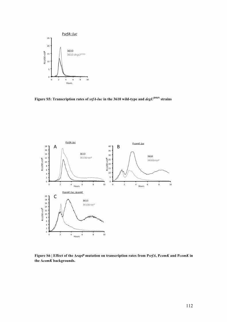
112
Figure S5: Transcription rates of srfA-luc in the 3610 wild-type and degUD56N strains
Figure S6 | Effect of the !rapP mutation on transcription rates from PsrfA, PcomK and PcomK in the !comK backgrounds.

113
Figure S7: The basal expression from PcomK is comparable in IS75, PS216 and 3610. All three strains carry PcomK-luc and (comK.

114
Table S1 Strains Strain
Number Backgrounda Descriptionb
IS75 168 derivative his leu met BD3338 B. subtilis RO-OO-2 PcomK::gfp (Cm) BD3339 B. subtilis RO-FF-1 PcomK::gfp (Cm) BD3341 B. subtilis RO-E-2 PcomK::gfp (Cm) BD4374 B. subtilis (IS75) PcomK::cfp (Kan) BD4773 B. subtilis (IS75) PcomK::luc (Cm) BD4893 B. subtilis (IS75) "comK (Spc), PcomK::luc (Cm) BD6432 B. subtilis (PS216) PcomK::luc (Cm) BD6434 B. subtilis (PS216) PcomK::cfp (Kan) BD6437 B. subtilis (PS216) "comK (Spc), PcomK::luc (Cm) BD6438 B. subtilis (3610) "comI, "comK (Spc), PcomK::luc (Cm) BD6439 B. subtilis (3610) "comI, PcomK::luc (Cm) BD6441 B. subtilis (3610) "comI, PcomK::cfp (Kan) BD7125 B. subtilis (3610) "comI, PcomG::luc (Cm), "degQ (Tet) BD7447 B. subtilis (3610) "comI, PcomK::luc (Cm), degQ IS75 BD7448 B. subtilis (3610) "comI, PcomG::luc (Cm), degQ IS75 BD7456 B. subtilis (IS75) PcomK::luc (Cm), degQ3610 BD7457 B. subtilis (IS75) PcomG::luc (Cm), degQ3610 BD8276 B. subtilis (IS75) PsrfA::luc (Cm), degQ3610
BD8277 B. subtilis (3610) "comI, PsrfA::luc (Cm), degQ IS75 BD8278 B. subtilis (IS75) PsrfA::luc (Cm), "degS (Kan) BD8279 B. subtilis (IS75) PcomK::luc (Cm), "degS (Kan) BD8280 B. subtilis (168) PcomG::luc (Cm), "degS (Kan) BD8281 B. subtilis (3610) "comI, PsrfA::luc (Cm), "degS (Kan) BD8282 B. subtilis (3610) "comI, PcomK::luc (Cm), "degS (Kan) BD8283 B. subtilis (3610) "comI, PcomG::luc (Cm), "degS (Kan)
BD8284 B. subtilis (IS75) PsrfA::mCherry (Kan), PcomG::gfp (Cm)
BD8285 B. subtilis (IS75) PsrfA::mCherry (Kan), PcomG::gfp (Cm), degQ3610 BD8286 B. subtilis (3610) "comI, PsrfA::mCherry (Kan), PcomG::gfp (Cm) BD8287 B. subtilis (3610) " comI, PsrfA::mCherry (Kan), PcomG::gfp (Cm), degQIS75 BD8288 B. subtilis (IS75) Pxyl-comK (Ery), Phyper-spank-degQ (Kan) BD8289 B. subtilis (3610) "rapP, PsrfA::luc (Cm) BD8290 B. subtilis (3610) "rapP, PcomK::luc (Cm) BD8291 B. subtilis (3610) "rapP, "comK (Spc), PcomK::luc (Cm) BD8292 B. subtilis (3610) "comI, PsrfA::luc (Cm), degUD56N (Kan)
aThe IS75 derivatives are all his leu met auxotrophs. bAll of the fusion constructs and the Phyper-spank-degQ construct are inserted by single crossover at the native loci. The Pxyl-comK construct is at the amyE locus.
115
THESIS SUMMARY
CONCLUSION

116
We have presented data supporting the idea that domestication led to the loss
of a regulation pathway for the K-state in B. subtilis. The standard laboratory strains
of B. subtilis, derived from strain 168, have been selected during more than fifty years
of use for ease of manipulation as useful genetic models. This selection is responsible
for (i) the high frequency of transformability and (ii) the loss of ability to form
biofilms (McLoon et al., 2011). The inability to form biofilms is attributed to five
genomic mutations/deletions. sfp, epsC, swrA and degQ are mutated while the
extrachromosomally encoded rapP gene is missing due to loss of the pLS32 plasmid
(McLoon et al., 2011).
Over the past decade, the undomesticated strain 3610, considered as the parent
of strain 168, has been intensively investigated because of its ability to form biofilms
and is widely viewed as an “undomesticated model” of B. subtilis. Although 3610 is
genetically really closely related to the domesticated 168 derivative, IS75, it is poorly
transformable. This phenotype for DNA transformation is partially due to the
presence of comI, a gene encoded by pLS32, a plasmid absent from the laboratory
strain. (Konkol et al., 2013) showed that ComI decreases the uptake of DNA without
interfering with the expression of the K-state. Because 3610 expresses the K-state
poorly, these observations suggest that the domesticated strain IS75 has lost some
regulatory feature of competence and that, therefore, our understanding of the K-state
regulation in B. subtilis is incomplete.
The undomesticated B. subtilis PS216 was isolated from a Slovenian soil sample and
has been shown to form structured and robust biofilms. Also, it exhibits a
transformation frequency and K-state expression intermediate between that of IS75
and 3610. All three strains, IS75, 3610 and PS216 belong to the same quorum-sensing
phenotype group and therefore respond to the same ComX quorum sensing

117
pheromone (Tortosa et al., 2001) (Stefanic & Mandic-Mulec, 2009). We have
characterized the genomic sequence of PS216, and have attempted to understand the
differences in frequency of K-state expression in the three strains in order to uncover
the regulatory features that are missing in IS75.
Our initial analysis confirmed, unsurprisingly, that the core circuitry for
competence regulation is the same in both domesticated and undomesticated strains of
B. subtilis as inactivation of key competence regulators, e.g. comK, mecA, comA and
comS, has the same effect on K-state expression in the three backgrounds.
We then designed a genetic approach to identify genes reducing K-state
development in undomesticated strains of B. subtilis. The undomesticated B. subtilis
PS216 strain was transformed with chromosomal DNA from strain IS75 carrying a
comG promoter lacZ fusion linked to a kanamycin-resistance marker. PS216 was used
because it is more transformable than 3610. We selected for kanamycin resistance and
screened for blue colonies as an indication that an unlinked allele from IS75 that
increases expression from the comG promoter had been transferred by “congression”
(simultaneous transformation of B. subtilis by unlinked markers). 1% of the
kanamycin resistant colonies developed a blue color, a frequency consistent with
congression in B. subtilis. Although PS216 colonies are normally mucoid, all the blue
colonies were non-mucoid. This difference in mucoidy expression, pointed to a
difference in the B. subtilis “Deg” regulatory system. Indeed, the mucoidy phenotype
in B. subtilis indicates the production of poly-"-glutamic acid through the expression
of the pgs operon, that is dependent on DegU~P and DegQ (Stanley & Lazazzera,
2005). As previously reported, one of the genetic differences between domesticated
168 strains (e.g. IS75) and the undomesticated strains 3610 and PS216 is a mutation
in the promoter region of degQ, which negatively affects the transcription of degQ
118
(Yang, Ferrari, Chen, & Henner, 1986) (McLoon et al., 2011). All the PS216 blue
colonies were then shown to have the degQ promoter mutation from strain IS75
suggesting that the “Deg” pathway down regulates K-state regulation in
undomesticated strains of B. subtilis.
To confirm the role of the degQ promoter mutation in K-state regulation we
swapped the degQ allele between strains IS75 and 3610. The rates of transcription of
srfA, comK and comG were all increased dramatically in strain 3610 with the mutant
degQ allele and decreased in strain IS75 with the wild-type degQ allele. We also
showed that the degQ swap has an effect on the percentage of competent cells as the
number of cells entering the K-state is increased or decreased in the swapped strains
compared to the wild types in the 3610 and IS75 backgrounds, respectively.
degQ is described in the literature as a modulator of the phosphorylation state
of DegU, which increases the transfer of a phosphoryl group from the histidine kinase
DegS to its cognate response regulator DegU (Kobayashi, 2007b). To determine if the
differences in the K-state previously observed is a consequence of a different level of
DegU~P in the cells, we looked at the effect of a degS KO on the expression of
competence genes (srfA, comK and comGA). These experiments confirmed that the
degQ effect on the K-state expression is mediated by the expected changes in
DegU~P.
We then tried to identify how the level of DegU~P can modulate the
expression of the K-state in B. subtilis. Our lab has shown that a stable form of
DegU~P, which is dephosphorylated at a slow rate, decreases the expression of the
early srfA operon, suggesting that a high level of DegU~P might inhibit srfA
expression. Because the comS open reading frame is embedded in the srfA operon,
and because ComS protects ComK from degradation by the ATP-dependent

119
MecA/ClpC/ClpP protease, we hypothesized that an increased level of DegU~P
would decrease ComK stability by repressing the expression of srfA (and therefore
comS). Data presented in this work confirm that the enhanced transformability of the
domesticated strain IS75 can be explained by the increased stability of ComK, caused
by the derepression of srfA (comS).
We have shown here that DegU~P plays an important role in the
undomesticated strains of B. subtilis by inhibiting the expression of srfA and therefore
the K-state. It was shown previously that unphosphorylated DegU binds to sequences
upstream from ComK helping ComK to bind to its own promoter at the onset of
competence development. Because DegU and DegU~P have both positive and
negative effects on the expression of the K-state it appears that the ratio of their
concentrations must be tightly regulated. The different mechanisms and upstream
signals controlling the phosphorylation of DegU are not yet fully understood. Our
results revealed that a relatively high level of DegU~P inhibits the K-state by
repressing the expression of srfA. The molecular mechanism of this repression has not
been characterized. Indeed, while we know DegU is able to directly bind DNA, we
don’t know if DegU~P is able to bind directly to the srfA promoter region to inhibit
its expression.
Competence for DNA transformation in B. subtilis is a consequence of noise
in the basal expression of ComK. Competent cells are produced when the threshold
level of ComK needed to activate the auto induction of comK transcription is
exceeded by basal (ComK-independent) comK transcription. This basal level of comK
transcription is set by changing amounts of Spo0A~P. Here we have shown that the
probability that a cell will transition to the K-state is also determined by the ratio of
DegU to DegU-P, which determine the binding affinity of ComK to its own promoter

120
and the stability of ComK, respectively. This work highlights a previously unknown
pathway for K-state regulation: the control of the transcription of the srfA operon
through DegU~P affects the stability of ComK which in turn affects the fraction of
cells entering the K-state in undomesticated strains of B. subtilis. The working model
of this pathway is presented in the figure X.
Figure X: K-state regulation in domesticated and undomesticated strains of B. subtilis.
In domesticated strains of B. subtilis, a mutation in the promoter region of
degQ decreases the rate of transcription of degQ (Yang et al., 1986). This low level of
DegQ decreases the phosphorylation of DegU through its cognate kinase DegS which
inhibits the repression effect of DegU~P at the srfA promoter level. As a result, ComS
is synthetized and inhibits the degradation of ComK through the proteolytic complex
!"#$%$&'
()*+,-./012-*'3+,-./012-*'
!"#$%4' !"#$%
3-5(' 6'7'
3-5('
3-58'
3-5#'
3-5#'
6'7'
3-5('
3-58'3-58'3-58'
3-5#'
3-5(' 6'7'3-5(' 6'7'
3-5(' 6'7'
3-5#'
6'7'
&'())% &'()*%+,-.%
&'())% &'()*%+,-.%
9+,#'9+,#'
9+,#':-0%'9;<9=6' 9+,#'
:-0%'9;<9=6'
+,-/% +,-/%

121
MecA/ClpC/ClpP, allowing ComK to bind to its own promoter and develop
competence. On the other hand, the undomesticated strains of B. subtilis do not carry
the mutation in the degQ promoter region resulting in a higher synthesis of DegQ
resulting in the accumulation of DegU~P. As a result, srfA transcription is repressed
and so is competence expression.
This work provides one more illustration of the importance of using wild
isolates for the study of real-life biology.

122
REFERENCES

123
Albano, M., Breitling, R., & Dubnau, D. A. (1989). Nucleotide sequence and genetic
organization of the Bacillus subtilis comG operon. J Bacteriol, 171(10), 5386-
5404.
Albano, M., & Dubnau, D. A. (1989). Cloning and characterization of a cluster of
linked Bacillus subtilis late competence mutations. J Bacteriol, 171(10), 5376-
5385.
Albano, M., Hahn, J., & Dubnau, D. (1987). Expression of competence genes in
Bacillus subtilis. J Bacteriol, 169(7), 3110-3117.
Alm, E., Huang, K., & Arkin, A. (2006). The evolution of two-component systems in
bacteria reveals different strategies for niche adaptation. PLoS Comput Biol,
2(11), e143. doi:10.1371/journal.pcbi.0020143
Ansaldi, M., Marolt, D., Stebe, T., Mandic-Mulec, I., & Dubnau, D. (2002). Specific
activation of the Bacillus quorum-sensing systems by isoprenylated
pheromone variants. Mol Microbiol, 44(6), 1561-1573.
Avery, O. T., Macleod, C. M., & McCarty, M. (1944). Studies on the Chemical
Nature of the Substance Inducing Transformation of Pneumococcal Types :
Induction of Transformation by a Desoxyribonucleic Acid Fraction Isolated
from Pneumococcus Type Iii. J Exp Med, 79(2), 137-158.
Berka, R. M., Hahn, J., Albano, M., Draskovic, I., Persuh, M., Cui, X., . . . Dubnau,
D. (2002). Microarray analysis of the Bacillus subtilis K-state: genome-wide
expression changes dependent on ComK. Mol Microbiol, 43(5), 1331-1345.
Blair, K. M., Turner, L., Winkelman, J. T., Berg, H. C., & Kearns, D. B. (2008). A
molecular clutch disables flagella in the Bacillus subtilis biofilm. Science,
320(5883), 1636-1638. doi:10.1126/science.1157877

124
Branda, S. S., Chu, F., Kearns, D. B., Losick, R., & Kolter, R. (2006). A major
protein component of the Bacillus subtilis biofilm matrix. Mol Microbiol,
59(4), 1229-1238. doi:10.1111/j.1365-2958.2005.05020.x
Branda, S. S., Gonzalez-Pastor, J. E., Ben-Yehuda, S., Losick, R., & Kolter, R.
(2001). Fruiting body formation by Bacillus subtilis. Proc Natl Acad Sci U S
A, 98(20), 11621-11626. doi:10.1073/pnas.191384198
Branda, S. S., Vik, S., Friedman, L., & Kolter, R. (2005). Biofilms: the matrix
revisited. Trends Microbiol, 13(1), 20-26. doi:10.1016/j.tim.2004.11.006
Briley, K., Jr., Dorsey-Oresto, A., Prepiak, P., Dias, M. J., Mann, J. M., & Dubnau, D.
(2011). The secretion ATPase ComGA is required for the binding and
transport of transforming DNA. Mol Microbiol, 81(3), 818-830.
doi:10.1111/j.1365-2958.2011.07730.x
Burbulys, D., Trach, K. A., & Hoch, J. A. (1991). Initiation of sporulation in B.
subtilis is controlled by a multicomponent phosphorelay. Cell, 64(3), 545-552.
Burkholder, P. R., & Giles, N. H., Jr. (1947). Induced biochemical mutations in
Bacillus subtilis. Am J Bot, 34(6), 345-348.
Burton, B., & Dubnau, D. (2010). Membrane-associated DNA transport machines.
Cold Spring Harb Perspect Biol, 2(7), a000406.
doi:10.1101/cshperspect.a000406
Cairns, L. S., Marlow, V. L., Bissett, E., Ostrowski, A., & Stanley-Wall, N. R. (2013).
A mechanical signal transmitted by the flagellum controls signalling in
Bacillus subtilis. Mol Microbiol, 90(1), 6-21. doi:10.1111/mmi.12342
Capra, E. J., & Laub, M. T. (2012). Evolution of two-component signal transduction
systems. Annu Rev Microbiol, 66, 325-347. doi:10.1146/annurev-micro-
092611-150039

125
Chai, Y., Chu, F., Kolter, R., & Losick, R. (2008). Bistability and biofilm formation
in Bacillus subtilis. Mol Microbiol, 67(2), 254-263. doi:10.1111/j.1365-
2958.2007.06040.x
Chai, Y., Kolter, R., & Losick, R. (2009). Paralogous antirepressors acting on the
master regulator for biofilm formation in Bacillus subtilis. Mol Microbiol,
74(4), 876-887. doi:10.1111/j.1365-2958.2009.06900.x
Chai, Y., Kolter, R., & Losick, R. (2010). Reversal of an epigenetic switch governing
cell chaining in Bacillus subtilis by protein instability. Mol Microbiol, 78(1),
218-229. doi:10.1111/j.1365-2958.2010.07335.x
Chai, Y., Norman, T., Kolter, R., & Losick, R. (2010). An epigenetic switch
governing daughter cell separation in Bacillus subtilis. Genes Dev, 24(8), 754-
765. doi:10.1101/gad.1915010
Chen, I., Christie, P. J., & Dubnau, D. (2005). The ins and outs of DNA transfer in
bacteria. Science, 310(5753), 1456-1460. doi:10.1126/science.1114021
Chen, I., & Dubnau, D. (2004). DNA uptake during bacterial transformation. Nat Rev
Microbiol, 2(3), 241-249. doi:10.1038/nrmicro844
Chu, F., Kearns, D. B., McLoon, A., Chai, Y., Kolter, R., & Losick, R. (2008). A
novel regulatory protein governing biofilm formation in Bacillus subtilis. Mol
Microbiol, 68(5), 1117-1127. doi:10.1111/j.1365-2958.2008.06201.x
Chung, Y. S., & Dubnau, D. (1995). ComC is required for the processing and
translocation of comGC, a pilin-like competence protein of Bacillus subtilis.
Mol Microbiol, 15(3), 543-551.
Clark, A. J., & Adelberg, E. A. (1962). Bacterial conjugation. Annu Rev Microbiol,
16, 289-319. doi:10.1146/annurev.mi.16.100162.001445

126
Crutz, A. M., & Steinmetz, M. (1992). Transcription of the Bacillus subtilis sacX and
sacY genes, encoding regulators of sucrose metabolism, is both inducible by
sucrose and controlled by the DegS-DegU signalling system. J Bacteriol,
174(19), 6087-6095.
Dahl, M. K., Msadek, T., Kunst, F., & Rapoport, G. (1992). The phosphorylation state
of the DegU response regulator acts as a molecular switch allowing either
degradative enzyme synthesis or expression of genetic competence in Bacillus
subtilis. J Biol Chem, 267(20), 14509-14514.
Dahm, R. (2008). Discovering DNA: Friedrich Miescher and the early years of
nucleic acid research. Hum Genet, 122(6), 565-581. doi:10.1007/s00439-007-
0433-0
Dartois, V., Debarbouille, M., Kunst, F., & Rapoport, G. (1998). Characterization of a
novel member of the DegS-DegU regulon affected by salt stress in Bacillus
subtilis. J Bacteriol, 180(7), 1855-1861.
Davies, D. (2003). Understanding biofilm resistance to antibacterial agents. Nat Rev
Drug Discov, 2(2), 114-122. doi:10.1038/nrd1008
de la Cruz, F., & Davies, J. (2000). Horizontal gene transfer and the origin of species:
lessons from bacteria. Trends Microbiol, 8(3), 128-133.
Dubnau, D. (1991). Genetic competence in Bacillus subtilis. Microbiol Rev, 55(3),
395-424.
Dubnau, D., & Cirigliano, C. (1972). Fate of transforming DNA following uptake by
competent Bacillus subtilis. Formation and properties of products isolated
from transformed cells which are derived entirely from donor DNA. J Mol
Biol, 64(1), 9-29.

127
Erickson, R. J. (1970). New ideas and data on competence and DNA entry in
transformation of bacillus subtilis. Curr Top Microbiol Immunol, 53, 149-199.
Fernandez, S., Ayora, S., & Alonso, J. C. (2000). Bacillus subtilis homologous
recombination: genes and products. Res Microbiol, 151(6), 481-486.
Finkel, S. E., & Kolter, R. (2001). DNA as a nutrient: novel role for bacterial
competence gene homologs. J Bacteriol, 183(21), 6288-6293.
doi:10.1128/JB.183.21.6288-6293.2001
Fujita, M., Gonzalez-Pastor, J. E., & Losick, R. (2005). High- and low-threshold
genes in the Spo0A regulon of Bacillus subtilis. J Bacteriol, 187(4), 1357-
1368. doi:10.1128/JB.187.4.1357-1368.2005
Giraldo, R., Andreu, J. M., & Diaz-Orejas, R. (1998). Protein domains and
conformational changes in the activation of RepA, a DNA replication initiator.
EMBO J, 17(15), 4511-4526. doi:10.1093/emboj/17.15.4511
Gould, S. J., & Subramani, S. (1988). Firefly luciferase as a tool in molecular and cell
biology. Anal Biochem, 175(1), 5-13.
Griffith, F. (1928). The Significance of Pneumococcal Types. J Hyg (Lond), 27(2),
113-159.
Guttenplan, S. B., Blair, K. M., & Kearns, D. B. (2010). The EpsE flagellar clutch is
bifunctional and synergizes with EPS biosynthesis to promote Bacillus subtilis
biofilm formation. PLoS Genet, 6(12), e1001243.
doi:10.1371/journal.pgen.1001243
Hahn, J., & Dubnau, D. (1991). Growth stage signal transduction and the
requirements for srfA induction in development of competence. J Bacteriol,
173(22), 7275-7282.

128
Hahn, J., Inamine, G., Kozlov, Y., & Dubnau, D. (1993). Characterization of comE, a
late competence operon of Bacillus subtilis required for the binding and
uptake of transforming DNA. Mol Microbiol, 10(1), 99-111.
Hahn, J., Luttinger, A., & Dubnau, D. (1996). Regulatory inputs for the synthesis of
ComK, the competence transcription factor of Bacillus subtilis. Mol
Microbiol, 21(4), 763-775.
Hahn, J., Maier, B., Haijema, B. J., Sheetz, M., & Dubnau, D. (2005). Transformation
proteins and DNA uptake localize to the cell poles in Bacillus subtilis. Cell,
122(1), 59-71. doi:10.1016/j.cell.2005.04.035
Hahn, J., Tanner, A. W., Carabetta, V. J., Cristea, I. M., & Dubnau, D. (2015).
ComGA-RelA interaction and persistence in the Bacillus subtilis K-state. Mol
Microbiol, 97(3), 454-471. doi:10.1111/mmi.13040
Haijema, B. J., Hahn, J., Haynes, J., & Dubnau, D. (2001). A ComGA-dependent
checkpoint limits growth during the escape from competence. Mol Microbiol,
40(1), 52-64.
Hall-Stoodley, L., Costerton, J. W., & Stoodley, P. (2004). Bacterial biofilms: from
the natural environment to infectious diseases. Nat Rev Microbiol, 2(2), 95-
108. doi:10.1038/nrmicro821
Hamoen, L. W., Kausche, D., Marahiel, M. A., van Sinderen, D., Venema, G., &
Serror, P. (2003). The Bacillus subtilis transition state regulator AbrB binds to
the -35 promoter region of comK. FEMS Microbiol Lett, 218(2), 299-304.
Hamoen, L. W., Smits, W. K., de Jong, A., Holsappel, S., & Kuipers, O. P. (2002).
Improving the predictive value of the competence transcription factor (ComK)
binding site in Bacillus subtilis using a genomic approach. Nucleic Acids Res,
30(24), 5517-5528.

129
Hamoen, L. W., Van Werkhoven, A. F., Bijlsma, J. J., Dubnau, D., & Venema, G.
(1998). The competence transcription factor of Bacillus subtilis recognizes
short A/T-rich sequences arranged in a unique, flexible pattern along the DNA
helix. Genes Dev, 12(10), 1539-1550.
Hamoen, L. W., Van Werkhoven, A. F., Venema, G., & Dubnau, D. (2000). The
pleiotropic response regulator DegU functions as a priming protein in
competence development in Bacillus subtilis. Proc Natl Acad Sci U S A,
97(16), 9246-9251. doi:10.1073/pnas.160010597
Henner, D. J., Ferrari, E., Perego, M., & Hoch, J. A. (1988). Location of the targets of
the hpr-97, sacU32(Hy), and sacQ36(Hy) mutations in upstream regions of the
subtilisin promoter. J Bacteriol, 170(1), 296-300.
Hershey, A. D. (1952). Inheritance in bacteriophage. Ann N Y Acad Sci, 54(6), 960-
962.
Hoa, T. T., Tortosa, P., Albano, M., & Dubnau, D. (2002). Rok (YkuW) regulates
genetic competence in Bacillus subtilis by directly repressing comK. Mol
Microbiol, 43(1), 15-26.
Hobley, L., Ostrowski, A., Rao, F. V., Bromley, K. M., Porter, M., Prescott, A. R., . . .
Stanley-Wall, N. R. (2013). BslA is a self-assembling bacterial hydrophobin
that coats the Bacillus subtilis biofilm. Proc Natl Acad Sci U S A, 110(33),
13600-13605. doi:10.1073/pnas.1306390110
Hsueh, Y. H., Cozy, L. M., Sham, L. T., Calvo, R. A., Gutu, A. D., Winkler, M. E., &
Kearns, D. B. (2011). DegU-phosphate activates expression of the anti-sigma
factor FlgM in Bacillus subtilis. Mol Microbiol, 81(4), 1092-1108.
doi:10.1111/j.1365-2958.2011.07755.x
130
Inamine, G. S., & Dubnau, D. (1995). ComEA, a Bacillus subtilis integral membrane
protein required for genetic transformation, is needed for both DNA binding
and transport. J Bacteriol, 177(11), 3045-3051.
Jiang, M., Shao, W., Perego, M., & Hoch, J. A. (2000). Multiple histidine kinases
regulate entry into stationary phase and sporulation in Bacillus subtilis. Mol
Microbiol, 38(3), 535-542.
Johnsen, P. J., Dubnau, D., & Levin, B. R. (2009). Episodic selection and the
maintenance of competence and natural transformation in Bacillus subtilis.
Genetics, 181(4), 1521-1533. doi:10.1534/genetics.108.099523
Kearns, D. B., Chu, F., Branda, S. S., Kolter, R., & Losick, R. (2005). A master
regulator for biofilm formation by Bacillus subtilis. Mol Microbiol, 55(3),
739-749. doi:10.1111/j.1365-2958.2004.04440.x
Kobayashi, K. (2007a). Bacillus subtilis pellicle formation proceeds through
genetically defined morphological changes. J Bacteriol, 189(13), 4920-4931.
doi:10.1128/JB.00157-07
Kobayashi, K. (2007b). Gradual activation of the response regulator DegU controls
serial expression of genes for flagellum formation and biofilm formation in
Bacillus subtilis. Mol Microbiol, 66(2), 395-409. doi:10.1111/j.1365-
2958.2007.05923.x
Kobayashi, K., & Iwano, M. (2012). BslA(YuaB) forms a hydrophobic layer on the
surface of Bacillus subtilis biofilms. Mol Microbiol, 85(1), 51-66.
doi:10.1111/j.1365-2958.2012.08094.x
Konkol, M. A., Blair, K. M., & Kearns, D. B. (2013). Plasmid-encoded ComI inhibits
competence in the ancestral 3610 strain of Bacillus subtilis. J Bacteriol,
195(18), 4085-4093. doi:10.1128/JB.00696-13

131
Koonin, E. V., Makarova, K. S., & Aravind, L. (2001). Horizontal gene transfer in
prokaryotes: quantification and classification. Annu Rev Microbiol, 55, 709-
742. doi:10.1146/annurev.micro.55.1.709
Kovacs, A. T., & Kuipers, O. P. (2011). Rok regulates yuaB expression during
architecturally complex colony development of Bacillus subtilis 168. J
Bacteriol, 193(4), 998-1002. doi:10.1128/JB.01170-10
Kresge, N., Simoni, R. D., & Hill, R. L. (2011). The molecular genetics of
bacteriophage: the work of Norton Zinder. J Biol Chem, 286(25), e4-5.
Kuhsel, M. G., Strickland, R., & Palmer, J. D. (1990). An ancient group I intron
shared by eubacteria and chloroplasts. Science, 250(4987), 1570-1573.
Lacks, S., Greenberg, B., & Neuberger, M. (1975). Identification of a
deoxyribonuclease implicated in genetic transformation of Diplococcus
pneumoniae. J Bacteriol, 123(1), 222-232.
Lazazzera, B. A. (2000). Quorum sensing and starvation: signals for entry into
stationary phase. Curr Opin Microbiol, 3(2), 177-182.
Lazazzera, B. A., Solomon, J. M., & Grossman, A. D. (1997). An exported peptide
functions intracellularly to contribute to cell density signaling in B. subtilis.
Cell, 89(6), 917-925.
Lederberg, J., Cavalli, L. L., & Lederberg, E. M. (1952). Sex Compatibility in
Escherichia Coli. Genetics, 37(6), 720-730.
Leisner, M., Stingl, K., Radler, J. O., & Maier, B. (2007). Basal expression rate of
comK sets a 'switching-window' into the K-state of Bacillus subtilis. Mol
Microbiol, 63(6), 1806-1816. doi:10.1111/j.1365-2958.2007.05628.x

132
Lenhart, J. S., Schroeder, J. W., Walsh, B. W., & Simmons, L. A. (2012). DNA repair
and genome maintenance in Bacillus subtilis. Microbiol Mol Biol Rev, 76(3),
530-564. doi:10.1128/MMBR.05020-11
Levene, P.A. (1919) The structure of yeast nucleic acid: IV. Ammonia hydrolysis. J
Biol Chem 40:415-424
Logan, B. E. (2009). Exoelectrogenic bacteria that power microbial fuel cells. Nat Rev
Microbiol, 7(5), 375-381. doi:10.1038/nrmicro2113
Londono-Vallejo, J. A., & Dubnau, D. (1993). comF, a Bacillus subtilis late
competence locus, encodes a protein similar to ATP-dependent RNA/DNA
helicases. Mol Microbiol, 9(1), 119-131.
Londono-Vallejo, J. A., & Dubnau, D. (1994). Mutation of the putative nucleotide
binding site of the Bacillus subtilis membrane protein ComFA abolishes the
uptake of DNA during transformation. J Bacteriol, 176(15), 4642-4645.
Lopez, D., Fischbach, M. A., Chu, F., Losick, R., & Kolter, R. (2009). Structurally
diverse natural products that cause potassium leakage trigger multicellularity
in Bacillus subtilis. Proc Natl Acad Sci U S A, 106(1), 280-285.
doi:10.1073/pnas.0810940106
Lopez, D., Gontang, E. A., & Kolter, R. (2010). Potassium sensing histidine kinase in
Bacillus subtilis. Methods Enzymol, 471, 229-251. doi:10.1016/S0076-
6879(10)71013-2
Lopez, D., Vlamakis, H., & Kolter, R. (2009). Generation of multiple cell types in
Bacillus subtilis. FEMS Microbiol Rev, 33(1), 152-163. doi:10.1111/j.1574-
6976.2008.00148.x
Lopez, D., Vlamakis, H., Losick, R., & Kolter, R. (2009). Paracrine signaling in a
bacterium. Genes Dev, 23(14), 1631-1638. doi:10.1101/gad.1813709

133
Maamar, H., & Dubnau, D. (2005). Bistability in the Bacillus subtilis K-state
(competence) system requires a positive feedback loop. Mol Microbiol, 56(3),
615-624. doi:10.1111/j.1365-2958.2005.04592.x
Mah, T. F., & O'Toole, G. A. (2001). Mechanisms of biofilm resistance to
antimicrobial agents. Trends Microbiol, 9(1), 34-39.
Marlow, V. L., Porter, M., Hobley, L., Kiley, T. B., Swedlow, J. R., Davidson, F. A.,
& Stanley-Wall, N. R. (2014). Phosphorylated DegU manipulates cell fate
differentiation in the Bacillus subtilis biofilm. J Bacteriol, 196(1), 16-27.
doi:10.1128/JB.00930-13
McLoon, A. L., Guttenplan, S. B., Kearns, D. B., Kolter, R., & Losick, R. (2011).
Tracing the domestication of a biofilm-forming bacterium. J Bacteriol, 193(8),
2027-2034. doi:10.1128/JB.01542-10
Mejean, V., & Claverys, J. P. (1993). DNA processing during entry in transformation
of Streptococcus pneumoniae. J Biol Chem, 268(8), 5594-5599.
Mirouze, N., Desai, Y., Raj, A., & Dubnau, D. (2012). Spo0A~P imposes a temporal
gate for the bimodal expression of competence in Bacillus subtilis. PLoS
Genet, 8(3), e1002586. doi:10.1371/journal.pgen.1002586
Mirouze, N., Ferret, C., Yao, Z., Chastanet, A., & Carballido-Lopez, R. (2015).
MreB-Dependent Inhibition of Cell Elongation during the Escape from
Competence in Bacillus subtilis. PLoS Genet, 11(6), e1005299.
doi:10.1371/journal.pgen.1005299
Mitrophanov, A. Y., & Groisman, E. A. (2008). Signal integration in bacterial two-
component regulatory systems. Genes Dev, 22(19), 2601-2611.
doi:10.1101/gad.1700308

134
Mohan, S., Aghion, J., Guillen, N., & Dubnau, D. (1989). Molecular cloning and
characterization of comC, a late competence gene of Bacillus subtilis. J
Bacteriol, 171(11), 6043-6051.
Mordini, S., Osera, C., Marini, S., Scavone, F., Bellazzi, R., Galizzi, A., & Calvio, C.
(2013). The role of SwrA, DegU and P(D3) in fla/che expression in B. subtilis.
PLoS One, 8(12), e85065. doi:10.1371/journal.pone.0085065
Morrison, D. A., & Baker, M. F. (1979). Competence for genetic transformation in
pneumococcus depends on synthesis of a small set of proteins. Nature,
282(5735), 215-217.
Msadek, T., Kunst, F., Klier, A., & Rapoport, G. (1991). DegS-DegU and ComP-
ComA modulator-effector pairs control expression of the Bacillus subtilis
pleiotropic regulatory gene degQ. J Bacteriol, 173(7), 2366-2377.
Nakano, M. M., Xia, L. A., & Zuber, P. (1991). Transcription initiation region of the
srfA operon, which is controlled by the comP-comA signal transduction
system in Bacillus subtilis. J Bacteriol, 173(17), 5487-5493.
Nakano, M. M., & Zuber, P. (1991). The primary role of comA in establishment of
the competent state in Bacillus subtilis is to activate expression of srfA. J
Bacteriol, 173(22), 7269-7274.
Nester, E. W., & Stocker, B. A. (1963). Biosynthetic Latency in Early Stages of
Deoxyribonucleic Acidtransformation in Bacillus Subtilis. J Bacteriol, 86,
785-796.
Ochman, H., Lawrence, J. G., & Groisman, E. A. (2000). Lateral gene transfer and the
nature of bacterial innovation. Nature, 405(6784), 299-304.
doi:10.1038/35012500

135
Ogura, M., Shimane, K., Asai, K., Ogasawara, N., & Tanaka, T. (2003). Binding of
response regulator DegU to the aprE promoter is inhibited by RapG, which is
counteracted by extracellular PhrG in Bacillus subtilis. Mol Microbiol, 49(6),
1685-1697.
Ogura, M., & Tsukahara, K. (2010). Autoregulation of the Bacillus subtilis response
regulator gene degU is coupled with the proteolysis of DegU-P by ClpCP. Mol
Microbiol, 75(5), 1244-1259. doi:10.1111/j.1365-2958.2010.07047.x
Ogura, M., Yamaguchi, H., Kobayashi, K., Ogasawara, N., Fujita, Y., & Tanaka, T.
(2002). Whole-genome analysis of genes regulated by the Bacillus subtilis
competence transcription factor ComK. J Bacteriol, 184(9), 2344-2351.
Omer Bendori, S., Pollak, S., Hizi, D., & Eldar, A. (2015). The RapP-PhrP quorum-
sensing system of Bacillus subtilis strain NCIB3610 affects biofilm formation
through multiple targets, due to an atypical signal-insensitive allele of RapP. J
Bacteriol, 197(3), 592-602. doi:10.1128/JB.02382-14
Ostrowski, A., Mehert, A., Prescott, A., Kiley, T. B., & Stanley-Wall, N. R. (2011).
YuaB functions synergistically with the exopolysaccharide and TasA amyloid
fibers to allow biofilm formation by Bacillus subtilis. J Bacteriol, 193(18),
4821-4831. doi:10.1128/JB.00223-11
Pakula, R., & Walczak, W. (1963). On the nature of competence of transformable
streptococci. J Gen Microbiol, 31, 125-133. doi:10.1099/00221287-31-1-125
Parashar, V., Konkol, M. A., Kearns, D. B., & Neiditch, M. B. (2013). A plasmid-
encoded phosphatase regulates Bacillus subtilis biofilm architecture,
sporulation, and genetic competence. J Bacteriol, 195(10), 2437-2448.
doi:10.1128/JB.02030-12

136
Penalva, M. A., Moya, A., Dopazo, J., & Ramon, D. (1990). Sequences of
isopenicillin N synthetase genes suggest horizontal gene transfer from
prokaryotes to eukaryotes. Proc Biol Sci, 241(1302), 164-169.
doi:10.1098/rspb.1990.0081
Piazza, F., Tortosa, P., & Dubnau, D. (1999). Mutational analysis and membrane
topology of ComP, a quorum-sensing histidine kinase of Bacillus subtilis
controlling competence development. J Bacteriol, 181(15), 4540-4548.
Prepiak, P., & Dubnau, D. (2007). A peptide signal for adapter protein-mediated
degradation by the AAA+ protease ClpCP. Mol Cell, 26(5), 639-647.
doi:10.1016/j.molcel.2007.05.011
Provvedi, R., Chen, I., & Dubnau, D. (2001). NucA is required for DNA cleavage
during transformation of Bacillus subtilis. Mol Microbiol, 40(3), 634-644.
Redfield, R. J. (1993). Genes for breakfast: the have-your-cake-and-eat-it-too of
bacterial transformation. J Hered, 84(5), 400-404.
Roggiani, M., & Dubnau, D. (1993). ComA, a phosphorylated response regulator
protein of Bacillus subtilis, binds to the promoter region of srfA. J Bacteriol,
175(10), 3182-3187.
Rudner, D. Z., LeDeaux, J. R., Ireton, K., & Grossman, A. D. (1991). The spo0K
locus of Bacillus subtilis is homologous to the oligopeptide permease locus
and is required for sporulation and competence. J Bacteriol, 173(4), 1388-
1398.
Serror, P., & Sonenshein, A. L. (1996). CodY is required for nutritional repression of
Bacillus subtilis genetic competence. J Bacteriol, 178(20), 5910-5915.
Singh, R., Paul, D., & Jain, R. K. (2006). Biofilms: implications in bioremediation.
Trends Microbiol, 14(9), 389-397. doi:10.1016/j.tim.2006.07.001

137
Smits, W. K., Eschevins, C. C., Susanna, K. A., Bron, S., Kuipers, O. P., & Hamoen,
L. W. (2005). Stripping Bacillus: ComK auto-stimulation is responsible for the
bistable response in competence development. Mol Microbiol, 56(3), 604-614.
doi:10.1111/j.1365-2958.2005.04488.x
Solomon, J. M., Lazazzera, B. A., & Grossman, A. D. (1996). Purification and
characterization of an extracellular peptide factor that affects two different
developmental pathways in Bacillus subtilis. Genes Dev, 10(16), 2014-2024.
Solomon, J. M., Magnuson, R., Srivastava, A., & Grossman, A. D. (1995).
Convergent sensing pathways mediate response to two extracellular
competence factors in Bacillus subtilis. Genes Dev, 9(5), 547-558.
Sparling, P. F. (1966). Genetic transformation of Neisseria gonorrhoeae to
streptomycin resistance. J Bacteriol, 92(5), 1364-1371.
Spizizen, J. (1958). Transformation of Biochemically Deficient Strains of Bacillus
Subtilis by Deoxyribonucleate. Proc Natl Acad Sci U S A, 44(10), 1072-1078.
Spizizen, J., Reilly, B. E., & Evans, A. H. (1966). Microbial transformation and
transfection. Annu Rev Microbiol, 20, 371-400.
doi:10.1146/annurev.mi.20.100166.002103
Stanley, N. R., & Lazazzera, B. A. (2005). Defining the genetic differences between
wild and domestic strains of Bacillus subtilis that affect poly-gamma-dl-
glutamic acid production and biofilm formation. Mol Microbiol, 57(4), 1143-
1158. doi:10.1111/j.1365-2958.2005.04746.x
Stefanic, P., & Mandic-Mulec, I. (2009). Social interactions and distribution of
Bacillus subtilis pherotypes at microscale. J Bacteriol, 191(6), 1756-1764.
doi:10.1128/JB.01290-08

138
Stewart, P. S. (2002). Mechanisms of antibiotic resistance in bacterial biofilms. Int J
Med Microbiol, 292(2), 107-113. doi:10.1078/1438-4221-00196
Stewart, P. S., & Franklin, M. J. (2008). Physiological heterogeneity in biofilms. Nat
Rev Microbiol, 6(3), 199-210. doi:10.1038/nrmicro1838
Stock, A. M., Robinson, V. L., & Goudreau, P. N. (2000). Two-component signal
transduction. Annu Rev Biochem, 69, 183-215.
doi:10.1146/annurev.biochem.69.1.183
Stover, A. G., & Driks, A. (1999). Regulation of synthesis of the Bacillus subtilis
transition-phase, spore-associated antibacterial protein TasA. J Bacteriol,
181(17), 5476-5481.
Strauch, M., Webb, V., Spiegelman, G., & Hoch, J. A. (1990). The SpoOA protein of
Bacillus subtilis is a repressor of the abrB gene. Proc Natl Acad Sci U S A,
87(5), 1801-1805.
Suel, G. M., Kulkarni, R. P., Dworkin, J., Garcia-Ojalvo, J., & Elowitz, M. B. (2007).
Tunability and noise dependence in differentiation dynamics. Science,
315(5819), 1716-1719. doi:10.1126/science.1137455
Tanaka, T., & Ogura, M. (1998). A novel Bacillus natto plasmid pLS32 capable of
replication in Bacillus subtilis. FEBS Lett, 422(2), 243-246.
Tomasz, A. (1966). Model for the mechanism controlling the expression of competent
state in Pneumococcus cultures. J Bacteriol, 91(3), 1050-1061.
Tortosa, P., Logsdon, L., Kraigher, B., Itoh, Y., Mandic-Mulec, I., & Dubnau, D.
(2001). Specificity and genetic polymorphism of the Bacillus competence
quorum-sensing system. J Bacteriol, 183(2), 451-460.
doi:10.1128/JB.183.2.451-460.2001

139
Trieu-Cuot, P., Gerbaud, G., Lambert, T., & Courvalin, P. (1985). In vivo transfer of
genetic information between gram-positive and gram-negative bacteria.
EMBO J, 4(13A), 3583-3587.
Tsukahara, K., & Ogura, M. (2008). Promoter selectivity of the Bacillus subtilis
response regulator DegU, a positive regulator of the fla/che operon and sacB.
BMC Microbiol, 8, 8. doi:10.1186/1471-2180-8-8
Turgay, K., Hahn, J., Burghoorn, J., & Dubnau, D. (1998). Competence in Bacillus
subtilis is controlled by regulated proteolysis of a transcription factor. EMBO
J, 17(22), 6730-6738. doi:10.1093/emboj/17.22.6730
Turgay, K., Hamoen, L. W., Venema, G., & Dubnau, D. (1997). Biochemical
characterization of a molecular switch involving the heat shock protein ClpC,
which controls the activity of ComK, the competence transcription factor of
Bacillus subtilis. Genes Dev, 11(1), 119-128.
Uhl, M. A., & Miller, J. F. (1996). Central role of the BvgS receiver as a
phosphorylated intermediate in a complex two-component phosphorelay. J
Biol Chem, 271(52), 33176-33180.
van Sinderen, D., Luttinger, A., Kong, L., Dubnau, D., Venema, G., & Hamoen, L.
(1995). comK encodes the competence transcription factor, the key regulatory
protein for competence development in Bacillus subtilis. Mol Microbiol,
15(3), 455-462.
van Sinderen, D., & Venema, G. (1994). comK acts as an autoregulatory control
switch in the signal transduction route to competence in Bacillus subtilis. J
Bacteriol, 176(18), 5762-5770.

140
Veening, J. W., Smits, W. K., & Kuipers, O. P. (2008). Bistability, epigenetics, and
bet-hedging in bacteria. Annu Rev Microbiol, 62, 193-210.
doi:10.1146/annurev.micro.62.081307.163002
Verhamme, D. T., Kiley, T. B., & Stanley-Wall, N. R. (2007). DegU co-ordinates
multicellular behaviour exhibited by Bacillus subtilis. Mol Microbiol, 65(2),
554-568. doi:10.1111/j.1365-2958.2007.05810.x
Verhamme, D. T., Murray, E. J., & Stanley-Wall, N. R. (2009). DegU and Spo0A
jointly control transcription of two loci required for complex colony
development by Bacillus subtilis. J Bacteriol, 191(1), 100-108.
doi:10.1128/JB.01236-08
Vlamakis, H., Aguilar, C., Losick, R., & Kolter, R. (2008). Control of cell fate by the
formation of an architecturally complex bacterial community. Genes Dev,
22(7), 945-953. doi:10.1101/gad.1645008
Vlamakis, H., Chai, Y., Beauregard, P., Losick, R., & Kolter, R. (2013). Sticking
together: building a biofilm the Bacillus subtilis way. Nat Rev Microbiol,
11(3), 157-168. doi:10.1038/nrmicro2960
Weinrauch, Y., Msadek, T., Kunst, F., & Dubnau, D. (1991). Sequence and properties
of comQ, a new competence regulatory gene of Bacillus subtilis. J Bacteriol,
173(18), 5685-5693.
Wozniak, R. A., & Waldor, M. K. (2010). Integrative and conjugative elements:
mosaic mobile genetic elements enabling dynamic lateral gene flow. Nat Rev
Microbiol, 8(8), 552-563. doi:10.1038/nrmicro2382
Yadav, T., Carrasco, B., Hejna, J., Suzuki, Y., Takeyasu, K., & Alonso, J. C. (2013).
Bacillus subtilis DprA recruits RecA onto single-stranded DNA and mediates

141
annealing of complementary strands coated by SsbB and SsbA. J Biol Chem,
288(31), 22437-22450. doi:10.1074/jbc.M113.478347
Yadav, T., Carrasco, B., Serrano, E., & Alonso, J. C. (2014). Roles of Bacillus subtilis
DprA and SsbA in RecA-mediated genetic recombination. J Biol Chem,
289(40), 27640-27652. doi:10.1074/jbc.M114.577924
Yang, M., Ferrari, E., Chen, E., & Henner, D. J. (1986). Identification of the
pleiotropic sacQ gene of Bacillus subtilis. J Bacteriol, 166(1), 113-119.
Yuksel, M., Power, J. J., Ribbe, J., Volkmann, T., & Maier, B. (2016). Fitness Trade-
Offs in Competence Differentiation of Bacillus subtilis. Front Microbiol, 7,
888. doi:10.3389/fmicb.2016.00888
Zeigler, D. R., Pragai, Z., Rodriguez, S., Chevreux, B., Muffler, A., Albert, T., . . .
Perkins, J. B. (2008). The origins of 168, W23, and other Bacillus subtilis
legacy strains. J Bacteriol, 190(21), 6983-6995. doi:10.1128/JB.00722-08
Zinder, N. D., & Lederberg, J. (1952). Genetic exchange in Salmonella. J Bacteriol,
64(5), 679-699.

142
AUTHOR: Mathieu MIRAS
TITLE: Identification of a new regulatory pathway for the K-state in Bacillus subtilis THESIS SUPERVISOR: Patrice POLARD PLACE AND DATE OF PhD DEFENSE: Université Toulouse III, Paul Sabatier, Toulouse IBCG’s conference room, Friday April 28th 2017, 2pm ABSTRACT: Bacteria can acquire new traits either by mutation or by the acquisition of external genetic material. This external acquisition of DNA is termed horizontal gene transfer and can occur through three mechanisms: conjugation, transduction and transformation. Transformation is the gene transfer resulting from the uptake of exogenous (environmental) DNA. The Gram-positive model organism Bacillus subtilis develops competence for DNA transformation if, and only if, the master regulator ComK is produced. In domesticated strains (strains used and selected after years of genetic manipulation in laboratory) occurs in approximately 15% of the population. Although competence regulation mechanisms are well understood, the reason why wild isolates (ancestors of the domesticated strains) of B. subtilis are poorly transformable (~1-2%) remains unclear. We demonstrate in this work that it is largely due to an inactivating point mutation in the promoter region of degQ. DegQ is known to be involved in the regulation of several different developmental pathways such as biofilm formation, exoprotease synthesis and competence development. This discovery introduces a new regulatory pathway in the development of genetic competence that was missing from our understanding. Indeed, all domesticated strains of B. subtilis share the degQ mutation that down-regulates the synthesis of DegQ and thereafter stabilizes ComK allowing a higher percentage of the population to become competent compared to the wild isolates. It appears to us that domestication and intensive genetic manipulations over the past fifty years are responsible of the loss of the “degQ regulation pathway” of competence in B. subtilis illustrating the significance of using wild isolates for the study of real-life biology. KEY WORDS: Bacillus subtilis, DNA transformation, regulation, competence, DegU, ComK AREA OF EXPERTISE: Microbiology HOST LABORATORY Laboratoire de Microbiologie et Génétique Moléculaires (LMGM) Université Toulouse III - Paul Sabatier 118 Route de Narbonne 31062 TOULOUSE Cedex 9 FRANCE

!

AUTEUR: Mathieu MIRAS TITRE: Identification of a new regulatory pathway for the K-state in Bacillus subtilis DIRECTEUR DE THESE: Patrice POLARD LIEU ET DATE DE SOUTENANCE: Université Toulouse III, Paul Sabatier, Toulouse Salle de conférence de l’IBCG, le vendredi 28 Avril 2017 à 14h00 RESUME: Les bactéries peuvent développer de nouvelles caractéristiques par mutation génétique ou par l’acquisition d’un matériel génétique exogène. Ce dernier, aussi appelé transfert de gène horizontal, peut se produire selon trois mécanismes différents : la conjugaison, la transduction et la transformation. L’organisme modèle, Bacillus subtilis, est naturellement transformable (ou compétent) si, et seulement si, le facteur de transcription ComK est produit. Au sein des souches domestiques, environ 15% de la population bactérienne devient compétente. Bien que les mécanismes de régulation de la compétence soient bien caractérisés, la raison pour laquelle les souches non domestiques de B. subtilis sont peu transformables (1~2%) reste inconnue. Nous démontrons ici que c’est essentiellement dû à une mutation de transition dans le promoteur du gène degQ. La protéine DegQ est impliquée dans la régulation de la formation de biofilms, de la synthèse d’exoprotéases et de la transformation génétique. Ce travail permet de mettre en évidence une nouvelle voie de régulation dans le développement de la compétence génétique qui était jusque-là méconnue. En effet, toutes les souches domestiques de B. subtilis partagent la mutation ponctuelle dans la région du promoteur de degQ qui a pour conséquence de diminuer la synthèse protéique de DegQ et de favoriser, à plus long terme, la stabilité de ComK. Ainsi, dans le contexte des souches domestiques, ComK va permettre à un plus grand nombre de bactéries de devenir compétentes. MOTS-CLES: Bacillus subtilis, DNA transformation, regulation, competence, DegU, ComK DISCIPLINE: Microbiologie INTITULE ET ADRESSE DE L’UFR OU DU LABORATOIRE : Laboratoire de Microbiologie et Génétique Moléculaires (LMGM) Université Toulouse III - Paul Sabatier 118 Route de Narbonne 31062 TOULOUSE Cedex 9 FRANCE