identification of active-site histidine residues of a self
TRANSCRIPT

Plant Physiol. (1 997) 1 15: 1421-1 429
Identification of Active-Site Histidine Residues of a Self-lncompatibility Ribonuclease from a Wild Tomato’
Simon Parry, Ed Newbigin, Graeme Currie, Antony Bacic*, and David Oxley
Plant Cell Biology Research Centre, School of Botany, University of Melbourne, Parkville, Victoria 3052, Australia
The style component of the self-incompatibility (S) locus of the wild tomato Lycopersicon peruvianum (L.) Mill. is an allelic series of glycoproteins with ribonuclease activity (S-RNases). Treatment of the S,-RNase from L . peruvianum with iodoacetate at p H 6.1 led to a loss of RNase activity. In the presence of a competitive inhibitor, guanosine 3”Ionophosphate (3‘-CMP), the rate of RNase inactiva- tion by iodoacetate was reduced significantly. Analysis of the tryp- tic digestion products of the iodoacetate-modified S-RNase by reversed-phase high-performance liquid chromatography and electrospray-ionization mass spectrometry showed that histidine-32 was preferentially modified in the absence of 3’-GMP. Histidine-88 was also modified, but this occurred both in the presence and absence of 3’-CMP, suggesting that this residue is accessible when 3’-CMP is in the active site. Cysteine-150 was modified by iodoac- etate in the absence of 3‘-CMP and, to a lesser extent, in its presence. The results are discussed with respect to the related fungal RNase T, family and the mechanism of S-RNase action.
Gametophytic self-incompatibility acts as a prezygotic barrier to self-fertilization in many flowering plants (de Nettancourt, 1977). In solanaceous plants self- incompatibility is controlled by a single genetic locus, the S locus. The stylar product of the solanaceous S locus is an allelic series of RNases (S-RNases) found in the extracellu- lar matrix of transmitting tract cells (McClure et al., 1989). The RNase activity of the S-RNases appears to be required for the rejection of incompatible pollen tubes within the style (Huang et al., 1994; Kowyama et al., 1994), suggesting that S-RNases function as allele-specific cytotoxins (Mc- Clure et al., 1989).
Five short stretches of amino acid sequence are con- served in a11 of the solanaceous S-RNases studied (Haring et al., 1990; Tsai et al., 1992), two of which resemble the sequences at the active site of fungal RNases typified by RNase T, from Aspergillus oryzae (McClure et al., 1989). In RNase T, the two sequences forming the active site each contain a His residue that is essential for catalytic activity (Kawata et al., 1990). These two His residues are also found in the S-RNases, although little biochemical work has been
This work was supported by a Special Research Centre grant from the Australian Research Council. S.P. is the recipient of a Melbourne University Scholarship from the Faculty of Science and School of Botany.
* Corresponding author; e-mail antony-bacic9mac.unimelb. edu.au; fax 61-3-9347-1071.
1421
done to confirm that these residues are at the active site of the enzyme.
Experiments with transgenic petunia (Petunia inflata) plants and a naturally occurring variant of a wild tomato (Lycopersicon peruvianum) implicate both the conserved His residues of the S-RNases in catalysis (Huang et al., 1994; Royo et al., 1994b). Chemical modification with iodoacetate has also implicated the His residues at the active site of severa1 RNases, including RNase T, (Crestfield et al., 1963; Irie et al., 1986; Kawata et al., 1990). However, with the S,-RNase of tobacco (Nicotiana data), iodoacetate preferen- tially alkylated a free Cys residue (Ishimizu et al., 1995). The alkylating agent, diethyl pyrocarbonate, modified one of the presumed active site His residues, His-31, of the S,-RNase, resulting in a loss of enzymatic activity. This indicates that His-31 has an important role in RNA hydro- lysis, probably similar to that played by His-53 in RNase T, (Ishimizu et al., 1995). There is no biochemical evidence supporting a role in catalysis for the second, presumed active-site His residue of the S-RNases.
In this study we used iodoacetate to identify the residues at the active site of the S,-RNase from L. peruvianum. The purification of the S,-RNase was by HIC, and this method of purification was compared with cation-exchange chro- matography, the method used to purify S-RNases from N. alata (Jahnen et al., 1989).
MATERIALS AND M E T H O D S
Self-incompatible wild tomato (Lycopersicon peruvianum [L.] Mill.) plants (genotype S,S,) were obtained from the Victorian State Department of Agriculture, Burnley, Victo- ria, Australia. The plants were grown under greenhouse conditions as described previously (Mau et al., 1986).
Purification of S,-RNase from L. peruvianum
S,-RNase was purified from S,S, styles using either cation-exchange chromatography (Jahnen et al., 1989) or HIC. For the HIC method, styles were frozen in liquid N, and ground to a powder with a mortar and pestle with 10% (w/w) Polyclar AT. The powder was extracted with 0.1 M
Tris-HC1, pH 7.8, containing 14 mM P-mercaptoethanol,
Abbreviations: cm, carboxymethyl; ESI, electrospray-ionization; HIC, hydrophobic-interaction chromatography; pe, pyridylethyl; RP-HPLC, reversed-phase HPLC.
Dow
nloaded from https://academ
ic.oup.com/plphys/article/115/4/1421/6071344 by guest on 10 February 2022

1422 Parry et al. Plant Physiol. Vol. 11 5, 1997
and the insoluble material was pelleted by centrifugation (15,8008, 10 min, 4°C). The supernatant was adjusted to 50% saturation by adding solid (NH,),SO,, and stirred for 30 min at 4°C. The insoluble material was pelleted by centrifugation and the supernatant was filtered (0.22-pm pore size) and loaded onto a polypropylaspartamide HIC column (12 pm; 5 X 50 mm; PolyLC, Columbia, MD) fitted to an HPLC system (System Gold, Beckman) as described below. The HIC column was equilibrated with 2 M
(NH,),S04 in 0.1 M PO, buffer, pH 6.9, and bound protein was eluted with a linear salt gradient (2 to O M [NH,],SO, in 0.1 M PO, buffer over 30 min; flow rate 1.0 mL/min) and monitored at 215 and 280 nm. Protein determination was by the method of Bradford (1976) for complex mixtures, and by RP-HPLC for purified proteins. Purity of S,-RNase preparations was assessed by SDS-PAGE (Laemmli, 1970).
RP-HPLC
RP-HPLC was performed on a 4.6- X 130-mm column (Aquapore RP-300, Brownlee, Santa Clara, CA) fitted to an HPLC device (Beckman) comprising a model 126 solvent delivery system and a model 168 diode array detector. Solvent A was 0.1% (v/v) aqueous trifluoroacetic acid and solvent B was 0.089% trifluoroacetic acid in 60% aqueous acetonitrile (v/v). Chromatography was performed at a flow rate of 1 mL/min with a linear gradient of O to 100% solvent B over 30 min unless stated otherwise. The UV absorbance of the column effluent was continuously mon- itored at 215 and 280 nm.
lodoacetate Treatment of S,-RNase
L. peruvianum S,-RNase (200 pg), purified by the HIC method, was exchanged into 20 mM Mes buffer, pH 6.1, containing 0.1% (w/v) Brij 58 (Sigma) by gel filtration (PD10 column, Pharmacia) and treated with 0.1 M iodoac- etate at 37°C for 7 h in both the presence and absence of 15 mM 3’-GMP (Sigma). After treatment the S,-RNase was repurified by RP-HPLC as described above.
RNase Assay
RNase activity was measured as described previously (McClure et al., 1989), except that reactions were carried out in 100 mM P04 buffer, pH 7, and 50 mM KC1. Purified torula yeast RNA (Sigma) was used as the substrate.
Trypsin Digestion of lodoacetate-Modified S,-RNase
To unfold the protein in preparation for proteolysis, the iodoacetate-treated S,-RNase (200 pg) was denatured and reduced by incubating at 50°C for 30 min with 100 mM NH,HC03, 6 M guanidinium chloride (Pierce), 10 mM DTT, and 10 mM EDTA, and then alkylated with 4-vinylpyridine (50 mM) at room temperature for 30 min. The sample was purified by RP-HPLC as described above and, after drying, was resuspended in 100 mM NH,HCO, and digested with trypsin (sequencing grade, Sigma) for 16 h at 37°C at an enzyme-to-substrate ratio of 1:lOO (w/w). The resulting
peptide mixture was fractionated by RP-HPLC as de- scribed above except that a 60-min gradient was used.
ESI-MS
ESI-MS was performed on a mass spectrometer (MAT 95, Finnigan MAT, Bremen, Germany) equipped with an elec- trospray source. A constant stream of acetonitrile:0.5% aqueous acetic acid (1:1, v/v) was pumped into the source at 3 pL/min using a syringe pump (Harvard Apparatus, South Natick, MA). Samples dissolved in the same solvent were injected into the source (Rheodyne, Cotati, CA). Spec- tra were acquired by scanning from 400 to 2000 D at 10 s/decade.
Sequencing by MS
MS/MS spectra were acquired on an ion-trap mass spec- trometer (LCQ, Finnigan). Samples in 50% (v/v) aqueous methanol were infused into the source at 3 pL/min via an injector and a syringe pump. The sheath liquid was 70% (v/v) aqueous methanol flowing at 1 pL/min and the sheath gas was N, at 30 p.s.i. The needle was kept at a fixed potential of 4.6 kV at 220°C. MS/MS spectra were acquired from an ion accumulation of 500 to 1000 ms. A11 ions other than a tl D window around the parent ion were ejected. The parent ion was then collisionally activated by applying a single frequency signal to the end caps. The relative signal strength of this frequency was typically set at 40 to 45% of the maximum signal, corresponding to approxi- mately 99% fragmentation of the parent ion. This process was repeated for MS/MS/MS experiments.
N-Terminal Peptide Sequencing
Peptides were sequenced on an automated protein se- quencer (model LF3400, Beckman) according to the manu- facturer’s instructions.
RESULTS
Purification of S,-RNase from L. peruvianum Styles by HIC
L. peruvianum S,-RNase was initially purified by the cation-exchange chromatography method used to purify S-RNases from tobacco (Nicotiana alata) (Jahnen et al., 1989). Unfortunately, this method gave an extremely low recovery of protein (Table I, method B), so an alternative method based on HIC was developed (Table I, method A). The initial steps are essentially the same as those described by Jahnen et al. (1989), except that the 50% (NH,),SO, supernatant is loaded directly onto an HIC column (Fig. la) instead of being precipitated with 95% (NH,),SO,, de- salted, and fractionated by cation-exchange chromatogra- phy. The yield of S,-RNase, based on RNase activity, was approximately 65% after HIC (although this is an underes- timate because other non-S-RNases contribute to the total RNase activity in the crude extract), and the protein was approximately 80% pure as assessed by RP-HPLC and SDS-PAGE (Table I; Fig. 1, b and c). S,-RNase could be
Dow
nloaded from https://academ
ic.oup.com/plphys/article/115/4/1421/6071344 by guest on 10 February 2022

Chemical Modification of S-RNase . 1423
Table I. Purification of S^-RNase from the styles of L. peruvianumPurification of S3-RNase from L peruvianum was performed using
either HIC or cation-exchange chromatography, and the recovery oftotal protein and RNase activity is compared for each method.
Purification Stepa
Crude extractMethod A
HICRP-HPLC
Method BCation-exchange
chromatography
Proteinb
Kg4150
7660
2
Total RNaseActivityr
AJ60 min- '
10,375
6,695-
NDf
Yieldd Puritye
%
100
65 80>99
80
cl Purification steps are outlined in "Materials and Methods." Forcomparative purposes, L. peruvianum SrRNase was also purified bythe method of Jahnen et al. (1989) (method B). The same amount ofcrude extract was used in both purification schemes. h Proteinconcentration in the crude extract was estimated by the method ofBradford (1976). After chromatography, the area of the peak contain-ing the S,-RNase was used to estimate the recovery of protein.Lysozyme and BSA were used as protein standards. c RNaseactivity was measured essentially by the method of McClure et al.(1989). Protein obtained by chromatography was desalted beforeassay. Yield is based on percent recovery of RNase activity ateach step. RP-HPLC caused a loss of RNase activity. e Purity wasestimated from RP-HPLC by comparing the area of the peak contain-ing S3-RNase with the total peak area. ' ND, Not detected.
further purified by RP-HPLC but this caused a loss ofenzymatic activity. The chemical modification experimentsreported here used the HIC-purified S3-RNase after it hadbeen desalted. The chemically modified S-RNase was sub-sequently purified by RP-HPLC (see below) before struc-tural analysis.
Treatment of SrRNase with lodoacetate
S3-RNase from HIC was incubated with iodoacetate inthe presence or absence of the competitive inhibitor 3'-GMP. RNase activity was monitored periodically through-out the incubation (Fig. 2). In the absence of 3'-GMP, only15% of the RNase activity remained after 7 h of incubation,whereas in the presence of 3'-GMP, approximately 80% ofthe enzymatic activity remained. S3-RNase from each of thetwo reaction mixtures (with or without 3'-GMP) was puri-fied by RP-HPLC for further analysis.
Identification of lodoacetate-Modified Peptides
After purification, the S3-RNase samples treated withiodoacetate (with or without 3'-GMP) were denatured andreduced, and the Cys residues were alkylated with4-vinylpyridine. The pyridylethylated samples were de-salted and digested with trypsin, and the resulting pep-tides were resolved by RP-HPLC (Fig. 3). The two chro-matograms were similar except for peaks 7, 12, 15, 16, and17 (Fig. 3). Peaks 7, 16, and 17 were larger in the sampleincubated with 3'-GMP, whereas peaks 12 and 15 werelarger in the sample incubated without 3'-GMP. Individual
peptides were collected from both digests and analyzed byESI-MS (Table II). The observed molecular masses of thepeptides were compared with the calculated molecularmasses derived from the predicted amino acid sequence ofS3-RNase (Royo et al., 1994a) (Fig. 4) and, in many cases,this was sufficient to identify a peptide. In some instances,this assignment was verified by N-terminal sequencing ofthe peptide. S3-RNase is a glycoprotein and the assignmentof the glycopeptides corresponding to peaks 10 and 11 (Fig.
a
0.004-
0.002
S3-RNase
5 10
Time (min)
97.4 —
Figure 1. Purification of S3-RNase from L. peruvianum styles by HIC.a, HIC profile of the stylar proteins soluble in 50% (NH4KSO4. Theshaded area indicates the fraction that was collected, b, AnalyticalRP-HPLC profile of the fraction collected in a. c, SDS-PAGE analysisof proteins present at each stage of the purification. Sizes of markerproteins are shown to the left of the gel in kilodaltons.
Dow
nloaded from https://academ
ic.oup.com/plphys/article/115/4/1421/6071344 by guest on 10 February 2022

1424 Parry et al. Plant Physiol. Vol. 11 5, 1997
abundances of the carboxymethylated and unmodified forms of peptides Asn-28 to Arg-40 and Asn-28 to Lys-38 (peaks 10 and 11, respectively; Fig. 3) were found to be very different. A cm group was present on about 5% of these two peptides from the sample incubated with 3'-GMP, and on 45 and 30% of these peptides, respectively, in the sample incubated without 3'-GMP (Table 111). Similarly, for His-88 to Lys-109 (peak 14; Fig. 3), approximately 15% of the peptide was carboxymethylated in the sample incubated with 3'-GMP, and 30% of the peptide was carboxymeth- ylated in the sample treated without 3'-GMP (Table 111).
O 2 4 6 8
Incubation time (h)
Figure 2. S,-RNase inactivation by iodoacetate. S,-RNase from HIC was desalted and incubated at 37°C with iodoacetate (1 O0 mM) in the presence ( O ) and absence (O) of 15 mM 3'-GMP in 20 mM Mes buffer, pH 6.1, containing 0.1 YO (wh) Brij 58. As a control, S,-RNase was incubated under the same conditions without iodoacetate (O). The RNase activity of each reaction was assayed at the indicated times. Each data point is the mean of two replicate assays.
ldentification of Modified His Residues
Peptides Asn-28 to Lys-38 and His-88 to Lys-109 (peaks 11 and 14, respectively; Fig. 3) were analyzed by ESI-MS to identify the amino acid residues that had been carboxy- methylated. The ESI-MS spectrum of peptide His-88 to Lys-109 yielded a strong, doubly charged ion at m/z 1375.9 (Fig. 5A), which corresponded to the carboxymethylated peptide (cm-HGTZSVDLYNQEQYFDLAIELK, where Z is
3) was on the basis of the N-terminal sequence and our previous studies (S. Parry and D. Oxley, unpublished data).
Table I1 shows that some peptides arose from partial digestion of the S,-RNase by trypsin. For example, peptide 10 had the same N-terminal sequence as peptide 11, with the observed molecular masses indicating that tryptic di- gestion at Lys-38 was incomplete (Fig. 4). A partial cleav- age also occurred at Asn-149, although whether this was caused by a proteolytic artifact or a chemical cleavage is not known. This partial cleavage produced the overlapping peptides Cys-150 to Arg-169 and Glu-144 to Arg-169 (peaks 12 and 15; Fig. 3) in the S,-RNase sample incubated with iodoacetate in the absence of 3'-GMP (Fig. 4). In the S,- RNase sample incubated with iodoacetate in the presence of 3'-GMP, these same peptides (Cys-150 to Arg-169 and Glu-144 to Arg-169) eluted from the RP-HPLC column later as peaks 16 and 17 (Fig. 3). This increase in retention time was a result of pyridylethylation, rather than carboxy- methylation, of Cys-150. Low levels of peptide 17 were also present in the sample incubated without 3'-GMP, suggest- ing that carboxymethylation of Cys-150 was incomplete. The ratio of cm-Cys-150 to pe-Cys-150 could be estimated from the height of the respective peaks on the RP-HPLC chromatogram. Approximately 70% of Cys-150 was car- boxymethylated in the absence of 3'-GMP, whereas in the sample incubated with 3'-GMP, only 20% of this peptide was carboxymethylated (Table 111).
Three peptides, 10, 11, and 14 (Table 11), were identified as being carboxymethylated at residues other than Cys-150. The carboxymethylated forms of peptides 10, 11, and 14 co-eluted from the RP-HPLC column with the correspond- ing unmodified forms. N-terminal sequencing of the frac- tion did not reveal any modified amino acid residues, but the relative abundance of modified to unmodified peptide in the fraction could be estimated from the intensity of the relevant ions in ESI-MS. From this analysis, the relative
0.2
0.1
ln
" o < 0.2
0.1
O
- GMP
..i. +GMP 1
I I
Time (min) Figure 3. RP-HPLC chromatogram of the tryptic digestion products of S,-RNase incubated with iodoacetate in the absence (-GMP) or presence (+GMP) of 3'-GMP. S,-RNase was reduced and S-pyridylethylated before digestion. Peak numbers correspond to those in Table II. Peaks corresponding to His-32- and His-88- containing (g1yco)peptides are indicated. Fractions containing cm- Cys-150 or pe-Cys-150 (see text) are also shown.
Dow
nloaded from https://academ
ic.oup.com/plphys/article/115/4/1421/6071344 by guest on 10 February 2022

Chemical Modi f icat ion of S-RNase 1425
Table II. Analysis o f tryptic (g1yco)peptides of S3-RNase after treatment with iodoacetate S,-RNase was incubated with iodoacetate at p H 6.1 in the absence (-GMP) and presence (+GMP) of 3'-GMP. Both samples were reduced,
pyridylethylated, and digested with trypsin. The products of the tryptic digest were separated by RP-HPLC (peptides 1-18; Fig. 3) and analyzed by ESI-MS and N-terminal sequencing.
Molecular Mass N-Terminal Sequence
Fraction Observed Assignment Calculated" -CMP +GMP
-CMP +CMP
1 2
3
4
5
6 7 8
9
1 o=
11'
12
13
14
15
16
17
18
728.8 853.7
N D
1208.8
1053.4
1049.5 N D 892.6
1331.0
N D ~ 852.8
N D
1208.3
1053.7
1049.7 1 091.2 892.2
1331.0
(2506.0, 2707.6) 550/od (2505.2, 2707.0) 95Yod (2564.2, 2765.8) 45% (ND, 2766.6) 5%
(2220.8, 2422.8) 70?'od (2278.6, 2481.2) 30%
(2277.7, 2480.0) 5% (2220.8, 2423.6) 95'/od
2392.6 N D
241 5.4 241 5.4 (2692.2) 60% (2692.0) 70% (2749.8) 40% (2751.6) 30%
3059.7 N D
N D 2440.6
31 06.2 3107.1
a Molecular N-glycans and
1692.7 1692.6
728.8 852.9
1207.3
1054.2
1050.3 1 091.4 892.1
1331.5
1611.8
1327.5
2439.9
241 4.6 2692.0 2750.1 31 06.6
2439.9
31 06.6
Ser-Phe-pe-Cys
Xaa'-Phe-Thr-lle-His- GlY
GlY Xaa-Phe-Thr-lle-His-
cm-Cys-lle-Gly-Asp
H is-G I y-Thr-pe-Cys
C I u-Val-Pro-Asn Ser
Tyr-Gln-Tyr Tyr-83 to Lys-87 Asn-120 to Lys-
Ser-Phe-pe-Cys-Lys Ser-1 4 to Lys-17 127
or Arg-19 Trp-181 to Lys-
190 Glu-170 to Arg-
178 Tyr-20 to Arg-27 lle-41 to Lys-49 Glu-110 to Lys-
116
139 Thr-128 to Lys-
Asn-28 to Arg-40 + glycan ? cm FouP
+ glycan ? cm W U P
Xaa-Phe-Thr-lle-His- GlY
GlY
Asn-28 to Lys-38 Xaa-Phe-Thr-lle-His-
CG-150 to Arg- 169 [cm-Cys-
1661 150; P-CYS-
Leu-63 to Arg-82 His-88 to Lys-109
2 cm group Glu-144 to Arg-
169 [cm-Cys- 150; pe-Cys- 1661
Cys-150 to Arg-
150; pe-Cys- 1661
Glu-144 to Arg- 169 [pe-Cys- 150; pe-Cys- 1661
169 [P-CYS-
1692.9 Asp-1 to Arg-13 masses calculated from the amino acid sequence predicted from the S3-RNase cDNA (Royo et al., 1994a) (Fig. 4), not including assuming that all Cys residues are pyridylethylated (+105.1 D). Multiple molecular masses (within
parentheses) are due to N-glycan heterogeneity at Asn-28. Molecular mass of N-glycans are 892.8 and 1096.0 D (S . Parry and D. Oxley, unpublished data). e Xaa, No amino acid detected by N-terminal sequencing.
ND, Not detected.
dPercentages indicate the relative abundance of the modified and unmodified (g1yco)peptides.
pe-Cys). MS/MS of this ion yielded strong b-H, and y" ion fragment series, corresponding to peptide fragmentations (Biemann, 1990). The b-H, ion series indicated the sequence Leu-95 to Phe-102 and the strong y" ion series yielded the majority of the sequence Asn-97 to Ile-106 (Fig. 5A). Since a11 of the b-H, series ions and none of the y" series ions contained a cm group, the carboxymethylated amino acid residue must be on the N-terminal side of Leu-95, i.e. in the
sequence His-88 to Asp-94. Therefore, a MS/MS/MS ex- periment was carried out on the b,-H, ion at m/z 863.2, corresponding to the peptide cm-HGTZSVD (Fig. 5A). The MS/MS/MS spectra of this peptide fragment clearly indi- cated the sequence cm-HGTZS, revealing that His-88 was carboxymethylated.
Peptide Asn-28 to Lys-38 gave an ESI-MS spectrum with four significant doubly charged ions (Fig. 58). These ions
Dow
nloaded from https://academ
ic.oup.com/plphys/article/115/4/1421/6071344 by guest on 10 February 2022
1426
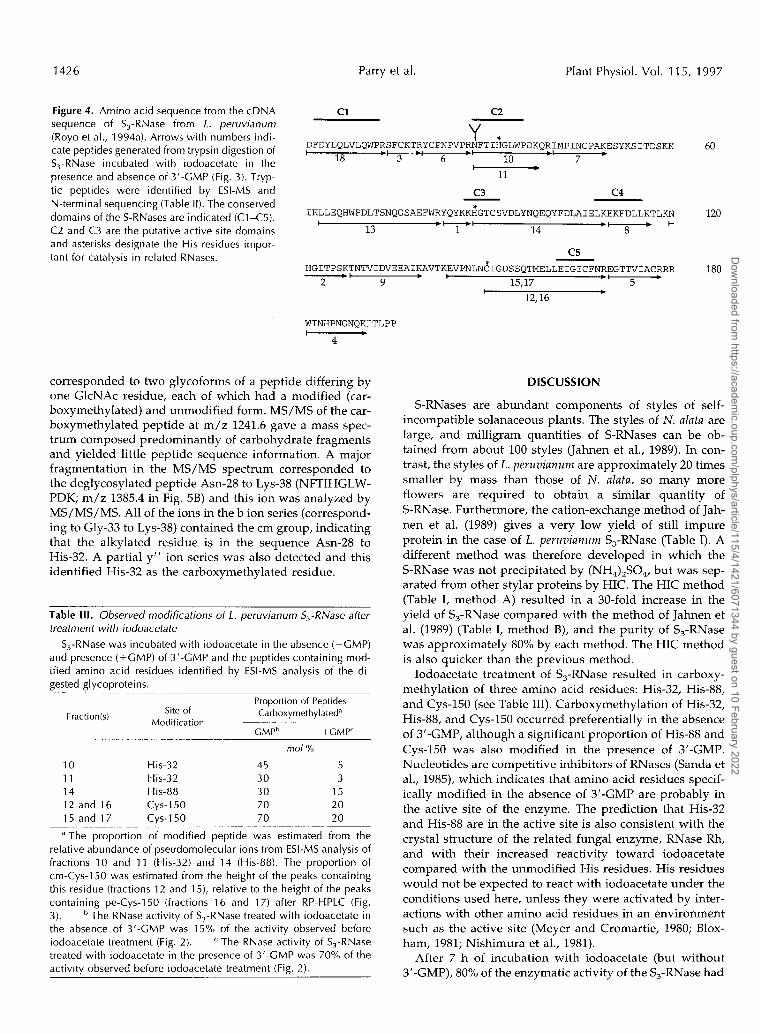
Figure 4. Amino acid sequence from the cDNA sequence of S,-RNase from L . peruvianum (Royo et al., 1994a). Arrows with numbers indi- cate peptides generated from trypsin digestion of S,-RNase incubated with iodoacetate in the presence and absence of 3’-GMP (Fig. 3). Tryp- tic peptides were identified by ESI-MS and N-terminal sequencing (Table 11). The conserved domains of the S-RNases are indicated (Cl-C5). C2 and C3 are the putative active site domains and asterisks designate the His residues impor- tant for catalysis in related RNases.
Parry et al. Plant Physiol. Vol. 115, 1997
c1 c2
Y * DFDYLQLVLQWPRSFCKTRYCPNPVPRNFTIHGLWPDKQRIMPINCPAKESYKSITDSKK I
18 3 6 - ‘ 10 7 . h+-.l - 11
60
c3 c4
IKLLEQHWPDLTSNQGSAEFWRYQYKK;~GTCSVDLYNQEQYFDLAIELKEKFDLLKTLKN 120 t .-I
13 1 14 - ‘ 8 . +
c5 HGITPSKTNTVIDVEEAIKAVTKEVPNLNC*IGDSSQTMELLEIGICFNREGTTVIACRRR 180
I 2 9 15,17 ‘ 5 .
I 12,16
*
WINHPNGNQKITLPP - 4
corresponded to two glycoforms of a peptide differing by one GlcNAc residue, each of which had a modified (car- boxymethylated) and unmodified form. MS/MS of the car- boxymethylated peptide at m/z 1241.6 gave a mass spec- trum composed predominantly of carbohydrate fragments and yielded little peptide sequence information. A major fragmentation in the MS/MS spectrum corresponded to the deglycosylated peptide Asn-28 to Lys-38 (NFTIHGLW- PDK; m/z 1385.4 in Fig. 5B) and this ion was analyzed by MS/MS/MS. A11 of the ions in the b ion series (correspond- ing to Gly-33 to Lys-38) contained the cm group, indicating that the alkylated residue is in the sequence Asn-28 to His-32. A partia1 y“ ion series was also detected and this identified His-32 as the carboxymethylated residue.
Table 111. Observed modifications o f L. peruvianum S3-RNase after treatment with iodoacetate
S,-RNase was incubated with iodoacetate in the absence (-GMP) and presence (+CMP) of 3’-CMP and the peptides containing mod- ified amino acid residues identified by ESI-MS analysis of the di- aested aIvcoDroteins.
Proportion of Peptides Fraction(s) Site of Carboxymethylated”
Modification -CMPb +CMP‘
mo/ %
10 His-32 45 5 11 His-32 30 3 14 His-88 30 15 12 and 16 CyS-150 70 20 15 and 17 CyS-150 70 20
a The proportion of modified peptide was estimated from the relative abundance of pseudomolecular ions from ESI-MS analysis of fractions 10 and 11 (His-32) and 14 (His-88). The proportion of cm-Cys-150 was estimated from the height of the peaks containing this residue (fractions 12 and 15), relative to the height of the peaks containing pe-Cys-150 (fractions 16 and 17) after RP-HPLC (Fig. 3). The RNase activity of S,-RNase treated with iodoacetate in the absence of 3’-GMP was 15% of the activity observed before iodoacetate treatment (Fig. 2). The RNase activity of S,-RNase treated with iodoacetate in the presence of 3’-CMP was 70% of the activity observed before iodoacetate treatment (Fig. 2).
DISCUSSION
S-RNases are abundant components of styles of self- incompatible solanaceous plants. The styles of N. alata are large, and milligram quantities of S-RNases can be ob- tained from about 100 styles (Jahnen et al., 1989). In con- trast, the styles of L. peruvianum are approximately 20 times smaller by mass than those of N. alata, so many more flowers are required to obtain a similar quantity of S-RNase. Furthermore, the cation-exchange method of Jah- nen et al. (1989) gives a very low yield of still impure protein in the case of L. peruvianum S,-RNase (Table I). A different method was therefore developed in which the S-RNase was not precipitated by (NH,),SO,, but was sep- arated from other stylar proteins by HIC. The HIC method (Table I, method A) resulted in a 30-fold increase in the yield of S,-RNase compared with the method of Jahnen et al. (1989) (Table I, method B), and the purity of S,-RNase was approximately 80% by each method. The HIC method is also quicker than the previous method.
Iodoacetate treatment of S,-RNase resulted in carboxy- methylation of three amino acid residues: His-32, His-88, and Cys-150 (see Table 111). Carboxymethylation of His-32, His-88, and Cys-150 occurred preferentially in the absence of 3’-GMP, although a significant proportion of His-88 and Cys-150 was also modified in the presence of 3’-GMP. Nucleotides are competitive inhibitors of RNases (Sanda et al., 1985), which indicates that amino acid residues specif- ically modified in the absence of 3’-GMP are probably in the active site of the enzyme. The prediction that His-32 and His-88 are in the active site is also consistent with the crystal structure of the related funga1 enzyme, RNase Rh, and with their increased reactivity toward iodoacetate compared with the unmodified His residues. His residues would not be expected to react with iodoacetate under the conditions used here, unless they were activated by inter- actions with other amino acid residues in an environment such as the active site (Meyer and Cromartie, 1980; Blox- ham, 1981; Nishimura et al., 1981).
After 7 h of incubation with iodoacetate (but without 3’-GMP), 80% of the enzymatic activity of the S,-RNase had
Dow
nloaded from https://academ
ic.oup.com/plphys/article/115/4/1421/6071344 by guest on 10 February 2022

Chemical Modification of S-RNase 1427
1w-
100 A
I x4 d 1323~2'21 '
(parent ion
!! 1375.9 3 40-
B 1331.6 1529.9 1693.8
20 -
l0OJ
80 - o $ 60- .- c - I 40- 2 .-
20 -
5w 1WO 1500 2wo mass/charge
MSlMS on mlz 1375.9 ion 1
1368.4 bii
1om.o b8
910.5
o Bo- '2 !! 3 .2 60 - 2 40-
20-
o 863.2
5 Y"14
p 60-
.- b7 y'9
h 40- 1801.4
B B
20 -
845.1 (parent ion
-%O) 562.2
b4 667.9 Y7
650.2 351.3
253.3 b3 bl
1060 2000 mass/charge
MSlMSlMS on mlz 863.2 ion 1 c m ~ s ~ ~ i w j p ~ y + + a l . , + s p
bz b3 b4 b5
I 611.0 ' 758'1 '16 (parent ion -pe group) X20
100 I
400 i masdcharge
li00 li00 li00 mass/charge
1660
MSlMS on d z 1241.6 ion
Y 1 Asn28-Phe.Thr-Ue-HirCly-Leu-Trp-Pra-Asp-Lys~
1 100-
80- De-glycosylated Asna-Lys38 (+ m gmup) o
.Ei c 'S 40- B
3 a-
20-
500 l0W 1500
I mass'drarge MSlMSIMS on d z 1385.4 ion 4
Y9 Y? YL
400 800 1200
mass/charge
Figure 5. MS sequencing data of peptides containing cm-His-88 and cm-His-32. A, ESI-MS of peptide mixture containing the cm form of peptide 14 (His-88 to Lys-109) (Fig. 3; Table 11). The pseudomolecular ion [M+H]' 1375.9 was subjected to MS/MS to yield the b-H2 and y" series of ions shown. The ions labeied with an asterisk represent loss of water. The b,-H2 fragment ion at m/z 863.2 (cm-His-88 to Asp-94) was subjected to MS/MS/MS. B, Fraction 11 (Fig. 3; Table I I ) , containing His-32, was analyzed by ESI-MS. The doubly charged ions at m/z 11 11.4 and 121 2.4 correspond to glycoforms of Asn-28 to Lys-38 and the ions at m/z 11 40.3 and 1241.6 correspond to cm forms of these glycopeptides, respectively. The branched structure indicates the N-glycosylation site at Asn-28. The ion at m/z 1241.6 was subjected to MS/MS, resuiting in extensive fragmentation of the glycan component of the glycopeptide. These fragment ions are marked by filled circles. The fragment ion (m/z 1385.4) corresponding to the deglycosylated form of Asn-28 to Lys-38 was subjected to MS/MS/MS to yield b and y" ion series. The sequences of the parent ions are shown above the MS/MS and MS/MS/MS spectra in A and B.
Dow
nloaded from https://academ
ic.oup.com/plphys/article/115/4/1421/6071344 by guest on 10 February 2022

1428 Parry et al. Plant Physiol. Vol. 1 1 5, 1997
been lost. Based on the levels of modifications (Table 111), it is possible that modification of Cys-150 was the sole cause of this loss of activity. Cys-150 is conserved in all charac- terized S-RNases (Tsai et al., 1992), and in S,-RNase this residue is not involved in a disulfide bond (S. Parry and D. Oxley, unpublished data). Although these results are con- sistent with a catalytic role for Cys-150, there is stronger evidence to support a structural role for this residue. In the S,- and S,-RNases from N. alata, Cys-150 is disulfide- bonded to a Cys residue located near the C terminus (Ish- imizu et al., 1996; Oxley and Bacic, 1996). This C-terminal Cys residue is conserved in all S-RNases except the L. peruviunum S,-RNase, suggesting that Cys-150 is usually involved in a disulfide bond. Furthermore, a three- dimensional model of RNase Rh predicts a structural role for Cys-150 (Kurihara et al., 1996).
Analysis of inactive S-RNases with changes at either His-32 or His-88 (S,-RNase numbering) indicates that both of these residues are needed for activity (Huang et al., 1994; Royo et al., 1994b). It is therefore probable that the cm-His residues observed in this study were responsible for the inactivation of S,-RNase. Recent chemical modification ex- periments by Ishimizu et al. (1995) on the N. alata S,-RNase found alkylation at His-32 (S,-RNase numbering) and a Cys residue at position 92 (S,-RNase has a Ser residue at this position; see Fig. 4). No modification of His-88 was observed, bringing the role of this residue in the catalytic activity of S-RNases into question. In this study we used MS sequencing to identify the modified His residues, be- cause N-terminal sequencing through the active-site His residues identified only a peak corresponding to an un- modified His residue (fractions 10, 11, and 14) (Fig. 4; Table 11). Whether the cm-His residue co-elutes with its unmod- ified form or is not recoverable under the Edman sequenc- ing conditions used is not known.
MS sequencing found that both His residues were mod- ified, and indicated that 40% of the His-32 residues (aver- age of modifications on glycopeptides 10 and 11 account- ing for the relative peak areas in Fig. 3) and 30% of the His-88 residues were modified in the absence of 3’-GMP. Carboxymethylation of His-32 and His-88 therefore ac- counts for 40 and 30%, respectively, of the loss of initial RNase activity. The sum of the two could account for the total loss in RNase activity but only if the two modifica- tions were mutually exclusive. Studies on fungal RNases related to the S-RNases also suggested that modification of one of the two catalytically active His residues prevented modification of the other (Irie et al., 1986; Kawata et al., 1990). Approximately equimolar amounts of the two cata- lytic His residues in the fungal RNases were modified. This is in agreement with the results presented in this study and suggests that the mode of catalysis of the fungal RNases and the S-RNases is similar.
If His-32 and His-88 were the catalytically active resi- dues, why was Cys-150 preferentially modified during our experiments in the absence of 3‘-GMP? One possibility is that carboxymethylation of either His residue altered the protein’s conformation. One effect of this conformational change may be to expose the thiol group on Cys-150 to the solvent, increasing the chance of carboxymethylation. Ac-
cordingly, the total percentage of modified His residue (cm-His-32 and cm-His-88) closely matches the percentage of cm-Cys-150, both in the presence and absence of 3‘-GMP (Table 111). Alternatively, 3’-GMP may cause a conforma- tional change that decreases solvent accessibility of Cys- 150. The finding that His-88 was modified in both the absence of 3’-GMP (30%) and the presence (15%) of 3’-GMP suggests that 3’-GMP does not occupy the entire substrate- binding site of the RNase.
The low specific activity of some S-RNases (including the S,-RNase) relative to RNase Rh, may be attributable to the absence of amino acid residues corresponding to His-104 and Glu-105 in RNase Rh. His-104 in this enzyme is im- portant for substrate binding (Ohgi et al., 1992), and Glu- 105 has a role in either polarizing the P = O bond of the PO, moiety or stabilizing the reaction intermediate (Irie et al., 1994). In vitro mutagenesis of these two amino acid residues caused a significant decrease in the specific activ- ity of the recombinant protein (Ohgi et al., 1992, 1993). In S,-RNase, Tyr-83 and Gln-84 are present at the positions occupied by His-104 and Glu-105 in RNase Rh. Few S-RNases have His at position 83 (S,-RNase numbering) and, although many have Glu in the next position, it is probable that the wide range of specific activities found among the S-RNases is attributable to the lack of conser- vation in the noncatalytic amino acid residues in the active site.
Degradation of RNA is central to models explaining the mechanism of pollen rejection during self-incompatibility (Matton et al., 1994; Lush and Clarke, 1997). The loss of RNA, particularly rRNA, compromises a pollen tube’s abil- ity to make proteins and, therefore, its capacity for rapid growth. The observation that some S-RNases are far less enzymatically active than others, yet have the same biolog- ical activity, appears to contradict this model. However, it is possible that the in vitro RNase activity assay does not reflect the in vivo RNase activity of the S-RNases. For example, the fungal RNase a-sarcin digests RNA nonspe- cifically in vitro, but can cleave rRNA at a specific site when the substrate is a ribosome (Endo et al., 1983). If the S-RNases had a specific target sequence, its ability to cleave RNA nonspecifically in vitro would be less biologically significant. Alternatively, given the variability in the spe- cific activities of the S-RNases, perhaps a high specific activity is not needed to slow pollen tube growth. It is possible that only a small amount of RNA has to be di- gested in the pollen tube to markedly affect its growth through the style. In this case, S-RNases having low spe- cific activities would still be able to reject incompatible pollen tubes. More work directed at determining how much S-RNase is taken up into the pollen tube, and the substrate this protein acts upon inside the pollen tube, is necessary before we can fully understand the mechanism of pollen tube rejection.
ACKNOWLEDCMENTS
We acknowledge support for the purchase of the ESI-MS from the Clive and Vera Ramaciotti Foundations, the Ian Potter Foun- dation, The Australian Research Council, and the University of
Dow
nloaded from https://academ
ic.oup.com/plphys/article/115/4/1421/6071344 by guest on 10 February 2022

Chemical Modification of S-RNase 1429
Melbourne. We also thank Ms. K. Dunse for assistance with the peptide sequencing.
Received April22, 1997; accepted August 26, 1997. Copyright Clearance Center: 0032-0889/97/115/1421/09.
LITERATURE ClTED
Biemann K (1990) Sequencing of peptides by tandem mass spec- trometry and high energy collision induced dissociation. Meth- ods Enzymol 193: 455-479
Bloxham DP (1981) The chemical reactivity of the histidine-195 residue in lactate dehydrogenase thiomethylated at the cysteine- 165 residue. Biochem J 193: 93-97
Bradford MM (1976) A rapid and sensitive method for the quan- titation of microgram quantities of protein utilizing the principle of protein-dye binding. Ana1 Biochem 72: 248-254
Crestfield AM, Stein WH, Moore S (1963) Alkylation and identi- fication of the histidine residues at the active site of ribonucle- ases. J Biol Chem 238: 2413-2420
de Nettancourt (1977) Incompatibility in Angiosperms. Springer- Verlag, New York
Endo Y, Huber PW, Wool IG (1983) The ribonuclease activity of the cytotoxin a-sarcin: the characteristics of the enzymatic ac- tivity of a-sarcin with ribosomes and ribonucleic acids as sub- strates. J Biol Chem 258: 2662-2667
Haring V, Gray JE, McClure BA, Anderson MA, Clarke AE (1990) Self-incompatibility: a self-recognition system in plants. Science
Huang S, Lee H-S, Karunanandaa B, Kao T-h (1994) Ribonuclease activity of Petunia inflata S proteins is essential for rejection of self-pollen. Plant Cell 6: 1021-1028
Irie M, Ohgi K, Watanabe H, Iwama M, Nakamura KT, Kurihara H, Nonaka T, Mitsui Y, Horiuchi H, Takagi M (1994) pH profile of kinetic constants of RNase Rh from Rhizopus niveus and its mutant enzymes towards UpU, and possible mechanisms of RNase Rh. J Biochem 115: 1083-1087
Irie M, Watanabe H, Ohgi K, Harada M (1986) Site of alkylation of the major ribonuclease from Aspergillus saitoi with iodoac- etate. J Biochem 99: 627-633
Ishimizu T, Miyagi M, Norioka S, Liu Y-H, Clarke AE, Sakiyama F (1995) Identification of histidine 31 and cysteine 95 in the active site of self-incompatibility associated S,-RNase in Nicoti- ana alata. J Biochem 118: 1007-1013
Ishimizu T, Norioka S, Kanai M, Clarke AE, Sakiyama F (1996) Location of cysteine and cystine residues in S-ribonucleases associated with gametophytic self-incompatibility. Eur J Bio- chem 242: 627-635
Jahnen W, Batterham MP, Clarke AE, Moritz RL, Simpson RJ (1989) Identification, isolation and N-terminal sequencing of style glycoproteins associated with self-incompatibility in Nico- tiana alata. Plant Cell 1: 493-499
Kawata Y, Sakiyama F, Hayashi F, Kyogoku Y (1990) Identifica- tion of two essential histidine residues of ribonuclease T, from Aspergillus oryzae. Eur J Biochem 187: 255-262
250: 937-941
Kowyama Y, Kunz C, Lewis I, Newbigin E, Clarke AE, Anderson MA (1994) Self-incompatibility in a Lycopersicon peruvianum vari- ant (LA2157) is associated with a lack of style S-RNase activity. Theor Appl Genet 88: 859-864
Kurihara H, Nonaka T, Mitsui Y, Ohgi K, Irie M, Nakamura KT (1996) The cr stal structure of ribonuclease Rh from Rhizopus
Laemmli UK (1970) Cleavage of structural proteins during the assembly of the head of bacteriophage T4. Nature 227: 680-685
Lush WM, Clarke AE (1997) Observations of pollen tube growth in Nicotiana alata and their implications for the mechanism of self-incompatibility. Sex Plant Reprod 10: 27-35
Matton DP, Nass N, Clarke AE, Newbigin E (1994) Self- incompatibility: how plants avoid illegitimate offspring. Proc Natl Acad Sci USA 91: 1992-1997
Mau S-L, Williams EG, Atkinson A, Anderson MA, Cornish EC, Grego B, Simpson RJ, Kheyr-Pour A, Clarke AE (1986) Style proteins of a wild tomato (Lycopersicon peruvianum) associated with expression of self-incompatibility. Planta 169: 184-191
McClure BA, Haring V, Ebert PR, Anderson MA, Simpson RJ, Sakiyama F, Clarke AE (1989) Style self-incompatibility gene products of Nicotiana alata are ribonucleases. Nature 342 955-957
Meyer SE, Cromartie TH (1980) Role of essential histidine resi- dues in L-a-hydroxy acid oxidase from rat kidney. Biochemistry 19 1874-1881
Nishimura H, Sempuku K, Iwashima A (1981) Possible functional roles of carboxyl and histidine residues in a soluble thiamine- binding protein of Saccharomyces cereuisiae. Biochim Biophys Acta 668: 333-338
Ohgi K, Horiuchi H, Watanabe H, Iwama M, Takagi M, Irie M (1992) Evidence that three histidine residues of a base non- specific and adenylic acid preferential ribonuclease from Rhizo- pus niveus are involved in the catalytic function. J Biochem 112:
Ohgi K, Horiuchi H, Watanabe H, Iwama M, Takagi M, Irie M (1993) Role of Asp51 and Glu105 in the enzymatic activity of a ribonuclease from Rhizopus niveus. J Biochem 113: 219-224
Oxley D, Bacic A (1996) Disulphide bonding in a stylar self- incompatibility ribonuclease of Nicotiana data. Eur J Biochem
Royo J, Kowyama Y, Clarke AE (1994a) Cloning and nucleotide sequence of two S-RNases from Lycopersicon peruvianum (L.) Mill. Plant Physiol 105 751-752
Royo J, Kunz C, Kowyama Y, Anderson M, Clarke AE, Newbigin E (199413) Loss of a histidine residue at the active site of S-locus ribonuclease is associated with self-compatibility in Lycopersicon peruvianum. Proc Natl Acad Sci USA 91: 6511-6514
Sanda A, Takizawa Y, Irie M (1985) Carboxymethylation of a ribonuclease from Rhizopus sp. Chem Pharm Bull33: 45154521
Tsai D-S, Lee H-S, Post LC, Kreiling KM, Kao T-h (1992) Se- quence of an S-protein of Lycopersicon peruvianum and compar- ison with other solanaceous S-proteins. Sex Plant Reprod 5:
niveus at 2.0 1 resolution. J Mo1 Biol 255 310-320
132-138
242: 75-80
256-263
Dow
nloaded from https://academ
ic.oup.com/plphys/article/115/4/1421/6071344 by guest on 10 February 2022