identification of an alternatively spliced site in human
TRANSCRIPT
Identification of an Alternatively Spliced Site in Human Plasma Fibronectin That Mediates Cell Type-specific Adhesion Mart in J. Humphries ,*~ Steven K. Akiyama ,* t Akira Komoriya,§ Kenneth Olden,*t and Kenneth M. Yamada~ * Howard University Cancer Center, Washington, DC 20060; * Membrane Biochemistry Section, Laboratory of Molecular Biology, National Cancer Institute, National Institutes of Health, Bethesda, Maryland 20892; and § Division of Molecular Biology Research, Biotechnology Research Center, Meloy Laboratories, Rockville, Maryland 20850
Abstract. We have compared the molecular specifici- ties of the adhesive interactions of melanoma and fibroblastic cells with fibronectin. Several striking differences were found in the sensitivity of the two cell types to inhibition by a series of synthetic peptides modeled on the Arg-Gly-Asp-Ser (RGDS) tetrapeptide adhesion signal. Further evidence for differences be- tween the melanoma and fibroblastic cell adhesion sys- tems was obtained by examining adhesion to proteo- lytic fragments of fibronectin. Fibroblastic BHK cells spread readily on f13, a 75-kD fragment representing the RGDS-containing, "cell-binding" domain of fibronectin, but B16-F10 melanoma cells could not. The melanoma cells were able to spread instead on f9, a ll3-kD fragment derived from the large subunit of fibronectin that contains at least part of the type III connecting segment difference region (or "V" region); f7, a fragment from the small fibronectin subunit that lacks this alternatively spliced polypeptide was inac- tive. Monoclonal antibody and f13 inhibition experi-
ments confirmed the inability of the melanoma cells to use the RGDS sequence; neither molecule affected melanoma cell spreading, but both completely abrogated fibroblast adhesion. By systematic analysis of a series of six overlapping synthetic peptides span- ning the entire type III connecting segment, a novel attachment site was identified in a peptide near the COOH-terminus of this region. The tetrapeptide se- quence Arg-Glu-Asp-Val (REDV), which is somewhat related to RGDS, was present in this peptide in a highly hydrophilic region of the type III connecting segment. REDV appeared to be functionally impor- tant, since this synthetic tetrapeptide was inhibitory for melanoma cell adhesion to fibronectin but was inactive for fibroblastic cell adhesion. REDV therefore represents a novel adhesive recognition signal in fibronectin that possesses cell type specificity. These results suggest that, for some cell types, regulation of the adhesion-promoting activity of fibronectin may oc- cur by alternative mRNA splicing.
C ELLULAR adhesion to extracellular matrices occurs during many important biological events, including growth, development, and disease pathogenesis (1, 4,
14, 18, 22, 26, 37, 45, 56, 58). A better understanding of the biochemical interactions involved in cell adhesion would yield valuable insights into the regulation of these processes. Considerable progress has been made recently by studying the molecular basis of the interaction of ceils with the proto- type adhesion factor fibronectin (for reviews see 4, 22, 25, 26, 46, 61).
Intact fibronectin binds with moderate affinity (Kd = 8 × 10 -7 M) to a relatively abundant fibroblastic cell surface receptor population (2). This direct binding interaction is in- hibited in a competitive manner both by well-characterized cell-binding fragments of fibronectin (5), and by synthetic peptides derived from the primary sequence of the fibronec- tin molecule (3). The sequence of the minimal cell recog- nition site in the "cell-binding" domain of fibronectin has
been recently identified as the tetrapeptide Arg-Gly-Asp-Ser (RGDS) (40-42, 63, 64). Synthetic peptides containing this sequence mediate cellular attachment when immobilized on a suitable substrate (40-42), and are autoinhibitory in assays monitoring the adhesion of fibroblastic cells to both intact fibronectin (40, 41, 63, 64) and its purified "cell-binding" domain (63).
The RGDS sequence in fibronectin is present in a highly hydrophilic region at a predicted ~-turn (40), which suggests that it is located at an exposed surface site accessible for binding to the fibronectin receptor. RGDS-containing pep- tides have recently been used in nonimmunological ap- proaches for identification of the fibronectin receptor (6, 21, 44), and for investigating the role of this adhesive recogni- tion signal in relevant biological processes in vivo such as experimental metastasis (24), embryonic development (9), and platelet function (13, 17).
Although evidence is strongest for the interaction of cells
© The Rockefeller University Press, 0021-9525/86/12/2637/11 $1.00 The Journal of Cell Biology, Volume 103 (No. 6, Pt. 2), Dec. 1986 2637-2647 2637
on November 29, 2018jcb.rupress.org Downloaded from http://doi.org/10.1083/jcb.103.6.2637Published Online: 1 December, 1986 | Supp Info:
with fibronectin being via the RGDS sequence, there are some indications of possible additional cell-binding regions or mechanisms within fibronectin. (a) The affinity of syn- thetic peptides, or even of an ll.5-kD cell-binding fragment, for fibroblasts is substantially lower than larger fragments or the intact molecule (3, 5), suggesting that sequences outside of RGDS are required for full biological activity of fibronec- tin. (b) Sialylated gangliosides have been reported to inter- fere with fibronectin-mediated adhesion in vitro (27, 67). Studies with a ganglioside-deficient mutant cell line, which is unable to retain endogenously synthesized fibronectin on its cell surface, have shown retention and reorganization of the pericellular fibronectin matrix after exogenous ganglio- side addition (50, 51, 66). Recently, a ganglioside-binding domain that may be responsible for this effect has been iden- tified in fibronectin (55). Binding activity resides in a region of fibronectin distinct from the "cell-binding" domain. (c) Comparisons of the adhesion-promoting activity of fibronec- tin, fibronectin fragments, and the heparan sulfate-binding protein, platelet factor 4, have suggested that heparan sulfate proteoglycan may mediate some aspects of the adhesion pro- cess, perhaps in concert with the "cell-binding" domain of fibronectin (8, 31, 60). Furthermore, attachment-promoting activity for mouse melanoma cells has been identified in a proteolytic fragment of fibronectin containing the COOH- terminal heparin-binding site (32). (d) Kinetic analysis of the incorporation of exogenous fibronectin into cell layers has suggested that assembly of the fibronectin matrix may be mediated by the NH2-terminal 70-kD domain (33).
In this study, we have compared the specificities of the adhesive interactions of B16-F10 murine melanoma cells and fibroblastic baby hamster kidney (BHK) cells with fibronec- tin, using a library of synthetic peptides modeled on the RGDS sequence. Differences were detected in the profile of inhibition of adhesion by the peptide homologues, which can be ascribed to the use of different adhesive recognition sig- nals by the two cell lines. BHK cells use the well-character- ized RGDS sequence, whereas B16-F10 ceils recognize a sec- ond site involving the type III connecting segment region of the fibronectin molecule that appears to be distinct from any of the other proposed cell interaction sites discussed above.
Materials and Methods
Materials Fibronectin was purified from freshly frozen, citrated human plasma (Na- tional Institutes of Health Blood Bank, Bethesda, MD) by gelatin affinity chromatography using elution with citric acid as described (35, 62). Tryptic fragments of fibronectin (t7, t9, and f13) were isolated as described (19). Labeling of fibronectin and f13 with carrier-free [3H]sodium borohydride (New England Nuclear, Boston, MA) was carried out as described (2, 52). 29-kD and 38-kD fragments of fibronectin containing the COOH-terminal, high affinity heparin-binding sites from the large and small subunits, respec- tively, were isolated by thermolysin digestion, chromatography on hydrox- ylapatite, and gel filtration (68). Custom synthesis of tetra- and pentapep- tides was carried out by Peninsula Laboratories, Inc. (Belmont, CA). Peptides were further purified as described (24). Synthesis of longer pep- tides is described below. The isolation and characterization of monoclonal antibody 333 (mAb333) has been described previously (5). Thr-Lys-Pro- Arg (TKPR; tuflsin), and Gly-His-Lys (GHK; liver cell growth factor) were from Bachem Inc. (Torrance, CA). Insulin A chain, fibrinopeptide A, Arg- Asp (RD), and Arg-Gly-Pro-Phe-Pro-lle (RGPFPI; sexual agglutination peptide) were from Sigma Chemical Co. (St. Louis, MO).
Cell-spreading Assay B16-FI0 murine melanoma cells (10) (generously provided by Dr. I. J. Fid- let, M. D. Anderson Hospital, University of Texas, Houston, TX) and fibroblastic BHK cells were cultured as described (U, 23, 63). Spreading assays on fibronectin or its proteolytic fragments were performed by a modification of the method of Yamada and Kennedy (63). Adhesion factors were diluted into Dulbecco's phosphate-buffered saline (PBS+) I (Gibco, Grand Island, NY), and 100-~tl aliquots were added to 6.4-mm (96-well) tis- sue culture clusters (Costar, Cambridge, MA). After incubation at room temperature for 60 rain, sites for nonspecific cell attachment to the plastic surface were blocked by incubation with 100 ~tl 10 nag/nil heat-denatured BSA. This blocking solution was prepared by dissolving crystalline BSA (Caibiochem-Behring Corp., La Jolla, CA) in divalent cation-free PBS + (PBS-), filtering the mixture, and heating in a water bath at 85"C for 10 rain. After a 30-rain incubation at room temperature, the BSA solution was removed and 25-1~1 peptide solutions in PBS + were added.
Cell cultures were washed with PBS-, and ceils were detached with 0.25% trypsin, 0.02% EDTA, then washed and resuspended to 1.3 x 10⁵ cells/ml in DME. Cell suspensions were allowed to recover from the proteo- lyric treatment for 10 min at 3"/*C before addition of 75-1~1 aliquots to the 96-well clusters. After incubation for 60 rain at 370C, attached cells were fixed with 3 % formaldehyde and 3% glutaraldehyde in PBS- and pho- tographed using phase-contrast microscopy. The percentage of cells adopt- ing a normal, well-spread morphology was estimated by counting 300 cells/well in a number of randomly selected fields.
The assay measuring cell spreading on immobilized peptides was carried out as described above except that 96-well clusters (Linbro Titertek; Flow Laboratories, Inc., McLean, VA) were coated with peptide for 2 h and nonspecific adhesion was reduced by blockage with 10 ~tg/ml BSA in PBS + f o r l h.
Synthesis of CS Peptides Overlapping peptides comprising the type III connecting segment (IIICS) of human plasma fibronectin were synthesized according to the method of Merrifield (34) using an Applied Biosystems 430A peptide synthesizer. The symmetric anhydrides of t-butyloxycarbonyl-protected amino acids (59) were used for dicyclohexylcarbodiimide-mediated coupling to the peptide chain. The recently developed phenylacetamidomethyl-resin (36) was used as a solid support. Peptides were cleaved from the support by treatment with anhydrous hydrogen fluoride for 1 h at 0°C in the presence of 10% (vol/vol) anisole and 1% (vol/vol) dimethylsulfide. After extraction with 50% (vol/ vol) acetic acid, the peptides were chromatographed on Bio-Gel P4 using 1 M acetic acid as eluant. Fractions containing peptide were lyophilized and further purified as described below.
Purity of Polypeptide Fragments and Synthetic Peptides The purity of tryptic fragments of fibronectin (17, 19, and f13) was assessed by serial dilution and SDS PAGE (using 4 % stacking and 8 % resolving gels) followed by Coomassie Blue staining. Fragments were 90-98% pure by this method; 19 was >/95% pure.
Synthetic peptides purified by Bio-Gel P4 chromatography were further purified to homogeneity by semi-preparative, reversed-phase HPLC. A Syn- chropak RP-P C18 column (250 mm x 10 nun; SynChrom, Inc., Linden, IN) was equilibrated with 0.1% trifluoroacetic acid at a flow rate of 1.0 ml/min and, after injection of peptide in the same solvent, the column was developed by a linear gradient of 0-10% acetonitrile in 0.1% trifluoroacetic acid over a 3-min period followed by a further linear gradient of 10-50% acetonitrile in 0.1% trifluoroacetic acid over a 30-rain period. By monitoring absorbance at 220 ran, peptides were found to elute as single major peaks. The main peaks were pooled, lyophilized, and rechromatographed on a Syn- chropak RP-P C18 analytical column (250 mm x 4.1 mm; SynChrom, Inc.) using the gradient conditions described above and a flow rate of 0.8 mi/min. Peptides eluted between 21 and 34% acetonitrile and were 95.1-99.4% pure (CS5 was 98.5% pure) after peak integration. CS peptides were labeled by reductive methylation of free primary amino groups with [3H]sodium borohydride (New England Nuclear) as described (52).
1. Abbreviations used in this paper: IIICS, type III connecting segment; PBS +, Dulbecco's phosphate-buffered saline; PBS-, divalent cation-free PBS +.
The Journal of Cell Biology. Volume 103, 1986 2638
A BHK B B16-F10
G R G D
\ \ \ \ ,,o"oo
0~) 0.2 0.'4 06 08 1-0 0~) 0:2 0.~4 0:6 ~0:8 ~--1.;0 PEPTIDE (mg/ml)
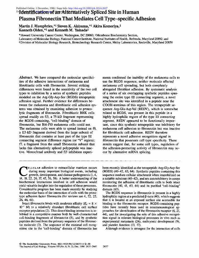
Figure 1. Comparison of the inhibitory effects of a series of tetra- and pentapeptides mod- eled on the RGDS sequence on cell spreading on fibronec- tin. The percentage of (A) BHK fibroblasts or (B) B16- F10 melanoma cells adopting a spread morphology on sub- strates coated with 2 txg/ml (A) or 4 ~tg/ml (B) human plasma fibronectin is shown at the indicated concentrations of synthetic peptide.
Results
Comparison of the Effects of Synthetic Peptides on Melanoma and Fibroblastic Cell Adhesion to Fibronectin
Recently, libraries of short, synthetic peptides have been used to examine the specificity of cellular interactions with fibronectin and other extracellular matrix components (13, 17, 40, 41, 43, 49, 63, 64). In general, peptides containing the RGDS sequence have been found to be the most effective inhibitors of adhesive function, whereas other homologues in which amino acids are deleted, substituted, or transposed are much less active. The relative activities of RGDS homo- logues can therefore be used to assess the specificity of a cell- adhesion protein interaction.
The data presented in Fig. 1 show a comparison of the rela- tive inhibitory activity of a series of homologous peptides on the fibronectin-mediated spreading of B16-F10 melanoma and BHK fibroblastic cells. A number of unexpected differ- ences were found in the ability of the peptides to interfere with the adhesion of these two cell lines. Consistent with previous reports using a variety of fibroblastic cell lines (40, 41, 49, 63, 64), fibronectin-mediated spreading of BHK cells was inhibited by (G)RGDS, a peptide that occurs in the pri- mary sequence of the "cell-binding" domain of fibronectin, and SDGR, a peptide with an inverted sequence (Fig. 1 A). For BHK cells, GRGDS was consistently more active on a molar basis than both RGDS and SDGR. The specificity of the inhibition of BHK cell spreading was apparent from the complete lack of inhibitory activity in three peptides con- taining either a deletion of the COOH-terminal residue of GRGDS (GRGD), a conservative substitution of a glutamic acid residue for an aspartic acid residue (GRGES), or a transposition of the central glycine and aspartic acid residues (GRDGS) (Fig. 1 A).
Parallel examination of the effects of each of these six pep- tides on B16-F10 melanoma cell spreading revealed several striking differences compared with BHK cells (Fig. 1 B). Al- though GRGDS, RGDS, and SDGR were again inhibitory, the relative activities of each were considerably different.
RGDS was the most active, followed by SDGR and GRGDS (Fig. 1 B). As for BHK cells, GRDGS was inactive, but for the melanoma cells both GRGD and GRGES were found to possess substantial activity. In particular, the concentrations of GRGDS and GRGES required for half-maximal inhibition were quite similar (230 I.tg/ml and 310 gg/ml respectively; Fig. 1 B). The marked inhibition by GRGES in Fig. 1 B con- trasts markedly with previous results with fibroblastic cells (40, 41, 63, 64).
The inhibition by peptides in Fig. 1 was competitive in na- ture, since increasing the level of substrate-adsorbed fibro- nectin to 50 Ixg/ml was in all cases sufficient to overcome the inhibitory activity of each peptide (data not shown). This re- sult also establishes that the data cannot be explained by selective cytotoxicity of the synthetic peptides for the mela-
NI I I
N
P
HEPARIN FIBRIN
I I I I I I IA
r
I COLLAGENI DNA I
f7 id " i I
14.38kD,IM I I I I I I I I I I
Bl, r i RGDS IIICS
I g/~ IIc I II I I " I I II I
1 2 9 k D I I I f13 ~ I | i
I I ~I I f 9 1 " " I
CELL HEPARIN I FIBRIN l
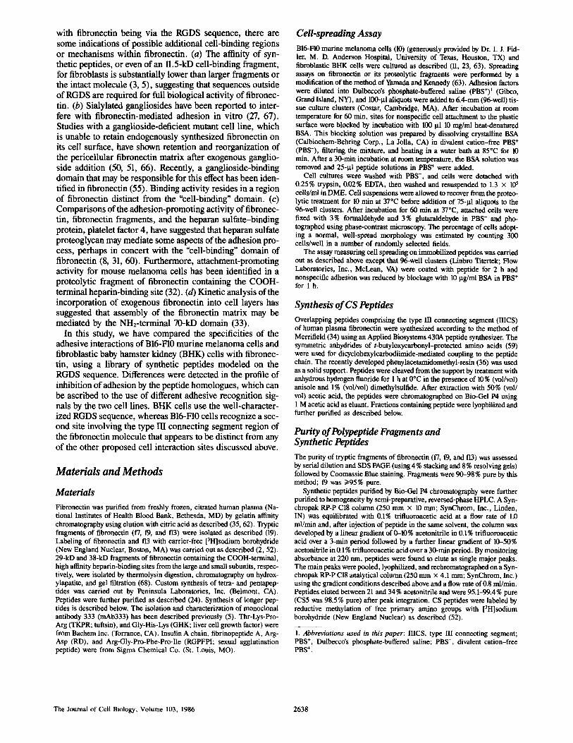
Figure 2. Map of the human plasma fibronectin dimer to show the derivation of the fragments used in this study. The sites of the RGDS tetrapeptide and the IIICS region are highlighted in the hatched areas. The binding specificities of the different fibronectin domains are listed under the diagram. The amino and carboxyl termini of the fibronectin chains are indicated by N and C, respectively.
Humphries et al. Novel Cell Adhesion Site in Human Fibronectin 2639
noma cells. As further controls, comparison of the semi- tivity to peptide inhibition of a mouse fibroblast cell line (BALB/c-3T3) with the mouse melanoma ceils (B16-F10) gave the same pattern of results as for BHK fibroblasts (data not shown); even primary cell strains of chick fibroblasts showed identical patterns of sensitivities (65), ruling out an artefact of the fibroblast species used. Similarly, culturing BHK fibroblasts for 1 wk in the same growth medium used for melanoma ceils did not affect their pattern of inhibition by the peptide library (data not shown), indicating that the difference in susceptibility to peptide inhibition was not at- tributable to the growth medium used for routine culture. Furthermore, the data in Fig. 1 did not arise from a complete breakdown in the specificity of peptide-mediated inhibition of the melanoma cells, since a number of synthetic peptide molecules unrelated to RGDS were completely inactive at concentrations up to 1 mg/ml (including TKPR, GHK, insu- lin A chain, fibrinopeptide A, RD, and RGPFPI [data not shown]; GRDGS was also inactive [Fig. 1 B]). These results in particular suggest that an RGD-type mechanism may be involved in B16-F10 melanoma cell spreading, but the differ- ent pattern of inhibition by RGDS homologues nevertheless reflects a novel profile of specificity for a cellular interaction with fibronectin.
Promotion of Melanoma and Fibroblastic Cell Spreading by Proteolytic Fragments of Fibronectin Two possible explanations for the differences in the profile of peptide inhibition in Fig. 1 were as follows: (a) the two cell types were recognizing different sites in fibronectin, or (b) the binding site of the fibronectin receptor of melanoma cells was altered, resulting in a less specific recognition of RGDS- like sequences compared with BHK cells. To investigate the first possibility, we compared the spreading of BI6-FI0 and BHK cells on different purified tryptic fragments of fibronec- tin. The fragments tested have been previously characterized in detail, and are designated f7, 19, and f13 (19). Fig. 2 is a map of the human plasma fibronectin dimer showing the ori- gin of these fragments, f13 is a 75-kD polypeptide constitut- ing the trypsin-resistant "cell-binding" domain, while f7 and 19 contain COOH-terminal extensions of f13 derived from the small and large subunits of the fibronectin dimer, respec-
tively, t9 (113 kD) incorporates f13 together with the adjacent heparin-binding domain and at least part of the type III con- necting segment (IIICS) or "V" region. IIICS is the region alternatively spliced in fibronectin mRNA that is thought to determine the size difference between each chain of the plasma fibronectin dimer; it appears to be located predomi- nantly in the large subunlt (25, 39, 47). t7 (146 kD) is the corresponding fragment derived from the small subunit. In addition to the heparin-binding domain, 17 contains the COOH-terminal fibrin-binding domain and therefore spans the site where IIICS is spliced out of the molecule (19).
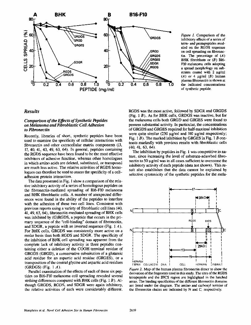
As previously reported (19), the concentration dependence of BHK fibroblast cell spreading was similar on fibronectin and the three tryptic fragments when the coating concentra- tion of polypeptide was expressed on a molar basis (Figs. 3 A and 4). The concentration required for 50% maximal spreading typically varied between 4 nM and 7 nM. The spreading of B16-F10 melanoma cells on intact fibronectin was comparable to BHK, with 50% maximal spreading ob- served at 7 nM and 4 nM, respectively (Fig. 3). However, the melanoma cells were strikingly poorly adhesive on a sub- strate of f13, with only 10% spreading occurring at even 53 nM (Figs. 3 B and 4). Examination of the effects of higher coating concentrations of f13 revealed that 50 % of maximal spreading was only obtained at >--530 nM, a concentration at least 75-fold higher than that needed for equivalent spreading on fibronectin, and 100-fold higher than the concentration re- quired for 50 % maximal spreading of BHK on f13 (Fig. 3 A). This finding suggests that for spreading on fibronectin, the melanoma cells either use a site distinct from RGDS or at least require adjacent contributory site(s) outside of the 75-kD "cell-binding" domain to be able to use the RGDS se- quence. Although the ability of BHK cells to respond to t"13 is a direct, positive control for the absence of activity of the melanoma cells, we also measured the binding of radiola- beled fibronectin and f13 to plastic to discount the possibility that the poor spreading of B16-F10 cells on f13 was due to the inability of the protein to bind to the wells. 3H-labeled f13 was found to bind ~l.2-fold better than [3H]fibronectin when expressed on a molar basis (data not shown).
When the spreading of melanoma ceils on the larger frag- ments 17 and 19 was examined, 19 was found to be as fully effective as intact fibronectin in supporting adhesion (50%
B H K BHK f13
c3 < L U
=_=4o r,/)
¢..)
A 8o
i i I I
40 sb
B B16-F10
- = r ' ' '
ADHESION PROTEIN (nM)
Figure 3. Analysis of the con- centration dependence of the spreading of (.4) BHK fibro- blasts or (B) B16-F10 mela- noma cells on fibronectin or its tryptic fragments, f7, t9, and f13. The coating concen- trations of all four molecules are expressed on a molar basis to permit comparison. 45 nM fibronectin monomer is equal to 12 t.tg/ml.
The Journal of Cell Biology, Volume 103, 1986 2640
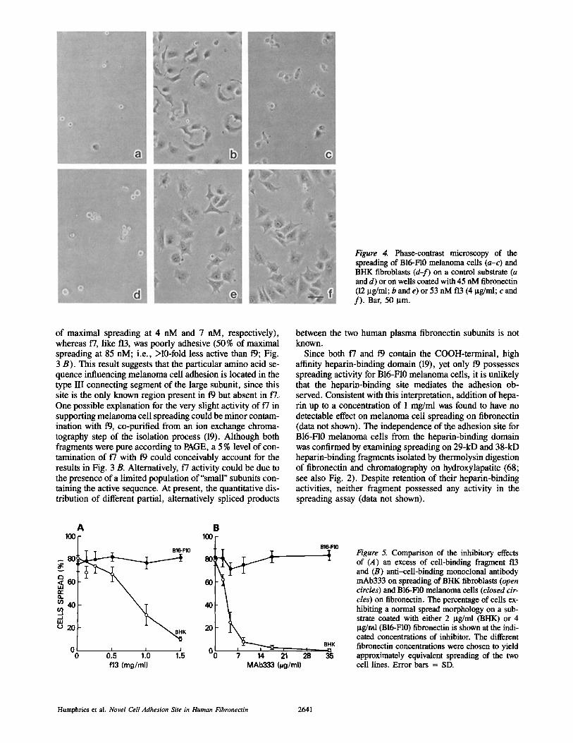
Figure 4. Phase-contrast microscopy of the spreading of B16-FI0 melanoma cells (a-c) and BHK fibroblasts (d-f) on a control substrate (a and d) or on wells coated with 45 nM fibroneetin (12 Ixg/ml; b and e) or 53 nM f13 (4 ~tg/ml; c and f) . Bar, 50 ~tm.
of maximal spreading at 4 nM and 7 nM, respectively), whereas 17, like f13, was poorly adhesive (50 % of maximal spreading at 85 riM; i.e., >10-fold less active than 19; Fig. 3 B). This result suggests that the particular amino acid se- quence influencing melanoma cell adhesion is located in the type lIl connecting segment of the large subunit, since this site is the only known region present in 19 but absent in 17. One possible explanation for the very slight activity of 17 in supporting melanoma cell spreading could be minor contam- ination with 19, co-purified from an ion exchange chroma- tography step of the isolation process (19). Although both fragments were pure according to PAGE, a 5 % level of con- tamination of 17 with 19 could conceivably account for the results in Fig. 3 B. Alternatively, 17 activity could be due to the presence of a limited population of"small" subunits con- taining the active sequence. At present, the quantitative dis- tribution of different partial, alternatively spliced products
between the two human plasma fibronectin subunits is not known.
Since both f7 and t9 contain the COOH-terminal, high affinity heparin-binding domain (19), yet only 19 possesses spreading activity for B16-F10 melanoma cells, it is unlikely that the heparin-binding site mediates the adhesion ob- served. Consistent with this interpretation, addition of hepa- rin up to a concentration of 1 mg/ml was found to have no detectable effect on melanoma cell spreading on fibronectin (data not shown). The independence of the adhesion site for B16-F10 melanoma cells from the heparin-binding domain was confirmed by examining spreading on 29-kD and 38-kD heparin-binding fragments isolated by thermolysin digestion of fibronectin and chromatography on hydroxylapatite (68; see also Fig. 2). Despite retention of their heparin-binding activities, neither fragment possessed any activity in the spreading assay (data not shown).
A 100
B16-F10
fT" n
m40 ~t ,
- J i i i
~ 2 0 K
0 I I J 0 0.5 1.0 1.5
f13 (mg/ml)
B 100
80:
6O
40
20
0~ 7 14 21
MAb333 (~g/ml)
B16-FlO
t
BHK I r~
28 35
I~gure 5. Comparison of the inhibitory effects of (A) an excess of cell-binding fragment f13 and (B) anti-cell-binding monoclonal antibody mAb333 on spreading of BHK fibroblasts (open circles) and B16-F10 melanoma cells (closed cir- cles) on fibronectin. The percentage of cells ex- hibiting a normal spread morphology on a sub- strate coated with either 2 ltg/ml (BHK) or 4 I~g/ml (B16-FI0) fibronectin is shown at the indi- cated concentrations of inhibitor. The different fibronectin concentrations were chosen to yield approximately equivalent spreading of the two cell lines. Error bars = SD.
H u m p h r i c s et al . Novel Cell Adhesion Site in Human Fibronectin 2 6 4 1
4 CSl I~ CS3 ~ - CS5
10 20 30 40 ~0 eO "lO 80 IO 100 110 120 D E L P Q L V T L P H P N L H G P E I L D V P S T V O K T p F V T H P G Y D T G N G I QL PGTSGQQPSVGQQM I F E E H G F R R T T P P T T A T P I RHRPRPYPPNVGEE I Q I G H I P R E D V D Y H L Y P H G P G L N P N A S T
I - CS2 ~1 1~ CS4 - I ' ° ~ ' C S 6 =
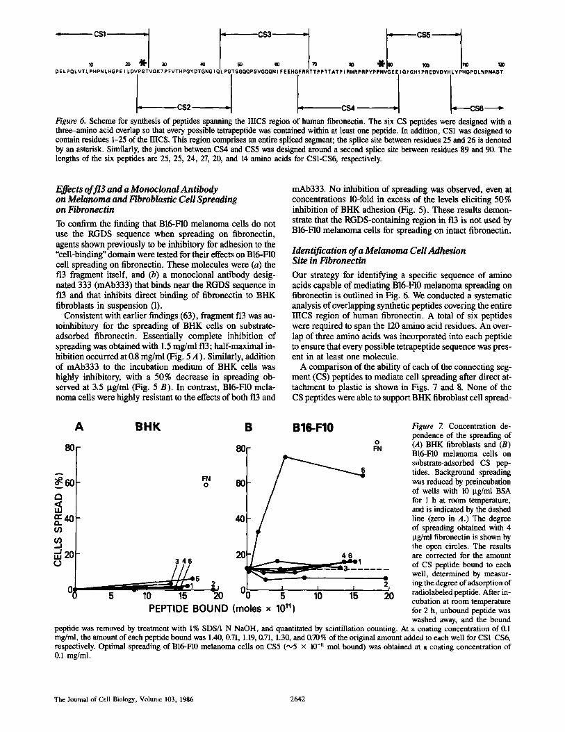
Figure 6. Scheme for synthesis of peptides spanning the IIICS region of human fibronectin. The six CS peptides were designed with a three-amino acid overlap so that every possible tetrapeptide was contained within at least one peptide. In addition, CSI was designed to contain residues 1-25 of the IIICS. This region comprises an entire spliced segment; the splice site between residues 25 and 26 is denoted by an asterisk. Similarly, the junction between CS4 and CS5 was designed around a second splice site between residues 89 and 90. The lengths of the six peptides are 25, 25, 24, 27, 20, and 14 amino acids for CS1-CS6, respectively.
Effects off13 and a Monoclonal Antibody on Melanoma and l~broblastic Cell Spreading on Fibronectin
To confirm the finding that B16-F10 melanoma cells do not use the RGDS sequence when spreading on fibronectin, agents shown previously to be inhibitory for adhesion to the "cell-binding" domain were tested for their effects on B16-F10 cell spreading on fibronectin. These molecules were (a) the f13 fragment itself, and (b) a monoclonal antibody desig- nated 333 (mAb333) that binds near the RGDS sequence in f13 and that inhibits direct binding of fibronectin to BHK fibroblasts in suspension (1).
Consistent with earlier findings (63), fragment f13 was au- toinhibitory for the spreading of BHK cells on substrate- adsorbed fibronectin. Essentially complete inhibition of spreading was obtained with 1.5 mg/ml f13; half-maximal in- hibition occurred at 0.8 mg/ml (Fig. 5 A). Similarly, addition of mAb333 to the incubation medium of BHK ceils was highly inhibitory, with a 50% decrease in spreading ob- served at 3.5 ttg/ml (Fig. 5 B). In contrast, BI6-F10 mela- noma cells were highly resistant to the effects of both t"13 and
mAb333. No inhibition of spreading was observed, even at concentrations 10-fold in excess of the levels eliciting 50% inhibition of BHK adhesion (Fig. 5). These results demon- strate that the RGDS-containing region in f13 is not used by B16-FI0 melanoma cells for spreading on intact fibronectin.
Identification of a Melanoma Cell Adhesion Site in Fibronectin
Our strategy for identifying a specific sequence of amino acids capable of mediating B16-F10 melanoma spreading on fibronectin is outlined in Fig. 6. We conducted a systematic analysis of overlapping synthetic peptides covering the entire IIICS region of human fibronectin. A total of six peptides were required to span the 120 amino acid residues. An over- lap of three amino acids was incorporated into each peptide to ensure that every possible tetrapeptide sequence was pres- ent in at least one molecule.
A comparison of the ability of each of the connecting seg- ment (CS) peptides to mediate cell spreading after direct at- tachment to plastic is shown in Figs. 7 and 8. None of the CS peptides were able to support BHK fibroblast cell spread-
A
80
A
~60 C~
¢ : 4 0 o. ¢/)
i J , , , 2 0
BHK B B16-F10
80
-45 ~l
10 1~ - ~ 0 5 10 15 20 P E P T I D E B O U N D ( m o l e s x 10111
Figure 7. Concentration de- pendence of the spreading of (A) BHK fibroblasts and (B) B16-F10 melanoma cells on substrate-adsorbed CS pep- tides. Background spreading was reduced by preincubation of wells with 10 I~g/ml BSA for 1 h at room temperature, and is indicated by the dashed line (zero in A.) The degree of spreading obtained with 4 I~g/ml fibronectin is shown by the open circles. The results are corrected for the amount of CS peptide bound to each well, determined by measur- ing the degree of adsorption of radiolabeled peptide. After in- cubation at room temperature for 2 h, unbound peptide was washed away, and the bound
peptide was removed by treatment with 1% SDS/1 N NaOH, and quantitated by scintillation counting. At a coating concentration of 0.1 mg/ml, the amount of each peptide bound was 1.40, 0.71, 1.19, 0.71, 1.30, and 0.70% of the original amount added to each well for CS1-CS6, respectively. Optimal spreading of BI6-FI0 melanoma cells on CS5 (",,5 x 10-" mol bound) was obtained at a coating concentration of 0.1 mg/ml.
The Journal of Cell Biology, Volume 103, 1986 2642
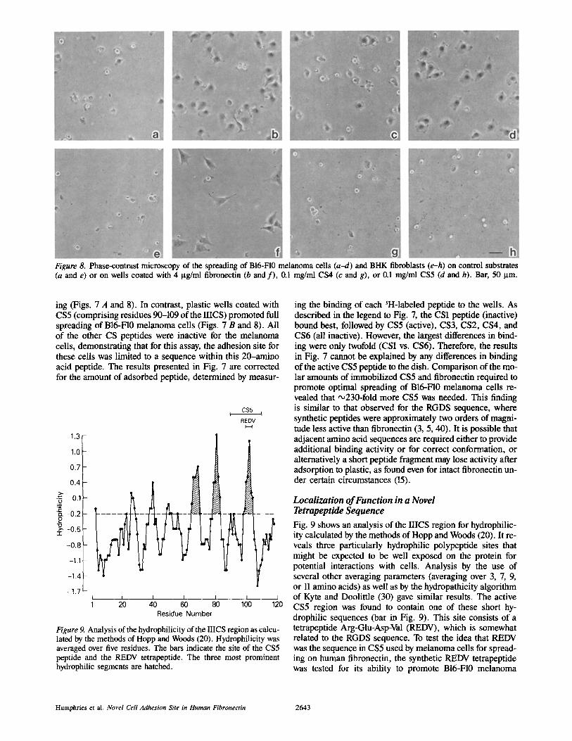
Figure 8. Phase-contrast microscopy of the spreading of B16-F10 melanoma cells (a-d) and BHK fibroblasts (e-h) on control substrates (a and e) or on wells coated with 4 gg/ml fibronectin (b and f) , 0ol mg/ml CS4 (c and g), or 0.1 mg/ml CS5 (d and h). Bar, 50 ~tm.
ing (Figs. 7 A and 8). In contrast, plastic wells coated with CS5 (comprising residues 90-109 of the IIICS) promoted full spreading of B16-F10 melanoma cells (Figs. 7 B and 8). All of the other CS peptides were inactive for the melanoma cells, demonstrating that for this assay, the adhesion site for these cells was limited to a sequence within this 20-amino acid peptide. The results presented in Fig. 7 are corrected for the amount of adsorbed peptide, determined by measur-
CS5 t I
REDV I-,-q
1.3
1.0
0.7
0.4
o.1 "5
:~ -0.2
~ -0 .5 "1-
-0 .8
-1.1
-1.4
-1.7 I I I I I I __.J 1 20 40 60 80 100 120
Residue Number
Figure 9. Analysis of the hydrophilicity of the IIICS region as calcu- lated by the methods of Hopp and Woods (20). Hydrophilicity was averaged over five residues. The bars indicate the site of the CS5 peptide and the REDV tetrapeptide. The three most prominent hydrophilic segments are hatched.
ing the binding of each 3H-labeled peptide to the wells. As described in the legend to Fig. 7, the CS1 peptide (inactive) bound best, followed by CS5 (active), CS3, CS2, CS4, and CS6 (all inactive). However, the largest differences in bind- ing were only twofold (CS1 vs. CS6). Therefore, the results in Fig. 7 cannot be explained by any differences in binding of the active CS5 peptide to the dish. Comparison of the mo- lar amounts of immobilized CS5 and fibronectin required to promote optimal spreading of B16-F10 melanoma ceils re- vealed that ~230-fold more CS5 was needed. This finding is similar to that observed for the RGDS sequence, where synthetic peptides were approximately two orders of magni- tude less active than fibronectin (3, 5, 40). It is possible that adjacent amino acid sequences are required either to provide additional binding activity or for correct conformation, or alternatively a short peptide fragment may lose activity after adsorption to plastic, as found even for intact fibronectin un- der certain circumstances (15).
Localization of Function in a Novel Tetrapeptide Sequence
Fig. 9 shows an analysis of the IIICS region for hydrophilic- ity calculated by the methods of Hopp and Woods (20). It re- veals three particularly hydrophilic polypeptide sites that might be expected to be well exposed on the protein for potential interactions with cells. Analysis by the use of several other averaging parameters (averaging over 3, 7, 9, or 11 amino acids) as well as by the hydropathicity algorithm of Kyte and Doolittle (30) gave similar results. The active CS5 region was found to contain one of these short hy- drophilic sequences (bar in Fig. 9). This site consists of a tetrapeptide Arg-Glu-Asp-Val (REDV), which is somewhat related to the RGDS sequence. To test the idea that REDV was the sequence in CS5 used by melanoma cells for spread- ing on human fibronectin, the synthetic REDV tetrapeptide was tested for its ability to promote B16-F10 melanoma
Humphries et al. Novel Cell Adhesion Site in Human Fibronectin 2643
A 80,
£D < ILl
(n
, ,--/, 20 (.J
~// REDV RGDS RGDV
100 200 300 400 500
B 8O
~ E D V
REDV + MAb"~"~k\ \ 40 -RGDS + MAb ' / " ~ \ \ \
20 RGDS'
o r I l 1 1 v o lOO 200 300 400 500
PEPTIDE (#g/ml )
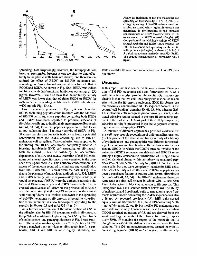
Figure 10. Inhibition of B16-F10 melanoma cell spreading on fibronectin by REDV. (A) The per- centage spreading of BI6-FI0 melanoma cells on a substrate coated with 4 I.tg/ml fibronectin was determined in the presence of the indicated concentrations of REDV (closed circle), RGDS (open circle), or RGDV (closed triangle). (B) Comparison of the inhibitory activity of REDV (closed symbols) and RGDS (open symbols) on B16-F10 melanoma cell spreading on fibronectin in the presence (triangles) or absence (circles) of 5 I.tg/ml monoclonal antibody mAb333 (MAb). The coating concentration of fibronectin was 4 ~g/rra.
spreading. Not surprisingly, however, the tetrapeptide was inactive, presumably because it was too short to bind effec- tively to the plastic wells (data not shown). We therefore ex- amined the effect of REDV on B16-F10 melanoma cell spreading on fibronectin and compared its activity to that of RGDS and RGDV. As shown in Fig. 10 A, REDV was indeed inhibitory, with half-maximal inhibition occurring at 150 Ixg/ml. However, it was also clear that the inhibitory activity of REDV was lower than that of either RGDS or RGDV for melanoma cell spreading on fibronectin (50% inhibition at ~100 ~tg/ml; Fig. 10 A).
From the results presented in Fig. 1, it was clear that RGDS-containing peptides could interfere with the adhesion of B16-F10 cells, and since peptides containing both RGDS and RGDV have been reported to promote adhesion of fibroblastic cells and to inhibit their attachment to fibronectin (40, 41, 63, 64), these two peptides appear to be able to act at both adhesion sites. The lower activity of REDV in Fig. 10 A may therefore be due to its inability to block a potential contribution from the RGDS-containing site to B16-F10 melanoma cell spreading. This conclusion was supported by the finding that REDV was almost completely inactive in blocking fibroblastic BHK cell spreading on fibronectin (data not shown). To test this possibility, the concentration dependence of REDV and RGDS inhibition of B16-F10 mela- noma cell spreading on fibronectin was examined in the pres- ence of 5 ~tg/ml mAb333. This antibody concentration is in excess of the amount required to eliminate any contribution from the RGDS site. It is clear from the data in Fig. 10 B that in the presence of monoclonal antibody mAb333, REDV and RGDS actually possess approximately equal activity, as would be expected if REDV were the authentic adhesion site for B16-F10 melanoma cells and RGDS cross-reacts. The in- creased effectiveness of REDV in the presence of mAb333 also demonstrates that the RGDS sequence in the central "cell-binding" domain is involved somewhat in the spreading of B16-FI0 melanoma on fibronectin, although its contribu- tion is not sufficient to allow blockage of spreading by the specific inhibitors t 3 and mAb333 (Fig. 5).
As a further confirmation of the identification of CS5 as the adhesion site for B16-F10 melanoma ceils in fibronectin, the profile of inhibition of spreading on CS5 by the library of synthetic tetra- and pentapeptides used in Fig. 1 was exam- ined. The relative inhibitory activities of the peptides on CS5 closely matched their activities on fibronectin itself; in par- ticular, GRGD and GRGES were highly inhibitory, and
RGDS and SDGR were both more active than GRGDS (data not shown).
Discussion
In this report, we have compared the mechanisms of interac- tion of B16-F10 melanoma cells and fibroblastic BHK cells with the adhesive glycoprotein fibronectin. Our major con- clusion is that the two cell lines recognize distinct adhesion sites within the fibronectin molecule; BHK fibroblasts use the previously characterized RGDS sequence located in the central "cell-binding" domain (40, 41, 63, 64), whereas B16- F10 melanoma cells recognize predominantly a novel, addi- tional adhesive region located in the type III connecting seg- ment of the molecule. At least part of this cell type-specific, adhesive activity is preserved in synthetic peptides contain- ing the active tetrapeptide REDV.
A number of different approaches provided evidence for this cell type-specific recognition of different adhesion sites. (a) The profile of the relative idhibitory activity of a library of synthetic tetra- and pentapeptides was different for spread- ing of melanoma and fibroblastic cells on fibronectin. In par- ticular, GRGD (in which the COOH-terminal residue of the authentic GRGDS sequence was deleted) and GRGES (con- taining a highly conservative substitution of a single amino acid of identical charge within an otherwise unaltered pep- tide) were of comparable activity to (G)RGDS for the mela- noma cells, but they were completely inactive for BHK cells. The lack of activity of GRGD- and GRGES-Iike peptides has been a consistent feature of studies with several fibroblastic cell lines (40, 41, 63, 64). The B16-FI0 melanoma therefore represents the first cell system in which GRGES has been found to be active in blocking adhesion to fibronectin. This unexpected result is discussed further below. (b) The ability of melanoma and fibroblastic cells to spread on tryptic frag- ments of fibronectin containing the RGDS sequence was also different. As reported previously (19), BHK cells spread equally well on fibronectin, f13 (the RGDS-containing "cell- binding" domain), f7, and t9, but the B16-F10 melanoma cells were able to use only fibronectin and t9 (17 and 19 contain COOH-terminal extensions of f13, and are derived from the small and large subunits of the fibronectin dimer, respec- tively [19]). t9 contains the region of the molecule that is thought to constitute the difference region between the two subunits. This 120-amino acid sequence, termed the type III connecting segment (IIICS) or "V" region, is alternatively
The Journal of Cell Biology, Volume 103, 1986 2644
spliced in fibronectin mRNA (7, 25, 28, 29, 38, 39, 47, 48, 54, 57). (c) Molecules which inhibit cell interactions with fibronectin near the RGDS recognition site (f13 and mAb333) completely blocked BHK fibroblast spreading on fibronectin but had no detectable effect on melanoma cell spreading.
McCarthy et al. (32) recently reported the existence of an adhesion site for B16-F10 cells in a 33-kD heparin-binding fragment of fibronectin that may represent a site for interac- tion with cell surface heparan sulfate. A similar fragment has been reported by Woods et al. (60) to be necessary for forma- tion of focal contacts by cells spreading on fibronectin. In our studies, however, B16-F10 cells were unable to spread on the high affinity heparin-binding fragments present in f7 and t9 (68), and their spreading on fibronectin was insensitive to the addition of heparin, thereby demonstrating the independence of B16-F10 cell adhesion from the heparin-binding site. In view of this result, and the reported lack of effect of RGDS in their adhesion assay, the site identified in this study, and the site described by McCarthy et al. (32) appear to be distinct.
Molecular identification of an amino acid sequence that could be involved in this cell type-specific spreading of B16- F10 melanoma ceils was accomplished by analyzing the adhesion-promoting activity of a series of six overlapping synthetic peptides comprising the entire human IIICS se- quence. One of these peptides, designated CS5, was able to mediate melanoma cell spreading after attachment to the plastic substrate, while the others were inactive. For fibro- blastic BHK cells, all six peptides were devoid of activity. This result parallels the finding that BHK ceils are unable to use the melanoma cell adhesion site in 19, a conclusion sup- ported by the ability of f13 and mAb333 to completely block the spreading of the fibroblastic cells on fibronectin. Since the use of the two adhesion sites in fibronectin appears to be cell type dependent, the intriguing possibility exists that different cell lineages may use different adhesion signals in fibronectin in vivo.
CS5 contains a sequence Arg-Glu-Asp-Val (REDV) that is quite hydrophilic and is somewhat related to the RGDS tetrapeptide. A similar analysis of hydrophilicity previously pointed to the RGDS-containing region in fibronectin as a potentially highly exposed sequence available for interaction with cells (40). Since synthetic REDV was found to be in- hibitory for melanoma cell spreading on both fibronectin and CS5, but not for BHK fibroblast spreading on fibronectin, this sequence appears to be an important feature of the mela- noma cell adhesive recognition site within the IIICS.
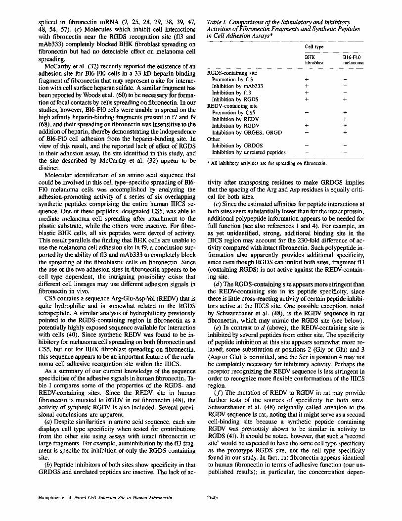
As a summary of our current knowledge of the sequence specificities of the adhesive signals in human fibronectin, Ta- ble I compares some of the properties of the RGDS- and REDV-containing sites. Since the REDV site in human fibronectin is mutated to RGDV in rat fibronectin (48), the activity of synthetic RGDV is also included. Several provi- sional conclusions are apparent.
(a) Despite similarities in amino acid sequence, each site displays cell type specificity when tested for contributions from the other site using assays with intact fibronectin or large fragments. For example, autoinhibition by the f13 frag- ment is specific for inhibition of only the RGDS-containing site.
(b) Peptide inhibitors of both sites show specificity in that GRDGS and unrelated peptides are inactive. The lack of ac-
Table I. Comparisons of the Stimulatory and Inhibitory Activities of Fibronectin Fragments and Synthetic Peptides in Cell Adhesion Assays*
Cell type
BHK BI6-FI0 fibroblast melanoma
RGDS-containing site Promotion by f13 + Inhibition by mAb333 + Inhibition by f13 + Inhibition by RGDS +
REDV-containing site Promotion by CS5 Inhibition by REDV Inhibition by RGDV + Inhibition by GRGES, GRGD
Other Inhibition by GRDGS Inhibition by unrelated peptides
m
+
+ + + +
* All inhibitory activities are for spreading on fibronectin.
tivity after transposing residues to make GRDGS implies that the spacing of the Arg and Asp residues is equally criti- cal for both sites.
(c) Since the estimated affinities for peptide interactions at both sites seem substantially lower than for the intact protein, additional polypeptide information appears to be needed for full function (see also references 1 and 4). For example, an as yet unidentified, strong, additional binding site in the IIICS region may account for the 230-fold difference of ac- tivity compared with intact fibronectin. Such polypeptide in- formation also apparently provides additional specificity, since even though RGDS can inhibit both sites, fragment f13 (containing RGDS) is not active against the REDV-contain- ing site.
(d) The RGDS-containing site appears more stringent than the REDV-containing site in its peptide specificity, since there is little cross-reacting activity of certain peptide inhibi- tors active at the IIICS site. One possible exception, noted by Schwarzbauer et al. (48), is the RGDV sequence in rat fibronectin, which may mimic the RGDS site (see below).
(e) In contrast to d (above), the REDV-containing site is inhibited by several peptides from either site. The specificity of peptide inhibition at this site appears somewhat more re- laxed; some substitution at positions 2 (Gly or Glu) and 3 (Asp or Glu) is permitted, and the Ser in position 4 may not be completely necessary for inhibitory activity. Perhaps the receptor recognizing the REDV sequence is less stringent in order to recognize more flexible conformations of the IIICS region.
( f ) The mutation of REDV to RGDV in rat may provide further tests of the sources of specificity for both sites. Schwarzbauer et al. (48) originally called attention to the RGDV sequence in rat, noting that it might serve as a second cell-binding site because a synthetic peptide containing RGDV was previously shown to be similar in activity to RGDS (41). It should be noted, however, that such a "second site" would be expected to have the same cell type specificity as the prototype RGDS site, not the cell type specificity found in our study. In fact, rat fibronectin appears identical to human fibronectin in terms of adhesive function (our un- published results); in particular, the concentration depen-
Humphries et al. Novel Cell Adhesion Site in Human Fibronectin 2645
dence of B16-F10 cell spreading on rat and human plasma fibronectins is identical, the pattern of inhibition of fibronec- tin-mediated cell spreading by short, synthetic peptides, including RGDV and REDV, is similar, and f13 is unable to autoinhibit B16-F10 cell adhesion on either fibronectin mole- cule. It is thus possible that the rat RGDV sequence is func- tionally equivalent to REDV and used only at the IIICS site in conjunction with the predicted modifying polypeptide in- formation or third site proposed in c (above).
The presence of an adhesion site for melanoma cells represents the first known function for the IIICS region. At present, five distinct fibronectin mRNA populations contain- ing different spliced products from the IIICS have been identified in human cells (7, 28, 29, 57), two of which contain the REDV sequence. The situation appears slightly less com- plex in the rat system, where three mRNA species have been identified (25, 38, 39, 47, 48, 54), two of which contain RGDV. Analysis of rat plasma fibronectin using antisera raised against fusion proteins containing the COOH-termi- na195 amino acids of the IIICS (or "V" region) revealed that only the large subunit(s) contained this sequence (39, 47). Although identical studies have not yet been performed with human fibronectin, the data from rat fibronectin appears to be consistent with the finding reported here that the mela- noma cell adhesion site is located only in the large subunit of human plasma fibronectin.
Although our results clearly identify a novel adhesive se- quence, containing REDV, in a IHCS region possessing cell type specificity, it should be emphasized that other specific adhesion sites may also exist. In particular, McCarthy et al. (32) have described an adhesion site in a COOH-terminal, heparin-binding fragment that was refractory to peptide inhi- bition. As noted above, our data may also suggest the exis- tence of an additional binding site in the IIICS that synergizes with the REDV site, in analogy to the model we proposed previously for function of the prototype RGDS region (3). The following speculative argument would be consistent with such a model: at least one fibrin-binding domain of hu- man fibronectin has been reported to contain a carboxyl- terminal portion of the IIICS region when separated proteo- lytically from the RGDS-containing domain (cf. references 7, 12, and 48), although the stoichiometry of this finding is unclear and the location of the REDV-containing region itself has not yet been mapped by amino acid sequencing. Since iS has also not been mapped by direct carboxyl-terminal se- quencing, it is conceivable that it lacks the REDV-containing region. In this case, an alternative hypothesis still consistent with all of our results would postulate the existence of two cell type-specific binding sites for melanoma cell adhesion, each displaying the same unusual pattern of susceptibility to inhibition by our peptide library. This hypothesis might also account for the >200-fold difference in activity between in- tact fibronectin (possibly containing two adhesion sites) and our synthetic peptide CS5 (containing only one of the sites). Only further extensive amino acid sequence and peptide analyses or in vitro mutagenesis studies will resolve whether such additional sites actually exist.
The identification of two distinct cell adhesion sites in the fibronectin molecule raises the question of whether the mela- noma cell receptor for fibronectin is related to the previously identified 140-kD complex that interacts with the RGDS site in fibronectin (6, 16, 21, 44, 53), or whether the two adhesion
sites interact with distinct receptors. Selective regulation of the quantities of two different fibronectin receptors on the surfaces of cells in vivo could conceivably be important bio- logically in modulation of adhesion during processes such as migration and embryonic development. Developmentally controlled splicing that dictates the presence of the REDV site in the IRCS region of fibronectin would also permit the local regulation of cell adhesion. It may be worthwhile inves- tigating whether the migration and adhesion of cell types from the same developmental lineage as melanoma cells (e.g., melanocytes, peripheral neurons, other neural crest cell derivatives) are via the REDV site, and hence may be regulated by the presence or absence of this sequence in the fibronectin matrix that constitutes the microenvironment of the cells.
This work was supported in part by grants CA-34918 and CA-14718 from the National Cancer Institute to K. O. M. H. was supported by a fellowship from the Cancer Research Institute.
Received for publication 23 July 1986, and in revised form 22 September
1986.
References
1. Akiyama, S. K., and K. M. Yarnada. 1982. Fibronectin in disease. In Connective Tissue Diseases. B. Wagner, R. Fleischmajer, and N. Kaufman, editors. Williams and Wilkins, Baltimore, Maryland. 55-96.
2. Akiyama, S. K., and K. M. Yamada. 1985. The interaction of plasma fibronectin with fibroblastic cells in suspension. J. Biol. Chem. 260:4492-4500.
3. Akiyama, S. K., and K. M. Yamada. 1985. Synthetic peptides competi- tively inhibit both direct binding to fibroblasts and functional biological assays for the purified cell-binding domain of fibronectin. J. Biol. Chem. 260:10402- 10405.
4. Akiyama, S. K., and K. M. Yamada. 1986. Fibronectin. Adv. Enzymol. Relat. Areas Mol. Biol. In press.
5. Akiyama, S. K., E, Hasegawa, T. Hasegawa, and K. M. Yamada. 1985. The interaction of fibronectin fragments with fibroblastic cells. J. Biol. Chem. 260:13256-13260.
6. Akiyama, S. K., S. S. Yamada, and K. M. Yamada. 1986. Characteriza- tion of a 140-kD avian cell surface antigen as a fibronectin-binding molecule. J. Cell Biol. 102:442-448.
7. Bernard, M. P., M. Kolbe, D. Well, and M-L. Chu. 1985. Human cellu- lar fibronectin: comparison of the carboxyl-terminal portion with rat identifies primary structural domains separated by hypervariable regions. Biochemistry. 24:2698-2704.
8. Beyth, R. J., and L. A. Culp. 1984. Complementary adhesive responses of human skin fibroblasts to the cell-binding domain of fibronectin and the hepa- ran sulfate-binding protein, platelet factor-4. Exp. Cell Res. 155:537-548.
9. Boucaut, J-C., T. Darribere, T. J. Poole, H. Aoyama, K. M. Yamada, and J. P. Thiery. 1984. Biologically active synthetic peptides as probes of em- bryonic development: a competitive peptide inhibitor of fibronectin function in- hibits gastrulation in amphibian embryos and neural crest cell migration in avian embryos. J. Celt Biol. 99:1822-1830.
I0. Fidler, I. J. 1973. Selection of successive tumor lines for metastasis. Na- ture (Lond. ) New Biol. 242:148-149.
11. Fidler, I. J. 1974. Inhibition of pulmonary metastasis by intravenous in- jection of specifically activated macrophages. Cancer Res. 34:1074-1078.
12. Garcia-Pardo, A., E. Pearlstein, and B. Frangione. Primary structure of human plasma fibronectin. Characterization of a 31,000-dalton fragment from the COOH-terminal region containing a free sulfhydryl group and a fibrin- binding site. J. Biol. Chem. 260:10320-10325.
13. Ginsberg, M., M. D. Pierschbacher, E. Ruoslahti, G. Marguerie, and E. Plow. 1985. Inhibition of fibronectin binding to platelets by proteolytic frag- ments and synthetic peptides which support cell adhesion. J. Biol. Chem. 260:3931-3936.
14. Grinnetl, F. 1984. Fibronectin and wound healing. J. Celt. Biochem. 26:107-I 16.
15. Grinnell, F., and M. K. Feld, 1981. Adsorption characteristics of plasma fibronectin in relationship to biological activity. J. Biomed. Mater. Res. 15:363-381.
16. Hasegawa, T., E. Hasegawa, W-T. Chen, and K. M. Yamada. 1985. Characterization of a membrane-associated glycoprotein complex implicated in cell adhesion to fibronectin. J. Cell. Biochem. 28:307-318.
17. Haverstick, D. M., J. F. Cowan, K. M. Yamada, and S. A. Santoro. 1985. Inhibition of platelet adhesion to fibronectin, fibrinogen and von Willebrand factor substrates by a synthetic tetrapeptide derived from the cell binding domain of fibronectin. Blood. 66:946-956.
The Journal of Cell Biology, Volume 103, 1986 2646
18. Hay, E. D., editor. 1981. Cell Biology of Extracellular Matrix. Plenum Publishing Corp., New York. 417 pp.
19. Hayashi, M., and K. M. Yamada. 1983. Domain structure of the carboxyl-terminal half of human plasma fibronectin. J. Biol. Chem. 258:3332- 3340.
20. Hopp, T. P., and K. R. Woods. 1981. Prediction of protein antigenic de- terminants from amino acid sequences. Proc. Natl. Acad. Sci. USA. 78:3824- 3828.
21. Horwitz, A., K. Duggan, R. Greggs, C. Decker, and C. A. Buck. 1985. The cell substrate attachment (CSAT) antigen has properties of a receptor for laminin and fibronectin. J. Cell Biol. 101:2134-2144.
22. Humphries, M, J., and K. M. Yamada. 1986. Non-collagenous glycopro teins. In Rheumatology, Vol. 10. K. Kuhn and T. Krieg, editors. Karger, Basel. 104-142.
23. Humphries, M. J., K. Matsumoto, S. L. White, and K. Olden. 1986. Oligosaccharide modification by swainsonine treatment inhibits pulmonary colonization by BI6-FI0 murine melanoma cells. Proc. Natl. Acad. Sci. USA. 83:1752-1756.
24. Humphries, M. J., K. Olden, and K. M. Yamada. 1986. A synthetic pep- tide from fibronectin inhibits experimental metastasis of murine melanoma cells. Science (Wash. DC). 233:467--470.
25. Hynes, R. 1985. Molecular biology of fibronectin. Annu. Rev. Cell Biol. 1:67-90.
26. Hynes, R. O. 1986. Fibronectins. Sci. Am. 254:42-51. 27. Kleinman, H. K., G. R. Martin, and P. H. Fishman. 1979. Ganglioside
inhibition of fibronectin-mediated cell adhesion to collagen. Proc. Natl. Acad. Sci. USA. 76:3367-3371.
28. Kornblihtt, A. R., K. Umezawa, K. Vibe-Pedersen, and F. E. Baralle. 1985. Primary structure of human fibronectin: differential splicing may gener- ate at least 10 polypeptides from a single gene. EMBO (Eur. Mol. Biol. Organ.) J. 4:1755-1759.
29. Kornblihtt, A. R., K. Vibe-Pedersen, and F. E. Baralle. 1984. Human fibronectin: cell-specific alternative mRNA splicing generates polypeptide chains differing in the number of internal repeats. Nucleic Acids Res. 12:5853- 5868.
30. Kyte, J., and R. F. Doolittle. 1982. A simple method for displaying the hydropathic character of a protein. J. Mol. Biol. 157:105-132.
31. Laterra, J., J. E. Silbert, and L. A. Culp. 1983. Cell surface heparan sul- fate mediates some adhesive responses to glycosaminoglycan-binding matrices, including fibronectin. J. Cell Biol. 96:112-123.
32. McCarthy, J. B., S. T. Hagen, and L. T. Furcht. 1986. Human fibronec- tin contains distinct adhesion- and motility-promoting domains for metastatic melanoma cells. J. Cell Biol. 102:179-188.
33. McKeown-Longo, P. J., and D. F. Mosher. 1985. Interaction of the 70,000-mol-wt amino-terminal fragment of fibronectin with the matrix-assem- bly receptor of fibroblasts. J. Cell Biol. 100:364-374.
34. Merrifield, R. B. 1963. Solid phase peptide synthesis. I. The synthesis of a tetrapeptide. J. Am. Chem. Soc. 85:2149-2154.
35. Miekka, S. I., K. C. Ingham, and D. Menache. 1982. Rapid methods for isolation of human plasma fibronectin. Thromb. Res. 27:1-14.
36. Mitchell, A. R., S. B. H. Kent, M. Engelhard, and R. B. Merrifield. 1978. A new synthetic route to tert-butyloxycarbonylaminoacylJ,-(oxymethyl)- phenylacetamidomethyl-resin, an improved support for solid phase peptide syn- thesis. J. Org. Chem. 43:2845-2852.
37. Mosher, D. F. 1984. Physiology of fibronectin. Annu. Rev. Med. 35:561-575.
38. Odermatt, E., J. W. Tamkun, and R. O. Hynes. 1985. Repeating modular structure of the fibronectin gene: relationship to protein structure and subunit variation. Proc. Natl. Acad. Sci. USA. 82:6571-6575.
39. Paul, J. I., J. E. Schwarzbauer, J. W. Tamkun, and R. O. Hynes. 1986. Cell type-specific fibronectin subunits generated by alternative splicing. J. Biol. Chem. 261:12258-12265.
40. Pierschbacher, M. D., and E. Ruoslahti. 1984. Cell attachment activity of fibronectin can be duplicated by small synthetic fragments of the molecule. Nature (Lond.). 309:30-33.
41. Pierschbacher, M. D., and E. Ruoslahti. 1984. Variants of the cell recog nition site of fibronectin that retain attachment-promoting activity. Proc. Natl. Acad. Sci. USA. 81:5985-5988.
42. Pierschbacher, M. D., E. G. Hayman, and E. Ruoslahti, 1982. Synthetic peptide with cell attachment activity of fibronectin. Proc. Natl. Acad. Sci. USA. 80:1224-1227.
43. Plow, E. F., M. D. Pierschbacher, E. Ruoslahti, G. A. Marguerie, and
M. H. Ginsberg. 1985. The effect of Arg-Gly-Asp-containing peptides on fibrinogen and yon Willebrand factor binding to platelets. Proc. Natl. Acad. Sci. USA. 82:8057-8061.
44. Pytela, R., M. D. Pierschbacher, and E. Ruoslahti. 1985. Identification and isolation of a 140-kD cell surface gtycoprotein with properties expected of a fibronectin receptor. Cell. 40:191-198.
45. Reddi, A. H., editor. 1985. Extracellular Matrix: Structure and Func- tion. Alan R. Liss, Inc., New York, 436 pp.
46. Ruoslahti, E., and M. D. Pierschbacher. 1986. Arg-Gly-Asp: a versatile cell recognition signal. Cell. 44:517-518.
47. Schwarzbauer, J. E., J. I. Paul, and R. O. Hynes. 1985. On the origin of species of fibronectin. Proc. Natl. Acad. Sci. USA. 82:1424-1428.
48. Schwarzbauer, J. E., J. W. Tamkun, I. R. Lemischka, and R. O. Hynes. 1983. Three different fibronectin mRNAs arise by alternative splicing within the coding region. Cell. 35:421--431.
49. Silnutzer, J. E., and D. W. Barnes. 1985. Effects of fibronectin-related peptides on cell spreading. In Vitro. 21:73-78.
50. Spiegel, S., K. M. Yamada, B. E. Hom, J. Moss, and P. H. Fishman. 1985. Fluorescent gangliosides as probes for the retention and organization of fibronectin by ganglioside-deficient mouse cells. J. Cell Biol. 100:721-726.
51. Spiegel, S., K. M. Yamada, B. E. Horn, J. Moss, and P. H. Fishman. 1986. Fibrillar organization of fibroneetin is expressed coordinately with cell surface gangliosides in a variant murine fibroblast. J. Cell Biol. 102:1898- 1906.
52. Tack, B. F., J. Dean, D. Eilat, P. E. Lorenz, andA. N. Schechter. 1980. Tritium labeling of proteins to high specific radioactivity by reduction methyla- tion. J. Biol. Chem. 255:8842-8847.
53. Tamkun, J. W., D. W. DeSimone, D. Fonda, R. S. Patel, C. Buck, A. F. Horwitz, and R. O. Hynes. ! 986. Structure of integrin, a glycoprotein involved in the transmembrane linkage between fibronectin and actin. Cell. 46:271-282.
54. Tamkun, J. W., J. E. Schwarzbauer, and R. O. Hynes. 1984. A single rat fibronectin gene generates three different mRNAs by alternative splicing of a complex exon. Proc. Natl. Acad. Sci. USA. 81:5140-5144.
55. Thompson, L. K., P. M. Horowitz, K. L. Bentley, D. D. Thomas, J. F. Alderete, and R. J. Klebe. 1986. Localization of the ganglioside-binding site of fibronectin. J. Biol. Chem. 261:5209-5214.
56. Trelstad, R. L., editor. 1985. The Role of Extracellular Matrix in Devel- opment. Alan R. Liss, Inc., New York. 643 pp.
57. Umezawa, K., A. R. Kornblihtt, and F. E. Baralle. 1985. Isolation and characterization ofcDNA clones for human liver fibronectin. FEBS (Fed. Eur. Biochem. Soc.) Lett. 186:31-34.
58. Vaheri, A., E. M. Salonen, and T. Vartio. 1985. Fibronectin in forma- tion and degradation of the pericellular matrix. Ciba Found. Syrup. 114:111- 126.
59. Wieland, T., C. Birr, and F. Flor. 1971. Symmetrical boc-amino acid anhydrides for economical peptide syntheses. Angew. Chem. Int. Ed. Engl. 10:336.
60. Woods, A., J. R. Couchman, S. Johansson, and M. H66k. 1986. Adhe- sion and cytoskeletal organisation of fibroblasts in response to fibronectin frag- ments. EMBO (Eur. Mol. Biol. Organ.) J. 5:665-670.
61. Yamada, K. M. 1983. Cell surface interactions with extracellular materials. Annu. Rev. Biochem. 52:761-799.
62. Yamada, K. M. 1983. Isolation of fibronectin from plasma and cells. In Immunochemistry of the Extracellular Matrix, Vol. I, H. Furthmayr, editor. CRC Press, Boca Raton, Florida. 111-123.
63. Yamada, K. M., and D. W. Kennedy. 1984. Dualistic nature of adhesive protein function: fibronectin and its biologically active peptide fragments can autoinhibit fibronectin function. J. Cell Biol. 99:29-36.
64. Yamada, K. M., and D. W. Kennedy. 1985. Amino acid sequence specificities of an adhesive recognition signal. J. Cell. Biochem. 28:99-104.
65. Yamada, K. M., and D. W. Kennedy. 1986. Peptide inhibitors of fibronectin, laminin, and other adhesion molecules: unique and shared features. J. Cell. Physiol. In press.
66. Yamada, K. M., D. R. Critchley, P. H. Fishman, and J. Moss. 1983. Exogenous gangliosides enhance the interaction of fibronectin with ganglioside- deficient cells. Exp. Cell Res. 143:295-302.
67. Yamada, K. M,, D. W. Kennedy, G. R. Grotendorst, and T. Momoi. 1981. Glycolipids: receptors for fibronectin? J. Cell. Physiol. 109:343-351.
68. Zardi, L., B. Carnemolla, E. Baiza, L. Borsi, P. Castellani, M. Rocco, and A. Siri. 1985. Elution of fibronectin proteolytic fragments from a hydrox- yapatite chromatography column. A simple procedure for the purification of fibronectin domains. Eur. J. Biochem. 146:571-579.
Humphries et al. Novel Cell Adhesion Site in Human Fibronectin 2647