identification of meat according to species by isoelectric focusing
TRANSCRIPT
of western New South Wales. In this region, sheep graze at stocking rates of one sheep to 5 hectares or lower, in paddocks of from 2000 to 8000 hectares. There is no supervision of lambing and in many flocks, lambs are not weaned. In one flock, 15 of 334 lambs at lamb marking had abnormally hairy birth coats. Lambs with hairy birth coats had been observed i n the flock previoutly. Of 6 hairy lambs examined, BVDV was recovered from the blood of one and BVDV antibody detected in serums from 3 others. Of 17 ewes examined, 8 had BVDV antibody. In the second flock, BVDV was isolated from the blood of a 9-week-old hairy lamb which was present in a flock of ewes due to start lambing within 2 weeks. No other affected lambs were observed.
In both flocks, the initial diagnosis was based on clinical signs of abnormally hairy birth coats and lower body weights than lambs of a similar age. There was no clinical evidence of central nervous system disturbances. Diagnosis was con- firmed by isolation of BVDV or detection of antibody to BVDV in blood samples from affected lambs (Plant e t al 1976a).
There were opportunities for contact between the chronically infected carrier lambs and ewes at a susceptible stage of pregnancy as lambs were not weaned, and during mating, there were other ewes lambing as a result of accidental matings. The sheep were normally dispersed while grazing, but they gathered daily at the limited watering points and this would provide the opportuni ty for the spread of the virus.
The significance of the disease in the flocks could not be assessed, and as there was no supervision of lambing abortions or perinatal mortalities due to BVDV would not be detected. I f affected lambs survived until lamb marking they could be iden ti fied.
Border disease can occur in sheep grazed under extensive conditions in a hot , arid environment, and this diagnosis in sheep in the western area of New South Wales offers an explanation of the occurrence o f BVDV antibody in sheep flocks in this region, as reported by St George (1971).
J . 'W. PLANT,
New South Wales Department of Agriculture, Veterinary Research Station, Glenfield, New South Wales, 2167
D. T. BYRNE', G. R. WOODS,
New South Wales Department of Agriculture, 32 Sulphide Street, Broken Hill, New South Wales, 2880 I 7 December 1981
* Present address: New South Wales Department of Agriculture, PO Box 417, Narrandera, N e w South Wales, 2700
References Plant, J . W., Acland, Helen M . and G a r d , G. P. (1976a)
Plant, J . W., Gard , G. P . and Acland, Helen M . (1976b) - Aust. vet. J . 52: 57.
- Ausr. vet. J . 52: 247. Plant, J . W., Gard, G . P . and Acland, Helen M. (1977) -
St. George, T . D. (1971) - Aust . vet. 1. 47: 370. Aust. vet. J . 53: 574.
IDENTIFICATION OF MEAT ACCORDING TO SPECIES BY ISOELECTRIC FOCUSING
The substitution of horse and kangaroo meat for beef has received much attention recently. All species-testing of meat at the Veterinary Research Institute is performed using the gel diffusion method (P room 1943). This test is based on the detection of serum proteins in the meat samples by use of either commercially available antiserums or antiserums pre- pared in this laboratory.
Using the antiserums available at the present time, the gel diffusion method is capable of distinguishing between the meats of cattle, horse, kangaroo, deer, pig, rabbit, cat, dog, guinea pig, hen, mouselrat and sheep, however it is unable to differentiate beef from buffalo o r sheep from goat meat. Another limitation of this method is that detection of mixtures of meat f rom 2 or more species is dependent on the use of a range of antiserums with each sample.
In a n attempt to overcome these limitations, we have examined the use of isoelectric focusing to separate a n d differentiate muscle proteins from different species. This technique has been used to distinguish between different fish species (Tingbergen and Olsman 1976; Mackie and Ritchie 1981) and different heat-processed food products containing beef, chicken, pig or horse meat (Tingbergen and Olsman 1976).
Samples of meat from 10 animals of each of the following species (cattle, buffalo, horse, kangaroo, sheep, goat and pig) were obtained from abattoirs. The samples were stored frozen at -2O'C prior to analysis or were analysed fresh. A 5 g portion of each sample was minced with scissors and then suspended in 5 ml of distilled water a n d disrupted using a stomacher. The homogenate was centrifuged a t I500 x g for 20 min and the supernatant fluid was used for analysis. The
Austral ian Veferinary Journal, Vol. 5 8 , Februa ry , 1982
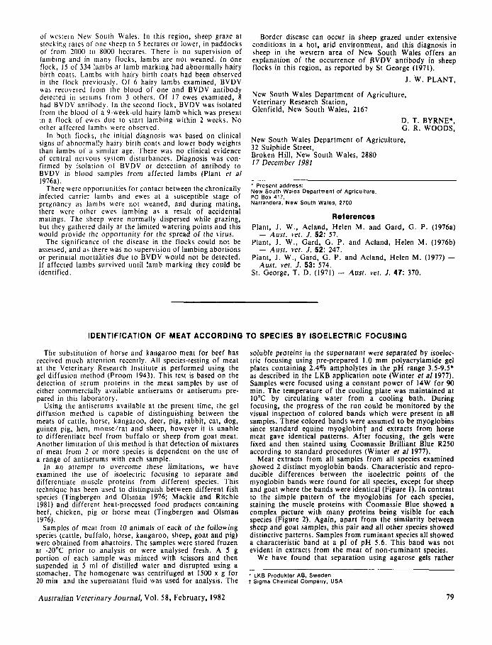
soluble proteins in the supernatant were separated by isoelec- tric focusing using pre-prepared 1 .O m m polyacrylamide gel plates containing 2.4% ampholytes in the p H range 3.5-9.5' as described in the LKB application note (Winter ef a1 1977). Samples were focused using a constant power of 14W for 90 min. The temperature of the cooling plate was maintained at 10°C by circulating water f rom a cooling bath. During focusing, the progress of the run could be monitored by the visual inspection of colored bands which were present in all samples. These colored bands were assumed t o be myoglobins since s tandard equine myoglobint and extracts f rom horse meat gave identical patterns. After focusing, the gels were fixed and then stained using Coomassie Brilliant Blue R25O according to s tandard procedures (Winter e t a/ 1977).
Meat extracts f rom all samples f rom all species examined showed 2 distinct myoglobin bands. Characterist ic and repro- ducible differences between the isoelectric points of the myoglobin bands were found for all species, except for sheep and goat where the bands were identical (Figure I) . In contrast t o the simple pattern of the myoglobins for each species, staining the muscle proteins with Coomassie Blue showed a complex picture with many proteins being visible for each species (Figure 2). Again, apar t f rom the similarity between sheep and goat samples, this pair and all other species showed distinctive patterns. Samples f rom ruminant species all showed a characteristic band a t a pl of p H 5.6. This band was not evident in extracts f rom the meat of non-ruminant species.
We have found that separation using agarose gels rather
* LKB Produkter AB, Sweden t Sigma Chemical Company, USA
19
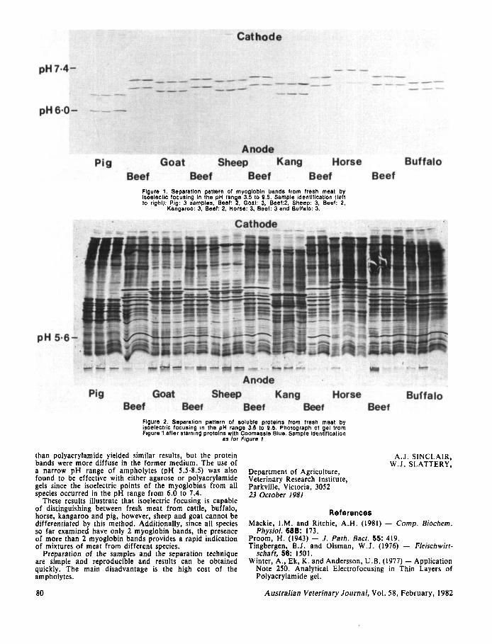
Figure 1. Separation pattern Of mYOglObin bands from fresh meat by lsoelectlc focusing in the pH range 3.5 to 9.5. Sample ldentlflcation (left lo right): Pig: 3 samples, Beef: 2, Goat: 3, Beef:2, Sheep: 3, Beef: 2,
Kangaroo: 3, Beef: 2, Horse: 3, Beef: 3 and Buffelo: 3.
Figure 2. Separation pattern of soluble proteins from fresh meat by isoelectric focusing In the pH range 3.5 to 9.5. Photograph of gel from Figure 1 after stalning protelns with Coomassle Blue. Sample ldentlflcation
as for Figure I .
than polyacrylamide yielded similar results, but the protein bands were more diffuse in the former medium. The use of a narrow pH range of ampholytes (pH 5 .5 -8 .5 ) was also found to be effective with either agarose or polyacrylamide gels since the isoelectric points of the myoglobins from all species occurred in the pH range from 6.0 to 7.4.
These results illustrate that isoelectric focusing is capable of distinguishing between fresh meat from cattle, buffalo, horse, kangaroo and pig, however, sheep and goat cannot be differentiated by this method. Additionally, since all species so far examined have only 2 myoglobin bands, the presence of more than 2 myoglobin bands provides a rapid indication of mixtures of meat from different species.
Preparation of the samples and the separation technique are simple and reproducible and results can be obtained quickly. The main disadvantage is the high cost of the ampholytes.
80
Department of Agriculture, Veterinary Research Institute, Parkville, Victoria, 3052 23 Ocrober 1981
A.J. SINCLAIR, W.J. SLATTERY,
References Mackie, I.M. and Ritchie, A.H. (1981) - Comp. Biochem.
Physiol. 68B: 173. Proorn, H. (1943) - J . Parh. Bacr. 55: 419. Tingbergen, B.J. and Olsman, W.J. (1976) - Fleischwirr-
schaft, 56: 1501. Winter, A,, Ek, K. and Anderson, U.B. (1977) - Application
Note 250. Analytical Electrofocusing in Thin Layers of Polyacrylamide gel.
Australian Veterinary Journal, Vol. 58 , February, 1982