identification of ranbp16 and ranbp17 as novel interaction partners for the bhlh transcription...
TRANSCRIPT

Journal of CellularBiochemistry
ARTICLEJournal of Cellular Biochemistry 111:195–206 (2010)
Identification of RANBP16 and RANBP17 as NovelInteraction Partners for the bHLH Transcription Factor E12
G
*
R
P
Jun-Ho Lee, Shengli Zhou, and Cynthia M. Smas*
Department of Biochemistry and Cancer Biology, University of Toledo College of Medicine, Toledo, Ohio 43614
ABSTRACTThe ubiquitously expressed basic helix-loop-helix (bHLH) transcription factors E12 and E47, products of alternative splicing of the E2A/TCF3
gene, regulate diverse biological processes including cell growth, differentiation and development. To search for novel protein interactions for
E12, we utilized the bHLH domain of E12 as a bait in yeast two-hybrid screening. Yeast two-hybrid, mammalian two-hybrid, and co-
immunoprecipitation analyses demonstrate specific interaction of E12 with RANBP17, a novel member of the importin-b superfamily; this
interaction maps to the CRM1 homology region of RANBP17. Ectopic expression of RANBP17 leads to a�3-fold increase in E2A/MyoD mediated
transactivation of an E-box regulated luciferase reporter gene. Interaction and transactivation studies also revealed similar functions for
RANBP16/XPO7. Furthermore, ectopic expression of either RANBP16 or RANBP17 resulted in increased level of endogenous transcript for the
cyclin-dependent kinase inhibitor, p21Waf1/Cip1, a well-characterized E2A target gene. Together, these biochemical and functional data reveal
RANBP16 and RANBP17 as novel regulators of E2A protein action. J. Cell. Biochem. 111: 195–206, 2010. � 2010 Wiley-Liss, Inc.
KEY WORDS: BHLH TRANSCRIPTION FACTOR; E2A PROTEINS (E12 AND E47); RANBP17; RANBP16; E-BOX; YEAST TWO-HYBRID; NUCLEOCYTO-PLASMIC TRANSPORT; CRM1
T he basic helix-loop-helix (bHLH) family of transcription
factors regulates gene transcription during cell differentia-
tion, proliferation, lineage commitment, and neoplastic transforma-
tion in a variety of cell types [Norton, 2000; Perry and Soreq, 2002;
Desprez et al., 2003]. Members of this family are defined by the
bHLH domain near their C-termini that is comprised of two
functionally distinct regions. The basic domain is rich in positively
charged amino acids critical for DNA binding while the HLH region
governs dimerization with other bHLH family members. The DNA
sequence bound by bHLH proteins is the hexanucleotide E-box
element (50-CANNTG-30), present in a variety of tissue-specific
enhancers [Murre et al., 1989; Funk et al., 1991; Yutzey and
Konieczny, 1992; Greenbaum and Zhuang, 2002]. The E2A proteins,
E12 and E47, are products of the E2A gene generated by alternate
splicing. As such, E12 and E47 possess distinct, albeit highly
conserved, sequences in their respective bHLH domains. Given their
ubiquitous expression, E12 and E47 are classified as class I bHLH
transcription factors [Murre et al., 1994; Massari and Murre, 2000;
Hikima et al., 2005]. E12 and E47 homo- or heterodimerize with
other bHLH transcription factors [Lassar et al., 1991; Massari and
Murre, 2000; Quong et al., 2002]; cell type selectivity of bHLH
protein action is conferred by tissue restricted bHLH factor(s) that
serve as dimerization partners for the E2A proteins [Massari and
Murre, 2000]. These include the myogenic regulatory factors MyoD,
rant sponsor: NIH; Grant sponsor: National Cancer Institute; Grant num
Correspondence to: Dr. Cynthia M. Smas, 3000 Arlington Ave, Toledo, OH 4
eceived 20 March 2009; Accepted 27 April 2010 � DOI 10.1002/jcb.226
ublished online 12 May 2010 in Wiley Online Library (wileyonlinelibrar
MRF-4, Myf-5, and myogenin [French et al., 1991; Lassar et al.,
1991; Venuti and Cserjesi, 1996]; BETA2/NeuroD that function in
neurogenesis, pancreatic development and insulin gene expression
[Lee, 1997; Mutoh et al., 1997; Glick et al., 2000]; and SCL/Tal1 in
hematopoietic lineage development [Hsu et al., 1994; Gould and
Bresnick, 1998]. The action of bHLH factors is also controlled by the
dominant negative regulatory Id proteins (Id1-Id4), a distinct group
of HLH factors that lack a basic region but possess an HLH
dimerization domain [Benezra et al., 1990; Langlands et al., 1997].
Furthermore, the physiological specificity of transcriptional
activation by bHLH factors is not solely dependent on their intrinsic
DNA binding specificities but also involves cooperative interactions
with other components of the transcriptional machinery [Eckner
et al., 1996; Massari et al., 1999; Turner et al., 2004].
In regard to protein interactions of bHLH factors, bHLH-bHLH
dimerization among bHLH protein family members is perhaps the
best studied [Massari and Murre, 2000]. However, there is ample
evidence that interactions of E2A proteins are not solely with other
members of the bHLH family, nor only within their HLH regions. A
number of non-bHLH interaction partners for E2A proteins have
been described. For example, E2A proteins interact with p300/CBP
and SAGA histone acetyltransferase complex [Eckner et al., 1996;
Massari et al., 1999; Bayly et al., 2004]; the MAPK-activated protein
kinases 3pK and MK2 interact with E47 to repress E-box-mediated
195ber: 5R01CA098141.
3614. E-mail: [email protected]
89 � � 2010 Wiley-Liss, Inc.
y.com).

transcriptional activity [Neufeld et al., 2000]; calmodulin binds the
basic region of E2A proteins to inhibit DNA binding [Saarikettu
et al., 2004]; and a C-terminal 31-kDa caspase-mediated cleavage
product of p130cas, which contains high sequence homology with Id
proteins, heterodimerizes with E2A proteins to result in inhibition of
p21Waf1/Cip1 transcription [Kim et al., 2004]. E2A protein–protein
interactions can thus serve as key integration points for a multitude
of intracellular signals that reflect and/or control overall cellular
status in a variety of biological settings.
Transport between nucleus and cytoplasm through the nuclear
pore complex is primarily mediated by importin b-related factors
which represent the largest class of nuclear transport receptors
[Kutay et al., 2000; Cook et al., 2007]. The common features of
importin b-related transport receptors include a molecular weight in
the range of �90 to �130 kDa and the presence of a conserved
region for RanGTP binding in their N-termini [Kutay et al., 2000;
Yoneda, 2000]. The closely related proteins RANBP16 (also termed
XPO7) and RANBP17 show an overall 67% amino acid identity and
phylogenetic analysis has determined that they are the most
evolutionarily distant members of the importin b superfamily
[Kutay et al., 2000]. The N-terminal importin b domain of RANBP17
is located at amino acids 30–95, which is found within a larger
region of homology with the CRM1 nuclear export protein, at amino
acids 8–167 of RANBP17. Northern blot analysis of polyAþ RNA
has led to RANBP17 transcript(s) expression described as testis-
enriched in mouse and human tissue samples [Koch et al., 2000].
However, Northern blot analyses have also detected RANBP17
transcript(s) in human heart, liver, kidney, pancreas and placenta
tissues, and in erythroid HEL and the megakaryocyte cell
lines Meg01 and M07E [Bernard et al., 2001; MacLeod et al.,
2003].
While the role of RANBP17 in nuclear transport has yet to be
reported, that of RANBP16/XPO7 has been addressed, albeit in only
a few studies [Kutay et al., 2000; Mingot et al., 2004]. Gorlich and
colleagues showed that RANBP16 has exportin function, with likely
broad substrate specificities [Mingot et al., 2004]. However, Bischoff
and coworkers observed an intermediate dissociation constant for
RanGTP hydrolysis and could therefore not define RANBP16
specifically as an importin or exportin [Kutay et al., 2000]. They
suggested it may have a unique function in bidirectional cargo
transport at the nuclear pore [Kutay et al., 2000]. Given that
RANBP16 did not recognize proteins that contain the classical type
of basic nuclear import signal, but rather interacted with positively
charged regions of several types of targets, it has been suggested that
RANBP16 may be a component of a novel type of nuclear transport
mechanism [Mingot et al., 2004]. The only additional information
on RANBP16/XPO7 function is a recent report on its role in the
localization of LKB1, a serine/threonine kinase that regulates cell
polarity, metabolism, and growth. STRADa induces relocalization of
LKB1 from the nucleus to the cytoplasm and stimulates its catalytic
activity. RANBP16/XPO7 associates with STRADa to impact LKB1
localization [Dorfman and Macara, 2008]. Direct interaction of
STRADa with RANBP16/XPO7 was reported in this study, however
RANBP17 was reported as negative for interaction with STRADa.
In a search for novel protein–protein interactions for the bHLH
transcription factor E12, we conducted yeast two-hybrid screening
196 NOVEL RANBP INTERACTION PARTNERS FOR E12
and identified RANBP17. Here we carry out studies that demonstrate
that RANBP17, and the closely related RANBP16, are novel E2A
binding partners that can function to enhance transcriptional
activation mediated by E2A proteins.
MATERIALS AND METHODS
CLONING AND EXPRESSION CONSTRUCTS
E2A protein expression constructs. The E12 bHLH region bait used in
yeast two-hybrid screening contains amino acids 508–654 of human
E12 in the pAS Gal4 DNA binding domain fusion vector (Clontech
Corp.). For mammalian two-hybrid assay, the same sequence of E12
and RANBP17 present in the yeast two-hybrid constructs were
subcloned into the mammalian two-hybrid expression vectors,
pVP16 and pM (Clontech Corp.), such that they maintained reading
frame with the VP16 transactivating and the GAL4-DNA binding
domains, respectively. A cDNA clone for full-length human E12
was obtained from Dr. C. Murre (Harvard University) and subcloned
into pcDNA3.1þ (Invitrogen Corp.). A full-length cDNA for human
E47 (GenBank accession number BC110579) was purchased from
Open Biosystems, sequence verified, and the insert subcloned into
pcDNA3.1þ. PCR-based cloning incorporating EcoRI and SalI
restriction sites was used to generate an expression construct for
amino acids 502–654 of E12 in the pCMV-Myc mammalian
expression plasmid (Clontech Corp.) such that a start codon was
provided with the epitope tag. Primer sequences used for this were:
50-GCCGAATTCAAGGAGGACGAGGAGAACACG-30 and 50-GGCG-
TCGACTCACATGTGCCCGGCGGGGTT-30.
RANBP16 and RANBP17 expression constructs. Mammalian expres-
sion constructs for full-length human RANBP16 and full-length
human RANBP17 that contained three copies of an HA tag at their
respective N-termini, RANBP16-pMT2SM and RANBP17-pMT2SM,
referred to herein as HA-RANBP17 and HA-RANBP16, were a kind
gift from Dr. W.G. Janssen (University of Heidelberg, Germany).
During the sequencing of RANBP17-pMT2SM, we found that a
single amino acid differed from the GenBank reference sequence
for human RANBP17, NM_022897.2. We corrected the RANBP17
sequence to match the RANBP17 GenBank reference sequence. An
I.M.A.G.E. cDNA clone for the short form of human RANBP17
(sRANBP17) in the mammalian expression vector pCMV-SPORT6
(GenBank accession number BI824159) was obtained from the
American Type Culture Collection (ATCC). HA-RANBP17-CRM1
was constructed in the pCMV-HA vector (Clontech Corp.) utilizing
PCR-based cloning with primers that incorporated EcoRI and
KpnI cloning sites: 50-GCCCGAATTCTGGCGCTGCACTTCCAGAGTT-
TG-30 and 50-GGCGGTACCTCATGTTCTCCAAGTTGTTGGAATCTG-30.
Luciferase reporter constructs. An E-box regulated firefly luciferase
reporter construct, termed E-box-pLuc, was generated via insertion
of three copies of the consensus E-box element, CAGGTG just 50 to
an SV40 minimal promoter in the pGL3-Promoter vector (Promega
Corp.). pGAL4-Luc was utilized as a reporter for protein–protein
interaction in mammalian two-hybrid assay and was provided by
Dr. B. Rowan (Tulane University, New Orleans LA). It contains a
GAL4 DNA binding site upstream of a minimal TATA box which
governs the expression of the firefly luciferase gene.
JOURNAL OF CELLULAR BIOCHEMISTRY

YEAST TWO-HYBRID SCREENING AND MATING ASSAYS
A DU145 prostate cancer cell two-hybrid library was generated in
the pGADT7-Rec vector using the Clontech Matchmaker 3 Library
Construction Kit (Clontech Corp.), following manufacturer’s
instructions. Approximately 2� 106 library clones were screened
by yeast mating with selection by growth for 10 days on agar
media lacking Leu, Trp, and His, followed by standard filter-lift
b-galactosidase assay of colonies. Plasmid DNA was isolated from
b-galactosidase positive yeast colonies, transformed into E. coli
DH5a, followed by preparation of E. coli plasmid DNA. DNA
plasmids for library cDNA clones were then individually trans-
formed into S. cerevisiae strain AH109 and tested for bait specificity
by mating with S. cerevisiae strain Y187 that harbored either the
E12 bait construct, a lamin negative control construct (provided in
the Matchmaker System 3 Kit), or pGBTK7 empty vector. Mating
mixtures were plated on the either double (Leu�, Trp�) dropout
(DDO) or triple (Leu�, Trp�, His�) dropout (TDO) nutrient selective
media and bait specificity of the interaction was scored by
appropriate growth pattern on selective media and by X-a-
galactosidase activity. Inserts for yeast two-hybrid library cDNA
clones that demonstrated bait-specific interaction were sequenced.
MAMMALIAN TWO-HYBRID ASSAY
HeLa cells were plated at 4� 104 per well in a 24 well plates and the
following day transfected with DNA constructs using Lipofectamine
2000 (Invitrogen Corp., Carlsbad, CA). Indicated combinations of
DNA constructs (0.2mg for each) were co-transfected with 0.4mg of
pGAL4-Luc and 0.2mg of pRLTK plasmid DNA per well. Forty-eight
hours after transfection, cells were lysed with passive lysis buffer
and dual luciferase activities were measured with a Turner Systems
luminometer in accordance with the manufacturer’s instruction
(Dual-Luciferase Reporter Assay System, Promega Corp.). Relative
luciferase activities were expressed as the ratio of firefly and Renilla
luciferase activities. Studies shown in each graph were performed a
minimum of three independent times with samples prepared as
independent triplicates for each assay. Representative data from a
minimum of three independently performed studies are shown.
Statistical significance was calculated using single factor ANOVA.
E-BOX REPORTER ASSAY
As indicated in the respective figure legend, combinations of the
following DNA expression constructs were used in an E-box
regulated luciferase reporter assay: E12 or E47 (0.004mg), MyoD
(0.1mg), RANBP (0.1mg of HA-RANBP17, HA-RANBP16,
sRANBP17, or HA-RANBP17-CRM1), Id1 (0.5mg), E-box-pLuc
(0.2mg), pRLTK (0.1mg). Numbers in parenthesis indicate the mass
of DNA construct used per well of a 24-well culture plate. Addition
of empty vector DNA was included as needed to equalize the
total combined mass of DNA per well to 1mg, or to 1.2mg when
transfections included the Id1 expression construct. Co-transfec-
tions were carried out in HeLa cells using Lipofectamine 2000
(Invitrogen Corp.), with media change at 4 h post-transfection.
Samples were assayed for firefly luciferase at 48 h post-transfection
with normalization by standard protein assay (Bio-Rad). Studies
presented in each graph were performed a minimum of three
independent times with samples prepared as independent triplicates
JOURNAL OF CELLULAR BIOCHEMISTRY
for each assay, with representative data shown. Statistical
significance was calculated using single factor ANOVA.
RNA PREPARATION AND Q-PCR ANALYSIS
293T cells were transiently transfected with HA-RANBP17 or HA-
RANBP16 DNA expression constructs, or with empty vector DNA as
a negative control. Transfections were carried out three independent
times with representative data shown. Transfection was via
Lipofectamine 2000 with media change 4 h post-transfection.
RNA was isolated from cells using TriZol Reagent (Invitrogen
Corp.), according to manufacturer’s instruction. First-strand cDNA
was synthesized using 5mg of DNase I-treated total RNA. Transcript
levels were analyzed by SYBR Green-based quantitative real-time
PCR (Q-PCR) in 25ml-reactions containing 1� SYBR Green PCR
Master Mix (Applied Biosystems, Foster City, CA), 200 nM each
forward and reverse primers, and 10 ng of cDNA. Primers used for
detection of human p21Waf1/Cip1 transcript were 50-GTCTT-
GTACCCTTGTGCCTC-30 and 50-GCTTCCTCTTGGAGAAGATCAG-30
and for human GAPDH 50-GAAGGTGAAGGTCGGAGTCA-30 and
50-TTCACACCCATGACGAACAT-30. Q-PCR was conducted with an
ABI 7500 Real-Time PCR System. PCR was carried out over 40 cycles
of 958C for 15 s, 608C for 30 s, and 728C for 40 s, with an initial cycle
of 508C for 2 min and 958C for 10 min to activate AmpliTaq Gold
DNA polymerase; a dissociation curve was generated over the range
of 60–958C. For the latter, a single sharp peak was observed in
each case, indicative of a single PCR product species. Transcript
expression was normalized against GAPDH. The cycle threshold
value was generated using ABI Prism 7500 SDS software, version
1.2. Fold changes were determined by the DDCT method and are
shown as means� SD of triplicates. Statistical significance was
calculated using single factor ANOVA.
IMMUNOCYTOCHEMISTRY AND INTRACELLULAR
LOCALIZATION STUDIES
For immunostaining studies, COS cells were plated on laminin-
coated coverslips and transfected with DNA for expression
constructs using Lipofectamine 2000. Forty-eight hours after
transfection, cells on coverslips were washed twice with PBS and
fixed with 100% ice-cold methanol for 10 min, washed in PBS, and
blocked by incubation in 0.1% BSA in PBS for 30 min at room
temperature. Cells were incubated with polyclonal E2A antibody for
detection of E12 (1:100, Santa Cruz Biotech.) and/or monoclonal HA
antibody (1:100, Covance Corp.) in 0.1% BSA in PBS for 1.5 h.
Following three washes in 0.1% BSA in PBS, cells were incubated
with Alexafluor 568-conjugated goat anti-mouse (1:800, Invitrogen
Corp.) and/or FITC-conjugated goat anti-rabbit (1:200, Bio-Rad)
secondary antibodies, as indicated. Following a 1 h incubation,
coverslips were washed and mounted on glass slides and cells
observed at 400� or 600� magnification using a Nikon Eclipse
E800 fluorescence microscope equipped with a digital camera.
Image acquisition and merging was performed with Image-Pro Plus
software (Media Cybernetics, Carlsbad, CA). For quantitative
assessment of intracellular localization, signals were scored as
predominately cytoplasmic, predominantly nuclear or equally
distributed in nucleus and cytoplasm, similar to previously
published methodology [Yagita et al., 2002]. For each experiment,
NOVEL RANBP INTERACTION PARTNERS FOR E12 197

50–100 cells were scored from three independent transfections, with
three independent experiments performed. Statistical significance
was calculated using single factor ANOVA.
WESTERN BLOT ANALYSIS, SUBCELLULAR FRACTIONATION, AND
CO-IMMUNOPRECIPITATION
For analysis of total cell lysates, HeLa cells were transfected with
4mg of HA-RANBP17 or empty vector per well of a 6-well plate
using Lipofectamine 2000 (Invitrogen Corp.). Cells were harvested at
48 h post-transfection by lysis in TNN(þ) cell lysis buffer (10 mM
Tris pH 8.0, 120 mM NaCl, 0.5% NP-40, 1 mM EDTA supplemented
with a protease inhibitor cocktail). Lysates were incubated on ice for
30 min with intermittent vortexing and after centrifugation at
20,800g for 15 min, supernatant was collected and protein content
determined (Bio-Rad, Hercules, CA). For subcellular fractionation,
cell lysates were prepared according to the instructions for
preparation of nuclear extracts (Active Motif, Carlsbad CA). For
Western blot analysis, 30mg of protein extract was fractionated on
10% SDS–PAGE gel, followed by electroblotting onto PVDF
membrane using 0.025 M Tris/0.192 M glycine transfer buffer with
20% methanol. Membranes were blocked for 1 h in 5% non-fat milk
in PBS containing 0.5% Tween 20 (PBS-T) followed by a 1 h
incubation at room temperature with a mouse monoclonal HA
antibody (1:2,000, Covance Research Products, Berkeley, CA), a
mouse monoclonal lamin A/C antibody (1:200, Affinity BioRea-
gents) or a rabbit polyclonal b-tubulin antibody (1:10,000, Covance
Research Products, Berkeley CA). Secondary antibody was HRP-
conjugated goat anti-mouse (1:2,000, Santa Cruz Biotech, Santa
Cruz CA) or anti-rabbit (1:2,000, Bio-Rad) antibody and signals were
detected using ECL Plus enhanced chemiluminescence reagent (GE
Healthcare). Western blot analyses were conducted a minimum of
three times with essentially same results and representative data
shown.
For co-immunoprecipitation, COS cells were transfected with the
indicated expression constructs and 48 h post-transfection total cell
lysates were prepared by sonication in TNN(þ) cell lysis buffer
(10 mM Tris pH 8.0, 120 mM NaCl, 0.5% NP-40, 1 mM EDTA
supplemented with a protease inhibitor cocktail). Fifty micrograms
of cell lysate was incubated with E2A antibody by rotation overnight
at 48C followed by incubation for 3 h with 25ml of 50% (v/v)
suspension of Protein-A-agarose beads (Santa Cruz Biotech, Inc.).
After centrifugation at 10,800g for 30 s, pelleted beads were washed
three times with TNN(þ) cell lysis buffer. Beads were resuspended in
SDS–PAGE loading buffer, boiled for 10 min, centrifuged and the
supernatant subjected to Western blot analysis. No signal was
detected when immunoprecipitation was performed in the absence
of antibody. A slight modification of this approach, found during the
course of our studies to reduce non-specific background, was used
for the experiment in Figure 6D. In this case lysates were pre-
incubated with Protein-A-agarose beads for 1 h at 48C, beads were
removed and protocol continued at the primary antibody addition
stage, as described above. For the Western blot analysis shown in
Figure 6D, we utilized protein-A-conjugated HRP, which recognizes
native but not denatured IgG, to minimize signals from immu-
noglobulin heavy and light chains. All co-immunoprecipitation
198 NOVEL RANBP INTERACTION PARTNERS FOR E12
studies were performed a minimum of three times with essentially
the same results and representative data are shown.
RESULTS
TWO-HYBRID SCREENING REVEALS RANBP17 IS A NOVEL
INTERACTION PARTNER FOR E12
Yeast two-hybrid screening was employed to search for novel
binding partners for E12. The transcriptional activation regions of
E12 (AD1 and AD2) that are located within the first 500 amino acids
of the protein render a yeast two-hybrid bait comprised of full-
length E12 unsuitable for screening as it autonomously induced
transcriptional activation of the yeast-two-hybrid reporter gene(s)
(data not shown). We therefore employed a bait that encoded amino
acids 508–654 of E12 and that includes the bHLH domain.
Figure 1A shows an illustration of the full-length E12 protein
consisting of 654 amino acids and the region of E12 used in the bait
construct. Following screening of 2� 106 clones of a DU145
prostate cancer cell yeast two-hybrid cDNA library, we identified a
clone that evidenced specific interaction with the E12 bait and that
maintained reading frame with the GAL4 activation domain
encoded by the cDNA library vector. It contained amino acids 1–
252 of RANBP17 (RANBP171–252).
Figure 1B shows the result of bait specificity testing of the
interaction between the E12 bait and RANBP171–252. Constructs for
the E12 bait or the RANBP171–252 prey were transformed into Y187
and AH109 strains of S. cerevisiae, respectively. We included a prey
for full length Id1, a known E12 interactor, as an E12 interactor
positive control. Empty vectors and a bait construct encoding lamin
C are negative controls. The left panel of Figure 1B indicates
effective mating of yeast harboring the indicated pair-wise
combinations of DNA binding and activation domain plasmids to
produce diploids that grow on Leu and Trp deficient DDO agar media
plates. The middle panel of Figure 1B demonstrates selective growth
on Leu, Trp, and His deficient TDO agar media plates; here
interaction is demonstrated by growth in the absence of His.
RANBP171–252 interaction with E12 is specific in that yeast that
harbor both the E12 bait construct and the RANBP171–252 library
clone (Fig. 1B, middle panel) grow on TDO media, whereas no
growth is noted for the respective negative controls. The
colorimetric detection of X-a-galactosidase activity, an additional
reporter gene for protein–protein interaction in this system, is
shown in the right panel of Figure 1B, further confirming bait-
specific interaction of RANBP171–252 with the E12 bait. We also find
the predicted interaction for the positive control combination of the
E12 bait and Id1 prey.
We next employed mammalian two-hybrid analysis to assess
protein–protein interaction in mammalian cells. The inserts from the
E12 bait and the RANBP171–252 prey constructs used in the yeast
two-hybrid study were subcloned into the mammalian two-hybrid
activation and binding domain vectors pVP16 and pM, respectively.
These expression constructs and a pGAL4-Luc reporter construct
were co-transfected into HeLa cells and luciferase activity assessed
at 48 h post-transfection. Figure 1C shows that, in comparison to
empty vector transfectants, a 5.7-fold increase (P< 0.01) in
luciferase activity was observed in cells co-transfected with fusion
JOURNAL OF CELLULAR BIOCHEMISTRY

Fig. 1. Yeast and mammalian two-hybrid analyses identify RANBP17 as a binding partner for E12. A: Schematic representation of full-length E12 (E121–654), full-length
RANBP17 (RANBP171–1088), the E12 bait (E12508–654) and the RANBP17 library prey clone (RANBP171–252); numbers indicate amino acid positions. B: The indicated pairwise
combinations of the bait (indicated at right) and prey constructs (indicated at top) and corresponding empty control vectors were separately transformed into yeast strains Y187 and
AH109, mated and protein–protein interaction was assayed as described in Materials and Methods Section. DDO, media lacking Leu and Trp; TDO, media lacking Leu, Trp and His; X-
a-Gal, X-a-galactosidase. C: Mammalian two-hybrid analysis. HeLa cells were transfected with the indicated expression constructs for E12508–654 and RANBP171–252 in the VP16
activation domain fusion or the pM binding domain fusion vectors, respectively. Luciferase activities were measured at 48h post-transfection. Data shown is mean� SD, �P< 0.01
for the E12 and RANBP17 co-transfected samples versus all others shown. Data are representative of three wholly independently conducted experiments, with transfections carried
out in triplicate for each such independent experiment. [Color figure can be viewed in the online issue, which is available at wileyonlinelibrary.com.]
constructs for E12508–654 and RANBP171–252, indicative of inter-
action of these two protein regions in mammalian cells.
As our yeast and mammalian two-hybrid studies only assessed
interaction of the bHLH region of E12 with amino acids 1–252 of
RANBP17, we employed co-immunoprecipitation studies to test for
association of full-length E12 with full-length RANBP17. Unfortu-
nately, assessment of multiple commercial antibodies for RANBP17
proved none were of utility for our studies, as such we used an HA-
tagged version. We first demonstrated that the expression construct
for HA-tagged RANBP17 was functional. The Western blot in Figure
2A indicates protein expression from the HA-RANBP17 construct
results in a single protein species consistent with the predicted
�120 kDa mass for RANBP17. Figure 2B demonstrates that E12 and
JOURNAL OF CELLULAR BIOCHEMISTRY
RANBP17 are present in the same protein complex and that this
signal is dependent on transfection of expression constructs for both
proteins. Figure 2C indicates, as anticipated from our yeast two-
hybrid screening result, that a region of E12 that was used in the
yeast two-hybrid bait is sufficient for co-immunoprecipitation with
full-length RANBP17.
CO-LOCALIZATION STUDIES OF E12 AND RANBP17
The results of the two-hybrid and co-immunoprecipitation inter-
action studies led us to address whether RANBP17 co-localized with
E12. We first assessed the intracellular localization of RANBP17
protein by subcellular fractionation and immunocytochemistry.
Nuclear and cytosolic fractions were prepared from cell lysates of
NOVEL RANBP INTERACTION PARTNERS FOR E12 199

Fig. 2. RANBP17 Co-Immunoprecipitates with E12 (A) Empty vector
pcDNA3.1þ (EV) or the HA-RANBP17 expression construct (RANBP17) was
transfected into HeLa cells and at 48 h post-transfection cell lysates were
subjected to Western blot analysis using monoclonal HA primary antibody. b-
tubulin (b-Tub) was used as a loading control. B: HA-RANBP17 and full-length
E12 protein was expressed separately or in combination by transfection of COS
cells. Fifty micrograms of each cell lysate was subjected to immunoprecipita-
tion and Western blot analysis with the indicated antibodies. 1/20 of total
protein (2.5mg) was analyzed for Western blot (INPUT). Equivalent protein
loading for Western blot was verified by Coomassie blue staining. (C) HA-
RANBP17 and E12502–654 protein was expressed separately or in combination
by transfection of COS cells and analyzed as in (B). For B and C, IP:
immunoprecipitation, WB: Western blot.
Fig. 3. Intracellular Localization of RANBP17 and E12. A: Nuclear and
cytoplasmic fractions of HeLa cell lysates transfected with empty vector
pcDNA3.1þ (EV) or HA-RANBP17 expression construct were subjected to
Western blot analysis with the indicated antibodies. b-tubulin (b-Tub) and
lamin A (Lam A) were used as markers for cytoplasmic and nuclear fraction,
respectively. B: Immunocytochemistry for RANBP17 localization. COS cells
were transfected with the HA-RANBP17 expression construct and at 48 h post-
transfection were processed as described in Materials and Methods Section.
Signal was detected utilizing HA primary antibody and Alexafluor 568 sec-
ondary antibody. Images presented are representative of data from five
independent experiments and are shown at 400�. C: Immunocytochemistry
for co-localization of RANBP17 and E12. COS cells were co-transfected with
HA-RANBP17 and E12 expression constructs and at 48 h post-transfection
cells were processed as described in Materials and Methods Section. Signals
were detected utilizing E12 primary antibody with FITC-conjugated secondary
antibody (for E12) and HA primary antibody with Alexafluor 568-conjugated
secondary antibody (for RANBP17). Images presented are representative of
data from five independent experiments and are shown at 200�. D: Analysis of
intracellular localization of RANBP17 in the absence (first three columns) or
presence (last three columns) of E12 transfection. Immunostaining for
RANBP17 and E12 was conducted as described in Materials and Methods
Section. The intracellular localization of RANBP17 protein in individual cells
was enumerated. C, cytoplasmic; N, nuclear; and B, both. Data are shown as the
percentage of total cells enumerated, with from 50 to 100 cells enumerated.
Localization was assessed in three independent transfections. �P< 0.01 for
pairwise comparisons of C, N, or B data for the RANBP17 plus E12 transfectants
versus RANBP17 only transfectants. [Color figure can be viewed in the online
issue, which is available at wileyonlinelibrary.com.]
HA-RANBP17-transfected HeLa cells and analyzed for RANBP17
protein utilizing Western blot with HA antibody. Figure 3A shows
that RANBP17 protein is detected in both the nuclear and cytosolic
fractions. Reprobing the membrane for b-tubulin and lamin A/C
indicates effective fractionation of cytosolic and nuclear proteins,
respectively. Immunocytochemistry of COS cells transfected with
full-length HA-RANBP17 reveal three types of cellular localization
patterns for RANBP17 protein (Fig. 3B). We define these as
predominantly cytoplasmic, predominantly nuclear or equally
distributed in both nucleus and cytoplasm. Among those cells
200 NOVEL RANBP INTERACTION PARTNERS FOR E12
exhibiting a predominantly nuclear signal for RANBP17 protein, we
observed a distinctive pattern of intense nuclear speckles, shown in
the middle panel of Figure 3B. A similar nuclear speckled pattern,
with an apparent exclusion of signal from nucleoli, had been
JOURNAL OF CELLULAR BIOCHEMISTRY

previously observed by Janssen and coworkers for an eGFP fusion of
murine RANBP17, although in that case localization of eGFP-
RANBP17 was reported to be nearly exclusively nuclear [Koch et al.,
2000]. Figure 3C illustrates that in cells showing a predominately
nuclear signal for RANBP17, such signal co-localizes with that for
E12; the latter is well characterized as an exclusively nuclear protein
[Lingbeck et al., 2008].
To determine whether the co-expression of E12 altered the
subcellular localization of RANBP17, we conducted immunocy-
tochemistry for RANBP17 in COS cells transfected with the HA-
RANBP17 expression construct in either the absence or presence of
co-transfection of an E12 expression construct. The subcellular
localization of RANBP17 was scored as cytoplasmic, nuclear or both.
The data in Figure 3D demonstrate that co-expression of E12 results
in increased nuclear localization of RANBP17 and decreased signal
for RANBP17 protein in the cytoplasm, consistent with interaction
between E12 and RANBP17. We also ascertained whether co-
expression of RANBP17 could influence E12 intracellular localiza-
tion. For this we utilized expression constructs for full-length E12
and for an N0-terminally truncated cytoplasmic-localized form of
E12 that lacks signal(s) for nuclear localization, termed E12502–654.
Co-expression of RANBP17 did not affect the distribution of either
full-length E12 or of E12502–654 from their exclusively nuclear and
cytoplasmic localization, respectively (data not shown). Thus while
nuclear E12 was shown to result in enhanced localization of
RANBP17 in the nucleus (Fig. 3D), the converse was not observed to
occur.
TRANSCRIPTIONAL ACTIVATION OF E12 BY RANBP17
To assess the functional consequences of the association of
RANBP17 with E12, we determined if RANBP17 affects E12-
mediated transcriptional activity. For this assay we generated an
E-box responsive luciferase reporter construct, E-box-pLuc that has
three copies of an E-box consensus enhancer element (CAGGTG)
present just 50 to an SV40 minimal promoter that drives expression
of a firefly luciferase reporter gene (Fig. 4A). E12 forms only weakly
active homodimers and in most cases exerts its transcriptional
action in a heterodimer with a second bHLH protein [Sun and
Baltimore, 1991; Shirakata and Paterson, 1995; Massari and Murre,
2000]. We therefore utilized the bHLH protein MyoD, a well-
characterized master regulator of skeletal muscle development
[Berkes and Tapscott, 2005], as a heterodimerization partner for E12
in these assays. Co-transfection of E-box-pLuc with expression
constructs for full-length E12 and MyoD, as anticipated, led to
�4-fold increase (P< 0.01) in luciferase activity (Fig. 4B, columns
1 and 2). In contrast, co-transfection of E12 and MyoD did not alter
luciferase activity of the pLuc reporter construct, that lacks an E-box
elements, was used as a negative control (Fig. 4B, columns 5 and 6),
indicating that the E12/MyoD-mediated transcriptional activation
of E-box-pLuc was E-box dependent. Having validated this assay,
we determined the effects of co-transfection of RANBP17 on the
ability of E12/MyoD to transactivate the E-box-pLuc luciferase
reporter. RANBP17 led to a 2.7-fold (P< 0.01) further stimulation of
transcriptional activity (Fig. 4B, column 2 vs. 3). No significant
effects on E-box-pLuc luciferase activity were noted upon co-
transfection of RANBP17 alone (Fig. 4B, column 4). Importantly, the
JOURNAL OF CELLULAR BIOCHEMISTRY
pLuc plasmid itself (i.e., no E-box) was not regulated by RANBP17
singly or in combination with E12/MyoD (last four columns of
Fig. 4B), indicating that the transcriptional response to RANBP17 is
specifically E-box dependent. This result supports the notion that
RANBP17 interacts with E12 in a functionally important manner
that enhances the transactivation activity of E12.
Id1 is a well-characterized inhibitory member of the HLH protein
family. Id1 has an HLH protein interaction interface, but lacks a
basic DNA binding domain [Benezra et al., 1990]. As such it
functions in a dominant negative manner via the sequestration of
E12 or other bHLH factors, into non-functional heterodimers. As a
consequence, Id1 can diminish E12-mediated transactivation of E-
box regulated genes [Norton et al., 1998; Yokota and Mori, 2002].
We determined whether RANBP17 might alter the ability of Id1 to
exert its dominant negative action. Figure 4C shows that, as
anticipated, co-transfection of an expression construct for Id1 with
that for E12 and MyoD results in abolishment of E-box responsive
transcriptional activity of E-box-pLuc (Fig. 4C, column 2 vs. 4). In
the presence of RANBP17, the dominant negative inhibitory effects
of Id1 expression are attenuated (Fig. 4C, column 5).
Lastly, we examined the ability of RANBP17 to affect endogenous
E2A-regulated gene transcription using transcript level of
p21Waf1/Cip1, a key cell cycle regulator important in all cells, as a
readout. The direct positive transcriptional regulation of this cyclin-
dependent kinase inhibitor by E2A proteins has been highly
characterized by multiple investigations [Prabhu et al., 1997; Funato
et al., 2001, 2003; Kim et al., 2004; Liu et al., 2004; Semerad et al.,
2009; Sun et al., 2009]. To test the ability of RANBP17 to impact
levels of endogenous p21Waf1/Cip1 transcript, we expressed
RANBP17 in 293T cells using transient transfection, with RNA
harvested 48 h later. Q-PCR analysis for p21Waf1/Cip1 shows that
expression of RANBP17 led to a 2.7-fold increase in level of
endogenous p21Waf1/Cip1 transcript compared with empty vector
transfectants.
E12 TRANSCRIPTIONAL ACTIVATION FUNCTION AND
PROTEIN–PROTEIN INTERACTION MAP TO THE CRM1 HOMOLOGY
REGION OF RANBP17
Limited studies of expression of RANBP17 transcript in human and
murine cells and tissues have been described [Koch et al., 2000;
Bernard et al., 2001; Hansen-Hagge et al., 2002; MacLeod et al.,
2003]. A 4.5 kb transcript is presumed to encode full-length
RANBP17 protein [Koch et al., 2000; Hansen-Hagge et al., 2002].
Additionally, Northern blots have revealed that transcripts of 2.5,
7.5, and/or 10 kb are expressed in various tissues and cell lines [Koch
et al., 2000; Bernard et al., 2001; MacLeod et al., 2003]. The 2.5 kb
sized transcript likely encodes a shorter form of RANBP17 protein,
as described below. The sequences and protein products of the other
RANBP17 transcript sizes are undetermined. Janssen and coworkers
identified a naturally occurring variant of RANBP17 transcript,
which they cloned from testis [Hansen-Hagge et al., 2002]. This
short form of RANBP17, termed herein sRANBP17, is generated via
alternate splicing that introduces a premature stop codon to result
in a C-terminally truncated protein of 576 amino acids, Figure 5A.
We assessed whether sRANBP17 could activate E12/MyoD E-box
mediated transcription and determined that it resulted in a �2-fold
NOVEL RANBP INTERACTION PARTNERS FOR E12 201

Fig. 4. Expression of RANBP17 Augments E12-Mediated Transcriptional Activity. A: Schematic diagram of luciferase reporter constructs used in the assay. Notation above the
boxed regions indicates: E-box, 3 copies of the CAGGTG E-box element; MP, SV40 minimal promoter element; Luc, luciferase reporter gene. B: The indicated combinations of
plasmids including E12, MyoD and full-length RANBP17 and either E-box pLuc or pLuc reporter constructs were transfected into HeLa cells and cell lysates were analyzed for
luciferase activity. C: The effect of RANBP17 on Id1 inhibition of E12/MyoD-mediated transcriptional activity. The combinations of expression constructs (described above) were
transfected into HeLa cells with co-transfection of an Id1 expression construct, as indicated. Value for transfection of E-box pLuc only was set to 1 (column 1). �P< 0.01 and��P< 0.05 for the indicated bracketed pairwise comparisons and for the respective data value versus column 1. For B and C, data are representative of three wholly independently
conducted experiments, with transfections carried out in triplicate for each such independent experiment. D: Regulation of endogenous p21Waf1/Cip1 transcript level by RANBP17.
Q-PCR analysis of RNA harvested from transfected 293T cells with �P< 0.01 compared to empty vector. Representative data from three independent transfection studies is shown.
increase (P< 0.01) in luciferase reporter gene expression (Fig. 5B).
Given this result and the fact that the RANBP17 sequence present in
the two-hybrid library cDNA clone was largely comprised of the
RANBP17 CRM1 region, we speculated that the transcriptional
response and protein interactions of RANBP17 might map to its
CRM1 homology region. To test this hypothesis, we generated an
HA-tagged expression construct containing the CRM1 homology
region of RANBP17 (amino acids 2–167), termed HA-RANBP17-
CRM1, and determined its ability to augment E12/MyoD-mediated
transcriptional response and to co-immunoprecipitate with E12502–654.
Figure 5C demonstrates that expression of HA-RANBP17-CRM1
results in a �2.5-fold increase in E-box mediated transcriptional
202 NOVEL RANBP INTERACTION PARTNERS FOR E12
activity. The co-immunoprecipitation data in Figure 5D reveals that
the CRM1 homology region of RANBP17 is sufficient to mediate
interaction with E12502–654. For the CRM1 protein, its CRM1 region
per se governs interaction with RanGTP. Our data indicate that in the
case of RANBP17, the CRM1 region also functions as an interface for
protein–protein interaction with E12.
EXPANSION OF THE E12-RANBP17 FUNCTIONAL NETWORK TO
INCLUDE E47 AND RANBP16
We next determined if the functional interaction we identified for
E12 and RANBP17 might also occur for proteins that are closely
related to E12 or RANBP17, namely E47 and RANBP16, respectively.
JOURNAL OF CELLULAR BIOCHEMISTRY
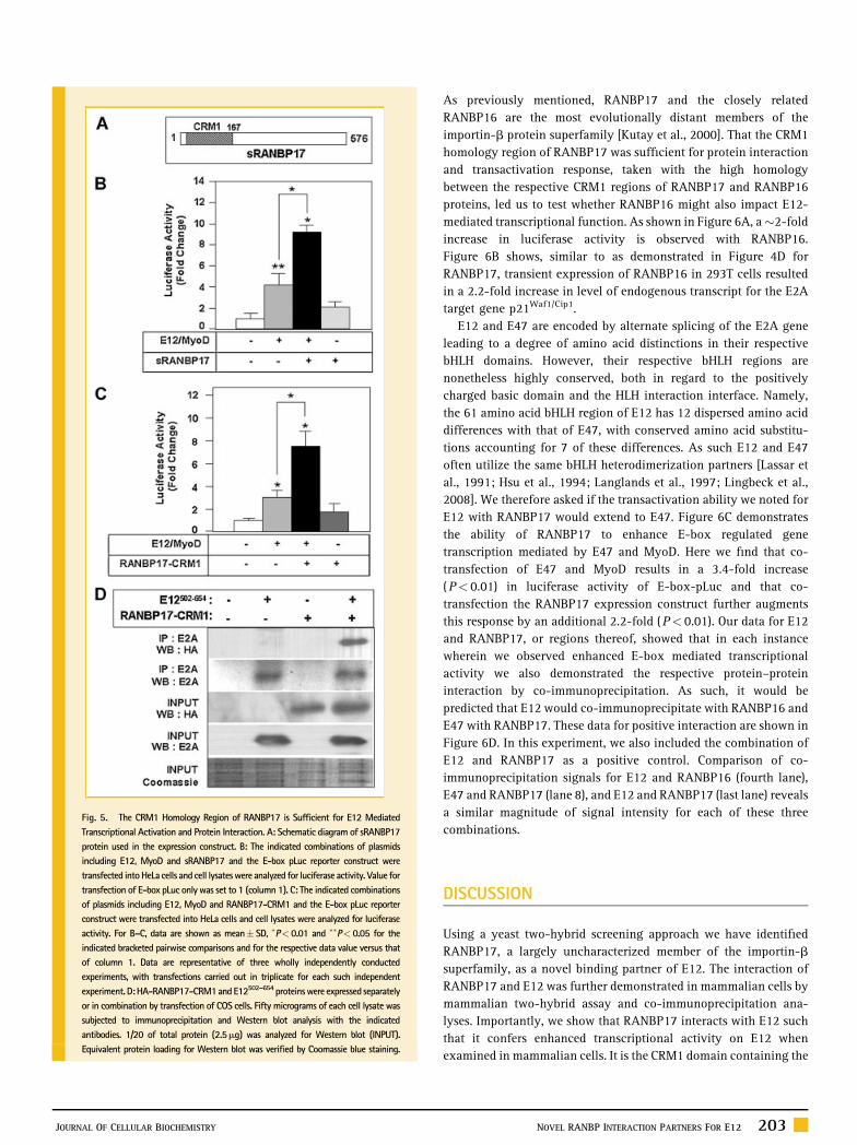
Fig. 5. The CRM1 Homology Region of RANBP17 is Sufficient for E12 Mediated
Transcriptional Activation and Protein Interaction. A: Schematic diagram of sRANBP17
protein used in the expression construct. B: The indicated combinations of plasmids
including E12, MyoD and sRANBP17 and the E-box pLuc reporter construct were
transfected intoHeLa cells andcell lysateswere analyzed for luciferase activity. Value for
transfection of E-box pLuc only was set to 1 (column 1). C: The indicated combinations
of plasmids including E12, MyoD and RANBP17-CRM1 and the E-box pLuc reporter
construct were transfected into HeLa cells and cell lysates were analyzed for luciferase
activity. For B–C, data are shown as mean� SD, �P< 0.01 and ��P< 0.05 for the
indicated bracketed pairwise comparisons and for the respective data value versus that
of column 1. Data are representative of three wholly independently conducted
experiments, with transfections carried out in triplicate for each such independent
experiment.D:HA-RANBP17-CRM1andE12502–654 proteinswere expressed separately
or in combination by transfection of COS cells. Fifty micrograms of each cell lysate was
subjected to immunoprecipitation and Western blot analysis with the indicated
antibodies. 1/20 of total protein (2.5mg) was analyzed for Western blot (INPUT).
Equivalent protein loading for Western blot was verified by Coomassie blue staining.
JOURNAL OF CELLULAR BIOCHEMISTRY
As previously mentioned, RANBP17 and the closely related
RANBP16 are the most evolutionally distant members of the
importin-b protein superfamily [Kutay et al., 2000]. That the CRM1
homology region of RANBP17 was sufficient for protein interaction
and transactivation response, taken with the high homology
between the respective CRM1 regions of RANBP17 and RANBP16
proteins, led us to test whether RANBP16 might also impact E12-
mediated transcriptional function. As shown in Figure 6A, a�2-fold
increase in luciferase activity is observed with RANBP16.
Figure 6B shows, similar to as demonstrated in Figure 4D for
RANBP17, transient expression of RANBP16 in 293T cells resulted
in a 2.2-fold increase in level of endogenous transcript for the E2A
target gene p21Waf1/Cip1.
E12 and E47 are encoded by alternate splicing of the E2A gene
leading to a degree of amino acid distinctions in their respective
bHLH domains. However, their respective bHLH regions are
nonetheless highly conserved, both in regard to the positively
charged basic domain and the HLH interaction interface. Namely,
the 61 amino acid bHLH region of E12 has 12 dispersed amino acid
differences with that of E47, with conserved amino acid substitu-
tions accounting for 7 of these differences. As such E12 and E47
often utilize the same bHLH heterodimerization partners [Lassar et
al., 1991; Hsu et al., 1994; Langlands et al., 1997; Lingbeck et al.,
2008]. We therefore asked if the transactivation ability we noted for
E12 with RANBP17 would extend to E47. Figure 6C demonstrates
the ability of RANBP17 to enhance E-box regulated gene
transcription mediated by E47 and MyoD. Here we find that co-
transfection of E47 and MyoD results in a 3.4-fold increase
(P< 0.01) in luciferase activity of E-box-pLuc and that co-
transfection the RANBP17 expression construct further augments
this response by an additional 2.2-fold (P< 0.01). Our data for E12
and RANBP17, or regions thereof, showed that in each instance
wherein we observed enhanced E-box mediated transcriptional
activity we also demonstrated the respective protein–protein
interaction by co-immunoprecipitation. As such, it would be
predicted that E12 would co-immunoprecipitate with RANBP16 and
E47 with RANBP17. These data for positive interaction are shown in
Figure 6D. In this experiment, we also included the combination of
E12 and RANBP17 as a positive control. Comparison of co-
immunoprecipitation signals for E12 and RANBP16 (fourth lane),
E47 and RANBP17 (lane 8), and E12 and RANBP17 (last lane) reveals
a similar magnitude of signal intensity for each of these three
combinations.
DISCUSSION
Using a yeast two-hybrid screening approach we have identified
RANBP17, a largely uncharacterized member of the importin-b
superfamily, as a novel binding partner of E12. The interaction of
RANBP17 and E12 was further demonstrated in mammalian cells by
mammalian two-hybrid assay and co-immunoprecipitation ana-
lyses. Importantly, we show that RANBP17 interacts with E12 such
that it confers enhanced transcriptional activity on E12 when
examined in mammalian cells. It is the CRM1 domain containing the
NOVEL RANBP INTERACTION PARTNERS FOR E12 203

Fig. 6. Expansion of the RANBP17 Interaction Network to Include Closely
Related Proteins. A: The indicated combinations of plasmids including E12,
MyoD and RANBP16 and the E-box pLuc reporter construct were transfected
into HeLa cells and cell lysates analyzed for luciferase activity. Value for
transfection of E-box pLuc only was set to 1 (column 1). �P< 0.01 for the
indicated bracketed pairwise comparisons and for the respective data value
versus that of column 1. B: Regulation of endogenous p21Waf1/Cip1 transcript
level by RANBP16. Q-PCR analysis of RNA harvested from transfected 293T cells
with �P< 0.01 compared to empty vector. Representative of three independent
transfection studies is shown. C: The indicated combinations of plasmids
including E47, MyoD, RANBP17 and the E-box pLuc reporter construct were
transfected into HeLa cells and cell lysates analyzed for luciferase activity. For A
and C, data are representative of three wholly independently conducted experi-
ments, with transfections carried out in triplicate for each such independent
experiment. Data are shown as the mean� SD. D: Co-immunoprecipitation
analysis reveals interaction of E12 with RANBP16 and E47 with RANBP17.
Various combinations of plasmids as indicated by ‘‘þ’’ signs at lane headings
were transfected into COS cells. Cell lysates were subjected to immunopreci-
pitation (IP) and Western blot (WB) analysis with the indicated antibodies. 1/20
of total protein (2.5mg) was analyzed for Western blot (INPUT). Equivalent
protein content per sample was verified by Coomassie blue staining of 20mg
sample aliquots. For comparison purposes, the far right lane shows interactionof
RANBP17 with E12, carried out in the same experiment and analyzed on the
same Western blot as the other combinations presented in this panel.
204 NOVEL RANBP INTERACTION PARTNERS FOR E12
N-terminal region amino acids (1-167) of RANBP17 and the C-
terminus including the bHLH domain (508–654 amino acids) of E12
that are responsible for this interaction. Interestingly, for the case of
the CRM1 protein itself, its CRM1 domain mediates the ability of
CRM1 to bind RanGTP [Ossareh-Nazari and Dargemont, 1999;
Petosa et al., 2004]. Our study suggests that the CRM1 domain may
have a wider role in various protein–protein interactions than has
previously been appreciated. It is of interest that we find that
transcriptional activity of both E47 and E12 are increased via
RANBP17 action and that each co-immunoprecipitate with
RANBP17. Thus it might be overall structural similarity, for
example the HLH motif, or the enrichment of positively charged
amino acids that mediates interaction of RANBP17 with E-proteins.
In their studies of RANBP16, Gorlich and coworkers concluded that
while cargoes of RANBP16 were structurally diverse, a common
theme appeared to be the presence of positively charged domains
[Mingot et al., 2004]. It is of interest to note that bHLH regions are
typified by a high density of positively charged amino acids
[Benezra et al., 1990]; it remains to be seen if the interactions we
describe herein will extend to other members of the bHLH protein
family. The fact that we demonstrate that E12 transcriptional
activity is also augmented by RANBP16, and that RANBP16 and E12
co-immunoprecipitate, reinforces the idea that positively charged
motifs are involved in functional interactions for RANBP16 and
suggests that this is also the case for RANBP17.
The ubiquitously expressed class I bHLH transcription factors,
including E2A proteins, for the most part, impact tissue specific gene
regulation via their interaction with cell type specific class II bHLH
transcription factors. Proteins that function in the nucleus,
including transcription factors, must be regulated in precise
temporal and cell type-specific manners. A multitude of studies
suggests that the regulation of nucleocytoplasmic shuttling of
transcription factors is an important mechanism by which their
activities can be regulated. Mutations affecting this process have
been identified in various cancers [Fabbro and Henderson, 2003;
Kau et al., 2004]. Transport between nucleus and cytoplasm through
the nuclear pore complex is largely mediated by the superfamily of
importin b-related factors which represents the largest class of
nuclear transport receptors [Kutay et al., 2000; Mingot et al., 2004].
This type of nucleocytoplasmic transport responds to the nucleo-
cytoplasmic RanGTP gradient [Gorlich et al., 1996; Kutay et al.,
1997]. Surprisingly little is known of the machinery specifically
involved in E2A protein transport. However, given their molecular
mass of �75, they exceed the estimated 20–40 kDa upper range of
mass for entering the nucleus by diffusion, and thus they are likely
to depend on facilitated receptor mediated translocation. Nuclear
localization of E2A proteins is required for their proper functioning
as transcription factors and E2A proteins contain potential nuclear
localization sequence in their N-terminus [Mitsui et al., 1993; Sloan
et al., 1996; Huggins et al., 1999; Chu and Kohtz, 2001], however the
specific details for the nucleocytoplasmic transport mechanism of
E2A proteins are not yet known. It is conceivable that if RANBP17
functions in nucleocytoplasmic transport, which to date has been
empirically demonstrated for RANBP16 but not well examined for
RANBP17, then E12 might be a cargo of RANBP17. On the other
hand, as RANBP17 and RANBP16 are both very evolutionarily
JOURNAL OF CELLULAR BIOCHEMISTRY

distant members of the importin-b superfamily, they may impact
E2A protein function via novel mechanisms.
Interestingly, the RANBP17 gene is the site of recurrent
chromosomal 5 breakpoints found in T-cell acute lymphoblastic
leukemia (T-ALL) with a (t5;14)(q35;q32) translocation the most
frequently observed [Bernard et al., 2001; Berger et al., 2003; Su
et al., 2006], and a (t5;14)(q34;q11) translocation also noted
[Hansen-Hagge et al., 2002]. It has been suggested that the
transcriptional activation of RANBP17, presumed to occur during
hematopoietic development, may favor genetic rearrangement by
opening up the chromatin in the RANBP17 region of chromosome 5
[Bernard et al., 2001]. To date, the biological effects of these
translocations have focused nearly exclusively on the resultant
activation of the transcription factor HOX11L2/TLX3 [MacLeod
et al., 2003; Bayly et al., 2004; Su et al., 2006]. However, the idea has
also been put forth that enhanced expression of RANBP17 transcript
could conceivably occur in the (t5;14)(q34;q11) rearrangement,
since this generates a head-to-tail juxtaposition of RANBP17 with
enhancer elements of the TCR delta gene [Bernard et al., 2001;
Hansen-Hagge et al., 2002]. This would be predicted to lead to
increased expression of full-length RANBP17 or a C-terminally
truncated RANBP17 protein, dependent on the breakpoint site
[Bernard et al., 2001; Hansen-Hagge et al., 2002]. It is possible that
in these and other settings RANBP17 plays a role in disease via its
ability to augment E12 and/or E47-mediated transcriptional signals.
In conclusion, we have identified RANBP16 and RANBP17 as new
players in the bHLH protein interaction network. E2A-regulated
transcriptional signals have key roles in numerous biological
settings. Taken with our finding that RANBP16 and RANBP17 can
enhance transcript levels of endogenous p21Waf1/Cip1, a well-
validated E2A transactivation target gene, our data suggests that in
certain biological settings RANBP16 or RANBP17 may impact
normal and disease processes via functional interaction with E2A
proteins.
ACKNOWLEDGMENTS
We thank Dr. W.G. Janssen (University of Heidelberg, Germany) forRANBP17-pMT2SM2 and RANBP16-pMT2SM, Dr. C. Murre(Harvard University) for E12, and Dr. B. Rowan (Tulane University)for the pGAL4-Luc plasmids. We thank Melissa Wilhelm and DavidMajors for help with yeast two-hybrid screening and Kun Liu fortechnical assistance. This work was supported by a grant from theNational Cancer Institute to C.M.S.
REFERENCES
Bayly R, Chuen L, Currie RA, Hyndman BD, Casselman R, Blobel GA, LeBrunDP. 2004. E2A-PBX1 interacts directly with the KIX domain of CBP/p300 inthe induction of proliferation in primary hematopoietic cells. J Biol Chem279:55362–55371.
Benezra R, Davis RL, Lockshon D, Turner DL, Weintraub H. 1990. The proteinId: A negative regulator of helix-loop-helix DNA binding proteins. Cell61:49–59.
Berger R, Dastugue N, Busson M, Van Den Akker J, Perot C, Ballerini P,Hagemeijer A, Michaux L, Charrin C, Pages MP, Mugneret F, Andrieux J,Talmant P, Helias C, Mauvieux L, Lafage-Pochitaloff M, Mozziconacci MJ,
JOURNAL OF CELLULAR BIOCHEMISTRY
Cornillet-Lefebvre P, Radford I, Asnafi V, Bilhou-Nabera C, Nguyen Khac F,Leonard C, Speleman F, Poppe B, Bastard C, Taviaux S, Quilichini B, Herens C,Gregoire MJ, Cave H, Bernard OA. 2003. t(5;14)/HOX11L2-positive T-cellacute lymphoblastic leukemia. A collaborative study of the Groupe Francaisde Cytogenetique Hematologique (GFCH). Leukemia 17:1851–1857.
Berkes CA, Tapscott SJ. 2005. MyoD and the transcriptional control ofmyogenesis. Semin Cell Dev Biol 16:585–595.
Bernard OA, Busson-LeConiat M, Ballerini P, Mauchauffe M, Della Valle,Monni V, Nguyen R, Khac F, Mercher T, Penard-Lacronique V, Pasturaud P,Gressin L, Heilig R, Daniel MT, Lessard M, Berger R. 2001. A new recurrentand specific cryptic translocation, t(5;14)(q35;q32), is associated withexpression of the Hox11L2 gene in T acute lymphoblastic leukemia. Leu-kemia 15:1495–1504.
Chu C, Kohtz DS. 2001. Identification of the E2A gene products as regulatorytargets of the G1 cyclin-dependent kinases. J Biol Chem 276:8524–8534.
Cook A, Bono F, Jinek M, Conti E. 2007. Structural biology of nucleocyto-plasmic transport. Annu Rev Biochem 76:647–671.
Desprez PY, Sumida T, Coppe JP. 2003. Helix-loop-helix proteins in mam-mary gland development and breast cancer. J Mammary Gland Biol Neo-plasia 8:225–239.
Dorfman J, Macara IG. 2008. STRADalpha regulates LKB1 localization byblocking access to importin-alpha, and by association with Crm1 andexportin-7. Mol Biol Cell 19:1614–1626.
Eckner R, Yao TP, Oldread E, Livingston DM. 1996. Interaction and functionalcollaboration of p300/CBP and bHLH proteins in muscle and B-cell differ-entiation. Genes Dev 10:2478–2490.
Fabbro M, Henderson BR. 2003. Regulation of tumor suppressors by nuclear-cytoplasmic shuttling. Exp Cell Res 282:59–69.
French BA, Chow KL, Olson EN, Schwartz RJ. 1991. Heterodimers ofmyogenic helix-loop- helix regulatory factors and E12 bind a complexelement governing myogenic induction of the avian cardiac alpha-actinpromoter. Mol Cell Biol 11:2439–2450.
Funato N, Ohtani K, Ohyama K, Kuroda T, Nakamura M. 2001. Commonregulation of growth arrest and differentiation of osteoblasts by helix-loop-helix factors. Mol Cell Biol 21:7416–7428.
Funato N, Ohyama K, Kuroda T, Nakamura M. 2003. Basic helix-loop-helixtranscription factor epicardin/capsulin/Pod-1 suppresses differentiation bynegative regulation of transcription. J Biol Chem 278:7486–7493.
Funk WD, Ouellette M, Wright WE. 1991. Molecular biology of myogenicregulatory factors. Mol Biol Med 8:185–195.
Glick E, Leshkowitz D, Walker MD. 2000. Transcription factor BETA2 actscooperatively with E2A and PDX1 to activate the insulin gene promoter.J Biol Chem 275:2199–2204.
Gorlich D, Pante N, Kutay U, Aebi U, Bischoff FR. 1996. Identification ofdifferent roles for RanGDP and RanGTP in nuclear protein import. EMBO J15:5584–5594.
Gould KA, Bresnick EH. 1998. Sequence determinants of DNA binding by thehematopoietic helix-loop-helix transcription factor TAL1: Importance ofsequences flanking the E-box core. Gene Expr 7:87–101.
Greenbaum S, Zhuang Y. 2002. Regulation of early lymphocyte developmentby E2A family proteins. Semin Immunol 14:405–414.
Hansen-Hagge TE, Schafer M, Kiyoi H, Morris SW, Whitlock JA, Koch P,Bohlmann I, Mahotka C, Bartram CR, Janssen JW. 2002. Disruption of theRanBP17/Hox11L2 region by recombination with the TCRdelta locus in acutelymphoblastic leukemias with t(5;14)(q34;q11). Leukemia 16:2205–2212.
Hikima J, Middleton DL, Wilson MR, Miller NW, Clem LW, Warr GW. 2005.Regulation of immunoglobulin gene transcription in a teleost fish: Identi-fication, expression and functional properties of E2A in the channel catfish.Immunogenetics 57:273–282.
Hsu HL, Huang L, Tsan JT, Funk W, Wright WE, Hu JS, Kingston RE, Baer R.1994. Preferred sequences for DNA recognition by the TAL1 helix-loop-helixproteins. Mol Cell Biol 14:1256–1265.
NOVEL RANBP INTERACTION PARTNERS FOR E12 205

Huggins GS, Chin MT, Sibinga NE, Lee SL, Haber E, Lee ME. 1999. Char-acterization of the mUBC9-binding sites required for E2A protein degrada-tion. J Biol Chem 274:28690–28696.
Kau TR, Way JC, Silver PA. 2004. Nuclear transport and cancer: Frommechanism to intervention. Nat Rev Cancer 4:106–117.
Kim W, Kook S, Kim DJ, Teodorof C, Song WK. 2004. The 31-kDa caspase-generated cleavage product of p130cas functions as a transcriptional repres-sor of E2A in apoptotic cells. J Biol Chem 279:8333–8342.
Koch P, Bohlmann I, Schafer M, Hansen-Hagge TE, Kiyoi H, Wilda M,Hameister H, Bartram CR, Janssen JW. 2000. Identification of a novelputative Ran-binding protein and its close homologue. Biochem BiophysRes Commun 278:241–249.
Kutay U, Bischoff FR, Kostka S, Kraft R, Gorlich D. 1997. Export of importinalpha from the nucleus is mediated by a specific nuclear transport factor. Cell90:1061–1071.
Kutay U, Hartmann E, Treichel N, Calado A, Carmo-Fonseca M, Prehn S, KraftR, Gorlich D, Bischoff FR. 2000. Identification of two novel RanGTP-bindingproteins belonging to the importin beta superfamily. J Biol Chem 275:40163–40168.
Langlands K, Yin X, Anand G, Prochownik EV. 1997. Differential interactionsof Id proteins with basic-helix-loop-helix transcription factors. J Biol Chem272:19785–19793.
Lassar AB, Davis RL, Wright WE, Kadesch T, Murre C, Voronova A, BaltimoreD, Weintraub H. 1991. Functional activity of myogenic HLH proteins requireshetero-oligomerization with E12/E47-like proteins in vivo. Cell 66:305–315.
Lee JE. 1997. NeuroD and neurogenesis. Dev Neurosci 19:27–32.
Lingbeck JM, Trausch-Azar JS, Ciechanover A, Schwartz AL. 2008. In vivointeractions of MyoD, Id1, and E2A proteins determined by acceptor photo-bleaching fluorescence resonance energy transfer. FASEB J 22:1694–1701.
Liu Y, Encinas M, Comella JX, Aldea M, Gallego C. 2004. Basic helix-loop-helix proteins bind to TrkB and p21(Cip1) promoters linking differentiationand cell cycle arrest in neuroblastoma cells. Mol Cell Biol 24:2662–2672.
MacLeod RA, Nagel S, Kaufmann M, Janssen JW, Drexler HG. 2003. Activa-tion of HOX11L2 by juxtaposition with 30-BCL11B in an acute lymphoblasticleukemia cell line (HPB- ALL) with t(5;14)(q35;q32.2). Genes ChromosomesCancer 37:84–91.
Massari ME, Murre C. 2000. Helix-loop-helix proteins: Regulators of tran-scription in eucaryotic organisms. Mol Cell Biol 20:429–440.
Massari ME, Grant PA, Pray-Grant MG, Berger SL, Workman JL, Murre C.1999. A conserved motif present in a class of helix-loop-helix proteinsactivates transcription by direct recruitment of the SAGA complex. Mol Cell4:63–73.
Mingot JM, Bohnsack MT, Jakle U, Gorlich D. 2004. Exportin 7 defines anovel general nuclear export pathway. EMBO J 23:3227–3236.
Mitsui K, Shirakata M, Paterson BM. 1993. Phosphorylation inhibits theDNA-binding activity of MyoD homodimers but not MyoD-E12 heterodi-mers. J Biol Chem 268:24415–24420.
Murre C, McCaw PS, Vaessin H, Caudy M, Jan LY, Jan YN, Cabrera CV, BuskinJN, Hauschka SD, Lassar AB., et al. 1989. Interactions between heterologoushelix-loop-helix proteins generate complexes that bind specifically to acommon DNA sequence. Cell 58:537–544.
Murre C, Bain G, van Dijk MA, Engel I, Furnari BA, Massari ME, Matthews JR,Quong MW, Rivera RR, Stuiver MH. 1994. Structure and function of helix-loop-helix proteins. Biochim Biophys Acta 1218:129–135.
Mutoh H, Fung BP, Naya FJ, Tsai MJ, Nishitani J, Leiter AB. 1997. The basichelix-loop-helix transcription factor BETA2/NeuroD is expressed in mam-malian enteroendocrine cells and activates secretin gene expression. ProcNatl Acad Sci USA 94:3560–3564.
Neufeld B, Grosse-Wilde A, Hoffmeyer A, Jordan BW, Chen P, Dinev D,Ludwig S, Rapp UR. 2000. Serine/Threonine kinases 3pK and MAPK-acti-vated protein kinase 2 interact with the basic helix-loop-helix transcription
206 NOVEL RANBP INTERACTION PARTNERS FOR E12
factor E47 and repress its transcriptional activity. J Biol Chem 275:20239–20242.
Norton JD. 2000. ID helix-loop-helix proteins in cell growth, differentiationand tumorigenesis. J Cell Sci 113(Pt 22): 3897–3905.
Norton JD, Deed RW, Craggs G, Sablitzky F. 1998. Id helix-loop-helixproteins in cell growth and differentiation. Trends Cell Biol 8:58–65.
Ossareh-Nazari B, Dargemont C. 1999. Domains of Crm1 involved in theformation of the Crm1, RanGTP, and leucine-rich nuclear export sequencestrimeric complex. Exp Cell Res 252:236–241.
Perry C, Soreq H. 2002. Transcriptional regulation of erythropoiesis. Finetuning of combinatorial multi-domain elements. Eur J Biochem 269:3607–3618.
Petosa C, Schoehn G, Askjaer P, Bauer U, Moulin M, Steuerwald U, Soler-Lopez M, Baudin F, Mattaj IW, Muller CW. 2004. Architecture of CRM1/Exportin1 suggests how cooperativity is achieved during formation of anuclear export complex. Mol Cell 16:761–775.
Prabhu S, Ignatova A, Park ST, Sun XH. 1997. Regulation of the expression ofcyclin-dependent kinase inhibitor p21 by E2A and Id proteins. Mol Cell Biol17:5888–5896.
Quong MW, Romanow WJ, Murre C. 2002. E protein function in lymphocytedevelopment. Annu Rev Immunol 20:301–322.
Saarikettu J, Sveshnikova N, Grundstrom T. 2004. Calcium/calmodulininhibition of transcriptional activity of E-proteins by prevention of theirbinding to DNA. J Biol Chem 279:41004–41011.
Semerad CL, Mercer EM, Inlay MA, Weissman IL, Murre C. 2009. E2A proteinsmaintain the hematopoietic stem cell pool and promote the maturation ofmyelolymphoid and myeloerythroid progenitors. Proc Natl Acad Sci USA106:1930–1935.
Shirakata M, Paterson BM. 1995. The E12 inhibitory domain preventshomodimer formation and facilitates selective heterodimerization with theMyoD family of gene regulatory factors. EMBO J 14:1766–1772.
Sloan SR, Shen CP, McCarrick-Walmsley R, Kadesch T. 1996. Phosphoryla-tion of E47 as a potential determinant of B-cell-specific activity. Mol Cell Biol16:6900–6908.
Su XY, Della-Valle V, Andre-Schmutz I, Lemercier C, Radford-Weiss I,Ballerini P, Lessard M, Lafage-Pochitaloff M, Mugneret F, Berger R, RomanaSP, Bernard OA, Penard-Lacronique V. 2006. HOX11L2/TLX3 is transcrip-tionally activated through T-cell regulatory elements downstream of BCL11Bas a result of the t(5;14)(q35;q32). Blood 108:4198–4201.
Sun XH, Baltimore D. 1991. An inhibitory domain of E12 transcription factorprevents DNA binding in E12 homodimers but not in E12 heterodimers. Cell64:459–470.
Sun R, Su Y, Zhao X, Qi J, Luo X, Yang Z, Yao Y, Xia Z. 2009. Humancalcium/calmodulin- dependent serine protein kinase regulates the expres-sion of p21 via the E2A transcription factor. Biochem J 419:457–466.
Turner EC, Cureton CH, Weston CJ, Smart OS, Allemann RK. 2004. Control-ling the DNA binding specificity of bHLH proteins through intramolecularinteractions. Chem Biol 11:69–77.
Venuti JM, Cserjesi P. 1996. Molecular embryology of skeletal myogenesis.Curr Top Dev Biol 34:169–206.
Yagita K, Tamanini F, Yasuda M, Hoeijmakers JH, van der Horst GT,Okamura H. 2002. Nucleocytoplasmic shuttling and mCRY-dependentinhibition of ubiquitylation of the mPER2 clock protein. EMBO J 21:1301–1314.
Yokota Y, Mori S. 2002. Role of Id family proteins in growth control. J CellPhysiol 190:21–28.
Yoneda Y. 2000. Nucleocytoplasmic protein traffic and its significance to cellfunction. Genes Cells 5:777–787.
Yutzey KE, Konieczny SF. 1992. Different E-box regulatory sequences arefunctionally distinct when placed within the context of the troponin Ienhancer. Nucleic Acids Res 20:5105–5113.
JOURNAL OF CELLULAR BIOCHEMISTRY