identification of swine by auricular vein patterns · the pig auricular vein is a branch from the...
TRANSCRIPT
Identification of Swine by Auricular Vein Patterns Ryan J. Harrell CNMT Nuclear Medicine and PET/CT University of St. Francis and Indiana Purdue Fort Wayne Fort Wayne, Indiana Dr. Clinton P. Rusk Associate Professor Department of Youth Development and Agricultural Education Purdue University West Lafayette, Indiana Dr. Brian T. Richert Associate Professor Swine Nutrition and Management Purdue University West Lafayette, Indiana Introduction The universal ear notching system was established in the 1900�s to standardize purebred and commercial swine identification (Rea, 1986). An adaptation of the universal ear notching system was developed by each breed association, identifying a pig by litter number and individual pig number (Neary and Yaher, 2002). The Yorkshire, Hamphire, Duroc and Landrace breeds have the same ear notching system with the largest litter notches being 161 in the right ear (The National Swine Registry, 2007). The Berkshire breed adopted a different ear notching system with the largest litter notch going up to 1199, utilizing both the left and right ear (American Berkshire Association, 2006). Even though it is classified as the universal standardized ear notching system, it is not the ideal identification system for swine with variations existing between breeds and geographic locations. Poor ear notching technique can result in misread identification if notches are placed in the middle of the ear, too close together or not deep enough in the ear (Rea, 1986). Besides ear notches, swine have also been identified by ear tags and tattoos. Ear tags can be removed, lost, chewed-on or switched between animals. Tattoos can fade and become hard to read over a period of time. Studies by Solis and Maala in 1975 determined that tattooing, ear notching and ear tagging are flawed systems that could be replicated, changed or modified. Biometrics became an alternative method of identification based on the anatomical and physical characteristics of animals. Optibrand, Ltd., LLC, created technology to capture retinal images along with a global positioning satellite system (GPS) used to verify location, date and time (Whittier et al., 2003). Retinal imaging has become a viable method of identification for beef and sheep (Rusk et al., 2006). Although it is possible to retinal scan swine for identification, it is very difficult to restrain the pig in order to capture the retinal image.
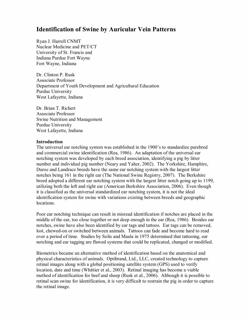
Blood vessel patterns in the ear are unique traits that can be used for identification. This method of identification is fairly non-invasive and accurate. Twenty laboratory rats were positively identified by defining twelve minutiae points created from analyzing the blood vessel branching structures in two sets of images from the same individual rat (Cameron et al., 2007). Eighteen out of twenty mice were successfully identified using an algorithm equation created from images of ear blood vessels patterns (Ellmauthaler and Wernsperger, 2007). Research by Nilsson et al. (2006), from Halmstad University, concluded that blood vessel patterns in mouse ears can be considered a suitable biometric identifier for laboratory mice. The pig auricular vein is a branch from the maxillary vein, caudal vein and bifurcates into the middle auricular vein (See Figure 1). The middle auricular vein can branch into various unique patterns. Figure 1: The general scheme of the veins with respect to the connections with the perihypophyseal cavernous sinus-carotid rete vascular complex. 1- external jugular vein, 2- maxillary vein, 9- caudal auricular vein, 10- middle auricular vein. Adaptations from (Ghoshal and Zguigal, 1986) and (Zezula-Szpyra and Grzegorzewski, 2000). This study investigated the use of auricular vein patterns as a potential identification tool compared to the universal ear notching system used as the gold standard in swine. Researchers developed a method of imaging auricular vein patterns in swine as a possible means of identification. Visual verification was determined to identify auricular vein pattern matches and mismatches. The objectives of this study were to:
1. Determine if swine auricular vein patterns can be imaged and used for identification.
2. Determine if each individual pig has a unique auricular vein pattern. 3. Determine whether auricular vein patterns change design or location over time. 4. Decide if a difference exists in auricular vein patterns between barrows and gilts.
5. Determine if any variations in auricular vein patterns exist between different breeds of swine.
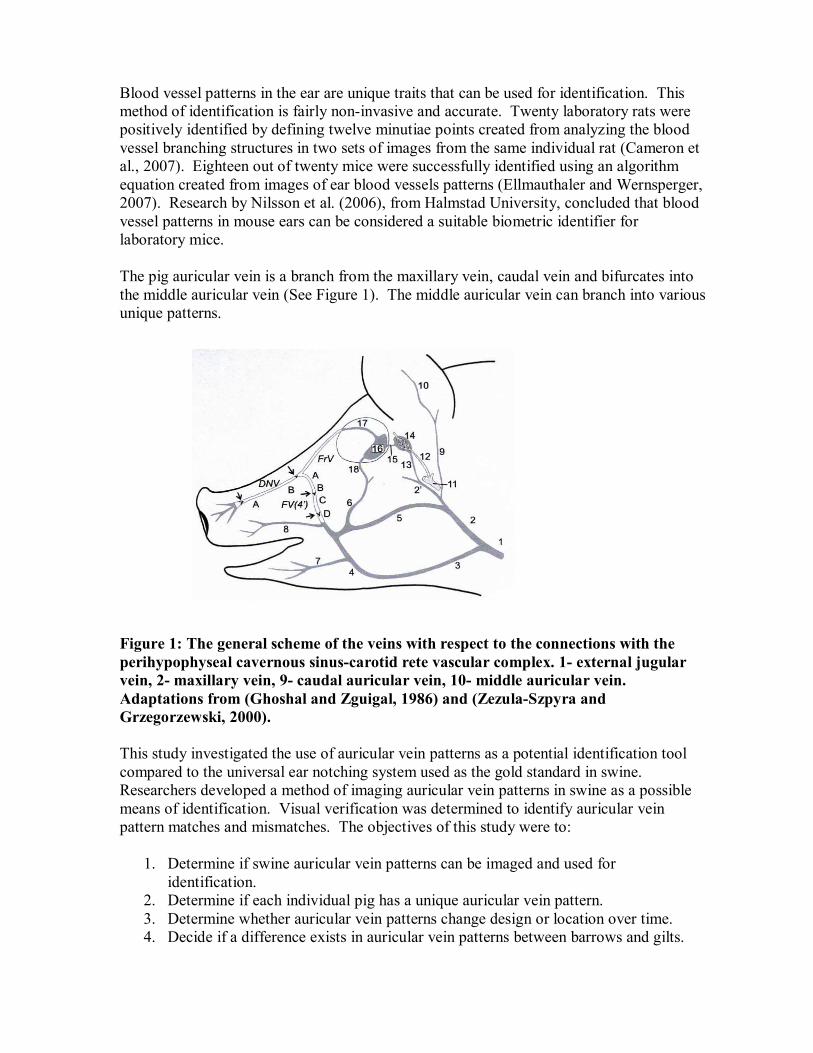
Hypothesis: HNull: Swine auricular vein patterns can be as viable for identification as the universal ear notching system. Methodology Background Information The OptiReader � Device (See Figure 2), developed by Optibrand Ltd., LLC, was used to capture swine auricular vein patterns and to provide a global positioning satellite system (GPS) for each record.
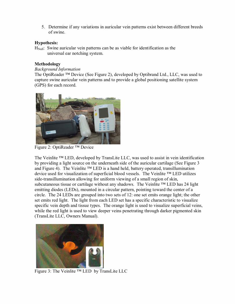
Figure 2: OptiReader � Device The Veinlite � LED, developed by TransLite LLC, was used to assist in vein identification by providing a light source on the underneath side of the auricular cartilage (See Figure 3 and Figure 4). The Veinlite � LED is a hand held, battery operated, transillumination device used for visualization of superficial blood vessels. The Veinlite � LED utilizes side-transillumination allowing for uniform viewing of a small region of skin, subcutaneous tissue or cartilage without any shadows. The Veinlite � LED has 24 light emitting diodes (LEDs), mounted in a circular pattern, pointing toward the center of a circle. The 24 LEDs are grouped into two sets of 12: one set emits orange light; the other set emits red light. The light from each LED set has a specific characteristic to visualize specific vein depth and tissue types. The orange light is used to visualize superficial veins, while the red light is used to view deeper veins penetrating through darker pigmented skin (TransLite LLC, Owners Manual).
Figure 3: The Veinlite � LED by TransLite LLC
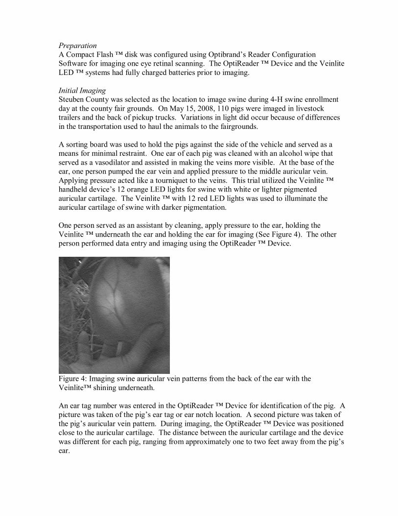
Preparation A Compact Flash � disk was configured using Optibrand�s Reader Configuration Software for imaging one eye retinal scanning. The OptiReader � Device and the Veinlite LED � systems had fully charged batteries prior to imaging. Initial Imaging Steuben County was selected as the location to image swine during 4-H swine enrollment day at the county fair grounds. On May 15, 2008, 110 pigs were imaged in livestock trailers and the back of pickup trucks. Variations in light did occur because of differences in the transportation used to haul the animals to the fairgrounds. A sorting board was used to hold the pigs against the side of the vehicle and served as a means for minimal restraint. One ear of each pig was cleaned with an alcohol wipe that served as a vasodilator and assisted in making the veins more visible. At the base of the ear, one person pumped the ear vein and applied pressure to the middle auricular vein. Applying pressure acted like a tourniquet to the veins. This trial utilized the Veinlite � handheld device�s 12 orange LED lights for swine with white or lighter pigmented auricular cartilage. The Veinlite � with 12 red LED lights was used to illuminate the auricular cartilage of swine with darker pigmentation. One person served as an assistant by cleaning, apply pressure to the ear, holding the Veinlite � underneath the ear and holding the ear for imaging (See Figure 4). The other person performed data entry and imaging using the OptiReader � Device.
Figure 4: Imaging swine auricular vein patterns from the back of the ear with the Veinlite� shining underneath. An ear tag number was entered in the OptiReader � Device for identification of the pig. A picture was taken of the pig�s ear tag or ear notch location. A second picture was taken of the pig�s auricular vein pattern. During imaging, the OptiReader � Device was positioned close to the auricular cartilage. The distance between the auricular cartilage and the device was different for each pig, ranging from approximately one to two feet away from the pig�s ear.
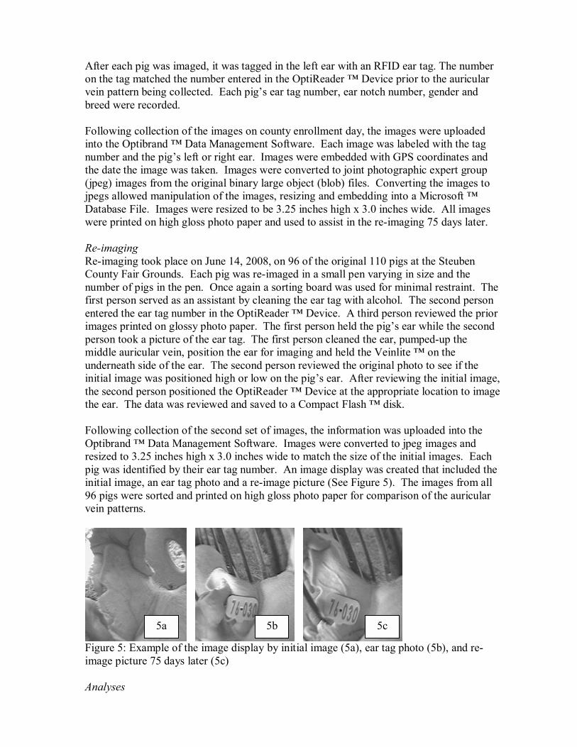
After each pig was imaged, it was tagged in the left ear with an RFID ear tag. The number on the tag matched the number entered in the OptiReader � Device prior to the auricular vein pattern being collected. Each pig�s ear tag number, ear notch number, gender and breed were recorded. Following collection of the images on county enrollment day, the images were uploaded into the Optibrand � Data Management Software. Each image was labeled with the tag number and the pig�s left or right ear. Images were embedded with GPS coordinates and the date the image was taken. Images were converted to joint photographic expert group (jpeg) images from the original binary large object (blob) files. Converting the images to jpegs allowed manipulation of the images, resizing and embedding into a Microsoft � Database File. Images were resized to be 3.25 inches high x 3.0 inches wide. All images were printed on high gloss photo paper and used to assist in the re-imaging 75 days later. Re-imaging Re-imaging took place on June 14, 2008, on 96 of the original 110 pigs at the Steuben County Fair Grounds. Each pig was re-imaged in a small pen varying in size and the number of pigs in the pen. Once again a sorting board was used for minimal restraint. The first person served as an assistant by cleaning the ear tag with alcohol. The second person entered the ear tag number in the OptiReader � Device. A third person reviewed the prior images printed on glossy photo paper. The first person held the pig�s ear while the second person took a picture of the ear tag. The first person cleaned the ear, pumped-up the middle auricular vein, position the ear for imaging and held the Veinlite � on the underneath side of the ear. The second person reviewed the original photo to see if the initial image was positioned high or low on the pig�s ear. After reviewing the initial image, the second person positioned the OptiReader � Device at the appropriate location to image the ear. The data was reviewed and saved to a Compact Flash � disk. Following collection of the second set of images, the information was uploaded into the Optibrand � Data Management Software. Images were converted to jpeg images and resized to 3.25 inches high x 3.0 inches wide to match the size of the initial images. Each pig was identified by their ear tag number. An image display was created that included the initial image, an ear tag photo and a re-image picture (See Figure 5). The images from all 96 pigs were sorted and printed on high gloss photo paper for comparison of the auricular vein patterns.
Figure 5: Example of the image display by initial image (5a), ear tag photo (5b), and re-image picture 75 days later (5c) Analyses
5a 5b 5c
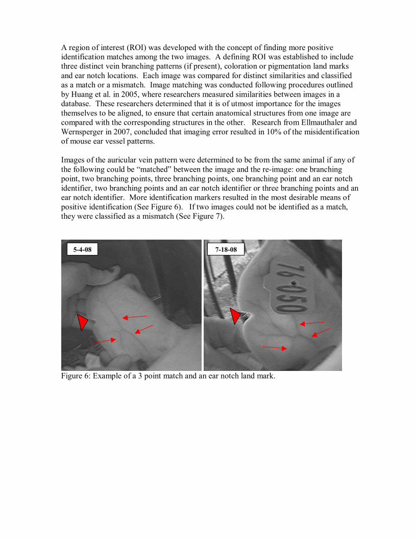
A region of interest (ROI) was developed with the concept of finding more positive identification matches among the two images. A defining ROI was established to include three distinct vein branching patterns (if present), coloration or pigmentation land marks and ear notch locations. Each image was compared for distinct similarities and classified as a match or a mismatch. Image matching was conducted following procedures outlined by Huang et al. in 2005, where researchers measured similarities between images in a database. These researchers determined that it is of utmost importance for the images themselves to be aligned, to ensure that certain anatomical structures from one image are compared with the corresponding structures in the other. Research from Ellmauthaler and Wernsperger in 2007, concluded that imaging error resulted in 10% of the misidentification of mouse ear vessel patterns. Images of the auricular vein pattern were determined to be from the same animal if any of the following could be �matched� between the image and the re-image: one branching point, two branching points, three branching points, one branching point and an ear notch identifier, two branching points and an ear notch identifier or three branching points and an ear notch identifier. More identification markers resulted in the most desirable means of positive identification (See Figure 6). If two images could not be identified as a match, they were classified as a mismatch (See Figure 7).
Figure 6: Example of a 3 point match and an ear notch land mark.
5-4-08 7-18-08
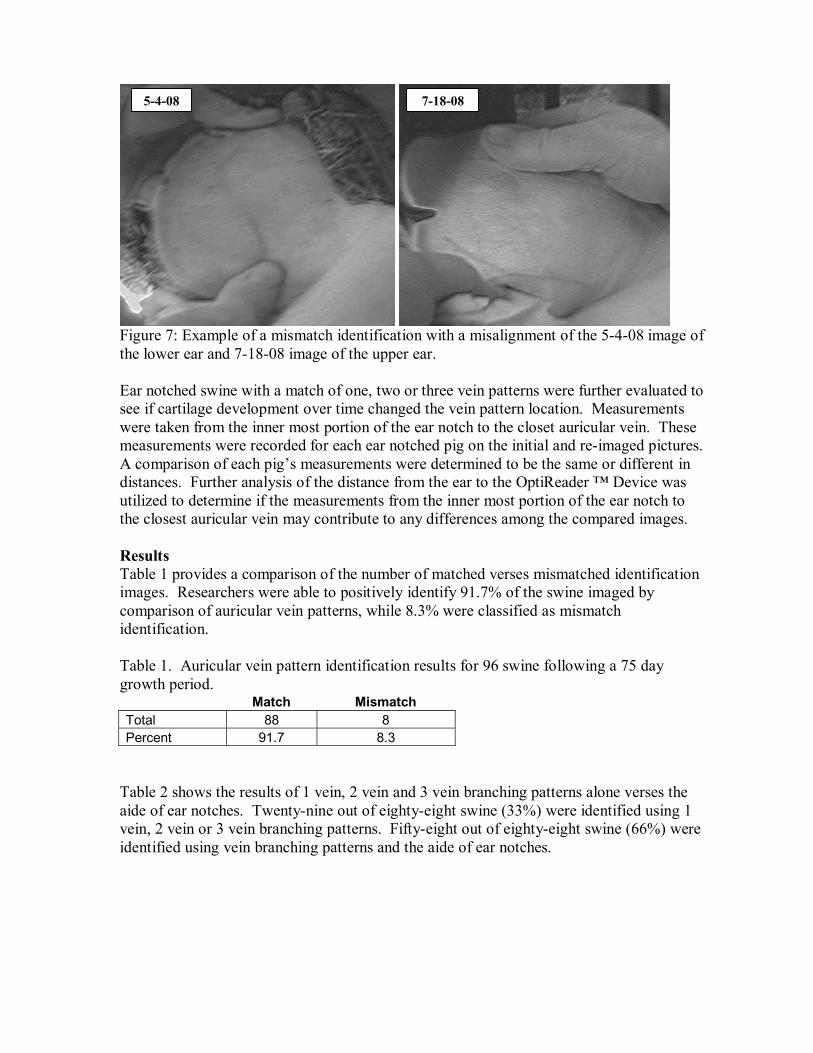
Figure 7: Example of a mismatch identification with a misalignment of the 5-4-08 image of the lower ear and 7-18-08 image of the upper ear. Ear notched swine with a match of one, two or three vein patterns were further evaluated to see if cartilage development over time changed the vein pattern location. Measurements were taken from the inner most portion of the ear notch to the closet auricular vein. These measurements were recorded for each ear notched pig on the initial and re-imaged pictures. A comparison of each pig�s measurements were determined to be the same or different in distances. Further analysis of the distance from the ear to the OptiReader � Device was utilized to determine if the measurements from the inner most portion of the ear notch to the closest auricular vein may contribute to any differences among the compared images. Results Table 1 provides a comparison of the number of matched verses mismatched identification images. Researchers were able to positively identify 91.7% of the swine imaged by comparison of auricular vein patterns, while 8.3% were classified as mismatch identification. Table 1. Auricular vein pattern identification results for 96 swine following a 75 day growth period. Match Mismatch Total 88 8 Percent 91.7 8.3
Table 2 shows the results of 1 vein, 2 vein and 3 vein branching patterns alone verses the aide of ear notches. Twenty-nine out of eighty-eight swine (33%) were identified using 1 vein, 2 vein or 3 vein branching patterns. Fifty-eight out of eighty-eight swine (66%) were identified using vein branching patterns and the aide of ear notches.
5-4-08 7-18-08
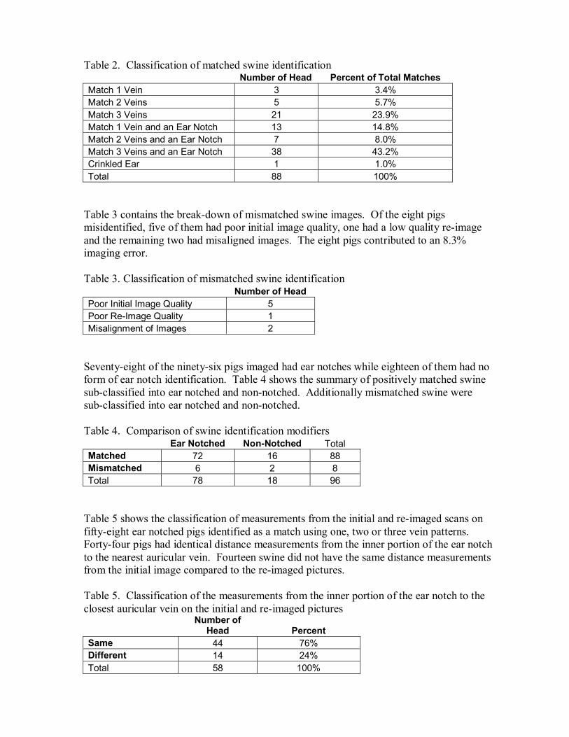
Table 2. Classification of matched swine identification Number of Head Percent of Total Matches Match 1 Vein 3 3.4% Match 2 Veins 5 5.7% Match 3 Veins 21 23.9% Match 1 Vein and an Ear Notch 13 14.8% Match 2 Veins and an Ear Notch 7 8.0% Match 3 Veins and an Ear Notch 38 43.2% Crinkled Ear 1 1.0% Total 88 100%
Table 3 contains the break-down of mismatched swine images. Of the eight pigs misidentified, five of them had poor initial image quality, one had a low quality re-image and the remaining two had misaligned images. The eight pigs contributed to an 8.3% imaging error. Table 3. Classification of mismatched swine identification Number of Head Poor Initial Image Quality 5 Poor Re-Image Quality 1 Misalignment of Images 2
Seventy-eight of the ninety-six pigs imaged had ear notches while eighteen of them had no form of ear notch identification. Table 4 shows the summary of positively matched swine sub-classified into ear notched and non-notched. Additionally mismatched swine were sub-classified into ear notched and non-notched. Table 4. Comparison of swine identification modifiers
Ear Notched Non-Notched Total Matched 72 16 88 Mismatched 6 2 8 Total 78 18 96
Table 5 shows the classification of measurements from the initial and re-imaged scans on fifty-eight ear notched pigs identified as a match using one, two or three vein patterns. Forty-four pigs had identical distance measurements from the inner portion of the ear notch to the nearest auricular vein. Fourteen swine did not have the same distance measurements from the initial image compared to the re-imaged pictures. Table 5. Classification of the measurements from the inner portion of the ear notch to the closest auricular vein on the initial and re-imaged pictures
Number of
Head Percent Same 44 76% Different 14 24% Total 58 100%
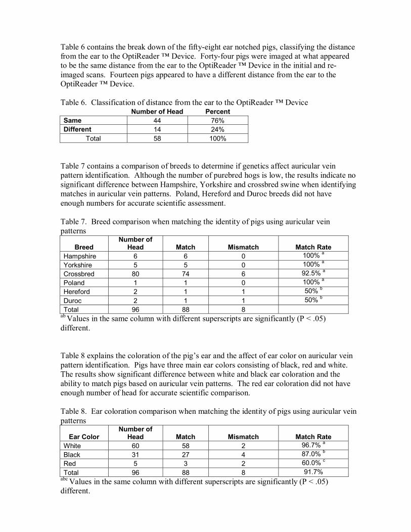
Table 6 contains the break down of the fifty-eight ear notched pigs, classifying the distance from the ear to the OptiReader � Device. Forty-four pigs were imaged at what appeared to be the same distance from the ear to the OptiReader � Device in the initial and re-imaged scans. Fourteen pigs appeared to have a different distance from the ear to the OptiReader � Device. Table 6. Classification of distance from the ear to the OptiReader � Device
Number of Head Percent Same 44 76% Different 14 24%
Total 58 100% Table 7 contains a comparison of breeds to determine if genetics affect auricular vein pattern identification. Although the number of purebred hogs is low, the results indicate no significant difference between Hampshire, Yorkshire and crossbred swine when identifying matches in auricular vein patterns. Poland, Hereford and Duroc breeds did not have enough numbers for accurate scientific assessment. Table 7. Breed comparison when matching the identity of pigs using auricular vein patterns
Breed Number of
Head Match Mismatch
Match Rate Hampshire 6 6 0 100% a
Yorkshire 5 5 0 100% a
Crossbred 80 74 6 92.5% a
Poland 1 1 0 100% a
Hereford 2 1 1 50% b
Duroc 2 1 1 50% b
Total 96 88 8 ab Values in the same column with different superscripts are significantly (P < .05) different. Table 8 explains the coloration of the pig�s ear and the affect of ear color on auricular vein pattern identification. Pigs have three main ear colors consisting of black, red and white. The results show significant difference between white and black ear coloration and the ability to match pigs based on auricular vein patterns. The red ear coloration did not have enough number of head for accurate scientific comparison. Table 8. Ear coloration comparison when matching the identity of pigs using auricular vein patterns
Ear Color Number of
Head Match Mismatch
Match Rate White 60 58 2 96.7% a
Black 31 27 4 87.0% b
Red 5 3 2 60.0% c
Total 96 88 8 91.7% abc Values in the same column with different superscripts are significantly (P < .05) different.
Barrows and gilts were compared to see if gender affects the ability to match pigs based on auricular vein patterns. There was no significant difference between genders for positive identification. Table 9. Gender comparison when matching the identity of pigs using auricular vein patterns
Gender Number of
Head Match Mismatch Match Rate Barrow 52 48 4 92.3% a
Gilt 44 40 4 90.9% a
a Values in the same column with the same superscripts are not significantly (P < .05) different. This study also found that a crinkle eared pig could be imaged and identified by its auricular vein pattern (See Figure 8).
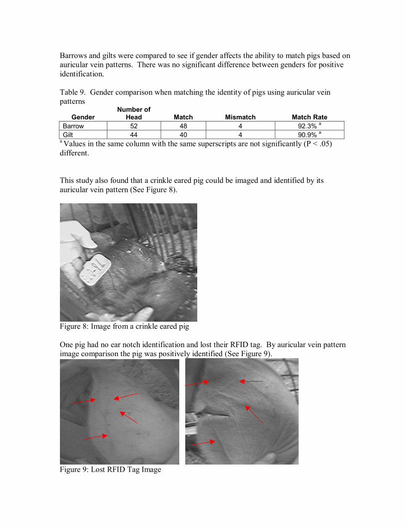
Figure 8: Image from a crinkle eared pig One pig had no ear notch identification and lost their RFID tag. By auricular vein pattern image comparison the pig was positively identified (See Figure 9).
Figure 9: Lost RFID Tag Image
Conclusion The findings from this study verify that swine can be identified by imaging auricular vein patterns and thus, we can accept the hypothesis: Auricular vein patterns can be as viable for identification of swine as the universal ear notching system. Identifying swine with ear notches and auricular vein patterns resulted in a match rate of 91.7%. The results show an 8.3% imaging error which contributed to the misidentification of eight out of ninety-six pigs. Previous mouse ear vein identification research established a 10% imaging error with the misidentification of two out twenty mice. The researchers also determined that each pig has their own distinct auricular vein pattern that does not appear to change over the course of time. Of the ninety-six swine imaged, each pig had its own auricular vein pattern consisting of one main vein, two main veins, three main veins or a branching pattern. During the time interval of 75 days, no change appeared in the auricular vein pattern location on forty-four out of fifty-eight ear notched pigs. Imaging the auricular vein patterns at the same distance from the ear to the OptiReader � Device on both sets of images, appears to make a difference in the accuracy of identifying a positive match and replicating identical measurements. Further research is needed to evaluate the uniqueness of each pigs auricular vein pattern and discovering the proper distance for imaging. No significant difference was detected when identifying barrows and gilts using auricular vein patterns. Researchers found a difference in their ability to determine matches using auricular vein patterns in hogs with white and black ear color. A higher percentage of hogs with white ears (96.7%) were able to be matched than those with black ears (87.0%). Additional research is needed for reviewing the red ear pigmentation. When comparing breeds of swine, no significant difference was determined between crossbreds, Hampshires and Yorkshires. Researchers determined that when imaging Yorkshires, Durocs or white crossbred pigs, it works best to use the Veinlite � LED in the orange LED light setting or to not use the LED lighting at all. Meanwhile, with Hampshires, Polands and black crossbreds, it works best to use the red light setting. More research is needed to evaluating the other existing breeds of swine in identifying auricular vein patterns.

Acknowledgements: Christine Blomeke Biometrics Laboratory Brittany Simmons Purdue University Keli Slack Purdue University

References: American Berkshire Association. (2006). Official Berkshire Ear-Notching System.
Membership Handbook. pg 9. http://www.americanberkshire.com/News&Information/TopPage.htm
Cameron, J., Jacobson, C., Nilsson, K., Rognvaldsson, T. (2007). Identifying Laboratory
Rodents Using Earprints. National Centre for Replacement, Refinement, and Reduction of Animals in Research (NC3Rs). Vol. 11:1-4.
Ellmauthaler, A., and Wernsperger, E. (2007). Biometric of Mice, Technical Report,
IDE0750. Masters Thesis in Computer Engineering. pg V. Ghoshal, N.G., and Zguigal, H. (1986). Dural Sinuses in the Pig and Their Extracranial
Venous Connections. American Journal of Veterinary Research. 47: 1165-1169. Huang, H.K., Nielsen, J.F., Nelson, M.D., and Liu L. (2005). Image �matching as a
Medical Diagnostic Support Tool (DST) for Brain Diseases in Children. Computerized Medical Imaging and Graphics. 29: 195-202.
Neary, M., and Yaher, A. (2002). Method of Livestock Identification. (Farm Animal
Management @ Purdue). West Lafayette, Indiana: Purdue University, Department of Animal Sciences.
Nilsson, K., Rognvaidsson, T., Cameron, J., and Jacobson, C. (2006). Biometric Identification of Mice. 18th International Conference of Pattern Recognition (ICPR�06) 4: 465-468.
Rea, J.C. (1986). Universal Ear Notching System in Swine. Agricultural Guide. Swine
Management. University of Missouri-Columbia Extension. 2505. Rusk, C.P., Blomeke, C.R., Balschweid, M.A., Elliott, S.J., Baker, D. (2006). An
Evaluation of Retinal Imaging Technology for 4-H Beef and Sheep Identification. Journal of Extension. 44(5). Article 5FEA7. http:www.joe.org/joe/2006october/a7.shtml
Solis, J.A., and Maaala, C.P. (1975). Muzzle Printing as a Method for Identification of
Cattle and Carabaos. Phillippine Journal of Veterinary Medicine, 14(1): 1-14. The National Swine Registry. (2007). Universal Ear Notching System. The Member
Resource Handbook. pg 8. http://www.nationalswine.com/formembersonly/formembersonly.html
Translite LLC, Veinlite � LED C E. Owners Manual. pg 2. www.veinlite.com

Whittier J., Shadduck J.A., and Golden, B.L. (2003). Secure Identification, Source Verification of Livestock � The Value of Retinal Images and GPS. Precision Livestock Farming. S COX (ed). Wageningen Acad. Pub. The Netherlands. 167-172.
Zezula-Szpyra, A., and Grzegorzewski, W., (2000). Morphology of the Dorsal Nasal
Frontal and Facial Veins in Adult Gilts. Via Medica. 59(3): 179-191.

Abstract Ear notching has been the gold standard of identification within the swine industry for several years. This study imaged the auricular vein patterns of 96 pigs using the OptiReader � Device and the Veinlite � LED. Eighty-eight of the 96 pigs imaged were matched for positive identification when comparing images from the initial and re-imaging scans. No significant difference was determined between crossbred, Hampshire and Yorkshire breeds of swine. There was no significant difference in the researcher�s ability to match barrows and gilts using auricular vein patterns. A high percentage of hogs with white ears (96.7%) were matched using auricular vein patterns, than those with black ears (87.0%). Each pig has a distinct auricular vein pattern that can be used for identification. In swine, imaging the auricular vein pattern can be a viable means of identification rather than utilizing ear notching alone.