identification of the bronchi for bronchoscopy in the
TRANSCRIPT
37
Identification of the Bronchi for Bronchoscopy in the Horse and
Segmentation of the Horse Lung
Ryuichi WADA*, Hiroko AIDA, Mikihiro KANEKO, Masa-aki OIKAWA, Toyohiko
YOSHIHARA, Yoshio TOMIOKA** and Masahiko NITTA**
Equine Research Institute, Japan Racing Association, 5-27-7 Tsurumaki, Setagaya-ku, Tokyo 154,
Japan
(Received 1 August 1991/Accepted 29 July 1992)
The bronchial system and pulmonary segments of 35 horse lungs were evaluated for
application of Bronchoscopy. A nomenclature system was proposed to identify individual
bronchi systematically. Corresponding to the bronchial system, the right and left lungs
were divided into 15 and 14 segments, respectively.
Key words: Bronchoscopy, horse, pulmonary segment, segmental bronchi
Jpn. J. Equine Sci. 3(1) : 37-43, 1992
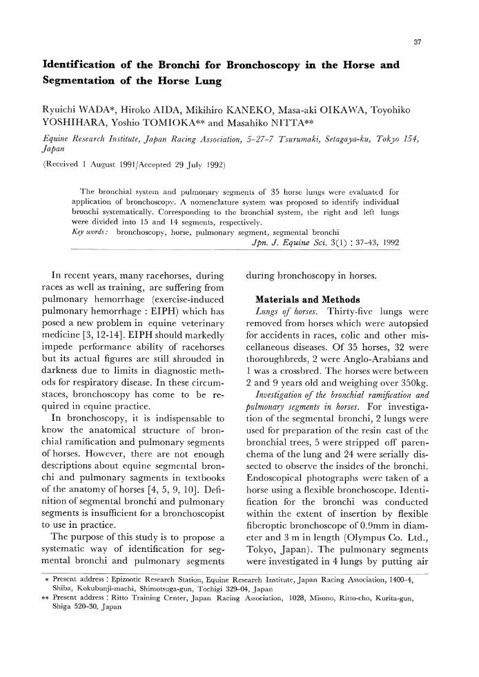
In recent years, many racehorses, during races as well as training, are suffering from
pulmonary hemorrhage (exercise-induced pulmonary hemorrhage : EIPH) which has posed a new problem in equine veterinary medicine [3, 12-14]. EIPH should markedly impede performance ability of racehorses but its actual figures are still shrouded in darkness due to limits in diagnostic methods for respiratory disease. In these circumstaces, bronchoscopy has come to be re
quired in equine practice. In bronchoscopy, it is indispensable to
know the anatomical structure of bronchial ramification and pulmonary segments of horses. However, there are not enough descriptions about equine segmental bronchi and pulmonary sagments in textbooks of the anatomy of horses [4, 5, 9, 10]. Definition of segmental bronchi and pulmonary segments is insufficient for a bronchoscopist to use in practice. The purpose of this study is to propose a
systematic way of identification for seg-mental bronchi and pulmonary segments
during bronchoscopy in horses.
Materials and Methods Lungs of horses. Thirty-five lungs were removed from horses which were autopsied for accidents in races, colic and other miscellaneous diseases. Of 35 horses, 32 were thoroughbreds, 2 were Anglo-Arabians and 1 was a crossbred. The horses were between 2 and 9 years old and weighing over 350kg. Investigation of the bronchial ramification and
pulmonary segments in horses. For investigation of the segmental bronchi, 2 lungs were used for preparation of the resin cast of the bronchial trees, 5 were stripped off parenchema of the lung and 24 were serially dissected to observe the insides of the bronchi. Endoscopical photographs were taken of a horse using a flexible bronchoscope. Identification for the bronchi was conducted within the extent of insertion by flexible fiberoptic bronchoscope of 0.9mm in diameter and 3 m in length (Olympus Co. Ltd., Tokyo, Japan). The pulmonary segments were investigated in 4 lungs by putting air
* Present address : Epizootic Research Station, Equine Researeh Institute, Japan Racing Association, 1400-4, Shiba, Kokubunji-machi, Shimotsuga-gun, Tochigi 329-04, Japan
** Present address : Ritto Training Center, Japan Racing Association, 1028, Misono, Ritto-cho, Kurita-gun, Shiba 520-30, Japan
38 R. WADA, H. AIDA, M. KANEKO ET AL.
'Table 1. Abbreviations in figures and tables
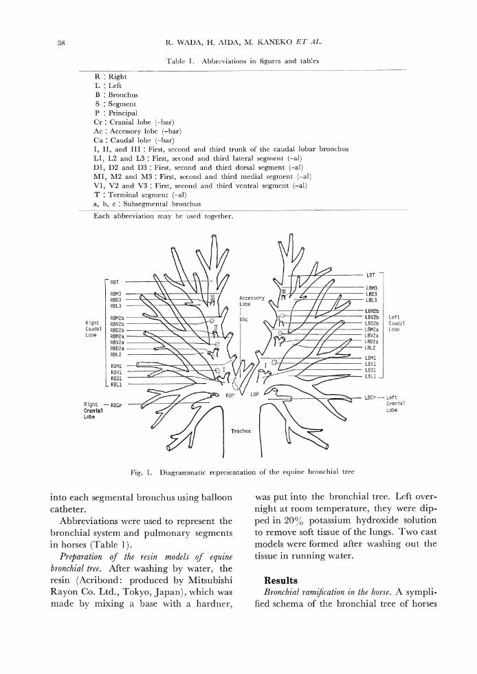
Fig, 1. Diagrammatic representation of the equine bronchial tree
into each segmental bronchus using balloon catheter.
Abbreviations were used to represent the bronchial system and pulmonary segments in horses (Table 1). Preparation of the resin models of equine
bronchial tree. After washing by water, the resin (Acribond: produced by Mitsubishi Rayon Co. Ltd., Tokyo, Japan), which was made by mixing a base with a hardner,
was put into the bronchial tree. Left over-night at room temperature, they were dip-
ped in 20% potassium hydroxide solution to remove soft tissue of the lungs. Two cast models were formed after washing out the
tissue in running water.
Results
Bronchial ram cation in the horse. A sympli-
fied schema of the bronchial tree of horses
39Segmental Bronchi and Pulmonary Segments
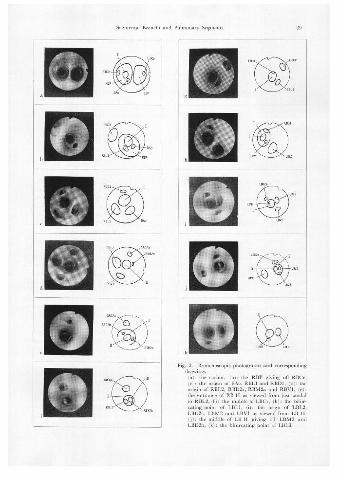
Fig. 2. Bronchoscopic photographs and corresponding
drawings
(a) : the carina, (b) : the RBP giving off RBCr, (c): the origin of BAc, RBLI and RBD1, (d): the origin of RBL2, RBD2a, RBM2a and RBV1, (e): the entrance of RB II as viewed from just caudal to RBL2, (f) : the middle of LBCr, (h) : the bifur-cating point of LBL1, (i) : the orign of LBL2, LBD2a, LBM2 and LBV1 as viewed from LB 11, (j) : the middle of LB II giving off LBM2 and LBD2b, (k): the bifurcating point of LBL3.
40 R. WADA, H. AIDA, M. KANEKO ET AL.
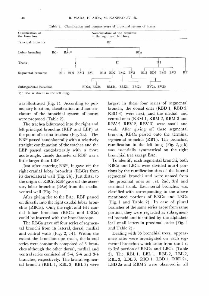
Table 2. Classification and nomenclature of bronchial system of horses
was illustrated (Fig. 1). According to pulmonary lobation, classification and nomenclature of the bronchial system of horses were proposed (Table 2).
The trachea bifurcated into the right and left principal bronchus (RBP and LBP) at the point of carina trachea (Fig. 2a). The RBP passed caudolaterally with a relatively straight continuation of the trachea and the LBP passed caudolaterally with a more acute angle. Inside diameter of RBP was a little larger than LBP.
Just after entering RBP, it gave off the right cranial lobar bronchus (RBCr) from its dorsolateral wall (Fig. 2b). Just distal to the origin of RBCr, RBP gave off the acces-sory lobar bronchus (BAc) from the media-ventral wall (Fig. 2c).
After giving rise to the BAc, RBP passed on directly into the right caudal lobar bron-chus (RBCa). Only the right and left cau-dal lobar bronchus (RBCa and LBCa) could be inserted with the bronchoscope.
The RBCa gave off four series of segmental bronchi from its lateral, dorsal, medial and ventral walls (Fig. 2, c-f). Within the extent the bronchoscope reach, the lateral series were constantly composed of 3 bran-ches although the other dorsal, medial and ventral series consisted of 3-4, 2-4 and 3-4 branches, respectively. The lateral segmental bronchi (RBL 1, RBL 2, RBL 3) were
largest in these four series of segmental bronchi, the dorsal ones (RBD 1, RBD 2, RBD 3) were next, and the medial and ventral ones (RBM 1, RBM 2, RBM 3 and RBV 2, RBV 2, RBV 3) were small and weak. After giving off these segmental bronchi, RBCa passed onto the terminal segmental bronchus (RBT). The bronchial ramification in the left lung (Fig. 2, g-k) was essentially symmetrical on the right bronchial tree except BAc.
To identify each segmental bronchi, both RBCa and LBCa were divided into 4 portions by the ramification sites of the lateral segmental bronchi and were named from the proximal ones as 1 st, 2nd, 3rd and terminal trunk. Each serial bronchus was classified with corresponding to the above mentioned portions of RBCa and LBCa (Fig. 1 and Table 2). In case of plural branches of the same series arose from same portion, they were regarded as subsegmental bronchi and identified by the alphabetical small letters in proximal order (Fig. 1 and Table 2).
Dealing with 35 bronchial trees, appearance rates were investigated on each segmental bronchus which arose from the 1 st to 3rd portion of RBCa and LBCa (Table 3). The RBL 1, LBL 1, RBL 2, LBL 2, RBL 3, LBL 3, RBD 1, LBD 1, RBD 2a, LBD 2a and RBM 2 were observed in all
41Segmental Bronchi and Pulmonary Segments
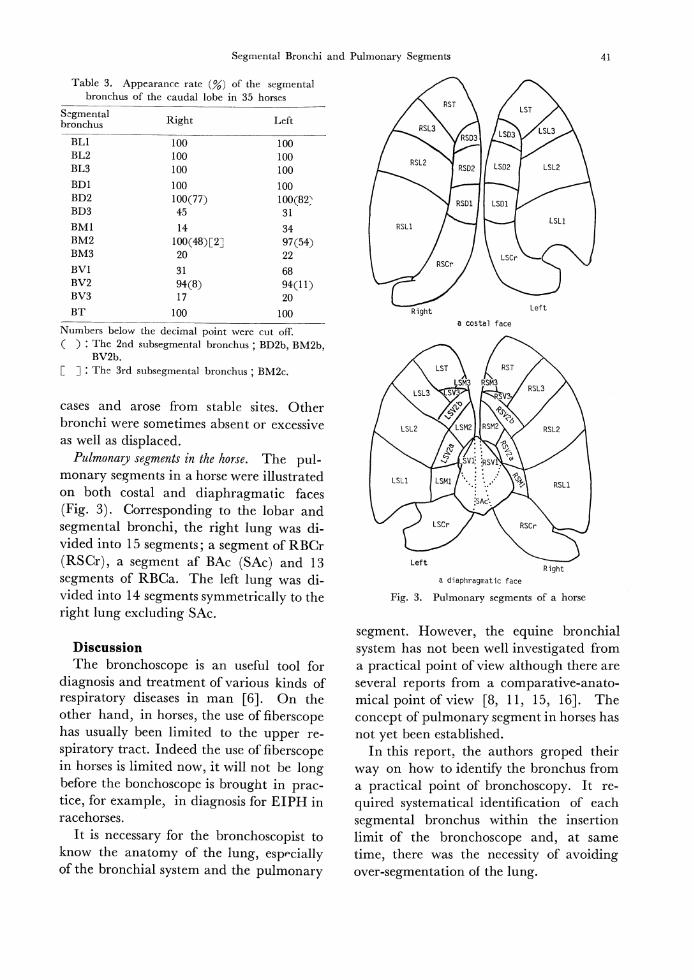
Table 3. Appearance rate (%) of the segmental bronchus of the caudal lobe in 35 horses
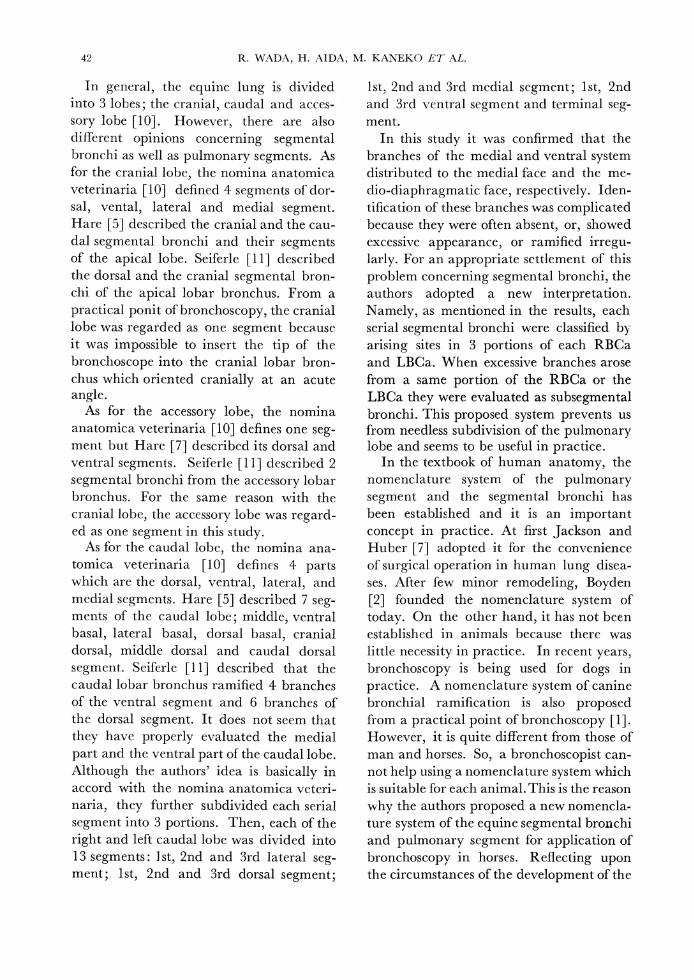
Fig. 3. Pulmonary segments of a horse
cases and arose from stable sites. Other bronchi were sometimes absent or excessive as well as displaced.
Pulmonary segments in the horse. The pul-monary segments in a horse were illustrated on both costal and diaphragmatic faces
(Fig. 3). Corresponding to the lobar and segmental bronchi, the right lung was di-vided into 15 segments ; a segment of RBCr (RSCr), a segment of BAc (SAc) and 13 segments of RBCa. The left lung was di-vided into 14 segments symmetrically to the right lung excluding SAc.
Discussion
The bronchoscope is an useful tool for diagnosis and treatment of various kinds of respiratory diseases in man [6]. On the other hand, in horses, the use of fiberscope has usually been limited to the upper re-
spiratory tract. Indeed the use of fiberscope in horses is limited now, it will not be long before the bonchoscope is brought in practice, for example, in diagnosis for EIPH in racehorses. It is necessary for the bronchoscopist to
know the anatomy of the lung, especially
of the bronchial system and the pulmonary
segment. However, the equine bronchial system has not been well investigated from a practical point of view although there are several reports from a comparative-anatomical point of view [8, 11, 15, 16]. The concept of pulmonary segment in horses has not yet been established. In this report, the authors groped their
way on how to identify the bronchus from a practical point of bronchoscopy. It re
quired systematical identification of each segmental bronchus within the insertion limit of the bronchoscope and, at same time, there was the necessity of avoiding over-segmentation of the lung.
42 R. WADA, H. AIDA, M. KANEKO ET AL.
In general, the equine lung is divided into 3 lobes; the cranial, caudal and acces-
sory lobe [10]. However, there are also different opinions concerning segmental bronchi as well as pulmonary segments. As for the cranial lobe, the nomina anatomica veterinaria [10] defined 4 segments of dor-sal, vental, lateral and medial segment. Hare [5] described the cranial and the cau-dal segmental bronchi and their segments of the apical lobe. Seiferle [11] described the dorsal and the cranial segmental bron-chi of the apical lobar bronchus. From a
practical ponit of bronchoscopy, the cranial lobe was regarded as one segment because it was impossible to insert the tip of the bronchoscope into the cranial lobar bron-chus which oriented cranially at an acute angle. As for the accessory lobe, the nomina anatomica veterinaria [10] defines one seg-ment but Hare [7] described its dorsal and ventral segments. Seiferle [11] described 2 segmental bronchi from the accessory lobar bronchus. For the same reason with the cranial lobe, the accessory lobe was regard-ed as one segment in this study.
As for the caudal lobe, the nomina ana-tomica veterinaria [10] defines 4 parts which are the dorsal, ventral, lateral, and medial segments. Hare [5] described 7 seg-ments of the caudal lobe; middle, ventral basal, lateral basal, dorsal basal, cranial dorsal, middle dorsal and caudal dorsal segment. Seiferle [11] described that the caudal lobar bronchus ramified 4 branches of the ventral segment and 6 branches of the dorsal segment. It does not seem that they have properly evaluated the medial
part and the ventral part of the caudal lobe. Although the authors' idea is basically in accord with the nomina anatomica veterinaria, they further subdivided each serial segment into 3 portions. Then, each of the right and left caudal lobe was divided into 13 segments: 1st, 2nd and 3rd lateral segment; 1st, 2nd and 3rd dorsal segment;
1st, 2nd and 3rd medial segment; 1st, 2nd and 3rd ventral segment and terminal segment. In this study it was confirmed that the branches of the medial and ventral system distributed to the medial face and the medio-diaphragmatic face, respectively. Identification of these branches was complicated because they were often absent, or, showed excessive appearance, or ramified irregularly. For an appropriate settlement of this
problem concerning segmental bronchi, the authors adopted a new interpretation. Namely, as mentioned in the results, each serial segmental bronchi were classified by arising sites in 3 portions of each RBCa and LBCa. When excessive branches arose from a same portion of the RBCa or the LBCa they were evaluated as subsegmental bronchi. This proposed system prevents us from needless subdivision of the pulmonary lobe and seems to be useful in practice. In the textbook of human anatomy, the
nomenclature system of the pulmonary segment and the segmental bronchi has been established and it is an important concept in practice. At first Jackson and Huber [7] adopted it for the convenience of surgical operation in human lung disea-
ses. After few minor remodeling, Boyden
[2] founded the nomenclature system of today. On the other hand, it has not been established in animals because there was little necessity in practice. In recent years, bronchoscopy is being used for dogs in
practice. A nomenclature system of canine bronchial ramification is also proposed from a practical point of bronchoscopy [1]. However, it is quite different from those of man and horses. So, a bronchoscopist can-not help using a nomenclature system which is suitable for each animal. This is the reason why the authors proposed a new nomencla-ture system of the equine segmental bronchi and pulmonary segment for application of bronchoscopy in horses. Reflecting upon the circumstances of the development of the
43Segmental Bronchi and Pulmonary Segments
human nomenclature system , further dis-cussion will be needed before the nomen-
clature system of equine segmental bronchi
and pulmonary segments is established.
References
1, Amic, T. C. and McKiernan B. C. (1986). Systematic identification of endobronchial anatomy
during bronchoscopy in the dog. Am. J. Vet. Res. 47: 2649-2657 .
2. Boyden, E. A. (1955). Segmental Anatomy of the Lungs. McGraw-Hill Book Co., New York .
3. Clarke, A. F. (1985). Review of exercise induced
pulmonary hemorrhage and its possible relationship with mechanical stress. Equine Vet. J. 17: 166-172.
4. Elenberger, W. and Banns, H. (1932). Hundbuch der vergleichenden Anatomie der Haustiere. 17
Aulf., Abb., ss. 733-741, julius Springer, Berlin.5. Getty, R. (1975). Sisson and Grossman's The
Anatomy of the Domestic Animals, 5 th ed., pp. 518-523, W. B. Saunders Company, Philaderphia,
London, Tront.6. Ikeda, S. (1975). Atlas of flexible bronchofiber-
scopy. pp. 58-72, University Park Press, Baltimore.7. Jackson, C. L. and Huber, J. F. (1943). Correlated
applied anatomy of the bronchial tree and lungs with a system of nomenclature. Dis. Chest. 9: 319-326
8. Nakakuki, S. (1980). Comparative anatomical studies on the mammalian lung. Bull. Fac. Agric.
Tokyo Univ. Agric. Technol. 21: 11-74.9. Nickel, R., Schummer, A., and Seiferle, E. (1973), The Viscera of the Domestic Mammals, pp. 242-
247, Verlag Paul Parey, Berlin, Hamburg.
10. Nomina Anaton ica Veterinaria japonica (2 nd
ed. of the Nomina Anatomica Veterinaria). pp.
138-139. (1981). Japanese Association of Veterinary
Anatomists, Kyoeishoji Co. Ltd., Tokyo.
11. Seiferle, E. (1956). Grundsatzliches zu Bau and
Benennung der Haussauger-Lunge. Okajimas Folia
Anat. Jpn. 28: 71-81.
12, Smith, J. D., Arthur, R., Barton, J., Bowen, E.,
Byars, D., Derksen, F.,Erikson, H., Gowen, B.,
Hinchcliff, K., Manohar, F., O•eCallaghan, M.,
Robinson, E., Sams, R., Soma, L., Sweeney, C.,
and Tobin, T. (1992). A workshop : Exercise
induced pulmonary hemorrhage findings, part 1.
Equine Practice 14(1): 19-25.
13. Smith, J. D., Arthur, R., Barton, J., Bowen, E.,
Byars, D., Derksen, F., Erikson, H., Gowen, B.,
Hinchcliff, K., Manohar, F., O'Callaghan, M.,
Robinson, E., Sams, R., Soma, L., Sweeney, C.,
and Tobin, T. (1992). A workshod : Exercise
induced pulmonary hemarrhage findings, part 2.
Equine Practice 14(2) : 9-15.
14. Smith, J. D., Arthur, R., Barton, J., Bowen, E.,
Byars, D., Derksen, F., Erikson, H., Gowen, B.,
Hinchcliff, K., Manohar, F.. O'Callaghan, M.,
Robinson, E., Sams, R., Soma, L., Sweeney, C.,
and Tobin, T. (1992). A workshop : Exercise
induced pulmonary hemorrhage findings, part 3.
Equine Practice 14(3) : 28-31.
15. Suzuki, T. and Ohkubo, M. (1977). Lobation of
the lungs of domestic animals, espesially dogs, cat
tle and horse. Jpn. J. Vet. Sci. 39: 59-67.
16, Yoshikawa, T. and Nakakuki, S. (1967). The
new lobulation of the lung. Proc. Jpn. Acad. 43:
1009-1011.
ウマの気管支鏡検査のための気管支同定法と肺区分― 和田隆一*,間 弘子,兼 子樹
広,及 川正明,吉 原豊彦,富 岡義雄**,新 田仁彦**(日 本中央競馬会競走馬総合研究所
〒154東 京都世田谷区弦巻5-27-7)
ウマの気管支鏡検査法を検討するため,35例 のウマの気管支分岐および肺区域を検索 し
た。気管支鏡はとくに肺後葉内の観察に有効であ り,後 葉気管支から派出する区域気管支
の命名法を提案した。これに基づき,ウ マの気管支鏡検査に有効な右肺15,左 肺14の 肺区
域を設定 した。
現勤務地*日 本中央競馬会競走馬総合研究所栃木支所 〒329-04栃 木県下都賀郡国分寺町柴1400-4
**日 本中央競馬会栗東 トレーニン グ ・センター 〒520-30滋 賀県栗太郡栗東町大字 御園1028