identificationofaroleforclasp2ininsulinaction s · tel.: 480-965-8365;...
TRANSCRIPT

Identification of a Role for CLASP2 in Insulin Action*□S
Received for publication, June 20, 2012, and in revised form, August 29, 2012 Published, JBC Papers in Press, September 19, 2012, DOI 10.1074/jbc.M112.394148
Paul Langlais§, James L. Dillon‡, April Mengos§, Debra P. Baluch‡, Ranna Ardebili‡, Danielle N. Miranda§, Xitao Xie§,Bradlee L. Heckmann§, Jun Liu§, and Lawrence J. Mandarino‡§1
From the ‡Center for Metabolic and Vascular Biology, Arizona State University, Tempe, Arizona 85287 and the §Mayo Clinic inArizona, Scottsdale, Arizona 85259
Background: Insulin stimulates glucose uptake in target tissues to promote blood glucose homeostasis, although themechanism is not fully understood.Results: siRNA-mediated knockdown of CLASP2 reduces insulin-stimulated glucose transport.Conclusion: CLASP2 is a new protein involved in insulin-stimulated glucose uptake.Significance: The identification of CLASP2 as a new player in insulin action provides an improved understanding of insulin-stimulated glucose uptake and GLUT4 trafficking.
Insulin stimulates the mobilization of glucose transporter 4(GLUT4) storage vesicles to the plasma membrane, resulting inan influx of glucose into target tissues such as muscle and fat.Wepresent evidence thatCLIP-associating protein 2 (CLASP2),a protein previously unassociated with insulin action, is respon-sive to insulin stimulation. Usingmass spectrometry-based pro-tein identification combined with phosphoantibody immuno-precipitation in L6 myotubes, we detected a 4.8-fold increase ofCLASP2 in the anti-phosphoserine immunoprecipitates uponinsulin stimulation. Western blotting of CLASP2 immunopre-cipitates with the phosphoantibody confirmed the finding thatCLASP2 undergoes insulin-stimulated phosphorylation, and anumber of novel phosphorylation sites were identified. Confo-cal imaging of L6 myotubes revealed that CLASP2 colocalizeswith GLUT4 at the plasma membrane within areas of insulin-mediated cortical actin remodeling. CLASP2 is responsible fordirecting the distal end of microtubules to the cell cortex, and ithas been shown thatGLUT4 travels alongmicrotubule tracks. Insupport of the concept that CLASP2 plays a role in the traffick-ing of GLUT4 at the cell periphery, CLASP2 knockdown bysiRNA in L6 myotubes interfered with insulin-stimulatedGLUT4 localization to the plasma membrane. Furthermore,siRNA mediated knockdown of CLASP2 in 3T3-L1 adipocytesinhibited insulin-stimulated glucose transport. We thereforepropose a new model for CLASP2 in insulin action, whereCLASP2 directs the delivery of GLUT4 to cell cortex landingzones important for insulin action.
Insulin binding to the cell surface insulin receptor activates ahighly coordinated series of signal transduction cascades,including the phosphatidylinositol 3-kinase (PI3K) pathway.Activation of PI3K by insulin results in a basolaterally localized
increase in phosphatidylinositol 3,4,5-trisphosphate (PIP3)2 atthe plasmamembrane (PM) (1). The insulin signal then splits toactivate the Akt and Rac1 pathways in parallel. Insulin stimula-tion of Akt releases the intracellular sequestration of GLUT4,allowing for GLUT4 cell surface translocation, PM insertion,and ultimately glucose uptake into the target cell. Insulin sig-naling to Rac1 within muscle cells induces cell periphery (cor-tical) actin reorganization and a ruffling of the cell membrane(2). TheAkt and Rac1 insulin signaling pathways then convergeat this point because Akt-responsive GLUT4 translocatesdirectly to the Rac1-induced, cortical actin-reorganized mem-brane ruffle (3). Intracellularly, GLUT4 resides within special-ized vesicles, referred to as “GLUT4 storage vesicles” or “insu-lin-responsive vesicles” (IRV) (4). The IRV have been shown totravel to the PM via the microtubule (MT) network, assisted bythe molecular motor kinesin (KIF5B) (5), although this mecha-nism is not fully understood.The microtubule-associated cytoplasmic linker protein
(CLIP)-associating protein 2 (CLASP2), belongs to the plus-endtracking protein (�TIPs) family, which are proteins that local-ize to the distal ends of MTs and function to regulate MTdynamics. CLASP2 has an established function in cell division(6, 7) and contributes to MT stabilization at leading edges ofmigrating fibroblasts (8). CLASP2 can also localize to theGolgi,acting as a microtubule organizing center for Golgi-derivedvesicle transport (9–11). CLASP2 attaches distal MT ends tothe cell cortex (9, 12), serving to direct MT-based transportfrom the Golgi to the PM. Current indirect evidence supports arole for CLASP2 in insulin action. Glycogen synthase kinase 3�(GSK3�), a kinase known to be deactivated by insulin-stimu-lated Akt, phosphorylates CLASP2, leading to disruption of theassociation of CLASP2 with MTs (13, 14). CLASP2 associateswith LL5�, a pleckstrin homology (PH) domain-containingprotein that interacts with PI3K-generated PIP3 (9, 15).CLASP2 also associates with LL5� (alternatively known as
* This work was supported, in whole or in part, by National Institutes of HealthGrant R01DK047936 (to L. J. M.).
□S This article contains supplemental Figs. 1– 4.1 To whom correspondence should be addressed: Mayo Clinic College of
Medicine, 13400 E. Shea Blvd., Scottsdale, AZ 85259. Tel.: 480-965-8365;Fax: 480-965-6899; E-mail: [email protected].
2 The abbreviations used are: PIP3, phosphatidylinositol 3,4,5-trisphosphate;ACN, acetonitrile; CLASP2, cytoplasmic linker protein (CLIP)-associatingprotein 2; FA, formic acid; GLUT4, glucose transporter 4; GSK3�, glycogensynthase kinase 3�; IRV, insulin-responsive vesicles; MS/MS, tandem massspectrometry; MT, microtubule; PM, plasma membrane.
THE JOURNAL OF BIOLOGICAL CHEMISTRY VOL. 287, NO. 46, pp. 39245–39253, November 9, 2012© 2012 by The American Society for Biochemistry and Molecular Biology, Inc. Published in the U.S.A.
NOVEMBER 9, 2012 • VOLUME 287 • NUMBER 46 JOURNAL OF BIOLOGICAL CHEMISTRY 39245
by guest on May 27, 2018
http://ww
w.jbc.org/
Dow
nloaded from
PHLDB1) (9), a protein that undergoes insulin-stimulatedtranslocation to the PM (16). Knockdown of LL5� in 3T3-L1adipocytes inhibits insulin-stimulated glucose uptake (16).We present the first direct evidence that CLASP2 responds
to insulin stimulation, through insulin-stimulated phosphory-lation and CLASP2 colocalization with GLUT4 at the corticalactin-reorganized PM ruffle. We present the finding thatsiRNA-mediated knockdownofCLASP2 in L6myotubes inhib-its insulin-stimulatedGLUT4 translocation to the PM. Further-more, siRNA-mediated knockdown of CLASP2 in 3T3-L1 adi-pocytes inhibits insulin-stimulatedglucose transport.The fact thatCLASP2, a protein known to attachMTs to the cell cortex, local-izes directly to the insulin-inducedmembrane ruffle sheds newlight onto the importance of theMTnetwork for the traffickingof GLUT4-containing vesicles at the PM.
EXPERIMENTAL PROCEDURES
Materials—The following suppliers were used: sequencing-grade trypsin, Sigma; C18 ZipTip, Millipore; phospho-MAPK/CDK substrate (PXS*P, S*PXR/K, or PXS*PXR/K) antibody,Cell Signaling Technologies; CLASP2 antibody, Novus Biolog-icals; GLUT4 antibody, Abcam; Alexa Fluor 568 phalloidin,Alexa Fluor 488 donkey anti-rabbit, and Alexa Fluor 568 don-key anti-mouse, Invitrogen.Cell Culture, Transfection, Immunoprecipitation, and West-
ern Blot Analysis—L6 rat myoblasts were maintained in�-MEM (Invitrogen) supplemented with 10% fetal bovineserum and 1% penicillin/streptomycin. Cells were subculturedby trypsinization (0.25% trypsin with ED TA) at approximately70% confluence. Myotube differentiation was induced by plac-ing 60–70% confluentmyoblasts in�-MEMcontaining 2% fetalbovine serum and 1% penicillin/streptomycin for 7–8 days.Medium for all cell cultures was replaced every 24–48 h, andcultures were maintained at 37 °C and 5% CO2. The 3T3-L1fibroblasts were grown in DMEM with 10% fetal bovine serumand differentiated into adipocytes as described (17). For celltreatment experiments, cells were starved for 4 h and theneither left untreated or stimulated with the indicated time andconcentration of insulin at 37 °C. Cells were lysed with lysisbuffer containing 50 mM HEPES, pH 7.6, 150 mM NaCl, 1%TritonX-100, 1mMNaF, 20mM sodiumpyrophosphate, 20mM
�-glycerol phosphate, 1 mM sodium orthovanadate, 10 �g/mlaprotinin, 10 �g/ml leupeptin, and 1mM phenylmethylsulfonylfluoride. Cell lysates were incubated on ice for 20 min followedby centrifugation (10,000 � g, 4 °C, 20 min), and the clarifiedsupernatants were used for immunoprecipitation. For immu-noprecipitation, cell lysates were incubated with specific anti-bodies conjugated to protein A or protein G-agarose beadsovernight at 4 °C with gentle rotation. Immunoprecipitateswere washed extensively with ice-cold PBS, and the proteinsbound to beads were eluted by heating at 95 °C for 4min in SDSsample loading buffer. Eluted proteins were separated by 10%SDS-PAGE and were either stained with Coomassie Blue ortransferred to a nitrocellulosemembrane for subsequentWest-ern blotting.In-gel Digestion—For the proteomics experiments, each lane
of the SDS-PAGE gel was cut into 15 slices, placed in a 0.6-mlpolypropylene tube, destained twice with 300 �l of 50% aceto-
nitrile (ACN) in 40 mM NH4HCO3 and dehydrated with 100%ACN for 15 min. After removal of ACN by aspiration, the gelpieces were dried in a vacuum centrifuge at 60 °C for 30 min.Trypsin (250 ng; Sigma) in 20 �l of 40 mM NH4HCO3 wasadded, and the samples were maintained at 4 °C for 15 minprior to the addition of 50 �l of 40 mM NH4HCO3 containingthree 10 fmol/�l peptides to serve as internal standards (brady-kinin fragment 2–9, B1901,�-sheet breaker peptide, S7563, andanaphylatoxinC3a fragment, A8651; Sigma). The digestionwasallowed to proceed at 37 °C overnight and was terminated byaddition of 10 �l of 5% formic acid (FA). After further incuba-tion at 37 °C for 30 min and centrifugation for 1 min, eachsupernatant was transferred to a clean polypropylene tube. Theextraction procedure was repeated using 40 �l of 0.5% FA, andthe two extracts were combined. The resulting peptide mix-tures were purified by solid phase extraction (C18 ZipTip) aftersample loading in 0.05% heptafluorobutyric acid:5% FA (v/v)and elution with 4 �l of 50% ACN:1% FA (v/v) and 4 �l of 80%ACN:1% FA (v/v), respectively. The eluates were combined anddried by vacuumcentrifugation and 10�l of 0.1%TFA/2%ACN(v/v) was added.Mass Spectrometry—HPLC-ESI-MS/MSn was performed on
a Thermo Finnigan (San Jose, CA) LTQ-FTICR fitted with aPicoViewTM nanospray source (NewObjective,Woburn,MA).On-line HPLC was performed using a Michrom BioResourcesParadigm MS4 micro two-dimensional HPLC (Alburn, CA)with a PicoFritTM column (New Objective, Woburn, MA,75-�m inner diameter, packed with ProteoPepTM II C18mate-rial, 300 Å); mobile phase, linear gradient of 2–27% ACN in0.1% FA in 65 min, a hold of 5 min at 27% ACN, followed by astep to 50% ACN, hold 5 min, and then a step to 80%; flow rate,400 nl/min.A “top 10” data-dependent MS/MS analysis was performed
(acquisition of a full scan spectrum followed by collision-in-duced dissociation mass spectra of the 10 most abundant ionsin the survey scan). The fragment mass spectra were thensearched against the human SwissProt_v 52.2 database (16,135entries) using Mascot (Matrix Science, London, UK; version2.1). The search variables that were used were: 10-ppm masstolerance for precursor ion masses and 0.5 Da for product ionmasses; digestion with trypsin; a maximum of twomissed tryp-tic cleavages; variablemodifications of oxidation ofmethionineand phosphorylation of serine, threonine, and tyrosine. Cross-correlation of Mascot search results with X! Tandem wasaccomplished with Scaffold (version Scaffold-01_06_19; Pro-teome Software, Portland, OR). Probability assessment of pep-tide assignments and protein identifications were madethrough the use of Scaffold. Only peptides with �95% proba-bility were considered. Assignments of the phosphopeptideswere confirmed by manual comparison of the MS/MS with thepredicted fragmentation generated in silico by theMS-Productcomponent of Protein Prospector, in addition toAscore assign-ment (18) using Scaffold PTM.Immunofluorescence and Confocal Microscopy—Myotubes
were fixed with 2% paraformaldehyde, permeabilized with 2%Tween 20, and either left untreated or incubated with antibod-ies to CLASP2 and GLUT4 overnight with gentle rocking at4 °C. After rinsing, the samples were incubated with either
CLASP2 in Insulin Action
39246 JOURNAL OF BIOLOGICAL CHEMISTRY VOLUME 287 • NUMBER 46 • NOVEMBER 9, 2012
by guest on May 27, 2018
http://ww
w.jbc.org/
Dow
nloaded from

Alexa Fluor 568 phalloidin for actin and/or secondary antibod-ies Alexa Fluor 488 or Alexa Fluor 568 for 2 h at room temper-ature and then subjected to a final series of rinsing. Imagingwasconducted using a Leica SP5 Multi-photon Scanning LaserMicroscope housed in the W. M. Keck Bioimaging Laboratoryat Arizona State University. Multiple lasers (argon, 488 nm;krypton, 561 nm; and He/Ne, 633 nm) allowed for sequentialimaging including collection of differential interference con-trast images of the sample. Settings for the gain and offset of thedetectors were kept constant for all experiments. Using a 63�oil objective, images were scanned at 0.7-�M intervals in the zaxis. The postacquisition image processing was performedusing the Leica SP5 software (Leica Microsystems Inc., Ban-nockburn, IL) andAdobe Photoshop software (MountainView,CA). For the quantification of the percentage of insulin-in-duced PM ruffles that contain GLUT4, cells were stimulatedwith insulin, and eight images of either negative control siRNAor CLASP2 siRNA-treated cells were obtained. Percentages ofthe number of ridges that contain GLUT relative to the totalnumber of ridges within each image were calculated, and thetwo groups were statistically analyzed. A ridge was defined aslacking GLUT4 if there was a complete absence of GLUT4immunofluorescence throughout the entire length of the ridge.For the -fold change of GLUT4 fluorescence per ridge calcula-tions, cells were stimulated with insulin, and eight images ofeither negative control siRNA or CLASP2 siRNA-treated cellswere obtained and “screen” overlaid within Adobe Photoshop,followed by analysis withNIH ImageJ software.Within the neg-ative control image, the immediate area surrounding ridgesthat contain both CLASP2 and GLUT4 were highlighted usingthe Freehand Selections tool, followed by Measuring RGBunder the PlugIns/Analyze tab. Four small background boxesnear the ridge, but not overlapping, were also measured forRGB and will be highlighted using the Rectangle tool. Uponmeasuring the RGB, values for the mean density per pixel(Mean) and integrated density (IntDen) were calculated anddisplayed. The average of the background value was then sub-tracted from the value for the area surrounding the ridge,resulting in Corrected Integrated Density. GLUT4 immunoflu-orescence was then normalized by the average value of the neg-ative control sample and then expressed as a -fold change overthe negative control � S.E.Fluorescence Resonance Energy Transfer (FRET)—L6 myo-
tubes were labeled with a combination of GLUT4 and CLASP2antibodies followed by secondary antibodies Alexa Fluor 488 or568. The myotubes were imaged on the Leica SP5 confocalmicroscope housed in the W. M. Keck Bioimaging facility atArizona State University. Multiple lasers allowed for simulta-neous imaging of Alexa Fluor 488 (argon (488 nm)) and AlexaFluor 568 (krypton (568 nm)) fluorophore-labeled myotubes.The Leica SP5 FRET Analysis software was used to determinethe energy efficiency by acceptor photo-bleaching (19). FRETacceptor photobleaching was conducted by double labeling themyotubeswith antibodies toCLASP2 andGLUT4.The second-ary antibody for GLUT4 was Alexa Fluor 568 and served as theacceptor fluorophore. The second antibody for CLASP2 wasAlexa Fluor488 and served as the donor fluorophore.Myotubeswere photobleached in the area of themembrane ruffle with the
Kr (568 nm) laser. Photobleaching prevents energy transfer tothe acceptor fluorophore which results in an increase in donorfluorophore emission.siRNA—Rat CLASP2 smart pool siRNA was purchased from
Dharmacon Research, and the targeting sequences were: AGACAT ACG GAC TAA GAA A; GCA CGA TCC TGA CGAGAG A; GGA AAT GCC AAG ACC GAT T; and AGA AAGCAG TGT CCG GAA A. Myotubes were transfected with 15nM siRNAon day 5 of differentiation using Lipofectamine 2000,according to the manufacturer’s protocol. 72 h after transfec-tion the cells were prepared for experimentation. For the3T3-L1 adipocytes, mouse CLASP2 stealth RNAi duplexeswere purchased from Invitrogen, and the targeting sequenceswere: GGA UGU GCA GCA AUC AGA UUU AUU U; CCCUAAGGUUGGAGGUCCUUCUAAA; andCCCAUGUACCUA GAC UUA UUC CUU U. Electroporation of either thenegative control siRNA duplex or the pool of 3 CLASP2 siRNAduplexes into 3T3-L1 adipocyte was performed as described (20).3–0-[Methyl-D-1-3H]Glucose Uptake—Differentiated 3T3-
L1 adipocytes seeded in 6-well plates were preincubated for 4 hin Krebs-Henseleit buffer (1.2 mM CaCl2, 4.4 mM KCl, 1.2 mM
MgSO4, 1.5 mMKH2PO4, 116mMNaCl, 29 mMNaHCO3) con-taining 4% bovine serum albumin and 0.5mM cold D-glucose. Inthe final 15 min of incubation, appropriate wells were treatedwith 0.2–20 nM insulin. The buffer was then replacedwith freshprewarmed Krebs-Henseleit buffer containing 0.4–1.0 �Ci/ml3–0-[methyl-D-1-3H]glucose and 0.4–1.0�Ci/ml L-[1-14C]glu-cose, andwere incubated for 10 s at room temperature. Glucoseuptakewas stopped by the addition of 0.2mM ice-cold phloretinin PBS. The cells were then washed with ice-cold phloretin inPBS and lysed with 1% SDS, and lysates were cleared by centrif-ugation. The radioactivity associated with the cells was thendetermined by liquid scintillation of aliquots of each sample(800�l). The uptakemeasurement was performed in duplicate.The protein concentration of each sample was determined bythe Lowrymethod, and results were normalized bymicrogramsof total protein.
RESULTS
CLASP2 Undergoes Insulin-stimulated Phosphorylation inRat L6Myotubes—The rationale at the beginning of this projectwas that the understanding of the signaling between the insulinreceptor and GLUT4 is incomplete. To address this, we per-formedmass spectrometry-driven proteomic profiling of phos-phoproteins from either serum-starved or insulin-treated ratL6 myotubes isolated with an antibody that targets phospho-serine in a PXS*P, S*PXR/K, or PXS*PXR/K motif (Fig. 1A). Ofthe 406 proteins identified, CLASP2 underwent the strongestinsulin-stimulated increase in abundance among proteins inthe phosphoserine antibody immunoprecipitates (4.8-fold, **p � 0.01) (Table 1 and Fig. 1B). Subsequent immunoprecipita-tion of CLASP2 from either serum-starved or insulin-treatedmyotubes and Western blotting of the immunoprecipitateswith the phosphoserine antibody confirmed the finding thatCLASP2 undergoes insulin-stimulated phosphorylation (Fig.1C). Western blotting of CLASP2 within L6myotubes revealeda doublet, which may represent different isoforms of the pro-tein (8). Mass spectrometry analysis of CLASP2 immunopre-
CLASP2 in Insulin Action
NOVEMBER 9, 2012 • VOLUME 287 • NUMBER 46 JOURNAL OF BIOLOGICAL CHEMISTRY 39247
by guest on May 27, 2018
http://ww
w.jbc.org/
Dow
nloaded from

cipitates from L6 myotube lysates resulted in 68% CLASP2sequence coverage (data not shown) and the detection of 15CLASP2 phosphorylation sites (Table 1). Calculation of Ascorevalues (18) to measure the probability of correct phospho-S/T/Y assignment validated Ser-319, Ser-322, Ser-1005, and Ser-1181 as novel, previously unidentified CLASP2 phosphoryla-tion sites (Table 2). Phosphorylation at Ser-467, Ser-469, Thr-475, Thr-477, Ser-570, and Ser-947 was also detected, althoughdue to low Ascore values, these sites cannot be assignedunequivocally.
To identify the signal transduction pathway(s) responsiblefor insulin-stimulated phosphorylation of CLASP2 within thePXS*P, S*PXR/K, or PXS*PXR/K motifs, L6 myotubes wereserum-starved and either left untreated or pretreated with 50nMwortmannin or 50�MPD98059 for 1 h to inhibit the insulin-stimulated activation of the PI3K andMAPK pathways, respec-tively. The cells were then either left untreated for basal analysisor treated with insulin. Whole cell lysates were probed for thephosphorylation of either AKT or MAPK to ensure efficientinsulin stimulation and either wortmannin or PD98059 inhibi-
FIGURE 1. CLASP2 undergoes insulin-stimulated phosphorylation. A, L6 myotubes were serum-starved and either left untreated or treated with 10 nM
insulin for 15 min. The myotubes were lysed, and the whole cell lysates were immunoprecipitated with phosphoserine antibody. The immunoprecipitates wereresolved by 10% SDS-PAGE and transferred to nitrocellulose membranes. The membranes containing the immunoprecipitated proteins were subjected toWestern blotting for phosphoserine in a PXS*P, S*PXR/K, or PXS*PXR/K motif. B, L6 myotubes were serum-starved and either left untreated or treated with 10nM insulin for 15 min. The myotubes were lysed, and the whole cell lysates were immunoprecipitated with phosphoserine antibody. The immunoprecipitateswere resolved by 10% SDS-PAGE followed by Coomassie staining. Each lane within the SDS-PAGE gel was divided into 15 slices, ranging the span of 250 to 15kDa, and the proteins were prepared and analyzed as described under “Experimental Procedures.” The data were then expressed as a -fold change over basal �S.E. (error bars; n � 3; **, p � 0.01; Student’s t test). C, L6 myotubes were serum-starved and either left untreated or treated with 10 nM insulin for 15 min. Themyotubes were lysed, and the whole cell lysates (WCL) were immunoprecipitated (IP) with CLASP2 antibody. The immunoprecipitates were resolved by 10%SDS-PAGE and transferred to nitrocellulose membranes. The membranes containing the immunoprecipitated CLASP2 were subjected to Western blotting forphosphoserine in a PXS*P, S*PXR/K, or PXS*PXR/K motif (upper panel) or with anti-CLASP2 antibodies (lower panel). D, L6 myotubes were serum-starved andeither left untreated, treated with insulin alone, or pretreated with 50 nM wortmannin (labeled W) or 50 �M PD98059 (labeled PD) for 1 h and then either leftuntreated or treated with 10�7
M insulin for 15 min. The myotubes were lysed, and the whole cell lysates were immunoprecipitated with CLASP2 antibody. Theimmunoprecipitates were resolved by 10% SDS-PAGE and transferred to nitrocellulose membranes. The membranes containing the immunoprecipitatedCLASP2 were subjected to Western blotting for phosphoserine in a PXS*P, S*PXR/K, or PXS*PXR/K motif (top panel) or with anti-CLASP2 antibodies (second panelfrom the top).Whole cell lysates were probed for the phosphorylation of either AKT (fifth panel from the top) or MAPK (third panel from the top), or the expressionof AKT (bottom panel) or MAPK (fourth panel from the top).
TABLE 1Identification of CLASP2 as an insulin-stimulated phosphoprotein
PhosphoproteinaExpt. 1 Expt. 2 Expt. 3
Basal Insulin Basal Insulin Basal Insulin
CLASP2 SC 8 56 6 46 14 35No. of IDs 4682 2842 5276 3463 1801 1965NormSC 1.71E-03 1.97E-02 1.14E-03 1.33E-02 7.77E-03 1.78E-02
a SC, spectrum counts; No. of IDs, number of assigned spectra; NormSC, normalized spectrum counts.
CLASP2 in Insulin Action
39248 JOURNAL OF BIOLOGICAL CHEMISTRY VOLUME 287 • NUMBER 46 • NOVEMBER 9, 2012
by guest on May 27, 2018
http://ww
w.jbc.org/
Dow
nloaded from

tion of the PI3K and MAPK pathways, respectively (Fig. 1D).The insulin-stimulated phosphorylation of CLASP2 within theproline-directed motifs was suppressed upon inhibition of theMAPK pathway and was not affected by inhibition of the PI3Kpathway.CLASP2 Colocalizes with GLUT4 at the Insulin-stimulated
Membrane Ruffle in Rat L6Myotubes—Because CLASP2 local-izes to MTs, particularly at the cell cortex (9), and GLUT4transport has been linked to traveling theMTnetwork (21–23),we reasoned that CLASP2 cellular localization in L6 myotubeswould provide insight regarding the possibility that CLASP2 isinvolved in GLUT4 trafficking. Cortical actin remodeling isknown to serve as a focal point for insulin signaling and insulin-stimulated GLUT4 localization (3), so we chose to use confocalmicroscopy to test whether CLASP2 colocalizes with areas of
insulin-stimulated cortical actin remodeling in L6 myotubes.Confocal imaging using rhodamine phalloidin allowed readyobservation of cortical actin remodeling, similar to previousreports (3, 24) (Fig. 2, top right panel, and supplemental Fig. 1).We then took advantage of the newly advanced differentialimage contrasting mode of the confocal microscope to focusonly on the cell surface z-plane corresponding to dorsal exten-sion of the PM (membrane ruffle) (24) proximal to the corticalactin remodeling. We were able to capture extremely sharpimages of insulin-induced membrane ruffling (Fig. 2, top leftpanel), whereas neither cortical actin remodeling nor mem-brane ruffling was observed in the serum-starved myotubes.Once an area of both cortical actin remodeling and cell surfacemembrane ruffling was established, we then tested CLASP2colocalization and discovered CLASP2 along the membrane
FIGURE 2. CLASP2 localizes to insulin-stimulated membrane ruffles. L6 myotubes were serum-starved and treated with 10 nM insulin for 15 min. Confocalimaging was performed as described under “Experimental Procedures.” DIC, differential interference contrast.
TABLE 2CLASP2 phosphorylation sites
Start Stop Peptide sequencea Phosphorylation site Ascorec
316 328 SsGsVAsLPQSDR Ser-317b 14.04Ser-319b 26.31Ser-322b 34.3
316 344 SSGSVASLPQSDRSSSSsQESLNRPFSSK Ser-333 18.53329 344 SSSSsQESLNRPFSSK Ser-333 10.11374 387 sRsDIDVNAAAGAK Ser-374 9.38
Ser-376 29.27467 488 sGsPGRVLtTtALSTVSSGAQR Ser-467b 0
Ser-469b 5.42Thr-475b 0.21Thr-477b 0.32
567 575 LSSsVSAmR Ser-570b 0935 957 GVTEAIQNFSFRsQEDmSEPLKR Ser-947b 5.3994 1007 ASLLHSVPLHSsPR Ser-1005b 27.961008 1024 SRDYNPYNYSDSIsPFNK Ser-1021 25.71174 1192 VLCPIIQtADYPINLAAIK Thr-1181b 28.73
a Lower case s/t represents phospho-site. Lower case m represents methionine oxidation site.b Novel, previously unreported phosphorylation site.c Ascore �20 signifies 99% certainty, 15–19 equals �90%, 3–15 is near 80% yet nonsufficient, and �3 lacks adequate information to make an assignment.
CLASP2 in Insulin Action
NOVEMBER 9, 2012 • VOLUME 287 • NUMBER 46 JOURNAL OF BIOLOGICAL CHEMISTRY 39249
by guest on May 27, 2018
http://ww
w.jbc.org/
Dow
nloaded from
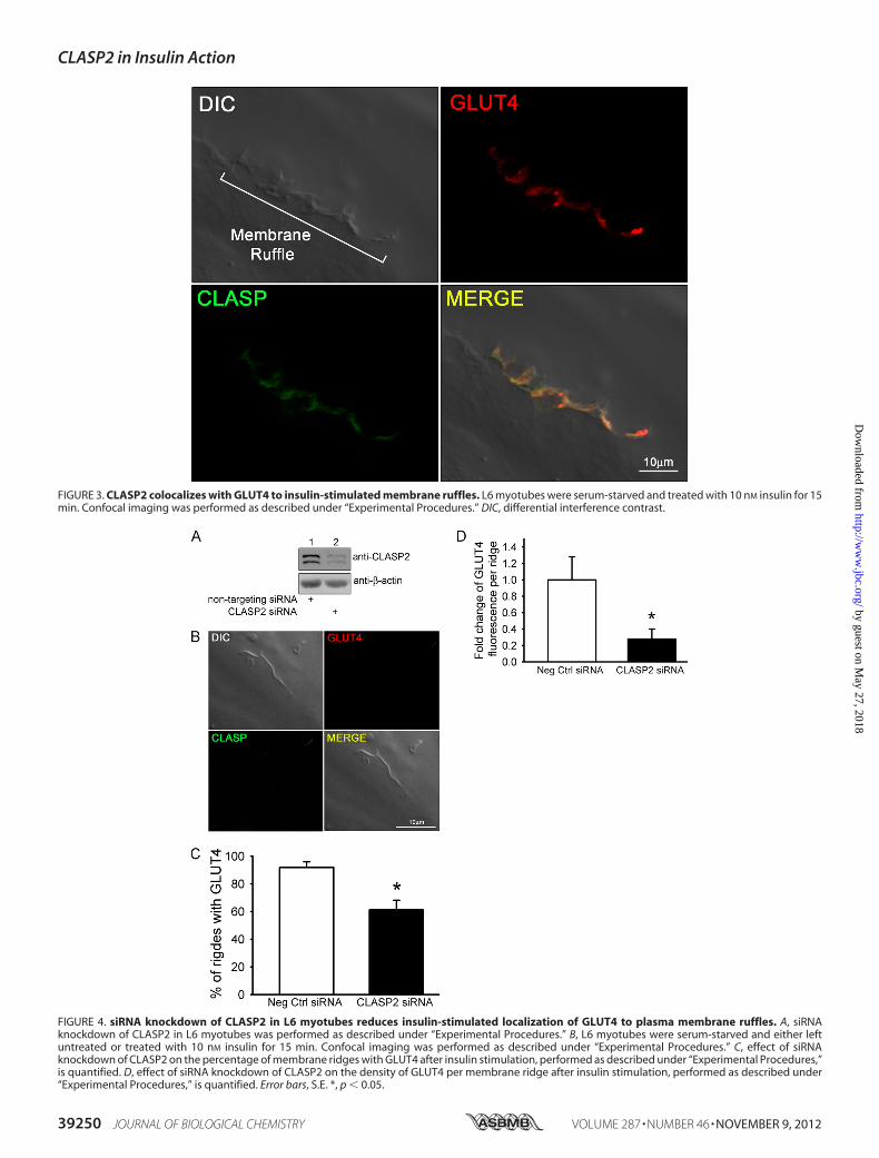
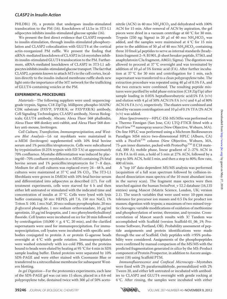
FIGURE 3. CLASP2 colocalizes with GLUT4 to insulin-stimulated membrane ruffles. L6 myotubes were serum-starved and treated with 10 nM insulin for 15min. Confocal imaging was performed as described under “Experimental Procedures.” DIC, differential interference contrast.
FIGURE 4. siRNA knockdown of CLASP2 in L6 myotubes reduces insulin-stimulated localization of GLUT4 to plasma membrane ruffles. A, siRNAknockdown of CLASP2 in L6 myotubes was performed as described under “Experimental Procedures.” B, L6 myotubes were serum-starved and either leftuntreated or treated with 10 nM insulin for 15 min. Confocal imaging was performed as described under “Experimental Procedures.” C, effect of siRNAknockdown of CLASP2 on the percentage of membrane ridges with GLUT4 after insulin stimulation, performed as described under “Experimental Procedures,”is quantified. D, effect of siRNA knockdown of CLASP2 on the density of GLUT4 per membrane ridge after insulin stimulation, performed as described under“Experimental Procedures,” is quantified. Error bars, S.E. *, p � 0.05.
CLASP2 in Insulin Action
39250 JOURNAL OF BIOLOGICAL CHEMISTRY VOLUME 287 • NUMBER 46 • NOVEMBER 9, 2012
by guest on May 27, 2018
http://ww
w.jbc.org/
Dow
nloaded from
ridge, corresponding to the zone of insulin-induced actin rear-rangement (Fig. 2, bottom two panels). We were not able toobtain FRET acceptor photobleaching between actin andCLASP2 (data not shown), suggesting that although these twoproteins both localize to the same membrane ruffle, they are�100 Å apart and are most likely not directly interacting at thez-plane tested. We then tested for colocalization of CLASP2with GLUT4 along the insulin-induced PM ruffle. Our strategywas to pinpoint a membrane ridge on the cell surface of aninsulin-stimulated myotube using the confocal differentialinterference contrast mode (Fig. 3, top left panel), followed byconfirmation ofGLUT4 localizationwithin the ridge (Fig. 3, topright panel). Using this approach, we discovered that CLASP2colocalizes with GLUT4 to the cortical actin reorganizedmem-brane ruffle (Fig. 3, bottom two panels, and supplemental Figs. 2and 3). This colocalization was confirmed by FRET acceptorphotobleaching which demonstrated up to a 19% FRET effi-ciency between CLASP2 and GLUT4 at the crest of the mem-brane ruffle (supplemental Fig. 4). These findings are strength-ened by the fact that the data are all from endogenous proteins,canceling out the possibility of artifactual results due to proteinoverexpression issues.CLASP2 Knockdown Reduces Insulin-stimulated GLUT4
Localization to the Cortical Actin-induced Plasma MembraneRuffle in Rat L6 Myotubes—The fact that CLASP2, a proteinknown to attach MTs to the cell cortex (8), localizes directly tothe insulin-induced membrane ruffle supports a role for theMT network in the trafficking of GLUT4-containing vesicles atthe membrane. Therefore, we chose to perform siRNA-mediated knockdown of CLASP2 protein in L6 myotubes totest the hypothesis that CLASP2 function is crucial for properinsulin-stimulated GLUT4 localization to the cortical actininduced PM ruffle. To suppress CLASP2 expression in L6myo-tubes, the cellswere treatedwith either 15 nMCLASP2-targetedsiRNA or nontargeting siRNA for 72 h. We first confirmedsiRNA-mediated knockdown of CLASP2 protein by Westernblotting (Fig. 4A). Analysis of CLASP2 mRNA by quantitativeRT-PCR revealed a 50% reduction of CLASP2 at the mRNAlevel (data not shown). We again focused our attention onmembrane ruffles and searched for ridges that lacked CLASP2immunofluorescence, as a result of siRNA-mediated knock-down of CLASP2 protein. Once a CLASP2-lacking membraneruffle was found, we then discovered that ruffles that possessedreduced CLASP2 protein levels also had a decrease in theamount of insulin-stimulated GLUT4 (Fig. 4B). We quantifiedthis phenomenon in two different ways: first, we compared thepercentage of ridges observed that possessedGLUT4 in controlversus CLASP2 siRNA myotubes and found that there was asignificant reduction in the number of ridges that possessedGLUT4 upon CLASP2 siRNA treatment (Fig. 4C); and second,we quantified GLUT4 fluorescence intensity within the ridgesthat did contain GLUT4 and found that CLASP2 siRNA alsosignificantly reduced the amount of GLUT4 per ridge (Fig. 4D).CLASP2 Knockdown Reduces Insulin-stimulated Glucose
Transport inMouse 3T3-L1 Adipocytes—To prove that CLASP2is vital for proper insulin action, we chose to study 3T3-L1adipocytes because this cell model possesses an excellentcapacity for insulin-stimulated glucose transport. We success-
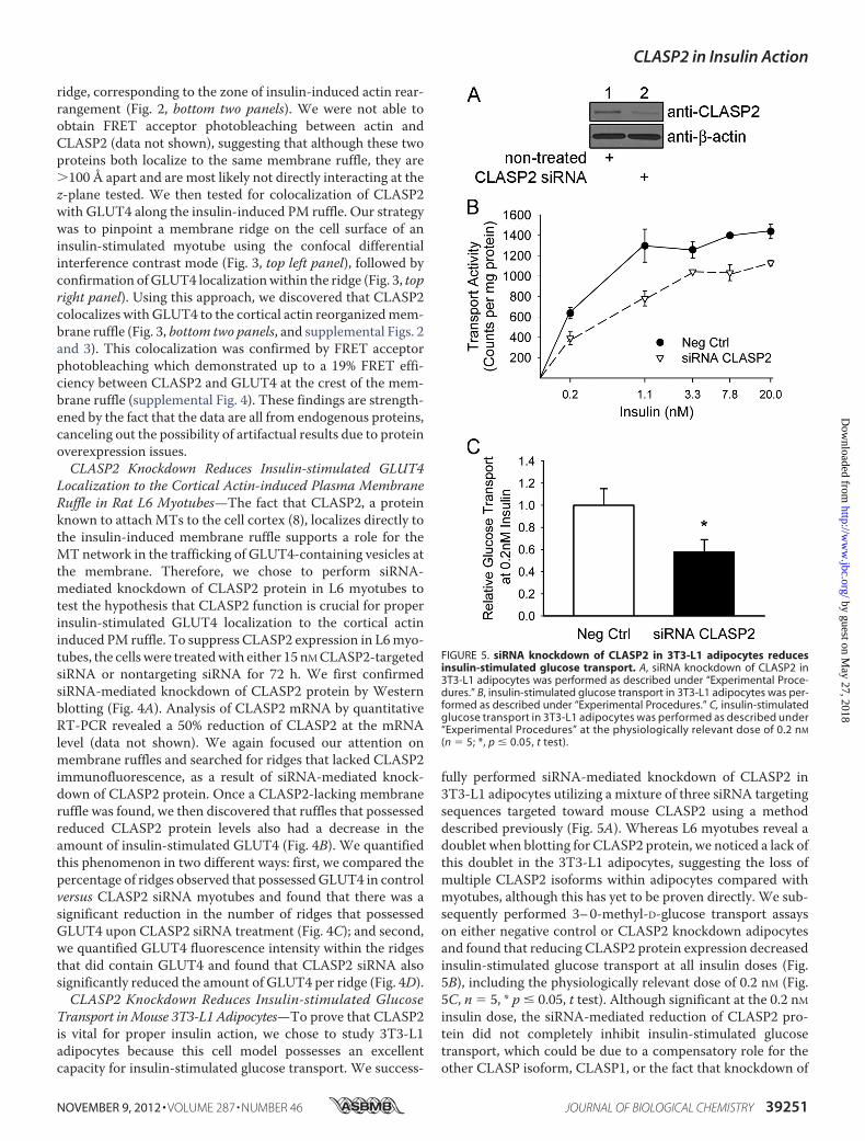
fully performed siRNA-mediated knockdown of CLASP2 in3T3-L1 adipocytes utilizing a mixture of three siRNA targetingsequences targeted toward mouse CLASP2 using a methoddescribed previously (Fig. 5A). Whereas L6 myotubes reveal adoublet when blotting for CLASP2 protein, we noticed a lack ofthis doublet in the 3T3-L1 adipocytes, suggesting the loss ofmultiple CLASP2 isoforms within adipocytes compared withmyotubes, although this has yet to be proven directly. We sub-sequently performed 3–0-methyl-D-glucose transport assayson either negative control or CLASP2 knockdown adipocytesand found that reducing CLASP2 protein expression decreasedinsulin-stimulated glucose transport at all insulin doses (Fig.5B), including the physiologically relevant dose of 0.2 nM (Fig.5C, n � 5, * p � 0.05, t test). Although significant at the 0.2 nMinsulin dose, the siRNA-mediated reduction of CLASP2 pro-tein did not completely inhibit insulin-stimulated glucosetransport, which could be due to a compensatory role for theother CLASP isoform, CLASP1, or the fact that knockdown of
FIGURE 5. siRNA knockdown of CLASP2 in 3T3-L1 adipocytes reducesinsulin-stimulated glucose transport. A, siRNA knockdown of CLASP2 in3T3-L1 adipocytes was performed as described under “Experimental Proce-dures.” B, insulin-stimulated glucose transport in 3T3-L1 adipocytes was per-formed as described under “Experimental Procedures.” C, insulin-stimulatedglucose transport in 3T3-L1 adipocytes was performed as described under“Experimental Procedures” at the physiologically relevant dose of 0.2 nM
(n � 5; *, p � 0.05, t test).
CLASP2 in Insulin Action
NOVEMBER 9, 2012 • VOLUME 287 • NUMBER 46 JOURNAL OF BIOLOGICAL CHEMISTRY 39251
by guest on May 27, 2018
http://ww
w.jbc.org/
Dow
nloaded from
CLASP2 was incomplete, and many cells were likely notaffected by siRNA treatment. In summary, the data as a wholesupport an important role for CLASP2 in insulin-stimulatedglucose transport.
DISCUSSION
Wepresent findings that themicrotubule-associated proteinCLASP2plays a central part inGLUT4 trafficking.We show thefirst direct and known evidence that CLASP2 responds toinsulin stimulation, through insulin-stimulated phosphory-lation. The phosphorylation was detected with a phosphoanti-body that targets phosphoserine in a PXS*P, S*PXR/K, orPXS*PXR/Kmotif, and this insulin-stimulated phosphorylationwas blocked by inhibition of the MAPK pathway. This findingdoes not imply that all phosphorylation of CLASP2 is con-trolled by the MAPK pathway, raising the possibility thatCLASP2 may undergo PI3K-controlled phosphorylation inresponse to insulin as well. In support of this hypothesis, it hasbeen shown that GSK3�, a kinase known to be deactivated byinsulin-stimulated Akt (25, 26), phosphorylates CLASP2, lead-ing to disruption of the association of CLASP2 with MTs (13).In this case, insulin would suppress GSK3� phosphorylationsites within CLASP2. In fact, many of the phosphorylation siteswe detected were not in classical MAPK phosphorylationmotifs, and current studies are under way to explore whetherinsulin-induced phosphorylation of CLASP2 regulates its func-tion with respect to GLUT4 translocation.We also present the finding that insulin stimulates CLASP2
colocalization with GLUT4 at the cortical actin reorganizedmembrane ruffle. The fact that CLASP2, a protein known toattach MTs to the cell cortex, localizes directly to the insulin-induced membrane ruffle sheds new light onto the possibleinvolvement MT network for the delivery of GLUT4-contain-ing vesicles to the membrane. Based on our findings, we havecreated a working model for MT-based delivery of GLUT4 to
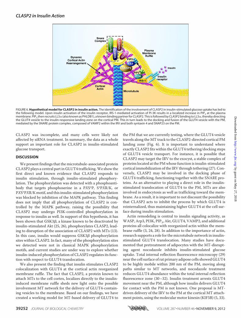
the PM that we are currently testing, where the GLUT4 vesicletravels along theMT track to the CLASP2-directed cortical PMlanding zone (Fig. 6). It is important to understand whereexactly CLASP2 fits within the GLUT4 tethering/docking stepsof GLUT4 vesicle transport. For instance, it is possible thatCLASP2 may target the IRV to the exocyst, a stable complex ofproteins located at the PMwhose function is insulin-stimulatedcortical immobilization of the IRV through tethering (27). Con-versely, CLASP2 may be involved in the docking phase ofGLUT4 trafficking, functioning together with the SNARE pro-teins. As an alternative to playing a direct role in the insulin-stimulated translocation of GLUT4 to the PM, MTs are alsoinvolved in endocytosis as well as trafficking toward the mem-brane. As a result, it is important to not discount the possibilitythat CLASP2 acts to inhibit the process by which GLUT4 isreinternalized, thus maintaining higher GLUT4 at the cell sur-face during insulin stimulation.Actin remodeling is central to insulin signaling activity, as
WASP, Arp3, PI3K, PIP3, Akt, GLUT4, VAMP2, and additionalproteins all colocalize with reorganized actin within the mem-brane ruffle (3, 24, 28). In addition to the importance of actin,research supports a role for themicrotubule network in insulin-stimulated GLUT4 translocation. Many studies have docu-mented that pretreatment of adipocytes with the MT-disrupt-ing agent nocodazole inhibits insulin-stimulated glucoseuptake. Total internal reflection fluorescence microscopy (29)near the cell surface of rat primary adipose cells showedGLUT4to be highly mobile within 200 nm of the PM, moving alongpaths similar to MT networks, and nocodazole treatmentreduces GLUT4 abundance within the total internal reflectionfluorescence zone (30–32). Insulin treatment arrests GLUT4movement near the PM, although how insulin delivers GLUT4for contact with the PM is not known. One proposal is MT-driven delivery of the IRV to the PM at the cortical MT attach-ment points, using themolecularmotor kinesin (KIF5B) (5, 33).
FIGURE 6. Hypothetical model for CLASP2 in insulin action. The identification of the involvement of CLASP2 in insulin-stimulated glucose uptake has led tothe following model. Upon insulin activation of the insulin receptor, IRS-1-mediated activation of PI-3K results in a localized increase in PIP3 at the plasmamembrane. PIP3 then recruits LL5� (also known as PHLDB1), a known binding partner for CLASP2. This is followed by CLASP2 binding to LL5�, thereby directingthe GLUT4 vesicle to the insulin-responsive landing zone on the cortical PM. This in turn leads to the docking and fusion of the GLUT4 vesicle with the PM,mediated by the SNARE protein complex, composed of VAMP2 within the IRV and both syntaxin 4 and SNAP23 on the PM.
CLASP2 in Insulin Action
39252 JOURNAL OF BIOLOGICAL CHEMISTRY VOLUME 287 • NUMBER 46 • NOVEMBER 9, 2012
by guest on May 27, 2018
http://ww
w.jbc.org/
Dow
nloaded from
Another possibility is that insulin-stimulated association ofGLUT4 with �-actinin 4 (ACTN4) transfers the IRV from theMT network over to cortical actin, where the IRV then travelsto the PM along actin using type I or V myosin (Myo1c andMyoVa and Vb, respectively) or nonmuscle myosin II (NM-MyoII) motors. It is important to note that there is no evidencefor the switching of the IRV fromMTs to cortical actin. Becausethe transport of GLUT4 to the PM is so important for insulin-stimulated glucose uptake, it is imperative to define the role ofactin and MT cytoskeletal elements in the insulin-controlleddelivery of GLUT4 to the muscle cell surface and the proteinsthat regulate this critical process.
REFERENCES1. Stöckli, J., Fazakerley, D. J., and James, D. E. (2011) GLUT4 exocytosis.
J. Cell Sci. 124, 4147–41592. Raftopoulou,M., andHall, A. (2004) Cellmigration: RhoGTPases lead the
way. Dev. Biol. 265, 23–323. Khayat, Z. A., Tong, P., Yaworsky, K., Bloch, R. J., and Klip, A. (2000)
Insulin-induced actin filament remodeling colocalizes actin with phos-phatidylinositol 3-kinase and GLUT4 in L6 myotubes. J. Cell Sci. 113,279–290
4. Rowland, A. F., Fazakerley, D. J., and James, D. E. (2011) Mapping insulin/GLUT4 circuitry. Traffic 12, 672–681
5. Semiz, S., Park, J. G., Nicoloro, S. M., Furcinitti, P., Zhang, C., Chawla, A.,Leszyk, J., and Czech, M. P. (2003) Conventional kinesin KIF5B mediatesinsulin-stimulated GLUT4 movements on microtubules. EMBO J. 22,2387–2399
6. Galjart, N. (2005) CLIPs and CLASPs and cellular dynamics. Nat. Rev.Mol. Cell Biol. 6, 487–498
7. Maiato, H., Fairley, E. A., Rieder, C. L., Swedlow, J. R., Sunkel, C. E., andEarnshaw, W. C. (2003) Human CLASP1 is an outer kinetochore compo-nent that regulates spindle microtubule dynamics. Cell 113, 891–904
8. Akhmanova, A., Hoogenraad, C. C., Drabek, K., Stepanova, T., Dortland,B., Verkerk, T., Vermeulen, W., Burgering, B. M., De Zeeuw, C. I., Gros-veld, F., and Galjart, N. (2001) CLASPs are CLIP-115 and -170 associatingproteins involved in the regional regulation of microtubule dynamics inmotile fibroblasts. Cell 104, 923–935
9. Lansbergen, G., Grigoriev, I., Mimori-Kiyosue, Y., Ohtsuka, T., Higa, S.,Kitajima, I., Demmers, J., Galjart, N., Houtsmuller, A. B., Grosveld, F., andAkhmanova, A. (2006) CLASPs attach microtubule plus ends to the cellcortex through a complex with LL5�. Dev. Cell 11, 21–32
10. Efimov, A., Kharitonov, A., Efimova, N., Loncarek, J., Miller, P. M., An-dreyeva, N., Gleeson, P., Galjart, N., Maia, A. R., McLeod, I. X., Yates, J. R.,3rd, Maiato, H., Khodjakov, A., Akhmanova, A., and Kaverina, I. (2007)Asymmetric CLASP-dependent nucleation of noncentrosomal microtu-bules at the trans-Golgi network. Dev. Cell 12, 917–930
11. Miller, P. M., Folkmann, A. W., Maia, A. R., Efimova, N., Efimov, A., andKaverina, I. (2009) Golgi-derived CLASP-dependent microtubules con-trol Golgi organization and polarized trafficking in motile cells. Nat. CellBiol. 11, 1069–1080
12. Mimori-Kiyosue, Y., Grigoriev, I., Lansbergen, G., Sasaki, H.,Matsui, C., Sev-erin, F., Galjart, N., Grosveld, F., Vorobjev, I., Tsukita, S., and Akhmanova, A.(2005) CLASP1 andCLASP2 bind to EB1 and regulatemicrotubule plus-enddynamics at the cell cortex. J. Cell Biol. 168, 141–153
13. Kumar, P., Lyle, K. S., Gierke, S.,Matov, A., Danuser, G., andWittmann, T.(2009) GSK3� phosphorylation modulates CLASP-microtubule associa-tion and lamella microtubule attachment. J. Cell Biol. 184, 895–908
14. Adachi, A., Kano, F., Tsuboi, T., Fujita, M., Maeda, Y., and Murata, M.(2010) Golgi-associated GSK3� regulates the sorting process of post-Golgi membrane trafficking. J. Cell Sci. 123, 3215–3225
15. Takabayashi, T., Xie,M. J., Takeuchi, S., Kawasaki,M., Yagi, H., Okamoto,M., Tariqur, R. M., Malik, F., Kuroda, K., Kubota, C., Fujieda, S., Nagano,T., and Sato, M. (2010) LL5� directs the translocation of filamin A and
SHIP2 to sites of phosphatidylinositol 3,4,5-triphosphate (PtdIns(3,4,5)P3)accumulation, and PtdIns(3,4,5)P3 localization is mutually modified byco-recruited SHIP2. J. Biol. Chem. 285, 16155–16165
16. Zhou, Q. L., Jiang, Z. Y., Mabardy, A. S., Del Campo, C. M., Lambright,D. G., Holik, J., Fogarty, K. E., Straubhaar, J., Nicoloro, S., Chawla, A., andCzech,M. P. (2010) A novel pleckstrin homology domain-containing pro-tein enhances insulin-stimulated Akt phosphorylation and GLUT4 trans-location in adipocytes. J. Biol. Chem. 285, 27581–27589
17. Baumann, C.A., Ribon, V., Kanzaki,M., Thurmond,D. C.,Mora, S., Shige-matsu, S., Bickel, P. E., Pessin, J. E., and Saltiel, A. R. (2000) CAP defines asecond signalling pathway required for insulin-stimulated glucose trans-port. Nature 407, 202–207
18. Beausoleil, S. A., Villén, J., Gerber, S. A., Rush, J., and Gygi, S. P. (2006) Aprobability-based approach for high-throughput protein phosphorylationanalysis and site localization. Nat. Biotechnol. 24, 1285–1292
19. Zimmermann, T., Rietdorf, J., Girod, A., Georget, V., and Pepperkok, R.(2002) Spectral imaging and linear unmixing enables improved FRET ef-ficiency with a novel GFP2-YFP FRET pair. FEBS Lett. 531, 245–249
20. Inoue, M., Chiang, S. H., Chang, L., Chen, X. W., and Saltiel, A. R. (2006)Compartmentalization of the exocyst complex in lipid rafts controlsGLUT4 vesicle tethering.Mol. Biol. Cell 17, 2303–2311
21. Huang, J., Imamura, T., Babendure, J. L., Lu, J. C., andOlefsky, J. M. (2005)Disruption of microtubules ablates the specificity of insulin signaling toGLUT4 translocation in 3T3-L1 adipocytes. J. Biol. Chem. 280,42300–42306
22. Eyster, C. A., Duggins, Q. S., Gorbsky, G. J., and Olson, A. L. (2006) Mi-crotubule network is required for insulin signaling through activation ofAkt/protein kinase B: evidence that insulin stimulates vesicle docking/fusion but not intracellular mobility. J. Biol. Chem. 281, 39719–39727
23. Chen, Y.,Wang, Y., Ji,W., Xu, P., and Xu, T. (2008) A pre-docking role formicrotubules in insulin-stimulated glucose transporter 4 translocation.FEBS J. 275, 705–712
24. Patel, N., Rudich, A., Khayat, Z. A., Garg, R., and Klip, A. (2003) Intracellularsegregation of phosphatidylinositol 3,4,5-trisphosphate by insulin-dependentactin remodeling in L6 skeletal muscle cells.Mol. Cell. Biol. 23, 4611–4626
25. Jiang, Z. Y., Zhou, Q. L., Coleman, K. A., Chouinard, M., Boese, Q., andCzech, M. P. (2003) Insulin signaling through Akt/protein kinase B ana-lyzed by small interfering RNA-mediated gene silencing. Proc. Natl. Acad.Sci. U.S.A. 100, 7569–7574
26. Katome, T., Obata, T., Matsushima, R., Masuyama, N., Cantley, L. C.,Gotoh, Y., Kishi, K., Shiota, H., and Ebina, Y. (2003) Use of RNA interfer-ence-mediated gene silencing and adenoviral overexpression to elucidatethe roles of AKT/protein kinase B isoforms in insulin actions. J. Biol.Chem. 278, 28312–28323
27. Inoue,M., Chang, L., Hwang, J., Chiang, S. H., and Saltiel, A. R. (2003) Theexocyst complex is required for targeting of GLUT4 to the plasma mem-brane by insulin. Nature 422, 629–633
28. Zaid, H., Antonescu, C. N., Randhawa, V. K., and Klip, A. (2008) Insulinaction on glucose transporters through molecular switches, tracks, andtethers. Biochem. J. 413, 201–215
29. Lizunov, V. A., Matsumoto, H., Zimmerberg, J., Cushman, S. W., andFrolov, V. A. (2005) Insulin stimulates the halting, tethering, and fusion ofmobile GLUT4 vesicles in rat adipose cells. J. Cell Biol. 169, 481–489
30. Xu, Y. K., Xu, K. D., Li, J. Y., Feng, L. Q., Lang, D., and Zheng, X. X. (2007)Bi-directional transport of GLUT4 vesicles near the plasma membrane ofprimary rat adipocytes. Biochem. Biophys. Res. Commun. 359, 121–128
31. Chen, S., Murphy, J., Toth, R., Campbell, D. G., Morrice, N. A., andMack-intosh, C. (2008) Complementary regulation of TBC1D1 and AS160 bygrowth factors, insulin, and AMPK activators. Biochem. J. 409, 449–459
32. Eyster, C. A., Duggins, Q. S., and Olson, A. L. (2005) Expression of consti-tutively active Akt/protein kinase B signals GLUT4 translocation in theabsence of an intact actin cytoskeleton. J. Biol. Chem. 280, 17978–17985
33. Imamura, T., Huang, J., Usui, I., Satoh, H., Bever, J., and Olefsky, J. M.(2003) Insulin-induced GLUT4 translocation involves protein kinase C�-mediated functional coupling between Rab4 and the motor protein kine-sin.Mol. Cell. Biol. 23, 4892–4900
CLASP2 in Insulin Action
NOVEMBER 9, 2012 • VOLUME 287 • NUMBER 46 JOURNAL OF BIOLOGICAL CHEMISTRY 39253
by guest on May 27, 2018
http://ww
w.jbc.org/
Dow
nloaded from

MandarinoDanielle N. Miranda, Xitao Xie, Bradlee L. Heckmann, Jun Liu and Lawrence J. Paul Langlais, James L. Dillon, April Mengos, Debra P. Baluch, Ranna Ardebili,
Identification of a Role for CLASP2 in Insulin Action
doi: 10.1074/jbc.M112.394148 originally published online September 19, 20122012, 287:39245-39253.J. Biol. Chem.
10.1074/jbc.M112.394148Access the most updated version of this article at doi:
Alerts:
When a correction for this article is posted•
When this article is cited•
to choose from all of JBC's e-mail alertsClick here
Supplemental material:
http://www.jbc.org/content/suppl/2012/09/19/M112.394148.DC1
http://www.jbc.org/content/287/46/39245.full.html#ref-list-1
This article cites 33 references, 19 of which can be accessed free at
by guest on May 27, 2018
http://ww
w.jbc.org/
Dow
nloaded from