identifying the targets of progesterone in human breast cancer
TRANSCRIPT

Identifying the Targets of Progesterone
in Human Breast Cancer
By
Mukul Sacchit Godbole
LIFE09201204009
Tata Memorial Centre
Mumbai
A thesis submitted to the
Board of Studies in Life Sciences
In partial fulfillment of requirements
for the Degree of
DOCTOR OF PHILOSOPHY
of
HOMI BHABHA NATIONAL INSTITUTE
June, 2018


STATEMENT BY AUTHOR
This dissertation has been submitted in partial fulfillment of requirements for an
advanced degree at Homi Bhabha National Institute (HBNI) and is deposited in the
Library to be made available to borrowers under rules of the HBNI.
Brief quotations from this dissertation are allowable without special permission,
provided that accurate acknowledgement of source is made. Requests for permission
for extended quotation from or reproduction of this manuscript in whole or in part
may be granted by the Competent Authority of HBNI when in his or her judgment the
proposed use of the material is in the interests of scholarship. In all other instances,
however, permission must be obtained from the author.
Navi Mumbai Mukul Sacchit Godbole
Date: 26th
June 2018

DECLARATION
I, hereby declare that the investigation presented in the thesis has been carried out by
me. The work is original and has not been submitted earlier as a whole or in part for a
degree / diploma at this or any other Institution / University.
Navi Mumbai Mukul Sacchit Godbole

List of Publications arising from the thesis
Journal:
1. ―Progesterone suppresses the invasion and migration of breast cancer cells
irrespective of their progesterone receptor status – a short report.‖, Godbole M,
Tiwary K, Badwe R, Gupta S, Dutt A, Cellular Oncology (Dordr), 2017, 40(4): 411-
417. PMID: 28653288 (Thesis work).
2. ―miR-129-2 mediates down-regulation of progesterone receptor in response to
progesterone in breast cancer cells‖, Godbole M, Chandrani P, Gardi N, Dhamne H,
Patel K, Yadav N, Gupta S, Badwe R, Dutt A, Cancer Biology & Therapy, 2017,
18(10): 801-805. PMID: 28876975 (Thesis work).
Chapters in books and lectures notes: None
Conferences:
1. M Godbole, K Tiwary, N Gardi, K Patel, R Chaubal, K Karve, V Parmar, N
Nair, S Gupta, R Badwe, A Dutt. Progesterone Suppresses Breast Cancer
Invasion and Migration by Up-regulating a Serine/Threonine Protein Kinase
SGK1; ENZYMES Conference, ACTREC (2017) (Poster presentation).
2. M Godbole, K Tiwary, N Gardi, K Patel, R Chaubal, K Karve, V Parmar, N
Nair, S Gupta, R Badwe, A Dutt. Progesterone up-regulates and activates a
tumor metastasis suppressor gene NDRG1 in human breast cancer cells; All
India Cell Biology Conference (AICBC), Gwalior (2016) (Poster presentation)

3. M Godbole, R Badwe, N Gardi, K Patel, K Tiwary, R Chaubal, K Karve, V
Parmar, N Nair, S Gupta, A Dutt. Progesterone up-regulates and activates a
tumor metastasis suppressor gene NDRG1 in human breast cancer cells; Tata
Memorial Centre Platinum Jubilee, A conference of new ideas in cancer—
challenging dogmas (2016) (Poster presentation).
4. M Godbole, P Chandrani, H Dhamne, K Patel, N Gardi, K Tiwary, S Gupta, R
Badwe, A Dutt. Dual Regulatory Model for Regulation of SGK1 by
Progesterone in Human Breast Cancer Cells; Tata Memorial Centre Platinum
Jubilee, A conference of new ideas in cancer—challenging dogmas (2016)
(Poster presentation).
5. M Godbole, K Patel, N Gardi, R Chaubal, K Karve, V Trivedi, V Parmar, N
Nair, S Gupta, R Badwe, A Dutt. Progesterone up-regulates NDRG1, a tumor
suppressor gene, in human breast cancer cells; MOSCon Pune (2016) (Poster
presentation).
6. M Godbole, K Patel, N Gardi, R Chaubal, K Karve, V Trivedi, V Parmar, N
Nair, S Gupta, R Badwe, A Dutt. Progesterone up-regulates NDRG1, a tumor
suppressor gene, in human breast cancer cells; 34th
Annual Convention of
Indian Association for Cancer Research (IACR), Jaipur (2015) (Poster
presentation).
7. K Patel, M Godbole, V Trivedi, K Karve, S Gupta, R Badwe, A Dutt. Identify
the transcriptional targets of progesterone in human breast cancer; Second
Global Cancer Genomics Consortium Symposium at ACTREC (2012) (Poster
presentation).

Others:
1. ―Notch pathway activation is essential for maintenance of stem-like cells in
early tongue cancer‖, Upadhyay P*, Nair S*, Kaur E, Aich J, Dani P,
Sethunath V, Gardi N, Chandrani P, Godbole M, Sonawane K, Prasad R,
Kannan S, Agarwal A, Kane S, Gupta S, Dutt S, Dutt A, Oncotarget, 2016,
7(31):50437-50449. PMID: 27391340.
2. ―CRE: a cost effective and rapid approach for PCR-mediated concatenation of
KRAS and EGFR exons‖, Ramteke MP, Patel KJ, Godbole M, Vyas M,
Karve K, Choughule A, Prabhash K, Dutt A, F1000Research, 2016, 4:160. doi:
10.12688/f1000research.6663.2. PMID: 27127615.
Mukul Sacchit Godbole

ACKNOWLEDGEMENTS
ACKNOWLEDGEMENTS
From reveille to retreat of my PhD tenure, I have had the honor and privilege to seek support, guidance, help, suggestions and best wishes from a countless souls. I take this opportunity to thank them all for being a part of this journey. At the very outset, I wish to thank my PhD mentor Dr. Amit Dutt with my deepest gratitude. He inculcated the thought in me that PhD is not just another degree; rather it’s an informed step towards creation of a thoughtful and philosophical human being who can stand for himself with his wisdom and training. Dr. Dutt has been the source of inspiration since the beginning of my PhD and shall stay so always. He has encouraged and guided me throughout and I thank him with all my heart for his belief in me and his support. He is a far-sighted mentor, who has played an important role in shaping my scientific and overall personality. He has always inspired me for the planning and execution of my research work. I take this opportunity to thank him for encouraging me in developing my presentation and writing skills as well, with the lab meet presentations, manuscripts, discussions and a unique methodology termed ‘pick-of-the-day’, all of which I have thoroughly enjoyed. Dr. Dutt has expanded my scientific vision and I am thankful to him for providing me the opportunity to collaborate with students in the lab and with research groups outside ACTREC. I also thank him for raising my limits and making an environment conducive to work in the lab through all the phases of my PhD tenure. I have had the great honor and privilege to work under the guidance of two of the finest human beings and clinician-scientists in the field of Cancer, Dr. Rajendra Badwe (Director, TMC) and Dr. Sudeep Gupta (Dy. Director, CRC, ACTREC). They have been a role model for me and I have been immensely benefited from all the meetings and discussions with them. I take this opportunity to express my heartfelt gratitude to Dr. Badwe and Dr. Gupta for their constant support, suggestions and insights in my project, which was initiated as an approach to unravel the mechanisms of progesterone in breast cancer in the Dutt laboratory. I would like to thank my doctoral committee members Dr. Girish Maru (Ex-chairperson), Dr. Sorab N Dalal (Chairperson), Dr. Prasanna Venkatraman, Dr. Dibyendu Bhattacharyya and Dr. Harsha Gowda (IOB, Bangalore) for their guidance and suggestions in my work. Their critical outlook has helped me in shaping my project and me as a student in all these years at ACTREC. Also I would like to thank Dr. Shilpee Dutt for her insights, suggestions and support in my work. I have always enjoyed discussing my work with her and obtaining her critical comments that have been of immense benefit to me. She has always encouraged me to strive for more and I am grateful to her for her best wishes. I would like to express my special thanks to Dr. Shubhada Chiplunkar (Director, ACTREC), Dr. Rajiv Sarin (Ex-Director, ACTREC) and Dr. Surekha Zingade (Ex-Dy.Director, CRI, ACTREC), for providing an excellent infrastructure and facilities at ACTREC and their constant support to budding researchers like me. I would

ACKNOWLEDGEMENTS
also like to thank the ACTREC-HBNI for providing PhD fellowship, Tata Memorial Centre (TMC) Project-2712 for funding the project, and Wellcome Trust/DBT India Alliance for their financial support. I would like to thank Dr. Milind Vaidya and Mrs. Sharada Sawant (Vaidya Lab, ACTREC) for providing me the opportunity to work as a master’s dissertation trainee in their lab in the year 2010. The five months spent in their laboratory motivated me to apply and pursue my doctoral degree at ACTREC. Also I would like to thank Dr. Shaida Andrabi (University of Kashmir) for providing me the pWZL-Myr-Neo-SGK1 construct which I have used in my thesis work. I wish to thank Prof. Tapas Kumar Kundu and his team (JNCASR, Bangalore) for giving me the opportunity to collaborate with them for their work. I extend my sincere thanks to Mr. Uday Dandekar (Incharge, CIR) and Mr. Durgadas Kulkarni for all their help and efforts in maintaining the common instrument rooms and facilities which I have used during my thesis work. I thank the ACTREC security and fire-fighting team for maintaining a safe environment for performing research and the canteen and retreat and hostel facilities. I specially thank the digital imaging facility members Vaishali madam, Tanuja madam and Mr. Jayraj for their help and support in all the microscopy related experiments, and also the Flow cytometry facility team, the Proteomics and the Genomics facility team at ACTREC. I am equally thankful to the Administrative department for their constant support and help in my tenure, especially in the last phase during my applications for International conference and the Passport. I also thank the IT team, the Accounts, Stores, Dispatch and Purchase departments for all the help and support. I would like to extend my special thanks to Mr. Mote, Mrs. Sharma, Mr. V. K. Singh, SCoPE cell and the HBNI-Academic office, especially Maya madam, for their guidance and support in all academic matters. I thank Ojaswini madam and Prerna madam for their best wishes and blessings. I also thank the Wellcome trust/DBT India Alliance and The Hindu newspaper for highlighting two of my research publications describing my PhD work and for encouraging me and the entire Dutt lab team for our work. In my opinion, research lab is like a microenvironment that nurtures the thoughts and work process of each student who is an integral component of the niche. Dutt laboratory is more than a microenvironment for me and this family has provided me with the most crucial support system for my PhD tenure and I am thankful to each member of this family. Firstly, I would like to thank the entire Progesterone-Breast cancer group for all the help and support throughout my tenure. I am thankful to Dr. Kuldeep, Kunal, Kanishka, Dr. Pratik, Rohan, Nilesh, Dr. Hemant, Dr. Manoj, Sharan, Prachi, Ratnam, Mallika, Vaishakhi and Neelima who have been the strong pillars of this team and I have thoroughly enjoyed working alongside them and all the brain-storming sessions. I owe a great debt to Dr. Kuldeep for helping me become an independent researcher and always supporting and guiding me in my work. I had the great privilege of having friends in the Dutt lab like Prajish, Dhananjay Sir, Dr. Pawan, Dr. Jyoti, Trupti, Asim, Sanket, Vaibhav, Bikram, Deepak Iyer, Bhasker, Hitesh, and Suhail and I thank them for their suggestions in my work, for being the support system in the

ACKNOWLEDGEMENTS
Up’s and Down’s of my tenure and maintaining a joyful atmosphere in the lab. We have shared great moments together including the scientific work presentations, conferences, and workshops, and the fun-filled events like birthdays, lab outings, parties, and celebrations for special moments. I would like to thank Prachi, Kanishka, Dr. Pratik, Asim, Sanket, Neelima, Ratnam, Prajish, Bhasker, Trupti, and Dhananjay Sir for helping me in my work during the down phase of my health. A special thanks to Dr. Pratik for guiding and helping me in the preparation of manuscript drafts, academic presentations and work reports and to Prachi for all her help in the most crucial phase of my tenure. I take this opportunity to thank Asim and Sanket for their timely help, for tolerating me in my down phases and being close friends since their entry in the lab! Also, I thank Ratnam for being the undeterred pillar of the Dutt lab and her help in lab management and support in my manuscripts and experiments. I am also thankful to my fellow mates from the Shilpee laboratory Sameer, Shraddha, Smita madam, Shailesh, Ekjot, and Jyothi for providing a friendly atmosphere and all their help and suggestions in my work. Working in the lab would not have been possible without the most important support system provided by Mr. Deepak Amburle, Mr. Shailesh Parvate, Mr. Deepak Chavan and Mr. Rane. I extend a heartfelt thanks to all of them for their technical support in my work. It has been delightful experience to have worked alongside Deepak, Dhananjay Sir and Shailesh Sir and all the fun-filled chats with them. I would like to specially thank Prajish, who was not only been a lab mate and a batch mate, but also a close friend. Since the beginning we have enjoyed working alongside, laughed our hearts out, helped in work and academic matters, discussed, criticized and at the same time stood for each other in all the Up’s and Down’s! Music is an eternal part of my life and I wish to thank the Barefeet Project members and my fellow ACTREC mates with whom I thoroughly enjoyed practicing and performing on stage for various concerts. The time spent with all the friends at ACTREC shall always stay close to my heart. Everyone needs those special friends who share all your happy and sad moments. I have been extremely privileged to have close friends and I would like to thank all of them for their constant support and care. This journey has been possible with the love, care and support of all my family members. I am deeply thankful to my Aai-Baba, Ajji-Ajoba, Maushi-Kaka, Mama-Mami, Kaka-Kaku, and my cousins Shruti, Anu tai, Kaushik dada and Harsh. I also thank them all for understanding me and joyfully accepting my absence on several family occasions! In these years I have had my down phases but my family has been my rock-solid support throughout and has stood by me mentally and physically. A special thanks to my aunt Sujata maushi for being my mother away from home! I take this opportunity to thank Dr. Anita, Dr. Manoj and Dr. Vidya, who are not only my doctors but also my family members, for helping me maintain my health, for their correct diagnosis and timely medication, and being my constant support throughout!

ACKNOWLEDGEMENTS
My dream to pursue PhD has come true all thanks to my parents. They have guided me through the path, nurtured my admiration for biology and have always boosted me to ask questions and seek answers to them. Their constant support, love, care and teachings has helped me throughout all the journeys. I thank them whole-heartedly for their encouragement and blessings! I proudly and lovingly dedicate my PhD thesis to my parents.
TABLE OF CONTENTS
TABLE OF CONTENTS
LIST OF FIGURES ................................................................................................... XIV
LIST OF TABLES .................................................................................................... XVI ABBREVIATIONS ................................................................................................. XVII SUMMARY OF PHARMACOLOGICAL INHIBITORS/MICRORNA INHIBITORS
USED IN THE THESIS ......................................................................................... XVIII SYNOPSIS ................................................................................................................ XIX
SUMMARY ........................................................................................................ XXXVI I. INTRODUCTION AND REVIEW OF LITERATURE ............................................ 1 1.1 An Introduction to Cancer: ....................................................................................... 1
1.2 Breast cancer: ........................................................................................................... 2 1.2.1 Classification of breast cancer: ..................................................................... 4 1.2.2 Endocrine therapy for breast cancer: ............................................................. 5 1.2.3 Recurrence of breast cancer: ......................................................................... 7
1.2.4 Correlation of PR expression with prognosis of breast cancer: .................... 8 1.2.5 Progesterone and Progesterone receptor (PR): .............................................. 9
1.2.6 Phenotypic, genomic and proteomic effects of progesterone ..................... 12 1.3 Progesterone influences survival of breast cancer patients: ................................... 14 1.4 Rationale of the study: ............................................................................................ 17
II. OBJECTIVE OF THE STUDY ............................................................................... 19 2.1 Genomic approach to identify targets of progesterone in breast cell lines ............ 19
2.2 Proteomic analysis of breast cell lines upon progesterone treatment ..................... 19 2.3 Functional validation of progesterone candidate genes in breast cancer cell line by
rescuing phenotype ....................................................................................................... 20 III. PROGESTERONE SUPPRESSES THE INVASION AND MIGRATION OF
BREAST CANCER CELLS IRRESPECTIVE OF THEIR PROGESTERONE
RECEPTOR STATUS – A SHORT REPORT ............................................................ 21
Abstract ........................................................................................................................ 21 3.1 Introduction ............................................................................................................ 22 3.2 Materials and methods ........................................................................................... 23
3.2.1 Breast cancer-derived cells .......................................................................... 23 3.2.2 Progesterone treatment ................................................................................ 24
3.2.3 Protein sample preparation .......................................................................... 24 3.2.4 Phospho-kinase activation profiling ............................................................ 25 3.2.5 Western blotting .......................................................................................... 26 3.2.6 RNA extraction and real-time PCR ............................................................. 26 3.2.7 Cell invasion assay ...................................................................................... 27
3.2.8 Scratch wound healing assay ....................................................................... 27 3.3 Results and discussion ............................................................................................ 28
IV. miR-129-2 MEDIATES DOWN-REGULATION OF PROGESTERONE
RECEPTOR IN RESPONSE TO PROGESTERONE IN BREAST CANCER CELLS
...................................................................................................................................... 36 Abstract ........................................................................................................................ 36 4.1 Introduction ............................................................................................................ 37
TABLE OF CONTENTS
4.2 Materials and Methods ........................................................................................... 38 4.2.1 Breast Cell lines .......................................................................................... 38 4.2.2 Progesterone treatment, RNA isolation and protein sample preparation .... 38 4.2.3 Small RNA sequencing analysis ................................................................. 39
4.2.4 Quantitative Real-time PCR ........................................................................ 39 4.2.5 Cloning of microRNA/PR 3′UTR and Luciferase assay ............................. 40 4.2.6 Transfection of microRNA inhibitor in breast cancer cells: ....................... 41
4.3 Results: ................................................................................................................... 41 4.3.1 Identification of progesterone responsive microRNAs targeting PR
expression in breast cancer cells .......................................................................... 41 4.3.2 Functional validation of miR-129-2 based regulation of progesterone
receptor ................................................................................................................. 46 4.4 Discussion .............................................................................................................. 49 V. DIFFERENTIAL REGULATION OF SGK1 BY PROGESTERONE ACTIVATES
AP-1/NDRG1 GENOMIC AXIS IN PR-POSITIVE AND NEGATIVE BREAST
CANCER CELLS ........................................................................................................ 52
Abstract ........................................................................................................................ 52 5.1 Introduction: ........................................................................................................... 53 5.2 Materials and Methods: .......................................................................................... 54
5.2.1 Breast Cell lines .......................................................................................... 54
5.2.2 Progesterone treatment and RNA isolation ................................................. 56 5.2.3 Gene Expression profiling ........................................................................... 56
5.2.4 Integrated Analysis ...................................................................................... 58 5.2.5 Small RNA sequencing analysis ................................................................. 59 5.2.6 Quantitative Real-time PCR ........................................................................ 60
5.2.7 Over-expression and knockdown studies .................................................... 61 5.2.8 Protein sample preparation and Western blot analysis ................................ 62
5.2.9 Treatment with SGK1 inhibitor................................................................... 63 5.2.10 Invasion Assay .......................................................................................... 63
5.2.11 Wound healing assay ................................................................................. 64 5.2.12 Dual-luciferase assay with microRNAs/SGK1 3‘UTR ............................. 64 5.2.13 Transfection of microRNA inhibitor in breast cancer cells ...................... 65 5.2.14 Statistical analysis ..................................................................................... 65
5.3 Results: ................................................................................................................... 65 5.3.1 Gene expression analyses reveal a novel dual-phase regulation of SGK1 by
progesterone in breast cancer cells ....................................................................... 65 5.3.2 SGK1 over expression mimics progesterone treatment to up-regulate
NDRG1 ................................................................................................................. 70
5.3.3 AP-1 transcription factors mediate up-regulation of NDRG1 ..................... 75 5.3.4 SGK1/NDRG1-axis inactivates EGFR- MAPK pathway to inhibit migration
and invasion of breast cancer cells ....................................................................... 78 5.4 Discussion: ............................................................................................................. 81 VI. DISCUSSION: ....................................................................................................... 83 6.1 Phenotypic and proteomic changes induced by progesterone ................................ 84 6.2 Genomics analysis of progesterone-induced changes in microRNAs and their
target genes ................................................................................................................... 85 6.3 Functional genomics analysis of progesterone-treated breast cancer cell lines ..... 87 6.4 Conclusion: ............................................................................................................. 90

TABLE OF CONTENTS
VII. REFERENCES: .................................................................................................... 93 VIII. APPENDIX ....................................................................................................... 104
8.1 APPENDIX: List of differentially expressed microRNAs in breast cancer
cells in response to progesterone. ....................................................................... 104
8.2 APPENDIX: List of differentially expressed genes upon progesterone
treatment of breast-derived cell lines. ................................................................ 105 IX. REPRINTS OF PUBLICATIONS ....................................................................... 106

LIST OF FIGURES
XIV
LIST OF FIGURES
I-Figure- 1: Overall incidence of most common female cancers worldwide ................. 3 I-Figure- 2: Basic structure of the two isoforms of human progesterone receptor (hPR)
........................................................................................................................................ 9
I-Figure- 3: The classical and non-classical signaling pathways regulated by
progesterone ................................................................................................................. 11 I-Figure- 4: Survival analysis with pre-operative progesterone treatment ................... 16
III-Figure- 1: Kinase phosphorylation is modulated by progesterone in breast cancer
cells ............................................................................................................................... 29 III-Figure- 2: Differentially phosphorylated kinases in response to progesterone
treatment of breast cancer cells .................................................................................... 30 III-Figure- 3: Progesterone suppresses phosphorylation of kinases involved in cell
migration and invasion in breast cancer cells by up-regulating DUSP1 (modified from
the manuscript) ............................................................................................................. 31 III-Figure- 4: Progesterone inhibits breast cancer cell invasion .................................. 33 III-Figure- 5: Migration of breast cancer cells decreases in response to progesterone
treatment ....................................................................................................................... 34
III-Figure- 6: Mifepristone antagonizes the effect of progesterone on cell migration . 35
IV-Figure- 1: Progesterone receptor is down-regulated in breast cancer cell lines in
response to progesterone (modified from the manuscript) ........................................... 45 IV-Figure- 2: Validation of miR-129-2-based regulation of PR .................................. 47
IV-Figure- 3: Expression of miR-129-2 in breast cancer patients in TCGA dataset
(Modified from manuscript) ......................................................................................... 49
V-Figure- 1: Gene expression profile of breast cell lines with different receptor
statuses ......................................................................................................................... 58
V-Figure- 2: Hypothetical model for all possible pattern of gene expression in
microarray analysis with three treatment conditions .................................................... 59
V-Figure- 3: Identification and validation of candidate genes and standardization of
progesterone treatment in breast cancer cells ............................................................... 66
V-Figure- 4: Validation of expression of SGK1 and NDRG1, and miR-29a and miR-
101-1 expression in breast cell lines treated with progesterone ................................... 67 V-Figure- 5: Functional validation of miR-29a and miR-101-1 mediated regulation of
expression of SGK1 ...................................................................................................... 69 V-Figure- 6: Ectopic expression of SGK1 mimics the effect of progesterone in breast
cancer cells ................................................................................................................... 71 V-Figure- 7: Knockdown of SGK1 decreases expression of NDRG1 and increases cell
migration and invasion in breast cancer cells ............................................................... 72 V-Figure- 8: Depletion of SGK1 renders breast cancer cells partially responsive to
progesterone ................................................................................................................. 73 V-Figure- 9: SGK1-inhibitor phenocopies the effect of depletion of SGK1 in breast
cancer cells ................................................................................................................... 74
V-Figure- 10: Progesterone up-regulates expression of the AP-1 network genes in
breast cell lines ............................................................................................................. 75

LIST OF FIGURES
XV
V-Figure- 11: SGK1 regulates expression of the AP-1 network genes in breast cancer
cells ............................................................................................................................... 76 V-Figure- 12: SGK1 regulates expression of the AP-1 network genes in MDA-MB-
231 cells ........................................................................................................................ 77
V-Figure- 13: Knockdown of EGR1 decreases expression of NDRG1 in breast cancer
cells ............................................................................................................................... 78 V-Figure- 14: NDRG1 regulates the activation of multiple cellular kinases and cell
migration in T47D cells ................................................................................................ 79 V-Figure- 15: Depletion of NDRG1 activates multiple cellular kinases and increases
migration of MDA-MB-231 cells ................................................................................ 80
VI-Figure- 1: Proposed dual-regulatory model for regulation of expression of SGK1 89 VI-Figure- 2: Working model for progesterone-mediated regulation of coding and
non-coding genes leading to suppression of cell migration and invasion in breast
cancer ........................................................................................................................... 91

LIST OF TABLES
XVI
LIST OF TABLES
I-Table- 1: Summary of intrinsic subtypes of breast cancer .......................................... 5
III-Table- 1: Selection of breast cell lines and validation of PR/ER/Her2 hormone
receptor status ............................................................................................................... 29
IV-Table- 1: Primer sequences used for real-time PCR validation of genes and
microRNAs ................................................................................................................... 40 IV-Table- 2: MicroRNAs de-regulated by progesterone in breast cancer cells can
target 3'UTR of PR ....................................................................................................... 44
V-Table- 1: Selection of breast cell lines and validation of PR/ER/Her2 hormone
receptor status ............................................................................................................... 55 V-Table- 2: STR profiling of breast cell lines .............................................................. 55 V-Table- 3: Primer sequences used for real-time PCR validation of genes and
microRNAs ................................................................................................................... 61
V-Table- 4: Relative log-fold change of expression of genes-of-interest in response to
progesterone as identified in microarray gene expression analysis and fold change
from real-time PCR analysis ........................................................................................ 69
V-Table- 5: Relative log-fold change of expression of microRNAs-of-interest in
response to progesterone as identified in small RNA sequencing and fold change from
real-time PCR analysis ................................................................................................. 70

ABBREVIATIONS
XVII
ABBREVIATIONS
ACTREC Advanced Centre for Treatment, Research and Education in
Cancer
HBNI Homi Bhabha National Institute
PR Progesterone receptor
ER Estrogen receptor
PRE Progesterone response element
SGK1 Serum- and glucocorticoid-regulated kinase 1
NDRG1 N-Myc downstream regulated gene 1
Her2 Human epidermal growth factor receptor 2
RNA Ribose nucleic acid
DNA Deoxy-ribose nucleic acid
miR microRNA
UTR Untranslated region
EGFR Epidermal growth factor receptor
ERK1/2 Extracellular signal-regulated kinase 1/2
AKT1 RAC-alpha serine/threonine-protein kinase
GR Glucocorticoid receptor
MR Mineralocorticoid receptor
AR Androgen receptor
mPR Membrane progesterone receptor
PGRMC1 Progesterone Receptor Membrane Component 1
SERBP1 Serpine1 mRNA binding protein 1
AP-1 Activating protein-1
EGR1 Early growth response gene 1
DUSP1 Dual-specificity phosphatase 1
MPA Medroxy-progesterone acetate
TNBC Triple negative breast cancer
STAT5A Signal transducer and activator of transcription 5A
EZF Epithelial zinc finger protein
F3 Tissue factor
MAPK Mitogen-activated protein kinase
CUEDC2 CUE Domain-Containing Protein
KDa Kilodalton
Src SRC proto-oncogene, non-receptor tyrosine kinase
Hrs Hours
ml Millilitre
μl Microlitre
nM Nanomolar
μg Microgram
FBS Fetal bovine serum
BCA Bicinchoninic acid
DMEM Dulbecco‘s Modified Eagle‘s Medium
RPMI 1640 Medium Roswell Park Memorial Institute Medium

PHARMACOLOGICAL/MICRORNA INHIBITORS USED IN THESIS
XVIII
SUMMARY OF PHARMACOLOGICAL INHIBITORS/MICRORNA
INHIBITORS USED IN THE THESIS
Sr.
No.
Inhibitor/
pharmacological
agent
Function(s) Concentration
used Purpose of usage
1. Mifepristone
Progesterone
receptor (PR)
antagonist [1]
100nM
To block the activity of PR
in cells to identify the PR-
independent modes of
action of progesterone
2. SGK1-inhibitor
(GSK650394A)
Inhibition of
SGK1 kinase
activity [2]
1μM
To check whether SGK1
kinase regulates the
expression of NDRG1 and
cell migration and
invasion
3. anti-miR-129-2 Inhibition of
miR-129-2
50nM, for
transfection in
breast cancer
cells
Inhibit the activity of miR-
129-2 to confirm its role in
regulation of PR
4. anti-miR-29a
anti-miR-101-1
Inhibition of
miR-29a and
miR-101-1
50nM, for
transfection in
breast cancer
cells
Inhibit the activity of miR-
29a and miR-101-1 to
confirm their role in
regulation of SGK1
5. Puromycin
Selection of
transduction
positive clones
1μg/ml
Selection of stable
transduction clones
derived after over-
expression or knockdown
of SGK1, NDRG1, and
EGR1
6. Neomycin/G418
Selection of
transduction
positive clones
1400μg/ml
Selection of stable
transduction clones
derived after over-
expression of SGK1
7. Mitomycin-C Inhibiting cell
proliferation [3] 5mg/ml
Inhibition of cell
proliferation prior to
wound scratch assay

SYNOPSIS
XIX
Homi Bhabha National Institute
SYNOPSIS OF Ph.D. THESIS
1. Name of the Student: Mukul Sacchit Godbole
2. Name of the Constituent Institution: Tata Memorial Centre, ACTREC
3. Enrolment No.: LIFE09201204009
4. Title of the Thesis: Identifying the Targets of Progesterone in Human Breast
Cancer
5. Board of Studies: Life Sciences
SYNOPSIS
1. Introduction:
Breast cancer is leading cause of death in women [1], with increasing incidences
globally [2]. The heterogeneity of this cancer type makes it challenging to diagnose
and treat patients and the five year survival rate is about 52% [3]. Breast cancer is
classified into five major sub-types—luminal A, luminal B, Her2-overexpressing,
basal-like and normal-like subtypes, as evidenced from various genomic
characterization studies [4]. Along with the conventional mode of treatment, patients
are subjected to endocrine therapy (like tamoxifen or trastuzumab) based on the IHC
analysis for the classical estrogen receptor (ER), progesterone receptor (PR) and
epidermal growth factor receptor-2 (Her2). Despite the initial response to these
endocrine therapies and the advances in early diagnosis, disease relapse remains a
major problem, especially for node-positive patients [5]. However, pre-operative
progesterone treatment of breast cancer patients with node-positive disease has shown

SYNOPSIS
XX
better survival outcome irrespective of the PR status of patients [6]. These results
corroborate early reports where surgery performed in the luteal phase (progestogenic
phase) of menstrual cycle provided survival benefit to breast cancer patients [7, 8].
Moreover, in vitro observations about the effect of progesterone support the clinical
findings [9, 10]. However the mechanistic role of progesterone in conferring survival
benefit to breast cancer patients independent of the PR status remains to be understood.
On the other hand, progesterone has been shown to decrease the expression of its own
receptor (PR) [11-13]. Also the utility of progestogens (progesterone-like compounds)
in clinical settings has been topic of immense debate [14]. Thus it is important to
understand the role played by progesterone and PR in breast cancer.
Along with resistance to therapy, another causal factor for relapse of breast cancer is
the metastasis of the disease [15]. Of note, previous reports suggest that progesterone
decreases the migration and invasion of only the PR-positive breast cancer cells in
vitro [9], indicating requirement of PR expression to mediate this effect [16]. However,
whether progesterone affects metastasis of cancer cells, independent of PR status,
remains elusive. Mechanistically, metastasis of breast cancer cells is known to be
affected by multiple molecular factors including activation of protein kinases [17]. For
instance, protein kinases like EGFR, AKT or FAK are known to activate the processes
of migration and invasion of breast cancer cells [18, 19]. Additionally, these kinases
act synergistically and it has been shown that the invasive capacity of breast cancer
cells can be suppressed by abrogating their activation [20]. Also, pathways
downstream to these kinases may serve to either promote or restrain processes of cell
invasion and migration. Thus suppressing activation of these kinases and downstream
SYNOPSIS
XXI
pathways could potentially mitigate migration and invasion of cancer cells.
Interestingly, steroid hormones and their receptors have been shown to affect the
activity of such kinases [16, 21]. However, whether other receptors like glucocorticoid
receptor or membrane progesterone receptor could mediate responses to progesterone
in breast cancer cells remains to be studied.
Additionally, progesterone has been shown to affect the transcriptional activation of
genes in breast cancer cells in a PR-isoform dependent manner [9, 22]. However,
molecular targets of progesterone in breast cancer independent of the PR status of
cells have not been characterized and an in-depth genomic analysis in PR-positive and
PR-negative breast cancer cells needs to be performed. Such analysis would help in
identifying coding and non-coding targets of progesterone to aid in understanding
effect of progesterone in breast cancer.
2. Specific Objectives:
To understand the role of progesterone in breast cancer, we have taken a
functional genomics and proteomics approach. We intend to identify the targets of
progesterone in breast cancer, independent of the PR status of cells, with the following
specific objectives:
Specific objective 1: Genomic approach to identify targets of progesterone in breast
cell lines.
Specific objective 2: Proteomic analysis of breast cell lines upon progesterone
treatment

SYNOPSIS
XXII
Specific objective 3: Functional validation of progesterone candidate genes in breast
cancer cell line by rescuing phenotype.
Specific objective 1: Genomic approach to identify targets of progesterone in
breast cell lines.
Work plan:
a. Selection of breast cell lines with varying receptor expression
b. Standardization of progesterone treatment
c. Genome-wide expression array and microRNA profiling
d. Real time based validation of candidate genes and microRNAs
To begin with, we selected a panel of seven breast cell lines with different
PR/ER/Her2 receptor statuses for identifying the targets of progesterone and
the identity of cell lines was confirmed by STR profiling. The progesterone
treatment conditions, 10nM concentration and treatment for 6h, were
standardized based on consistent up-regulation of three known progesterone
target genes [22] in real time PCR analysis.
Next we performed expression array analysis of the breast cell lines treated
with progesterone and combination of mifepristone and progesterone to
identify targets of progesterone. Differential gene expression analysis
identified Serum- and glucocorticoid-regulated kinase 1, SGK1, as the top up-
regulated gene while N-Myc Downstream-regulated gene 1, NDRG1, as one of
the recurrently up-regulated genes in breast cell lines upon treatment with
progesterone. Both these genes were interesting candidates for our study
owing their biological role reported previously [9, 23] and their genetic link
SYNOPSIS
XXIII
whereby SGK1 has been shown to phosphorylate NDRG1 [24]. Moreover
SGK1 and NDRG1 were found to be co-expressed in our expression array
analysis. We could validate the expression of both these genes using real-time
PCR and western blot analysis in response to progesterone.
Furthermore, we studied the expression of AP-1 network genes which are
known to regulate the transcription of NDRG1 [25, 26]. Real-time PCR
analysis of FOS, JUN, EGR1 and DUSP1 suggested an increase in expression
of these AP-1 network genes in response to progesterone in breast cell lines
independent of the PR-status of cells. This suggests that progesterone could
potentially regulate expression of the tumor suppressor gene NDRG1 via the
AP-1 network genes in breast cancer cells.
Apart from the coding targets of progesterone, we also studied the non-coding
microRNA gene targets of progesterone in breast cancer cells. We performed small
RNA sequencing of four breast cancer cell lines (T47D, BT474, MCF7 and MDA-
MB-231) treated with progesterone, using Illumina HiSeq 1000 with eight
multiplex libraries. Differential expression analysis of microRNAs suggested 20
microRNAs to be commonly up-regulated and 19 microRNAs to be commonly
down-regulated in breast cancer cells. Of note, our analysis predicted SGK1 to be
target of miR-29a and miR-101-1, while PR to be target of miR-129-2. miR-29a and
miR-101-1 were found to be down-regulated while miR-129-2 was found to be up-
regulated in response to progesterone treatment. These results were validated using
real-time PCR analysis of breast cancer cells treated with progesterone.
Next, we functionally validated the anti-correlation between expression of genes
and their respective microRNA partners, by performing luciferase assay with co-

SYNOPSIS
XXIV
expression of the microRNAs and 3‘UTR of SGK1 and PR. Of note, our analysis
suggests that inhibition of these microRNAs relieved the repression in luciferase
activity in vitro.
Functional characterization of the coding and non-coding microRNA targets of
progesterone has been performed in the third objective.
Specific objective 2: Proteomic analysis of breast cell lines upon progesterone
treatment
Work plan:
a. Quantitative proteomic analysis of breast cancer cells treated with
progesterone
b. Western blot based validation of candidate proteins in breast cancer
We initially performed 2D gel electrophoresis of proteins isolated from breast
cancer cells treated with progesterone. Our analysis identified cathepsin-D and
glutathione-S-transferase, which are known targets of progesterone, to be up-
regulated in breast cancer cells. However we did not pursue 2D gel electrophoresis
owing the low resolution. Next, we performed iTRAQ analysis of four breast
cancer cell lines treated with progesterone to identify the differentially expressed
proteins in response to progesterone. However, no conclusive set of proteins could
be identified in this quantitative proteomic approach. Also the re-analysis of
iTRAQ data was planned but could not be performed.
To study the effect of progesterone on activation of kinases, we performed
proteome profiling of PR-positive and PR-negative breast cancer cells in
response to progesterone using a phospho-kinase array platform. In both cell
SYNOPSIS
XXV
lines together we observed 7 of 43 kinases to be de-phosphorylated in response
to progesterone viz. Akt1/2/3, STAT3, p70 S6 Kinase, RSK1/2/3, PLC-γ1,
FAK and Fgr. In addition we observed significant de-phosphorylation of
ERK1/2, MSK1/2, EGFR, p27, TOR and p38α in response to progesterone.
It is known that majority of these kinases are known regulators of cell
migration and invasion and that blocking the activity of these kinases can
attenuate these phenotypes [18, 19]. Thus our results suggest that progesterone
suppresses the phosphorylation of 12 kinases out of 43 in a PR independent
manner and that this could affect the cell migration and invasion of breast
cancer cells. We performed western blot based validation of phosphorylation
changes of candidate kinases in breast cancer cells upon treatment with
progesterone. In T47D and MDA-MB-231 cells, our western blot analysis
suggests significant reduction in phosphorylation of EGFR, AKT and ERK1/2
kinases in response to progesterone, consistent with earlier reports [27].
Interestingly, our real time PCR analysis suggests an increased expression of
dual specificity phosphatase, DUSP1, in breast cancer cells upon treatment
with progesterone. DUSP1 has been shown to de-phosphorylate these kinases
in breast cancer cells [27]. Next we performed in vitro cell migration and
invasion assay using panel of breast cancer cells to analyze the effect of
progesterone on these cell phenotypes. In continuation to our earlier findings,
our results suggest that blocking PR using mifepristone did not affect the
activity of progesterone to suppress the cell migration or invasion of breast
cancer cells. This suggests that progesterone suppresses these cellular
phenotypes in a PR-independent manner. Our results corroborate the findings

SYNOPSIS
XXVI
of the clinical trial where progesterone was shown to reduce recurrence of
node-positive breast cancer patients independent of their PR-status [6].
Specific objective 3: Functional validation of progesterone candidate genes in
breast cancer cell line by rescuing phenotype.
Work plan:
a. Gain or loss of function study of candidate genes in breast cell lines.
b. Cell based assays for studying the function of candidate progesterone target
genes
We have performed extensive functional characterization of genomic (coding and
non-coding) and proteomic targets of progesterone in this objective.
Functional validation of microRNA-gene interaction:
In order to validate the physical interaction between miR-129-2 and PR in breast
cancer cells, we inhibited the expression of miR-129-2 and performed western blot
analysis of PR. The results obtained suggested that upon inhibition of miR-129-2,
PR expression is stabilized and remains unaltered even upon progesterone
treatment as compared to expression of PR in cells treated with negative control.
Thus our analysis validates interaction of miR-129-2 with PR in breast cancer. Next
we studied the expression of PR in the TCGA cohort (n=359) in patients with high
miR-129-2 expression and in absence of miR-129-2 expression. Our analysis
suggests that PR expression was significantly elevated in patients with absence of
miR-129-2 expression as compared to patients with high expression of miR-129-2.
Taken together, we show that PR expression is controlled by miR-129-2 in
response to progesterone in breast cancer. These observations are of biological and
SYNOPSIS
XXVII
clinical significance since use of microRNA inhibitors can help stabilize the target
protein levels in patients and aid in the treatment under the adjuvant and neo-
adjuvant settings along with the conventional mode of treatment. However the
clinical efficacy of microRNA inhibitors needs to be tested for further usage.
Establishing cell-based assays to study the effect of progesterone and candidate
genes:
1. We performed cell invasion and migration assays in breast cancer cell lines
treated with progesterone. Our in vitro analysis using a panel of breast cancer
cells with different PR/ER/Her2 expression suggested that progesterone
suppressed the invasion and migration of these cells irrespective of the hormone
receptor status.
Next, treatment of breast cancer cells with combination of mifepristone (PR-
antagonist) and progesterone also led to decrease in invasion and migration as
compared to untreated cells. This suggests that blocking PR using mifepristone
did not affect the activity of progesterone to suppress the cell migration or
invasion of breast cancer cells. This suggests that progesterone suppresses
these cellular phenotypes in a PR-independent manner. Our results corroborate
the findings of the clinical trial where progesterone was shown to reduce
recurrence of node-positive breast cancer patients independent of their PR-
status [6].
Genetic and pharmacological perturbation of SGK1 in breast cancer cells:
As shown in objective-1, up-regulation of SGK1 and NDRG1 was validated using
western blot analysis. However the increase in phosphorylation of NDRG1 was not

SYNOPSIS
XXVIII
significant in response to progesterone. Further, we have shown that progesterone
up-regulates the expression of the AP-1 network genes in breast cancer cells. As
reported previously, the expression and activity of AP-1 family members are
regulated by various cellular kinases in response to stress and mitogenic stimulus
[28]. In our cell line based expression array analysis, SGK1, a serine/threonine
kinase, was found to be up-regulated in breast cancer cells in response to
progesterone treatment. The members of AP-1 network (EGR1 and FOS) regulate
the expression of NDRG1 via binding sites in the promoter region. Thus to
understand whether SGK1 can regulate the expression of the AP-1 network genes
and hence NDRG1, we set to over-express and deplete or pharmacologically inhibit
SGK1 expression in breast cancer cell lines. These genetic and pharmacological
perturbations of SGK1 were performed in PR-positive (T47D) and PR-negative
(MDA-MB-231) breast cancer cells.
1. Over-expression of SGK1:
We have used constructs that over-express wild-type SGK1 and myristoylated
SGK1 in cells. Upon over-expression of SGK1, expression and phosphorylation of
NDRG1 was increased in both the cell lines. Thus, in addition to earlier reports
where SGK1 was shown to phosphorylate NDRG1 [24, 29], SGK1 also up-
regulated expression of NDRG1. Further, SGK1 was found to up-regulate the
expression of AP-1 network genes in breast cancer cells. Next wound migration
and invasion assays performed with these cells suggested that SGK1 decreased
these cellular phenotypes in both the cell lines. Thus our results suggest that
SGK1 mimics the effect of progesterone in breast cancer cells.
2. Genetic depletion and pharmacological inhibition of SGK1:
SYNOPSIS
XXIX
a. Upon knockdown of SGK1, the expression and phosphorylation of NDRG1
was decreased. Also the expression of AP-1 network genes was decreased upon
depletion of SGK1. Moreover, cell migration and invasion of breast cancer cells
was increased in these cells as compared to sh-NT (non-targeting) clone.
b. Similar results were obtained upon pharmacological inhibition of SGK1 in both
PR-positive and PR-negative cell lines.
c. Thus it can be concluded that SGK1 mimics the effect of progesterone in
regulating the expression of NDRG1 which potentially regulates cell migration
and invasion of breast cancer cells.
To delineate the role played by NDRG1 in breast cancer, we depleted the
expression of NDRG1 in breast cancer cells. Consistent with earlier reports [30,
31], our western blot analysis suggests that knockdown of NDRG1 increased the
phosphorylation of EGFR, AKT and ERK1/2 kinases. Of note, breast cancer cells
showed an increase in cell migration upon depletion of NDRG1. These results
help strengthen our hypothesis that progesterone suppresses breast cancer cell
migration via NDRG1.
Next, we depleted the expression of EGR1, member of the AP-1 network,
in both T47D and MDA-MB-231 cells to study role of EGR1 in regulating
expression of NDRG1. Interestingly, our western blot analysis suggests
that knockdown of EGR1 led to depletion in expression of NDRG1.
Moreover, the cells show increased cell migration, consistent with previous
reports [25, 32]. Since we have earlier shown that SGK1 regulates the
expression of EGR1 and NDRG1 in breast cancer cells, our study suggests
that progesterone potentially utilizes the SGK1/AP-1 network/NDRG1 axis

SYNOPSIS
XXX
to de-phosphorylate ERK1/2, AKT and EGFR kinases and suppress breast
cancer cell invasion and migration in a PR-independent manner.
3. Conclusion:
We used genomic and proteomic approaches to identify the targets of
progesterone in a panel of breast cancer cell lines followed by their functional
validation. The finding of this study furthers our understanding on how
progesterone functions in vitro in breast cancer cells. We show that progesterone
suppresses cell invasion and cell migration in a panel of breast cancer cells
independent of their PR statuses by inactivating kinases. Expression array
analysis of breast cancer cells treated with progesterone led to an identification of
SGK1 and NDRG1 as genomic targets of progesterone. Using genetic and
pharmacological perturbation experiments, we show that SGK1 regulates the
expression of NDRG1 affecting cell invasion and cell migration independent of
the PR-status of breast cancer cells, detailing a mechanistic basis for pre-operative
progesterone intervention in breast cancer patients.
Furthermore, we show that progesterone-mediated up-regulation of miR-129-2
leads to down-regulation of PR and that inhibiting miR-129-2 could stabilize
expression of PR in breast cancer. Since absence of PR could make tumors
resistant to endocrine therapy, our study suggests that stabilization of PR by
inhibiting microRNAs such as miR-129-2, along with standard treatment
modalities, could help in enhancing clinical response to endocrine therapies
among breast cancer patients. This study also identifies dual-regulation of SGK1
in response to progesterone. My work led to an understanding of an intricate

SYNOPSIS
XXXI
genetic interaction up-regulating the expression of SGK1 by down-regulating the
expression of two novel microRNA miR-29a and miR-101-1 that target the
3‘UTR of SGK1.
Taken together, our study provides the first lead to model a randomized
clinical trial, by using an in vitro study, to systematically elucidate the role of pre-
operative progesterone intervention in breast cancer patients by targeting novel
coding and non-coding genes.
References:
1. Gupta, S., Breast cancer: Indian experience, data, and evidence. South Asian J
Cancer, 2016. 5(3): p. 85-6.
2. Ginsburg, O., et al., The global burden of women's cancers: a grand challenge in
global health. Lancet, 2017. 389(10071): p. 847-860.
3. Sankaranarayanan, R., et al., Cancer survival in Africa, Asia, and Central
America: a population-based study. Lancet Oncol, 2010. 11(2): p. 165-73.
4. Vargo-Gogola, T. and J.M. Rosen, Modelling breast cancer: one size does not fit
all. Nat Rev Cancer, 2007. 7(9): p. 659-72.
5. Cardoso, F., et al., Locally recurrent or metastatic breast cancer: ESMO Clinical
Practice Guidelines for diagnosis, treatment and follow-up. Ann Oncol, 2010. 21
Suppl 5: p. v15-9.
6. Badwe, R., et al., Single-injection depot progesterone before surgery and survival
in women with operable breast cancer: a randomized controlled trial. J Clin
Oncol, 2011. 29(21): p. 2845-51.
7. Badwe, R.A., et al., Timing of surgery during menstrual cycle and survival of
premenopausal women with operable breast cancer. Lancet, 1991. 337(8752): p.
1261-4.
8. Saad, Z., et al., Timing of surgery in relation to the menstrual cycle in
premenopausal women with operable breast cancer. Br J Surg, 1994. 81(2): p.
217-20.
9. Mohammed, H., et al., Progesterone receptor modulates ERalpha action in breast
cancer. Nature, 2015. 523(7560): p. 313-7.
10. Singhal, H., et al., Genomic agonism and phenotypic antagonism between
estrogen and progesterone receptors in breast cancer. Sci Adv, 2016. 2(6): p.
e1501924.
11. Zhang, P.J., et al., CUE domain containing 2 regulates degradation of
progesterone receptor by ubiquitin-proteasome. Embo j, 2007. 26(7): p. 1831-42.
12. Lange, C.A., T. Shen, and K.B. Horwitz, Phosphorylation of human progesterone
receptors at serine-294 by mitogen-activated protein kinase signals their
degradation by the 26S proteasome. Proc Natl Acad Sci U S A, 2000. 97(3): p.
SYNOPSIS
XXXII
1032-7.
13. Cochrane, D.R., et al., Progestin regulated miRNAs that mediate progesterone
receptor action in breast cancer. Mol Cell Endocrinol, 2012. 355(1): p. 15-24.
14. Carroll, J.S., et al., Deciphering the divergent roles of progestogens in breast
cancer. Nat Rev Cancer, 2017. 17(1): p. 54-64.
15. Weigelt, B., J.L. Peterse, and L.J. van't Veer, Breast cancer metastasis: markers
and models. Nat Rev Cancer, 2005. 5(8): p. 591-602.
16. Bellance, C., et al., Progesterone receptor isoforms PRA and PRB differentially
contribute to breast cancer cell migration through interaction with focal adhesion
kinase complexes. Mol Biol Cell, 2013. 24(9): p. 1363-74.
17. Steeg, P.S., Targeting metastasis. Nat Rev Cancer, 2016. 16(4): p. 201-218.
18. Yang, Z., et al., The epidermal growth factor receptor tyrosine kinase inhibitor
ZD1839 (Iressa) suppresses c-Src and Pak1 pathways and invasiveness of human
cancer cells. Clin Cancer Res, 2004. 10(2): p. 658-67.
19. Li, W., et al., Binding of MMP-9-degraded fibronectin to beta6 integrin promotes
invasion via the FAK-Src-related Erk1/2 and PI3K/Akt/Smad-1/5/8 pathways in
breast cancer. Oncol Rep, 2015. 34(3): p. 1345-52.
20. Chen, Y.J., et al., Gallic acid abolishes the EGFR/Src/Akt/Erk-mediated
expression of matrix metalloproteinase-9 in MCF-7 breast cancer cells. Chem
Biol Interact, 2016. 252: p. 131-40.
21. Piperigkou, Z., et al., Estrogen receptor beta modulates breast cancer cells
functional properties, signaling and expression of matrix molecules. Matrix Biol,
2016. 56: p. 4-23.
22. Richer, J.K., et al., Differential gene regulation by the two progesterone receptor
isoforms in human breast cancer cells. J Biol Chem, 2002. 277(7): p. 5209-18.
23. Bandyopadhyay, S., et al., Role of the putative tumor metastasis suppressor gene
Drg-1 in breast cancer progression. Oncogene, 2004. 23(33): p. 5675-81.
24. Murakami, Y., et al., Identification of sites subjected to serine/threonine
phosphorylation by SGK1 affecting N-myc downstream-regulated gene 1
(NDRG1)/Cap43-dependent suppression of angiogenic CXC chemokine
expression in human pancreatic cancer cells. Biochem Biophys Res Commun,
2010. 396(2): p. 376-81.
25. Zhang, P., K.M. Tchou-Wong, and M. Costa, Egr-1 mediates hypoxia-inducible
transcription of the NDRG1 gene through an overlapping Egr-1/Sp1 binding site
in the promoter. Cancer Res, 2007. 67(19): p. 9125-33.
26. Salnikow, K., et al., The Regulation of Hypoxic Genes by Calcium Involves c-
Jun/AP-1, Which Cooperates with Hypoxia-Inducible Factor 1 in Response to
Hypoxia. Mol Cell Biol, 2002. 22(6): p. 1734-41.
27. Chen, C.C., D.B. Hardy, and C.R. Mendelson, Progesterone Receptor Inhibits
Proliferation of Human Breast Cancer Cells via Induction of MAPK Phosphatase
1 (MKP-1/DUSP1). J Biol Chem, 2011. 286(50): p. 43091-102.
28. Lopez-Bergami, P., E. Lau, and Z. Ronai, Emerging roles of ATF2 and the
dynamic AP1 network in cancer. Nat Rev Cancer, 2010. 10(1): p. 65-76.
29. Schmid, E., et al., Serum- and glucocorticoid-inducible kinase 1 sensitive NF-
kappaB signaling in dendritic cells. Cell Physiol Biochem, 2014. 34(3): p. 943-
54.
30. Kovacevic, Z., et al., The Metastasis Suppressor, N-MYC Downstream-regulated
Gene-1 (NDRG1), Down-regulates the ErbB Family of Receptors to Inhibit

SYNOPSIS
XXXIII
Downstream Oncogenic Signaling Pathways. J Biol Chem, 2016. 291(3): p. 1029-
52.
31. Dixon, K.M., et al., Dp44mT targets the AKT, TGF-β and ERK pathways via the
metastasis suppressor NDRG1 in normal prostate epithelial cells and prostate
cancer cells. Br J Cancer, 2013. 108(2): p. 409-19.
32. Wang, X.X., et al., PAK5-Egr1-MMP2 signaling controls the migration and
invasion in breast cancer cell. Tumour Biol, 2013. 34(5): p. 2721-9.
Publications in Referred Journal:
a. Published—
1) Godbole M, Tiwary K, Badwe R, Gupta S, Dutt A. Progesterone suppresses the
invasion and migration of breast cancer cells irrespective of their progesterone
receptor status – a short report. Cellular Oncology, 40(4):411-417, 2017; PMID:
28653288; doi: 10.1007/s13402-017-0330-z (Thesis work).
2) Upadhyay P*, Nair S*, Kaur E, Aich J, Dani P, Sethunath V, Gardi N, Chandrani
P, Godbole M, Sonawane K, Prasad R, Kannan S, Agarwal A, Kane S, Gupta S,
Dutt S, Dutt A. Notch pathway activation is essential for maintenance of stem-
like cells in early tongue cancer. Oncotarget, 2016. doi:
10.18632/oncotarget.10419.
3) Ramteke MP, Patel KJ, Godbole M et al. CRE: a cost effective and rapid
approach for PCR-mediated concatenation of KRAS and EGFR exons.
F1000Research, 2016, 4:160. doi: 10.12688/f1000research.6663.2.
b. Accepted—
Godbole M, Chandrani P, Gardi N, Dhamne H, Patel K, Yadav N, Gupta S,
Badwe R, Dutt A. miR-129-2 mediates down-regulation of progesterone receptor
in response to progesterone in breast cancer cells. (Accepted for publication in
Cancer Biology & Therapy) (Thesis work).
c. Communicated/In preparation—
Godbole M et al, Progesterone up-regulates and activates a tumor metastasis
suppressor gene NDRG1 in human breast cancer cells (In preparation) (Thesis
work).

SYNOPSIS
XXXIV
Other Publications—
a. Book/Book Chapter: N.A
b. Conference/Symposium (Oral/Poster presentation):
1. M Godbole, K Tiwary, N Gardi, K Patel, R Chaubal, K Karve, V Parmar, N Nair,
S Gupta, R Badwe, A Dutt. Progesterone Suppresses Breast Cancer Invasion and
Migration by Up-regulating a Serine/Threonine Protein Kinase SGK1; ENZYMES
Conference, ACTREC (2017) (Poster presentation).
2. M Godbole, K Tiwary, N Gardi, K Patel, R Chaubal, K Karve, V Parmar, N Nair,
S Gupta, R Badwe, A Dutt. Progesterone up-regulates and activates a tumor
metastasis suppressor gene NDRG1 in human breast cancer cells; All India Cell
Biology Conference (AICBC), Gwalior (2016) (Poster presentation).
3. M Godbole, R Badwe, N Gardi, K Patel, K Tiwary, R Chaubal, K Karve, V Parmar,
N Nair, S Gupta, A Dutt. Progesterone up-regulates and activates a tumor metastasis
suppressor gene NDRG1 in human breast cancer cells; Tata Memorial Centre
Platinum Jubilee, A conference of new ideas in cancer—challenging dogmas (2016)
(Poster presentation).
4. M Godbole, P Chandrani, H Dhamne, K Patel, N Gardi, K Tiwary, S Gupta, R
Badwe, A Dutt. Dual Regulatory Model for Regulation of SGK1 by Progesterone in
Human Breast Cancer Cells; Tata Memorial Centre Platinum Jubilee, A conference of
new ideas in cancer—challenging dogmas (2016) (Poster presentation).
5. M Godbole, K Patel, N Gardi, R Chaubal, K Karve, V Trivedi, V Parmar, N Nair,
S Gupta, R Badwe, A Dutt. Progesterone up-regulates NDRG1, a tumor suppressor
gene, in human breast cancer cells; MOSCon Pune (2016) (Poster presentation).
6. M Godbole, K Patel, N Gardi, R Chaubal, K Karve, V Trivedi, V Parmar, N Nair,
S Gupta, R Badwe, A Dutt. Progesterone up-regulates NDRG1, a tumor suppressor
gene, in human breast cancer cells; 34th
Annual Convention of Indian Association for
Cancer Research (IACR), Jaipur (2015) (Poster presentation).
7. Patel K, Godbole M, Trivedi V, Karve K, Gupta S, Badwe R, Dutt A. Identify the
transcriptional targets of progesterone in human breast cancer; Second Global Cancer
Genomics Consortium Symposium at ACTREC (2012) (Poster presentation).

SYNOPSIS
XXXV

SUMMARY
XXXVI
SUMMARY
Breast cancer is the most commonly occurring cancer in females worldwide and the
outcome, amongst the other causal factors, is influenced by hormones. Surgery
performed in the progestogenic luteal phase of menstrual cycle showed beneficial
effect on the survival of pre-menopausal breast cancer patients. Based on these early
observations, a clinical trial with pre-operative progesterone intervention was
conducted in a cohort of breast cancer patients, which suggested an increase in overall
and disease-free survival of patients independent of their menopausal and PR status.
However, the molecular mechanism of action of progesterone remained to be explored.
To understand the effect of progesterone in breast cancer, I performed a functional
genomics and proteomics analysis of breast-derived cell lines representing different
hormone receptor statuses. First, I studied the effect of progesterone on the cell
migration and invasion of breast cancer cells by performing cell-based assays and
proteome profiling for activation of kinases. I observed that progesterone suppresses
the activation of multiple kinases and concomitantly inhibits the migratory properties
of breast cancer cells. I also observed that PR plays a non-essential role in regulation
of these phenotypes since blocking the activity of PR did not hamper the effect of
progesterone.
Secondly, genomic analysis for identification of microRNAs de-regulated by
progesterone led to the discovery of miR-129-2 which regulates expression of PR in
breast cancer cells. I found that miR-129-2 and PR hold an inverse correlation in
breast cancer patients, as observed in the TCGA analysis. Interestingly, upon

SUMMARY
XXXVII
inhibition of miR-129-2 activity in breast cancer, expression of PR was reinstated and
such anti-microRNA strategies may hold promise to stabilize PR expression in breast
cancer patients and potentially improve the response to endocrine therapies.
In the final part of my work, I performed a functional genomics analysis of seven
breast-derived cell lines using microarray gene expression analysis. I observed an
increased expression of Serum- and glucocorticoid-regulated kinase 1, SGK1 and N-
Myc downstream regulated gene 1, NDRG1, in response to progesterone treatment of
breast cell lines irrespective of their PR statuses. Using genetic perturbation
approaches, I observed that SGK1 regulates the expression of NDRG1 and cell
migration and invasion phenotype in breast cancer. In an effort to identify mediators
downstream to SGK1, I observed that members of the AP-1 network genes can
respond to progesterone treatment and SGK1 to regulate the expression of NDRG1. I
also observed that NDRG1 regulates the activation of kinases like EGFR, AKT1 and
ERK1/2 and thus regulates cell migration and invasion of breast cancer cells
independent of their PR status. Thus the regulation of these phenotypic properties of
breast cancer cells could be mediated by increased expression of SGK1 and NDRG1 in
response to progesterone. Furthermore, I also observed that progesterone suppresses
the expression of two novel microRNAs miR-29a and miR-101-1 that target 3‘UTR of
SGK1. Since progesterone is known to increase expression of SGK1 via progesterone
response elements in the promoter region, I postulate that progesterone maintains a
sustained expression of SGK1 in a dual-regulatory manner in breast cancer cells.

SUMMARY
XXXVIII
In overall, my study helps in identification of a tight network of genes regulated by
progesterone which suppresses the cell invasion and migration of breast cancer, where
the effects of progesterone are primarily mediated via the SGK1 and NDRG1 axis. Of
note, I show that progesterone mediates its activity independent of the PR status of
breast cancer cells. My study presents the first leads to model the randomized clinical
trial studying the effect of progesterone in breast cancer.

INTRODUCTION AND REVIEW OF LITERATURE
1
I. INTRODUCTION AND REVIEW OF LITERATURE
1.1 An Introduction to Cancer:
Majority of the human cells possess the capacity to grow, divide, differentiate and
become part of a complex system that makes a functional human body. Complex
processes govern the smooth functioning of the cells, controlling derailment of the
system [4]. However, sometimes the random alterations in the machinery, which
otherwise is corrected by the cells, prevail over the normalcy and give rise to a
diseased state [5]. This process can be rightly explained, in a dramatic fashion, as,
―You either die a hero, or you live long enough to see yourself become the villain…‖
(a quote from ‘The Dark Knight’ movie). Cancer is one such old-fashioned villain that
originates from own cells of the body, surpasses the immune system, and evade the
neighboring tissue structure to colonize and expand its empire. It remains to be one of
the toughest enemies to defeat even today, while we still are way behind in
understanding the ever-evolving mechanisms utilized by cancer. Cancer is amongst
the leading causes of death and accounts for one in eight deaths globally.
Development of cancer is a complex multistep process, which involves acquisition of
heritable genetic alterations and natural selection processes which transforms a
seemingly normal cell in to cancerous [6, 7]. There are numerous types of cancers,
with several different subtypes arising in a tissue-specific manner, each with own set
of genetic and epigenetic abnormalities. This uniqueness of each cancer type demands
development of more-so-unique treatment modalities and yet we are unable to catch-
up with this recalcitrant disease. Though the percentage of incidence and mortality for
each cancer type varies in a region- and economic-status specific manner, lung cancer

INTRODUCTION AND REVIEW OF LITERATURE
2
in males and breast cancer in females is the leading cause of death due to cancer
worldwide [8]. A variety of causes have been associated for the initiation and
progression of sporadic cancerous lesions for both the sexes, including environmental
and lifestyle-related components like the use of tobacco in any form, exposure to
occupational and environmental carcinogens, alcohol, diet and obesity, infection by
cancer-associated pathogens like viruses and bacteria, and reproductive and hormonal
factors that lead to development of cancer especially in women. Apart from these
external causative agents, hereditary genomic alterations in certain genes have been
found as causative factors for many familial cancer types [9].
The initiation and progression of cancers originating in organs like the breast, ovary,
endometrium, prostate and testis are influenced by the endogenous and externally
administered hormonal agents, apart from the genetic alterations associated with each
of these cancers. Hormones are known to alter the cellular properties of hormone-
responsive cells and influence normal physiological development of these organs.
However, through complex interactions and increased exposure to hormones, women
develop breast and endometrial cancers [10]. The properties of cancer development
and progression in these two organs are different and many studies have been directed
to understand the influence of hormones on female cancers.
1.2 Breast cancer:
Breast cancer is the second most common cancer and its incidence accounts for 25%
of all cancer types globally [11, 12]. Specifically, in women, breast cancer is the
leading cause of death and the death-rates are about 4-times higher than other cancer
types (I-Figure-1). On an average, women are prone to develop breast cancer in the
window period of 40-50 years of age and that second cancer may arise later in life,

INTRODUCTION AND REVIEW OF LITERATURE
3
showing a bimodal distribution of incidence [13, 14]. Some of the known causal
factors include prolonged exposure to endogenous hormones, late age at first
pregnancy, hereditary associations (mutations in BRCA1/2 genes), alcohol
consumption, obesity and physical inactivity [15, 16]. Like other cancer types, breast
cancer harbors mutations in key driver genes like TP53, AKT1, CDH1, GATA3,
PIK3CA, PTEN and RB1, along with AKT2, APC, ARID1A and ARID1B, CASP8,
CDKN1B, MLL3 and so on [17, 18]. In addition, hormone replacement therapy has
long been debated as a causal factor for breast cancer risk and thus the treatment
options for breast cancer patients vary based on their menopausal status [19, 20]. With
the advent of better diagnostic and screening techniques, the incidence of breast
cancer has reduced to a significant extent. The screening methods employed for breast
cancer include mammography, breast examination, imaging methods (MRI, PET-scan,
PET–CT and others), genetic testing and IHC [21, 22]. However, another school of
thought prevails that these early diagnostic tools have led to an over-diagnosis and
over-treatment of patients [23].
I-Figure- 1: Overall incidence of most common female cancers worldwide
INTRODUCTION AND REVIEW OF LITERATURE
4
[Adapted from GLOBOCAN 2012, (globocan.iacr.fr)]. The colors indicate the propensity of
each cancer in different parts of the world.
1.2.1 Classification of breast cancer:
Breast cancer is amongst the most heterogeneous class of cancers and is considered to
be a collection of different diseases, rather than just one disease with varying features
[24]. Breast cancer has been classified in to five major sub-types based on gene
expression signatures: luminal-A, luminal-B, Her2-enriched, basal-like and claudin-
low subtypes [25, 26]. All of these subtypes have unique gene expression patterns and
vary with respect to their hormone receptor statuses, such as the luminal class is
usually ER-positive with varying PR expression, Her2-enriched show high expression
of the Her2 receptor, while basal-like and claudin-low subtype is usually triple
negative (PR-/ER-/Her2-), along with other molecular differences and metastatic
preferences for these subtypes (I-Table-1) [27-30]. Classification of breast cancers
into different subtypes has helped in an improved understanding of intrinsic molecular
characteristics of tumors and stratification of patients to provide them with tailor-
made treatments and thus aid in better survival by avoiding unnecessary exposures to
chemotherapeutic agents. In general, patients with the luminal-A tumors have a better
survival outcome, while those with the basal-like or Her2-enriched tumors show poor
survival outcome [26].
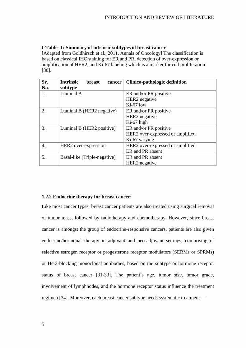
INTRODUCTION AND REVIEW OF LITERATURE
5
I-Table- 1: Summary of intrinsic subtypes of breast cancer
[Adapted from Goldhirsch et al., 2011, Annals of Oncology] The classification is
based on classical IHC staining for ER and PR, detection of over-expression or
amplification of HER2, and Ki-67 labeling which is a marker for cell proliferation
[30].
Sr.
No.
Intrinsic breast cancer
subtype
Clinico-pathologic definition
1. Luminal A ER and/or PR positive
HER2 negative
Ki-67 low
2. Luminal B (HER2 negative) ER and/or PR positive
HER2 negative
Ki-67 high
3. Luminal B (HER2 positive) ER and/or PR positive
HER2 over-expressed or amplified
Ki-67 varying
4. HER2 over-expression HER2 over-expressed or amplified
ER and PR absent
5. Basal-like (Triple-negative) ER and PR absent
HER2 negative
1.2.2 Endocrine therapy for breast cancer:
Like most cancer types, breast cancer patients are also treated using surgical removal
of tumor mass, followed by radiotherapy and chemotherapy. However, since breast
cancer is amongst the group of endocrine-responsive cancers, patients are also given
endocrine/hormonal therapy in adjuvant and neo-adjuvant settings, comprising of
selective estrogen receptor or progesterone receptor modulators (SERMs or SPRMs)
or Her2-blocking monoclonal antibodies, based on the subtype or hormone receptor
status of breast cancer [31-33]. The patient‘s age, tumor size, tumor grade,
involvement of lymphnodes, and the hormone receptor status influence the treatment
regimen [34]. Moreover, each breast cancer subtype needs systematic treatment—
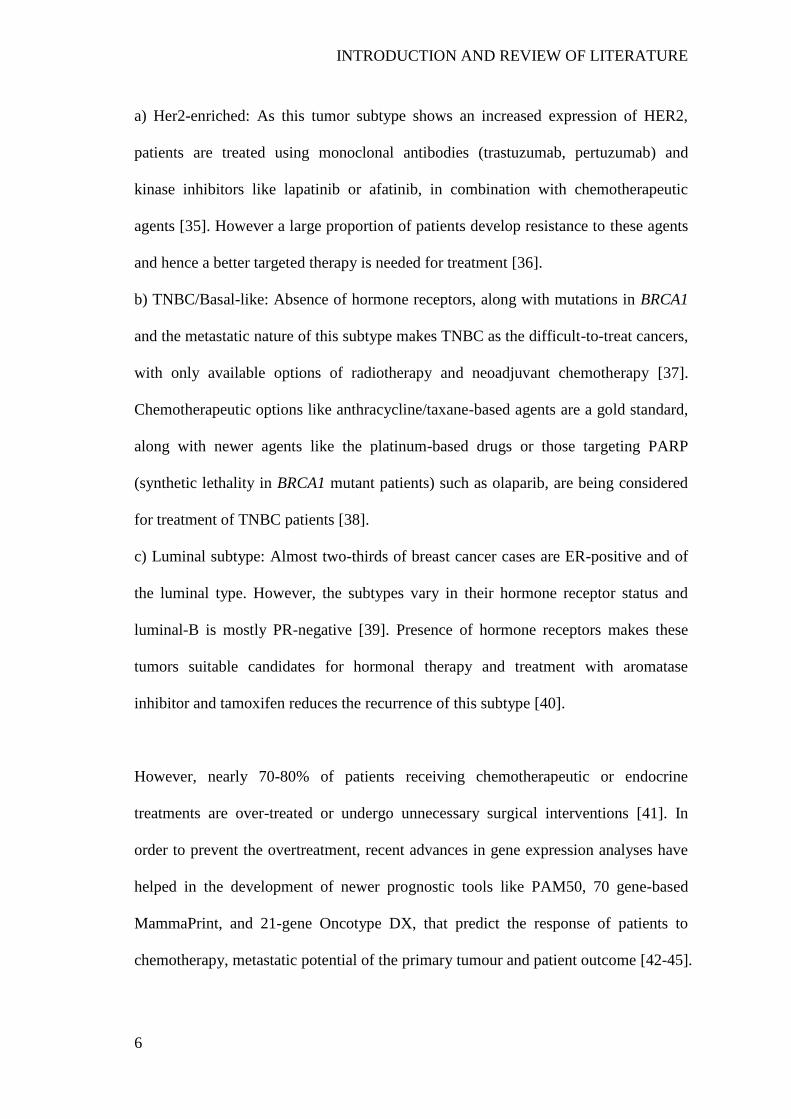
INTRODUCTION AND REVIEW OF LITERATURE
6
a) Her2-enriched: As this tumor subtype shows an increased expression of HER2,
patients are treated using monoclonal antibodies (trastuzumab, pertuzumab) and
kinase inhibitors like lapatinib or afatinib, in combination with chemotherapeutic
agents [35]. However a large proportion of patients develop resistance to these agents
and hence a better targeted therapy is needed for treatment [36].
b) TNBC/Basal-like: Absence of hormone receptors, along with mutations in BRCA1
and the metastatic nature of this subtype makes TNBC as the difficult-to-treat cancers,
with only available options of radiotherapy and neoadjuvant chemotherapy [37].
Chemotherapeutic options like anthracycline/taxane-based agents are a gold standard,
along with newer agents like the platinum-based drugs or those targeting PARP
(synthetic lethality in BRCA1 mutant patients) such as olaparib, are being considered
for treatment of TNBC patients [38].
c) Luminal subtype: Almost two-thirds of breast cancer cases are ER-positive and of
the luminal type. However, the subtypes vary in their hormone receptor status and
luminal-B is mostly PR-negative [39]. Presence of hormone receptors makes these
tumors suitable candidates for hormonal therapy and treatment with aromatase
inhibitor and tamoxifen reduces the recurrence of this subtype [40].
However, nearly 70-80% of patients receiving chemotherapeutic or endocrine
treatments are over-treated or undergo unnecessary surgical interventions [41]. In
order to prevent the overtreatment, recent advances in gene expression analyses have
helped in the development of newer prognostic tools like PAM50, 70 gene-based
MammaPrint, and 21-gene Oncotype DX, that predict the response of patients to
chemotherapy, metastatic potential of the primary tumour and patient outcome [42-45].

INTRODUCTION AND REVIEW OF LITERATURE
7
1.2.3 Recurrence of breast cancer:
Breast cancer shows metastatic behavior and is known to metastasize to adjacent
lymph-nodes and distant locations like the lung, bone, liver and brain [46, 47].
Metastatic property of breast cancer causes more deaths than the primary tumor itself.
Since predicting the metastasis of breast cancer is difficult, majority of the patients
receive adjuvant chemotherapy of which only half of the patients relapse and the other
half are over treated [48]. Even with the advances in diagnostic and treatment
modalities, node-positive patients relapse with a metastatic disease [21]. The classical
metastasis theory of breast cancer suggests that a subset of primary site tumor acquires
properties to metastasize to distant locations and that such cells are selected over the
period of progression of the disease. However, newer theories have challenged this
belief and it is now believed that breast cancer cells acquire mutations in metastasis-
causing genes at a much early stage of tumorigenesis [49].
Thus, the metastatic nature and resistance to therapy account for the relapse and
recurrence of breast cancer. Generally, patients with luminal-type tumors have a lower
risk of recurrence and those with the Her2-enriched and basal-like (TNBC) tumors
have an increased risk for local and regional recurrence of the disease [50]. Moreover,
breast cancer poses a unique threat that the disease can recur even after first 10 years
after diagnosis and that after initial response to endocrine agents, a significant
percentage of patients relapse with metastatic tumor. Thus patients may have to be
kept on maintenance therapy for a prolonged period, even after remission of the tumor,
to remove traces of the disease and reduce chances of recurrence [51].

INTRODUCTION AND REVIEW OF LITERATURE
8
1.2.4 Correlation of PR expression with prognosis of breast cancer:
From a prognostic point-of-view, patients with PR positive tumors show better
response to therapy and delayed metastasis [52]. For patients on endocrine therapy
targeting ER, PR is considered an important predictor for clinical response to the
therapy [53]. However, during progression of the disease, breast tumors lose
expression of hormone receptors, while some show over-expression of growth factor
receptors leading to resistance to endocrine therapy [54-56].
PR expression correlates with prognosis of breast cancer patients [53, 57, 58]; and,
patients with PR/ER-positive tumors respond better to endocrine therapy as compared
with patients with PR-negative/ER-positive or -negative tumors [59]. Of note,
presence of PR has been shown to associate with lower metastasis of tumors to distant
sites [52]. However, patients show resistance to endocrine therapy with influence by
factors like loss of either of the hormone receptors (PR/ER), or by over-expression of
Her2 receptor which circumvents the endocrine suppression or due to over-expression
of proteins like cyclin-D1 [59, 60]. A slightly intriguing observation shows that
hormones can down-regulate their own receptors. For instance, estrogen has been
shown to decrease expression of ER via up-regulation of microRNAs [61]. Similarly,
progesterone regulates the degradation of PR by phosphorylation regulated by MAPK
and ubiquitination induced by CUEDC2. Apart from this, PR is down-regulated by
methylation of promoter of PR and microRNA-mediated down-regulation in breast
and endometrium cells [62-64]. However, microRNAs regulating PR expression in
response to progesterone have not been studied in detail [65]. Thus, a better
understanding of mechanisms leading to PR down-regulation may aid in maintaining
the expression of PR in breast cancer. Thus these clinical observations mandate a

INTRODUCTION AND REVIEW OF LITERATURE
9
detailed investigation of hormone receptor status of patients receiving endocrine
therapy.
1.2.5 Progesterone and Progesterone receptor (PR):
Progesterone is a membrane soluble steroid hormone, secreted by the ovary, that
produces intracellular signals and aids in formation of epithelial cells, differentiation
and side-branching of cells, maturation and alveologenesis of the mammary gland [66,
67]. Cyclical variations in the levels of estrogen and progesterone make the menstrual
cycle, which comprises of an unopposed estrogenic phase and luteal progestogenic
phase. These cyclical variations have varying effect on the hormone-responsive
tissues like the uterus, ovary and the breast. At the breast tissue level, progesterone
hypothetically acts in a paracrine manner by stimulating the PR-positive cells in the
tissue to activate signaling in the PR-negative cells and shows differential effects on
cells based on their PR status [68, 69].
I-Figure- 2: Basic structure of the two isoforms of human progesterone receptor
(hPR)
[Adapted from Chabbert-Buffet et al., 2005, Hum Reprod Update]. Different domains
of the PR have been depicted where each abbreviation stands for— AF, activation
function; ID, inhibitory domain; LBD, ligand binding domain; DBD, DNA binding
domain; ATG, initiation of transcription codon [70].
PR is a core member of the superfamily of nuclear steroid hormone receptors [71]. PR
is a single gene, located on chromosome position 11q21-11q23 and encodes two

INTRODUCTION AND REVIEW OF LITERATURE
10
isoforms— PR-A and PR-B, with PR-B being the complete protein (116KDa)
synthesized from 933 amino acids, while PR-A lacks the first 164 amino acids at the
N-terminal and is 769 amino acids in length (81KDa) (I-Figure-2) [72, 73]. PR is
expressed in reproductive organs like the breast, endometrium, ovary and other organs
like the brain where PR plays an important role in development, differentiation, and
proliferation of target tissues and the pathological processes in endocrine-based
cancers [74, 75]. The expression of PR is under tight regulation by estrogen and
progesterone [63, 76]. PR is localized to the cytoplasm in a non-functional state,
where it associates with Hsp90 chaperone. When progesterone enters the cell, it binds
and dimerises PR and the complex enters the nucleus to function as a transcription
factor by recognizing a consensus sequence GGT/AACAnnnTGTTCT, termed as
progesterone response elements (PRE), in the promoter of target genes. This mode of
activity is termed as the genomic pathway regulated by progesterone and PR.
However, of the many potential PRE-binding sites in the genome, PR occupies only a
small fraction upon progesterone stimulation, and that cooperation with transcription
factors like FOXA1, ETS, STAT or SP1 are essential for binding PR cistromes [74,
75]. Other steroid hormone receptors like the glucocorticoid receptor (GR),
mineralocorticoid receptor (MR) and androgen receptor (AR) recognize similar
consensus sequences, and work synergistically through overlapping cistromes
regulated by these hormone receptors [77-79]. Recently, progesterone-mediated
alteration in ER cistrome by PR has been shown in breast cancer [58, 80]. Also,
progesterone has been shown to associate with GR and AR to mediate its function in
breast and other cancer types [77]. Thus essentially, progesterone may function even
in the absence of PR by associating with other receptors; however, the pathways or
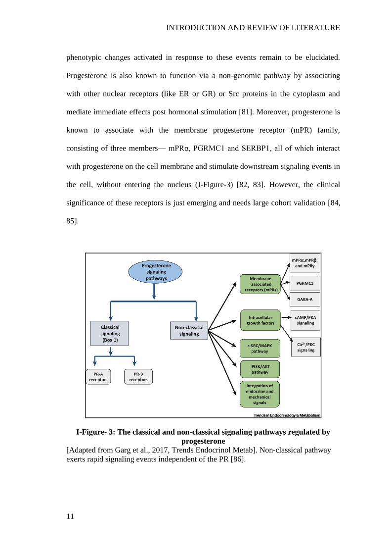
INTRODUCTION AND REVIEW OF LITERATURE
11
phenotypic changes activated in response to these events remain to be elucidated.
Progesterone is also known to function via a non-genomic pathway by associating
with other nuclear receptors (like ER or GR) or Src proteins in the cytoplasm and
mediate immediate effects post hormonal stimulation [81]. Moreover, progesterone is
known to associate with the membrane progesterone receptor (mPR) family,
consisting of three members— mPRα, PGRMC1 and SERBP1, all of which interact
with progesterone on the cell membrane and stimulate downstream signaling events in
the cell, without entering the nucleus (I-Figure-3) [82, 83]. However, the clinical
significance of these receptors is just emerging and needs large cohort validation [84,
85].
I-Figure- 3: The classical and non-classical signaling pathways regulated by
progesterone [Adapted from Garg et al., 2017, Trends Endocrinol Metab]. Non-classical pathway
exerts rapid signaling events independent of the PR [86].

INTRODUCTION AND REVIEW OF LITERATURE
12
1.2.6 Phenotypic, genomic and proteomic effects of progesterone
Progesterone is primarily involved in the differentiation and proliferation of target
cells in a context-dependent manner [87-89]. In the breast cells, progesterone induces
cell proliferation in a biphasic manner, by increasing cell proliferation in the first 24-
48hrs and then inhibiting proliferation during second cell cycle even after addition of
fresh progesterone to the cells [90]. Under in vitro settings, progesterone functions as
an anti-estrogenic moiety by reducing proliferation of breast cancer cells. However,
the anti-proliferative activity was found to be dependent on the expression of PR in
breast cancer cells [91, 92]. On the contrary, progesterone may induce proliferation of
cancer cells under in vivo conditions [93]. Progesterone has also been shown to
influence the growth of cancer cells, again in a PR-dependent manner [94, 95].
However these anti-proliferative and growth-repressing actions of progesterone are
challenged by the model for development of breast cancer, where progesterone has
been hypothesized to help in progression of the initial breast tumor lesions [69]. The
pro-angiogenic property of progesterone has been observed in cancer cells by up-
regulation of expression of vascular endothelial growth factor (VEGF) and platelet-
derived growth factor (PDGF) support this notion [96, 97]. However, progesterone
may have a dual role in regulating the progression of cancer cells. It has been
observed processes like migration and invasion of cancer cells are important for the
progression and metastasis of cancer. To this end, progesterone has been shown to
reduce the migration of PR-positive breast cancer cells in vitro. Not only does
progesterone influence migration and cell morphology of breast-derived cells but also
of cells derived from the lung and endometrium [58, 80, 98-101]. Whilst this
INTRODUCTION AND REVIEW OF LITERATURE
13
phenotypic influence, progestins have been shown to increase the migration of PR-
positive breast cancer cells [102]. This disparity in the action of progesterone and
progestin can be attributed to the differential effect imparted by these agents in breast
cancer cells [103, 104]. However, it remains to be elucidated in a mechanistic manner
whether progesterone can influence the migration and invasion of breast cancer cells
independent of the PR status of cells.
These phenotypic effects of breast cancer cells derive their origin from the genomic
and proteomic changes that progesterone induces in a context-dependent manner. The
genomic effects are transient transcriptional changes triggered by change in chromatin
architecture via recruitment of transcription factors [105]. Progesterone recruits
chromatin modifiers and induces transcriptional changes via association with PR and
other hormone receptors (like GR and ER) to either activate or repress the
transcription of target genes [58, 106, 107]. It has been proposed that progesterone can
regulate expression of gene sets in a similar manner in topologically associating
domains (TADs) [108]. Interestingly, PR can alter the transcriptional activity of ER in
breast cancer cells treated with estrogen and progesterone, suggestive of an anti-
estrogenic activity of progesterone in breast cancer [58, 80]. Moreover, studies have
been conducted to study genes regulated by progesterone receptor in an isoform-
specific manner. Majority of these targets of progesterone are related to regulation of
cell-cycle and proliferation [109, 110].
On the other hand, effect of progesterone on expression and activation of proteins has
remained under-studied. Proteomic analysis of bovine uterus has been performed
which suggests an essential role of progesterone in development of embryo during the
INTRODUCTION AND REVIEW OF LITERATURE
14
pre-implantation stage [111]. Recently, a LC-MS/MS-based analysis of the human
endometrial cells treated with progesterone in combination with calcitriol has
identified proteins involved in the anti-tumorigenic action of progesterone [112].
However, such global proteome profiling efforts have not been taken using the human
breast tissue cells. Mostly, candidate-based approaches have characterized the effect
of progesterone on proteins involved in regulation of cell-cycle or proliferation of
cells. Reports suggest that progesterone inhibits estrogenic activity by up-regulating
proteins that inhibit growth of the PR-positive breast cancer cells [95, 113]. Certain
studies have also shown that progesterone alters the phosphorylation of certain kinases
and increases migration of breast cancer cells [102, 114, 115]. A detailed
characterization of effect of progesterone on the activity of kinases also remains to be
explored.
1.3 Progesterone influences survival of breast cancer patients:
Physiologic concentrations of estrogen, progesterone, prolactin hormones influence
processes of pregnancy and development of hormone-responsive organs in women.
On the other hand, prolonged exposure to these hormones has also been associated
with development of cancer, especially estrogen [116]. However, few studies have
found no harm associated with endogenous progesterone or as hormone replacement
therapy given to postmenopausal women [117-120]. And a lot of debate surrounds the
use of progestin for hormone replacement therapy, the jury is still out [121].
Observational studies have been conducted to study the effect of the phases of
menstrual cycle on the outcome of breast cancer. The analysis suggests that surgery
performed in the progestogenic (luteal) phase associates with a favorable prognosis as
compared to surgery performed in other phases of the menstrual cycle [122-124].

INTRODUCTION AND REVIEW OF LITERATURE
15
Moreover, it was found that elevated serum progesterone levels correlated with better
survival of breast cancer patients [124, 125]. These early observational studies
conceived a randomized clinical trial in Tata Memorial Hospital (TMH), India, where
a cohort of operable breast cancer patients was subjected to single-depot progesterone
intervention 5-14 days before surgery. Patients with different menopausal stages, PR
expression and tumor grades were randomly grouped into the control group and the
pre-operative progesterone group. Overall in the cohort of 1000 patients, progesterone
did not influence the survival of patients; however, analysis in a selected cohort of
patients with node-positive disease showed a 10% increase in disease-free and overall
survival of patients receiving pre-operative progesterone treatment as compared to
patients in the control group. Interestingly, the beneficial effect was observed
independent of the PR status or the menopausal status of breast cancer patients (I-
Figure-4). Though pre-/peri-operative endocrine intervention has shown beneficial
effects in receptor-positive breast cancer patients [126, 127], the randomized clinical
trial by Badwe et al is the first of its kind with pre-operative progesterone intervention
and provides a hypothesis-generating outcome to be further tested on a larger cohort
[128].
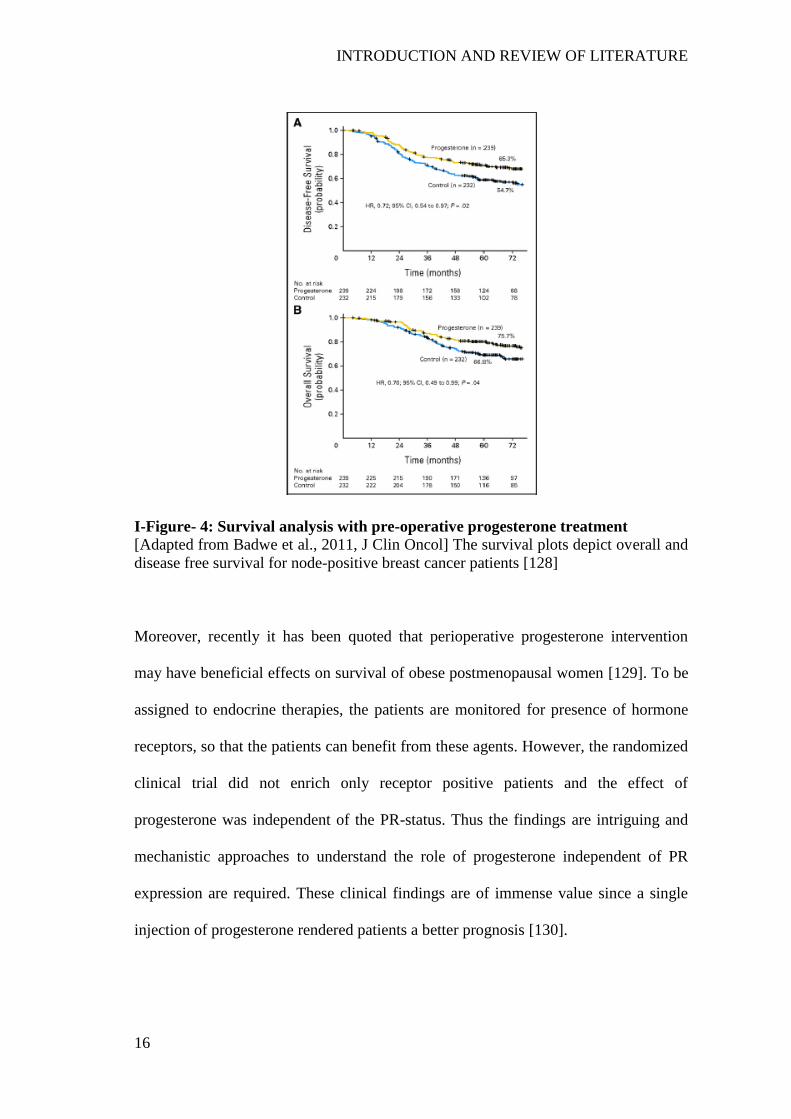
INTRODUCTION AND REVIEW OF LITERATURE
16
I-Figure- 4: Survival analysis with pre-operative progesterone treatment
[Adapted from Badwe et al., 2011, J Clin Oncol] The survival plots depict overall and
disease free survival for node-positive breast cancer patients [128]
Moreover, recently it has been quoted that perioperative progesterone intervention
may have beneficial effects on survival of obese postmenopausal women [129]. To be
assigned to endocrine therapies, the patients are monitored for presence of hormone
receptors, so that the patients can benefit from these agents. However, the randomized
clinical trial did not enrich only receptor positive patients and the effect of
progesterone was independent of the PR-status. Thus the findings are intriguing and
mechanistic approaches to understand the role of progesterone independent of PR
expression are required. These clinical findings are of immense value since a single
injection of progesterone rendered patients a better prognosis [130].

INTRODUCTION AND REVIEW OF LITERATURE
17
1.4 Rationale of the study:
Vast numbers of studies have focused on identifying the PR-dependent gene
expression profiles in breast cancer [109, 131]. Although many candidate-based
studies have been performed, no global proteomic and genomic changes have been
reported specifically in response to progesterone in breast cancer, independent of the
PR status. Moreover, majority of the studies have been performed in presence of other
hormones. Hence, whether progesterone can alter transcriptional signature of breast
cancer cells irrespective of the PR expression remains to be explored.
Of note, to identify targets of progesterone independent of PR, one approach could be
to perform transcriptional and proteomic profiling of breast cancer patient-derived
tissue samples from the randomized clinical trial with pre-operative progesterone and
compare the expression profiles with that of tumor samples derived from patients in
the control group. Such a study can provide immense details about the long-term
effects of progesterone on breast cancer and is currently being performed at Tata
Memorial Centre. However, the unique targets of progesterone may not be discerned
from this approach since it will be an additive effect of all hormones together with
progesterone, together with the heterogeneity of tumor samples. It has already been
suggested that progesterone can alter the transcriptional profile of breast cancer in the
presence of estrogen using in vitro studies [58]. Moreover, the effect observed in
samples derived from the clinical trial will be a delayed response to progesterone and
it may not be possible to validate the results using orthologous methodologies. An
alternative approach is to perform an in vitro study using a panel of breast-derived cell
lines which represent different hormone receptor statuses and breast cancer subtypes
and technically are a better system for performing functional genomic and proteomic

INTRODUCTION AND REVIEW OF LITERATURE
18
analysis. A large repertoire of breast-derived cell lines can be recruited for such a
study [132-134]. These cell lines are a homogenous system and provide the
opportunity for functional validation of candidate genes using genetic and
pharmacologic manipulation, in this case in response to progesterone. Of note, the
progesterone-specific effects can be monitored by regulating the progesterone
treatment conditions and by depletion of most of the interfering hormones and growth
factors from the conditioning medium and the cell lines provide an opportunity to
block the activity of PR receptor to identify PR-independent targets of progesterone
[70, 135, 136]. The effect of progesterone can be elucidated using short-term (from
few minutes to few hours) or long-term (24-72hrs) treatment. Most of the studies to
understand the genomic effects of progesterone have utilized short-term (3-6hrs)
treatment conditions since hormones induce gene expression changes immediately
after their addition to cells [58, 109]. Thus cell lines represent a better system to
elucidate the molecular action of progesterone in breast cancer and also are of
immense utility to study the role of candidate genes by genetic perturbation
approaches.

OBJECTIVES OF THE STUDY
19
II. OBJECTIVE OF THE STUDY
Specifically, few key questions need to be addressed to elucidate the targets of
progesterone in breast cancer:
1. What are the genomic and proteomic targets of progesterone in breast cancer cells
independent of the PR status?
2. Can progesterone abrogate the cell migration and invasion phenotype of breast
cancer?
3. What is the possible mode of action of progesterone that helped improve the
survival of breast cancer patients?
To provide a molecular basis for solving each of the above questions, it is imperative
to adopt a functional genomics and proteomics approach using a panel of breast-
derived cell lines to capture the short-term effects of progesterone, without bias to the
hormone receptor status of the cells.
To fill the void in the understanding of targets of progesterone, we have the following
specific objectives:
2.1 Genomic approach to identify targets of progesterone in breast cell lines
In this objective, we aim to identify the coding and non-coding targets, identified
using microarray gene expression analysis and small RNA sequencing analysis
respectively and performed in a panel of breast-derived cell lines, in response to
progesterone treatment.
2.2 Proteomic analysis of breast cell lines upon progesterone treatment
In this second objective, we have focused on the proteomic changes induced by
progesterone, independent of the PR status of cell lines.

OBJECTIVES OF THE STUDY
20
2.3 Functional validation of progesterone candidate genes in breast cancer cell
line by rescuing phenotype
In this final part of the study, we have performed functional validation of candidate
genes and microRNAs obtained from both the earlier objectives. Specifically, we have
performed genetic and pharmacological perturbations of the candidate genes or
microRNAs, phenotypic assays to understand the global effects induced by
progesterone and upon perturbation of candidate genes, irrespective of the hormone
receptor statuses.

PROGESTERONE SUPPRESSES INVASION AND MIGRATION OF BREAST
CANCER
21
III. PROGESTERONE SUPPRESSES THE INVASION AND
MIGRATION OF BREAST CANCER CELLS IRRESPECTIVE OF
THEIR PROGESTERONE RECEPTOR STATUS – A SHORT
REPORT
[An excerpt; as published in Cellular Oncology (2017); 40(4):411-417]
Abstract
Purpose Pre-operative progesterone treatment of breast cancer has been shown to
confer survival benefits to patients independent of their progesterone receptor (PR)
status. The underlying mechanism and the question whether such an effect can also be
observed in PR negative breast cancer cells remain to be resolved.
Methods We performed proteome profiling of PR-positive and PR-negative breast
cancer cells in response to progesterone using a phospho-kinase array platform.
Western blotting was used to validate the results. Cell-based phenotypic assays were
conducted using PR-positive and PR-negative breast cancer cells to assess the effect
of progesterone.
Results We found that progesterone induces de-phosphorylation of 12 out of 43
kinases tested, which are mostly involved in cellular invasion and migration
regulation. Consistent with this observation, we found through cell-based phenotypic
assays that progesterone inhibits the invasion and migration of breast cancer cells
independent of their PR status.
Conclusion Our results indicate that progesterone can inhibit breast cancer cell
invasion and migration mediated by the de-phosphorylation of kinases. This inhibition
appears to be independent of the PR status of the breast cancer cells. In a broader
context, our study may provide a basis for an association between progesterone
PROGESTERONE SUPPRESSES INVASION AND MIGRATION OF BREAST
CANCER
22
treatment and recurrence reduction in breast cancer patients, thereby providing a lead
for modelling a randomized in vitro study.
3.1 Introduction
Metastasis is a major cause of recurrence in breast cancer patients [48]. As a standard
mode of treatment, patients with metastatic breast cancer are subjected to adjuvant
hormonal therapy [137]. Also, pre-operative progesterone treatment has been shown
to reduce the recurrence rate among node-positive patients, independent of their
progesterone receptor (PR) status [128]. While the invasion and migration of breast
cancer cells play major roles in establishing metastases [138, 139], in vitro cell-based
observations on the effect of progesterone, restricted to PR positive cells, have
corroborated the clinical observations [58, 80, 102]. As yet, however, the effect of
progesterone on the invasion and migration of PR negative breast cancer cells remains
to be systematically explored [140].
The metastatic nature of breast cancer cells is known to be affected by multiple
molecular factors, including the activation of protein kinases [141]. The protein
kinases EGFR, AKT or FAK have, for instance, been found to activate the processes
of migration and invasion of breast cancer cells [142, 143]. In addition, it has been
found that these kinases may act synergistically and that abrogating their activation
may decrease the invasive capacity of breast cancer cells [144]. Also, pathways
downstream of these kinases may serve to restrain cell invasion and migration [141].
Although these kinases have been found to be affected by steroid hormone receptors
[145], it remains to be explored whether they mediate the responses to progesterone in
breast cancer cells.
PROGESTERONE SUPPRESSES INVASION AND MIGRATION OF BREAST
CANCER
23
To address the question whether progesterone can regulate cellular migration and
invasion of breast cancer cells independent of their PR status, we selected a panel of
breast cancer-derived cell lines with different PR statuses. Next, we performed a
phospho-proteomic screening of kinases associated with migration and invasion using
a human proteome phospho-kinase array platform, and studied their phosphorylation
status after treating the respective cells with progesterone. Our cell-based phenotypic
and biochemical analysis results suggest that progesterone may mitigate the invasion
and migration of breast cancer cells, irrespective of their PR status.
3.2 Materials and methods
3.2.1 Breast cancer-derived cells
The BT474, T47D, MCF7, ZR-75-1, MDA-MB-231 and BT-549 breast cancer-
derived cell lines were obtained as a gift from Dr. Slamon's Laboratory (Department
of Medicine, UCLA, USA). The cell lines were authenticated by DNA short tandem
repeat (STR) profiling using the Promega GenePrint 10 system in conjunction with
the GeneMarker HID software tool and the ATCC database. The cells were tested for
mycoplasma and, if necessary, made mycoplasma-free using an EZKill Mycoplasma
Removal reagent (HiMedia). BT474, T47D, MCF7 and MDA-MB-231 cells were
cultured in DMEM medium (Gibco), whereas ZR-75-1 and BT-549 were cultured in
RPMI-1640 medium (Gibco). BT-549 cells were supplemented with 0.023 IU/ml
insulin. All culture media were supplemented with 10% (v/v) FBS (Gibco), 2.5 mg/ml
Amphotericin-B (Abbott) and 1.25 µl/ml Gentamycin (Abbott). The cells were

PROGESTERONE SUPPRESSES INVASION AND MIGRATION OF BREAST
CANCER
24
cultured at 37°C in a 5% CO2 incubator. The PR/ER/Her2 receptor statuses of all the
cells as reported in [133] were validated by reverse transcriptase-PCR.
3.2.2 Progesterone treatment
Cells were grown to 70-80% confluence and then serum starved in DMEM low
glucose medium (HiMedia) for a period of 24h. Next, the cells were treated with 10
nM 17-α hydroxy-progesterone caproate (progesterone) (MP Biomedicals) in the same
medium for 6h. In case of mifepristone + progesterone (M+P) combination treatment,
100 nM RU486 (mifepristone) was added for 2h followed by 10 nM progesterone
treatment for 6h in the same medium. An equal volume of alcohol was used as vehicle
control.
3.2.3 Protein sample preparation
Cells were grown to a 70-80% confluence in a 100 mm culture dish and washed
thoroughly with sterile 1x PBS. Next, the cells were subjected to 24h serum starvation
(using DMEM low glucose phenol-red free medium) followed by progesterone
treatment for 8h. Alcohol was used as vehicle control. After progesterone treatment,
the cells were harvested using a sterile cell scraper and cell lysates were prepared in
RIPA Buffer (Sigma-Aldrich) supplemented with a protease-inhibitor cocktail
solution (Sigma-Aldrich) and 0.1 M DTT. After intermittently tapping and vortexing
the samples on ice, cell debris was pelleted by centrifugation at 14000 rpm after which
the protein concentrations were determined using BCA reagent (MP Biomedicals).
Bovine serum albumin was used as a standard and the estimations were performed in
triplicate.

PROGESTERONE SUPPRESSES INVASION AND MIGRATION OF BREAST
CANCER
25
3.2.4 Phospho-kinase activation profiling
Kinase activation profiling of T47D (PR-positive) and MDA-MB-231 (PR-negative)
breast cancer-derived cells was performed using a Human Phospho-kinase array kit
(ARY003B; R&D Systems) according to the manufacturer‘s instructions. Briefly,
cells were grown in T75 flasks till 70-80% confluence was reached, serum-starved for
another 24 h (in serum-free phenol-red-free DMEM medium) and treated with
progesterone for 8h. Next, the cells were harvested, washed with 1x PBS and lysed,
after which 400 μg protein from untreated and progesterone-treated samples was
incubated overnight at 4°C with a pre-blocked antibody array nitrocellulose
membrane. Subsequently, the membranes were incubated with detection antibodies
and probed using streptavidin-HRP, after which signals were developed using chemi-
reagents provided with the kit. Exposures to X-ray films were taken from 10 seconds
to 10 minutes (till saturation was reached). Signal densities of reference spots on both
membranes were compared between each pair of membranes used for the control and
progesterone-treated samples. The pixel density of each spot, in duplicate, was
calculated using ImageJ Array Analyzer plugin. The average pixel density for the
duplicate spots for each of the kinases was subtracted from the negative control
density. The average pixel densities for control and progesterone-treated samples were
plotted as percent phosphorylation for each phospho-kinase. The differential
phosphorylation cut-off value was set at 20% increase or decrease in phosphorylation
of kinases in response to progesterone.
PROGESTERONE SUPPRESSES INVASION AND MIGRATION OF BREAST
CANCER
26
3.2.5 Western blotting
Equal amounts of cell lysate were resolved by 10% SDS-PAGE and transferred to
PVDF membranes using a wet transfer method. Primary antibodies directed against p-
EGFR (Y1068) (Cell Signaling, 3777S; Dilution 1:500), p-AKT (S473) (Cell
Signaling, 4060S; Dilution 1:500), p-ERK1/2 (T202/Y204) (Cell Signaling, 9101S;
Dilution 1:1000), total EGFR (1005) (Santa Cruz Biotechnology, sc-03; Dilution
1:1000), total AKT (11E7) (Cell Signaling, 4685S; Dilution 1:1000), total ERK2 (c-
14) (Santa Cruz Biotechnology, sc-154; Dilution 1:1000) and β-actin (I-19)-R (Santa
Cruz Biotechnology, sc-1616-R; Dilution 1:3000) were diluted in 3% BSA solution
prepared in 1x TBST and incubated over-night at 4°C. A goat anti-rabbit IgG-HRP
secondary antibody (Santa Cruz Biotechnology, sc-2004; Dilution 1:3000) was used
for the detection of primary antibody binding. ECL Western Blotting Substrate (Pierce)
and Takara Chemiluminescence substrate (ClonTech Takara) were used for
visualization of the protein bands on X-ray films (Fuji Films).
3.2.6 RNA extraction and real-time PCR
For RNA extraction from alcohol and progesterone-treated breast cancer-derived cells,
the respective cells were treated with progesterone for 6h. Next, TRIzol reagent
(Invitrogen) was used to lyse the cells after which RNA was isolated according to the
manufacturer‘s protocol. RNA concentrations were measured using NanoDrop. For
assessment of the DUSP1 transcript levels, cDNA was synthesized using a High
capacity cDNA reverse transcription kit (Applied Biosystems) and subjected to
quantitative real-time PCR using a Roche Light-Cycler-II 480 instrument in
conjunction with a Roche real-time master mix (Roche). Expression changes were
PROGESTERONE SUPPRESSES INVASION AND MIGRATION OF BREAST
CANCER
27
calculated using the 2-ΔΔCT
method. GAPDH was used as internal control for
normalization. The primer sequences used for DUSP1 were Forward primer OAD-571:
CCTGCAGTACCCCACTCTACG; Reverse primer OAD-572:
CCCAAGGCATCCAGCATGTCC and for GAPDH Forward primer OAD-328:
AATCCCATCACCATCTTCCA; Reverse primer OAD-329:
TGGACTCCACGACGTACTCA.
3.2.7 Cell invasion assay
A Matrigel invasion assay was performed using 24-well Transwell inserts (Corning)
coated with 100 μg matrigel and allowed to settle for 24hr at 37ºC. Next, 35000 cells
suspended in 350 μl serum-free medium were seeded into the upper chamber and 600
μl of 10% serum-containing medium was added to the lower chamber. After this, the
cells were allowed to invade for 16-18h at 37ºC, followed by fixation of the invaded
cells and staining by crystal violet. After mounting the membrane using DPX on a
slide, the cells were observed under an upright microscope. Ten random fields were
chosen after which the number of cells in each field was counted and plotted as
percentage cell invasion.
3.2.8 Scratch wound healing assay
Confluent cell monolayers in 6-well plates, grown in phenol-red free DMEM for
18hrs without serum, were subjected to a scratch with a sterile pipette tip. After this,
the cells were briefly rinsed using 1X PBS to remove debris and subsequently
incubated with low-glucose phenol-red free DMEM medium containing 10%
charcoal-stripped FBS (Gibco). The cells were treated with 10 nM progesterone or

PROGESTERONE SUPPRESSES INVASION AND MIGRATION OF BREAST
CANCER
28
100 nM mifepristone or a combination of both. Alcohol was used as a vehicle control.
Cell migration at the wound surface was measured during a period of 20 h under an
inverted microscope. Quantification was performed using the ImageJ wound healing
plugin tool by measuring the distance of the wound edge of the migrating cells from
the start point to the migrated point in three separate wounds in three independent
experiments.
3.3 Results and discussion
The activation of kinases like EGFR and ERK1/2 has been reported to play an
important role in the de-regulation of cellular processes that are associated with the
metastatic capacity of breast cancer cells [146]. Here, we set out to assess the effect of
progesterone on the activation of kinases in breast cancer cells using a human
phospho-kinase array platform. To verify the effect of progesterone independent of the
progesterone receptor (PR) status of the cells, we selected both PR-positive (T47D)
and PR-negative (MDA-MB-231) breast cancer-derived cells for our study (III-Table
1). Untreated cells were used as negative controls.
Sr.
No. Cell Line
Literature reported
Receptor Status
Validation of Receptor
Status by RT-PCR
PR ER HER2 PR ER HER2
1. BT474 + + + + + +
2. T47D + + - + + -
3. MCF7 + + - + + -
4. ZR-75-1 - + - - + -
5. MDA-MB-231 - - - - - -
6. BT-549 - - - - - -
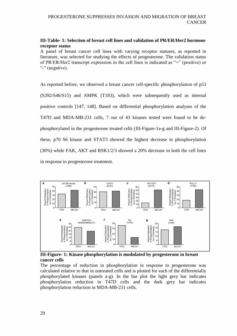
PROGESTERONE SUPPRESSES INVASION AND MIGRATION OF BREAST
CANCER
29
III-Table- 1: Selection of breast cell lines and validation of PR/ER/Her2 hormone
receptor status
A panel of breast cancer cell lines with varying receptor statuses, as reported in
literature, was selected for studying the effects of progesterone. The validation status
of PR/ER/Her2 transcript expression in the cell lines is indicated as ―+‖ (positive) or
―-‖ (negative).
As reported before, we observed a breast cancer cell-specific phosphorylation of p53
(S392/S46/S15) and AMPK (T183), which were subsequently used as internal
positive controls [147, 148]. Based on differential phosphorylation analyses of the
T47D and MDA-MB-231 cells, 7 out of 43 kinases tested were found to be de-
phosphorylated in the progesterone treated cells (III-Figure-1a-g and III-Figure-2). Of
these, p70 S6 kinase and STAT3 showed the highest decrease in phosphorylation
(30%) while FAK, AKT and RSK1/2/3 showed a 20% decrease in both the cell lines
in response to progesterone treatment.
III-Figure- 1: Kinase phosphorylation is modulated by progesterone in breast
cancer cells
The percentage of reduction in phosphorylation in response to progesterone was
calculated relative to that in untreated cells and is plotted for each of the differentially
phosphorylated kinases (panels a-g). In the bar plot the light grey bar indicates
phosphorylation reduction in T47D cells and the dark grey bar indicates
phosphorylation reduction in MDA-MB-231 cells.

PROGESTERONE SUPPRESSES INVASION AND MIGRATION OF BREAST
CANCER
30
In addition, we observed a reduction in phosphorylation of the ERK1/2 (T202/Y204,
T185/Y187), EGFR (Y1068), MSK1/2 (S376/S360), p38α (T180/Y182) and p27
(T198) kinases upon treatment with progesterone (III-Figure-2), as reported earlier
[91], and validated the results by Western blot analysis (III-Figure-3a). Consistent
with earlier reports [91], we also observed a significant up-regulation of a dual
specificity phosphatase, DUSP1, upon treatment with progesterone in breast cancer
cells that could possibly mediate the effect observed (III-Figure-3b).
III-Figure- 2: Differentially phosphorylated kinases in response to progesterone
treatment of breast cancer cells
The figure represents X-ray films exposed to nitrocellulose membrane for 5min with
untreated and progesterone-treated lysates from a) T47D and b) MDA-MB-231 cells.
Black lines and numbers on the right of the spots indicate the location of differentially
phosphorylated kinases in untreated and progesterone-treated sets for both cells. The
annotation of the spots is as follows:
1- p38α; 2- ERK1/2; 3- EGFR; 4- MSK1/2; 5- AKT1/2/3; 6- Fgr; 7- FAK; 8- p70S6K;
9- p27; 10- STAT3; 11- RSK1/2/3; 12- PLC-γ1.
Taken together, our results indicate that progesterone can reduce the phosphorylation
of 12 out of 43 kinases tested in a PR-independent manner, which could affect cellular

PROGESTERONE SUPPRESSES INVASION AND MIGRATION OF BREAST
CANCER
31
signaling pathways downstream to these kinases with a concomitant increase in the
expression of a dual-specificity phosphatase, DUSP1, that could mediate the de-
phosphorylation of these kinases [91].
III-Figure- 3: Progesterone suppresses phosphorylation of kinases involved in
cell migration and invasion in breast cancer cells by up-regulating DUSP1
(modified from the manuscript)
a) Western blot analysis of p-EGFR (Y1086), p-AKT (S473) and p-ERK1/2
(T202/Y204) was performed in T47D and MDA-MB-231 (MD231) cells treated with
progesterone. Total protein for each phospho-kinase was also probed. Numbers on
blot indicate ratio of intensity of phosphorylation for each kinase with respect to its
total protein levels. β-actin was used as internal loading control. ―*‖ mark on β-actin
indicates same β-actin blot used for p-ERK1/2 and p-EGFR panels. ―-‖ on the western
blot panel indicates control, while ―+‖ indicates progesterone treatment. b) Real-time
PCR analysis of DUSP1 was performed in breast cancer cells treated with
progesterone. Graph has been plotted as fold-change for DUSP1 with respect to
GAPDH for control and progesterone-treated cells; horizontal black line indicates
expression of DUSP1 in control cells. Figure is representative of three independent
experiments performed in triplicates. P-value was calculated using student's unpaired
t-test. ** indicates P-value <0.001; *** indicates P-value <0.0001.

PROGESTERONE SUPPRESSES INVASION AND MIGRATION OF BREAST
CANCER
32
Based on the known involvement of EGFR, AKT and ERK1/2 in the invasion and
migration of breast cancer cells and the finding that their inhibition may block this
phenotype [142, 143, 149] or vice versa, i.e., metastases inhibitors may inhibit the
phosphorylation of FAK in PR-negative MDA-MB-231 cells [150] or lung cancer
cells [99], we set out to analyze the in vitro effects of progesterone on breast cancer
cells. Using a Matrigel chamber assay in conjunction with cells with varying
PR/ER/HER2 statuses (III-Table-1) we found that progesterone could decrease the
invasion capacity of different breast cancer-derived cells (BT474, T47D, MCF7 and
MDA-MB-231), irrespective of their hormone receptor status (III-Figure-4a-d). This
result suggests that targeted activation of kinases by progesterone may bring about
phenotypic changes in breast cancer-derived cells independent of their PR-status,
potentially decreasing their metastatic capacity similar to combinatorial EGFR and
AKT inhibition, which is known to affect the invasion of breast and other cancer cells
mediated by matrix metalloproteinases [144, 151]. Importantly, we found that PR
blocking by mifepristone had no significant effect on the invasive capacities of the
cells, again suggesting that progesterone may induce suppression of invasion in breast
cancer cells in a predominantly PR-independent manner (III-Figure-4a-d).
Next we performed scratch wound healing assays to assess whether breast cancer cell
migration is affected by progesterone. Similar to the effect of progesterone on breast
cancer cell invasion, we observed a significant decrease in cellular migration in
response to progesterone over the period of 20h in a PR independent manner (III-
Figure-5). The non-essential role of PR that we observed in the inhibition of migration
of breast cancer-derived cells in response to progesterone, specifically in the PR-
negative MDA-MB-231, ZR-75-1 and BT-549 cells, may be mediated by interaction

PROGESTERONE SUPPRESSES INVASION AND MIGRATION OF BREAST
CANCER
33
of progesterone with the glucocorticoid receptor (GR) or the membrane progesterone
receptor (mPR), as has been reported before [77, 98, 99].
III-Figure- 4: Progesterone inhibits breast cancer cell invasion
Invasion assays were performed with breast cancer-derived cell lines a) BT474, b)
T47D, c) MCF7 and d) MDA-MB-231 treated with progesterone, mifepristone or a
combination of mifepristone and progesterone (M+P). The bar plot represents the
percentage of cell invasion for each panel. The figure is representative of three
independent experiments performed in triplicates. P-values were calculated using
student's unpaired t-test. ** p < 0.001; *** p < 0001; ns not significant.
Of note, it has also been reported that treatment with glucocorticoids may similarly
decrease the migration of PR-negative MDA-MB-231 cells [152], which suggests that
redundant pathways may underlie the progesterone response in a PR-independent
manner [83]. Consistent with these observations, we found that blocking PR by
mifepristone prior to exposing the cells to progesterone did not rescue the effect of
progesterone, suggesting that the progesterone-mediated suppression of migration in
breast cancer cells is predominantly mediated in a PR-independent manner (III-

PROGESTERONE SUPPRESSES INVASION AND MIGRATION OF BREAST
CANCER
34
Figure-6a and 6b). This result corroborates a clinical study in which it was found that
progesterone may reduce the recurrence of node-positive breast cancer in patients
independent of their PR status [128].
III-Figure- 5: Migration of breast cancer cells decreases in response to
progesterone treatment
Scratch wounds were made in breast cancer-derived cell lines a) T47D, b) ZR-75-1, c)
MDA-MB-231 and d) BT-549, with differing receptor statuses. Subsequently, the
cells were treated with alcohol (control) and progesterone for 20hrs and followed in
time for migration. The bar plots indicate the percentages of cellular migration, with
direct comparisons between control and progesterone treated cells. The figures are
representative of three independent experiments performed in triplicates. P-values
were calculated using student's unpaired t-test. ** p < 0.001; *** p < 0.0001.
A recent in vitro study, however, suggested that the PR status may play an essential
role as no significant effect was observed in PR-negative MDA-MB-231 cells in
response to R5020, which is a synthetic progestin [153]. But, it has also been shown
that the downstream effects of progesterone and progestin may be variable [103] and
this notion, together with possible variations that may occur during cell line passage,
could account for the phenotypic differences observed in MDA-MB-231 cells.
Moreover, we found that the progesterone-mediated suppression of migration and
invasion also occurred in other PR-negative breast cancer-derived cells, i.e., ZR-75-1
and BT-549, which has not been reported before.

PROGESTERONE SUPPRESSES INVASION AND MIGRATION OF BREAST
CANCER
35
III-Figure- 6: Mifepristone antagonizes the effect of progesterone on cell
migration
a) T47D and b) MDA-MB-231 cells were treated with alcohol (control), progesterone,
mifepristone and mifepristone+progesterone for 20hrs and followed for time-lapse
cellular migration assay. Bar plots indicate percentage cellular migration of the
cells, with a comparison between control and progesterone; mifepristone and
mifepristone+progesterone; and progesterone and mifepristone+progesterone
treated cells. Figures are representative of three independent experiments
performed in triplicates. P-value was calculated using student‘s unpaired t-test. **
indicates P-value<0.001; *** indicates P-value<0.0001.
In summary, we present a first lead to model a randomized in vitro study to
systematically elucidate the role of kinases that may underlie the clinical outcome of
pre-operative progesterone intervention in breast cancer patients.

GENOMICS ANALYSIS OF PROGESTERONE-INDUCED MICRORNA‘s
36
IV. miR-129-2 MEDIATES DOWN-REGULATION OF
PROGESTERONE RECEPTOR IN RESPONSE TO
PROGESTERONE IN BREAST CANCER CELLS
[An excerpt; as published in Cancer Biology and Therapy (2017); 18(10):801-805]
Abstract
Objective
Hormonal therapy is an important component of first line of treatment for breast
cancer. Response to hormonal therapy is influenced by the progesterone receptor
(PR)-status of breast cancer patients. However as an early effect, exposure to
progesterone decreases expression of PR in breast cancer cells. An understanding of
the mechanism underlying down-regulation of PR could help improve response to
hormonal therapy.
Methods
We performed small RNA sequencing of breast cancer cells for identification of
microRNAs targeting PR in response to progesterone treatment. Biochemical
approaches were used to validate the findings in breast cancer cells.
Results
Analysis of small RNA sequencing of four breast cancer cell lines treated with
progesterone revealed an up-regulation of miR-129-2 independent of the PR status of
the cells. We show that miR-129-2 targets 3‘UTR of PR to down-regulate its
expression. Furthermore, inhibition of miR-129-2 expression rescues the down-
regulation of PR in breast cancer cells. Also, the expression levels of miR-129-2 was
observed to be elevated in patients with low expression of PR in the TCGA cohort
(n=359).
GENOMICS ANALYSIS OF PROGESTERONE-INDUCED MICRORNA‘s
37
Conclusion
miR-129-2 mediates down-regulation of PR in breast cancer cells in response to
progesterone, while anti-miR-129-2 could potentiate PR expression levels among
patients with inadequate PR levels. Thus, modulation of activity of miR-129-2 could
stabilize PR expression and potentially improve response to hormonal therapy under
adjuvant or neo-adjuvant settings.
4.1 Introduction
Breast cancer is the most prevalent cancer among women worldwide. Despite all
advances in early diagnosis and treatment, nearly 30% node-negative and 70% node-
positive patients relapse with metastatic disease [21]. Treatment of breast cancer
patients is influenced by the presence of estrogen receptor (ER) and progesterone
receptor (PR). ER/PR positive patients tend to respond better to hormonal therapy and
have a lower risk of relapse compared to ER-positive, PR-negative patients [59]. The
down-regulation of PR expression in breast cancer cells is caused either by
methylation at PR promoter [154], or in response to progesterone by post translational
modification of the PR protein by CUEDC2 and MAPK [63, 64]. Growing evidence
also suggest microRNAs to respond to steroid hormones and suppress the activity of
respective hormone receptor [65]. For instance, miR-18a, miR-19b and miR-20b
(paralogous pri-microRNAs) down-regulate the expression of ER in response to
estrogen in breast cancer [61]. Comparatively, similar regulation of microRNA
expression in response to progesterone has been less explored [155]. In order to study
the progesterone-regulated microRNAs targeting PR, we performed small RNA
sequencing of breast cancer cell lines treated with progesterone. The differentially
expressed microRNAs were used to identify microRNAs that target 3'UTR of PR. Our

GENOMICS ANALYSIS OF PROGESTERONE-INDUCED MICRORNA‘s
38
analysis reveals miR-129-2 targets PR and is up-regulated in response to progesterone.
The association of miR-129-2 and PR was functionally validated by luciferase assay.
Also western blot analysis suggests that inhibition of miR-129-2 stabilizes PR in
breast cancer cells even in presence of progesterone. Moreover, patients with high
miR-129-2 levels had significantly lower expression of PR as compared to patients
with no miR-129-2 expression in The Cancer Genome Atlas cohort.
4.2 Materials and Methods
4.2.1 Breast Cell lines
T47D, BT474, MDA-MB-231 and MCF7 breast cancer cell lines were cultured,
grown and confirmed the ER/PR/Her2 receptor status as explained in Chapter-III
section 3.2.1. Additionally, the human embryonic kidney 293FT cells were obtained
from Invitrogen and cultured in DMEM medium supplemented with 10%FBS. All the
cell lines were authenticated using STR profiling and made mycoplasma-free as
described in section 3.2.1.
4.2.2 Progesterone treatment, RNA isolation and protein sample preparation
Progesterone treatment, RNA isolation and protein sample preparation were
performed as explained in sections 3.2.2, 3.2.3, 3.2.5 and 3.2.6 of Chapter-III. For
western blotting, primary antibodies used were PR-AB (sc-810, 1:300 dilution) and β-
actin (sc-1616-R, 1:4000 dilution). Secondary antibody used was goat anti-mouse (sc-
2005, 1:3000 dilution) and goat anti-rabbit (sc-2004, 1:3000 dilution).
GENOMICS ANALYSIS OF PROGESTERONE-INDUCED MICRORNA‘s
39
4.2.3 Small RNA sequencing analysis
Small RNA sequencing was performed on single lane of Illumina HiSeq 1000 with
eight multiplex libraries from the four breast cancer cell lines. The reads obtained
from deep sequencing of small RNAs were subjected to Illumina adaptor trimming
using FastX tool kit and were size filtered to select for candidate miRNA‘s (14 to 24
bases) from a pool of small RNA sequences using in-house perl script. The size
separated reads were then mapped onto human miRNA reads obtained from miRBase
(version 21) using Bowtie2 (version 2.1.0) [156] with 0 mismatches in the first 8
bases. MicroRNAs were quantified followed by normalisation by read per million
using in-house script. Deregulated miRNAs with >= 3fold change were retained for
further analysis. For searching microRNAs targeting PR 3'UTR, differentially
expressed microRNAs in response to progesterone were compared to microRNAs
predicted to target PR using 6 algorithms (TargetScan, miRanda, miRWalk, miRMap,
RNA22 and RNAhybrid).
4.2.4 Quantitative Real-time PCR
Transcript levels of candidate microRNA‘s were analyzed by quantitative real time
PCR. 1µg total RNA was used for cDNA synthesis using Mir-X miRNA First-Strand
Synthesis Kit (Clontech Takara). For analyzing transcript levels of de-regulated genes,
cDNA was synthesized using High capacity cDNA reverse transcription kit (Applied
Biosystems). cDNA from each cell line with the two treatment conditions were then
subjected to quantitative real-time PCR analysis using Roche Light-Cycler-II 480
instrument using the Mir-X miRNA qRT-PCR SYBR Kit (2X) Master Mix (Clontech
Takara) for microRNAs and Roche real-time master mix (Roche) for genes.
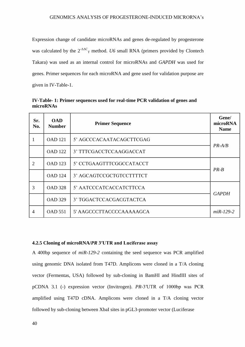
GENOMICS ANALYSIS OF PROGESTERONE-INDUCED MICRORNA‘s
40
Expression change of candidate microRNAs and genes de-regulated by progesterone
was calculated by the 2-ΔΔC
T method. U6 small RNA (primers provided by Clontech
Takara) was used as an internal control for microRNAs and GAPDH was used for
genes. Primer sequences for each microRNA and gene used for validation purpose are
given in IV-Table-1.
4.2.5 Cloning of microRNA/PR 3′UTR and Luciferase assay
A 400bp sequence of miR-129-2 containing the seed sequence was PCR amplified
using genomic DNA isolated from T47D. Amplicons were cloned in a T/A cloning
vector (Fermentas, USA) followed by sub-cloning in BamHI and HindIII sites of
pCDNA 3.1 (-) expression vector (Invitrogen). PR-3′UTR of 1000bp was PCR
amplified using T47D cDNA. Amplicons were cloned in a T/A cloning vector
followed by sub-cloning between XbaI sites in pGL3-promoter vector (Luciferase
Sr.
No.
OAD
Number Primer Sequence
Gene/
microRNA
Name
1 OAD 121 5‘ AGCCCACAATACAGCTTCGAG
PR-A/B
OAD 122 3‘ TTTCGACCTCCAAGGACCAT
2 OAD 123 5‘ CCTGAAGTTTCGGCCATACCT
PR-B
OAD 124 3‘ AGCAGTCCGCTGTCCTTTTCT
3 OAD 328 5‘ AATCCCATCACCATCTTCCA
GAPDH
OAD 329 3‘ TGGACTCCACGACGTACTCA
4 OAD 551 5' AAGCCCTTACCCCAAAAAGCA miR-129-2
IV-Table- 1: Primer sequences used for real-time PCR validation of genes and
microRNAs
GENOMICS ANALYSIS OF PROGESTERONE-INDUCED MICRORNA‘s
41
Expressing vector, Promega). For the Luciferase assay, 293FT cells (50,000 cells/well)
were transfected using lipofectamine 2000 reagent (Life Technologies) with the
combination of these constructs along with Renilla luciferase vector (for normalizing
transfection efficiency) and 5nM miR-inhibitors (SIGMA, HSTUD0162) in separate
wells. 48 hours post-transfection cells were lyzed and luciferase assay was performed
to measure luminescence (Berthold Luminometer, Germany). Experiment was
performed in triplicates and differences between group showing p-values <0.05
(calculated using an unpaired student‘s t-test) were considered significant.
4.2.6 Transfection of microRNA inhibitor in breast cancer cells:
T47D cells were grown up to 60% confluence and transfected with 25nM negative
control microRNA inhibitor (against miR-29a) and miR-129-2 inhibitor (SIGMA,
HSTUD0162). Post-transfection, cells were incubated for 48h and then treated with
progesterone for 6h. Cell lysate was prepared and western blot analysis was performed.
4.3 Results:
4.3.1 Identification of progesterone responsive microRNAs targeting PR
expression in breast cancer cells
Consistent with earlier reports, we observed down-regulation of PR transcripts when
T47D and BT474 cells were treated with 10nM progesterone for 6h (IV-Figure-1a).
Similarly, progesterone reduced the expression level of PR protein in T47D cells (IV-
Figure-1c). To understand the role of microRNA‘s involved in regulation of PR
expression, we performed small RNA sequencing of three PR-positive T47D, BT474,
MCF7 and one PR-negative MDA-MB-231 cell line for identifying microRNAs

GENOMICS ANALYSIS OF PROGESTERONE-INDUCED MICRORNA‘s
42
which could down-regulate PR expression in response to progesterone treatment for
6h. On an average we obtained 22 million reads per sample per cell line. The sequence
reads were mapped to human microRNA sequences obtained from miRBase (version
21) to identify median 800 mature microRNA sequences. These microRNA reads
were used for identification of differentially expressed microRNAs. We used a fold-
change cut-off of 3-fold difference and observed that progesterone had an effect in
both directions by up-regulating and down-regulating the microRNAs and all the four
cells had different number of de-regulated microRNAs (IV-Table-2). Of these, 98
microRNAs were up-regulated in T47D, 96 in BT474, 189 in MCF7 and 106 in
MDA-MB-231 cells in response to progesterone. Intriguingly, expression of miR-
513a-5p shown to be differentially up-regulated in response to synthetic progestin
(medroxy progesterone acetate, MPA) by microarray-based analysis in T47D cells
was not observed in any of the four breast cancer cells in this study [155], possibly
due to variable downstream effects elicited by synthetic progestin (MPA) and
progesterone [103] or distinct platform specific threshold involved in these studies.
The up-regulated microRNAs found across the four breast cancer cells were further
used to search microRNAs targeting 3'UTR of PR gene. Of the 6 different algorithms
used, we found three microRNAs (miR-3908, miR-129-2-3p and miR-3140-3p) that
were predicted to target 3' UTR of PR and showed an increased expression relative to
levels in control. When expression of these microRNAs was checked in the TCGA
breast cancer cohort (n=359), only miR-129-2 was found to be expressed. Next, the
up-regulation of miR-129-2 in response to progesterone could be validated by real-
time PCR in our panel of cells (IV-Figure-1b). While the progestin-regulated miR-
513a-5p could be validated only at 100nM progestin (MPA) as reported by Cochrane

GENOMICS ANALYSIS OF PROGESTERONE-INDUCED MICRORNA‘s
43
et al. [155], we could validate consistent up-regulation of miR-129-2 in response to
10nM progesterone that was used for small RNA sequencing analysis, inclusive of
PR-negative breast cancer cells (IV-Figure-1b). Thus we observed that progesterone
mediated up-regulation of miR-129-2 was independent of the PR expression of cells.
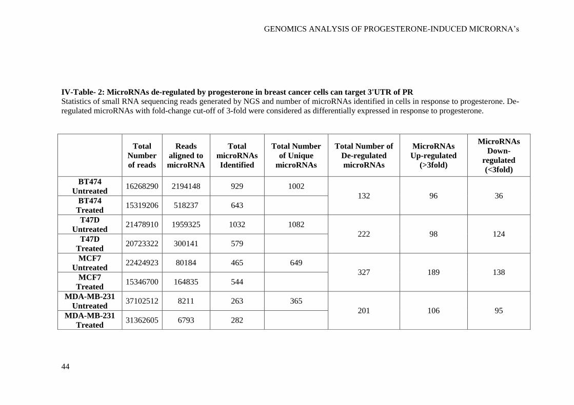
GENOMICS ANALYSIS OF PROGESTERONE-INDUCED MICRORNA‘s
44
IV-Table- 2: MicroRNAs de-regulated by progesterone in breast cancer cells can target 3'UTR of PR
Statistics of small RNA sequencing reads generated by NGS and number of microRNAs identified in cells in response to progesterone. De-
regulated microRNAs with fold-change cut-off of 3-fold were considered as differentially expressed in response to progesterone.
Total
Number
of reads
Reads
aligned to
microRNA
Total
microRNAs
Identified
Total Number
of Unique
microRNAs
Total Number of
De-regulated
microRNAs
MicroRNAs
Up-regulated
(>3fold)
MicroRNAs
Down-
regulated
(<3fold)
BT474
Untreated 16268290 2194148 929 1002
132 96 36 BT474
Treated 15319206 518237 643
T47D
Untreated 21478910 1959325 1032 1082
222 98 124 T47D
Treated 20723322 300141 579
MCF7
Untreated 22424923 80184 465 649
327 189 138 MCF7
Treated 15346700 164835 544
MDA-MB-231
Untreated 37102512 8211 263 365
201 106 95 MDA-MB-231
Treated 31362605 6793 282

GENOMICS ANALYSIS OF PROGESTERONE-INDUCED MICRORNA‘s
45
IV-Figure- 1: Progesterone receptor is down-regulated in breast cancer cell lines
in response to progesterone (modified from the manuscript)
a) Transcript levels of PR were measured using real-time PCR in T47D and BT474
cells treated with 10nM progesterone for 6h. Graph has been plotted as fold change
expression of PR normalized to GAPDH in progesterone-treated versus control.
Analysis is representative of three independent experiments and P-value was
calculated using student‘s unpaired t-test. b) Transcript levels of miR-129-2 under
similar progesterone treatment conditions were measured by real-time PCR and
plotted as fold change in progesterone versus control of T47D, BT474, MCF7 and
MDA-MB-231 cells obtained after normalization to expression of U6 small RNA.
Transcript levels in both control and progesterone-treated cells have been shown.
Analysis is representative of three independent experiments and P-value was
calculated using student‘s unpaired t-test. ** indicates P-value <0.001; *** indicates
P-value <0.0001. c) Western blot analysis of PR (PR-A and PR-B) in response to
progesterone treatment in T47D cells. β-actin was used internal protein loading
control. Numbers on blot indicate ratio of intensity of PR with respect to β-actin for
each lane. Western blot analysis is representative of two independent experiments. d)
Western blot analysis of PR (PR-A and PR-B) in T47D cells treated with either anti-
miR-control or anti-miR-129-2. As indicated in the panel, cells were either treated
with progesterone or untreated. β-actin was used as internal protein loading control.
Numbers on blot indicate ratio of intensity of PR with respect to β-actin for each lane.
Western blot analysis is representative of two independent experiments.

GENOMICS ANALYSIS OF PROGESTERONE-INDUCED MICRORNA‘s
46
4.3.2 Functional validation of miR-129-2 based regulation of progesterone
receptor
In our attempt to functionally characterize the association of miR-129-2 with PR, a
~1000 bp 3‘UTR of PR (containing seed sequence for miR-129-2) was cloned
downstream to luciferase gene in a pGL3-promoter vector. The sequence for
premature miR-129-2 (~400bp) was cloned in pcDNA3.1 vector. Three set of
transfections were performed— pGL3-PR 3‘UTR; pGL3-PR 3‘UTR with pcDNA-
3.1-miR-129-2; and combination of pGL3-PR 3‘UTR, pcDNA3.1-miR-129-2 and anti-
miR-129-2 (IV-Figure-2a). The expression of firefly luciferase gene was checked
using luciferase reporter assay system (Promega) and normalized to renilla expression,
which was used as an internal control, in each of these sets. Our analysis suggests that
upon over-expression of miR-129-2, the luciferase signal was significantly reduced as
compared to signal in vector only cells. Addition of microRNA inhibitor against miR-
129-2 (anti-miR-129-2), a double stranded RNA sequence which is complimentary to
and specifically targets miR-129-2, reversed the repression and showed an increase in
luciferase signal (IV-Figure-2b).

GENOMICS ANALYSIS OF PROGESTERONE-INDUCED MICRORNA‘s
47
IV-Figure- 2: Validation of miR-129-2-based regulation of PR
a) pCDNA3.1-miR-129-2 and pGL3- PR 3‘UTR in different combinations with anti-
miR-129-2 were co-transfected in 293FT cells and luciferase signal in each condition
was measured, as shown in the figure. b) Quantified luminescence units normalized to
renilla expression was plotted for each of the sets mentioned above. Analysis is
representative of three independent experiments and the P-value was calculated using
student‘s t-test. ** indicates P-value <0.001; *** indicates P-value <0.0001.
Next, we inhibited miR-129-2 in T47D cells and compared the PR expression in these
cells with the expression in cells transfected with negative control (targeting miR-29a)
inhibitor. Our western blot analysis suggests that upon exposure to progesterone,
T47D cells transfected with negative control showed decrease in PR expression, while
PR showed stable expression in cells transfected with miR-129-2 inhibitor even in the
presence of progesterone treatment (IV-Figure-1d). Thus our results provide basis for

GENOMICS ANALYSIS OF PROGESTERONE-INDUCED MICRORNA‘s
48
direct interaction of miR-129-2 with PR, where in addition to previous findings, we
demonstrate that over-expression of miR-129-2 mimics the effect of progesterone
treatment to down-regulate PR and that inhibition of miR-129-2 abrogates its
interaction with PR in breast cancer cells. Taken together, these studies emphasize the
plurality in microRNA-mediated feedback regulation of PR.
Next, we analyzed the TCGA breast cancer cohort (n=359) for studying expression of
PR in breast cancer patients with high miR-129-2 expression and in absence of miR-
129-2 expression. When expression of PR was checked in patients with high miR-129-
2 expression (n=134) versus patients with absence of miR-129-2 expression (n=225)
we observed a significantly higher expression of PR in patients with absence of miR-
129-2 expression as compared to patients with high expression of miR-129-2
(P=0.0002) as shown in IV-Figure-3. Thus a further in-depth analysis needs to be
carried out to ascertain the exact role of miR-129-2 in survival of breast cancer
patients.

GENOMICS ANALYSIS OF PROGESTERONE-INDUCED MICRORNA‘s
49
IV-Figure- 3: Expression of miR-129-2 in breast cancer patients in TCGA dataset
(Modified from manuscript)
Expression plot for PR in breast cancer patients with high miR-129-2 expression
(n=134) and with absence of miR-129-2 expression (n=225) in the TCGA cohort. The
box-plot is overlaid with dot-plot wherein each point represents patient sample. Y-axis
indicates normalized read count (RSEM) values for PR in a total of 359 breast cancer
patients where expression of PR and miR-129-2 was available. P-value (P=0.0002)
was calculated using student‘s unpaired t-test with Welch‘s correction. (Comment:
The TCGA provides only level-3 data as openly accessible, which lacks adjacent
normal sample data. Thus, to get the best possible contrast between the two groups
using available (only tumor) dataset, we restricted to top quartile (high) miR-129-2
expressing patients versus no miR-129-2 expressing patients for PR expression,
wherein we observe a significant anti-correlation in their expression)
4.4 Discussion
Small RNA sequencing analysis of progesterone treated breast cancer cell lines led to
the identification of a novel PR-targeting microRNA miR-129-2. Since the increased
expression of miR-129-2 was independent of the PR-status of breast cancer cells, a
possible role of other steroid hormone receptors like membrane progesterone receptor
or glucocorticoid receptor as suggested in literature [77, 99] to mediate the role of
progesterone in these cells remains to be systematically analyzed. Consistent with our
GENOMICS ANALYSIS OF PROGESTERONE-INDUCED MICRORNA‘s
50
finding, analysis of TCGA breast cancer dataset suggests a significantly decreased
expression of PR in patients with elevated expression of miR-129-2 as compared to
patients with no miR-129-2 expression, indicating a possibility for the decreased
expression of PR in patients with low PR expression. It has been observed that factors
like loss of PR or menopausal status of women can alter the response to hormonal
therapy [157]. Some studies have indicated that the absence of PR could underlie
tumors resistance to hormonal therapy [59], or could potentially increase the risk of
relapse [158]. Hence we propose that stabilization of PR expression in patients with
tumors expressing low PR levels by blocking activity of such microRNAs using
specific microRNA inhibitors, along with other treatment modalities, could be
potentially helpful in enhancing the response of patients to hormonal therapies. In
support of this notion, our in vitro luciferase assay and western blot results using miR-
129-2 inhibitor suggest that inhibition of miR-129-2 can increase the expression of PR.
Thus, we validate under in vitro settings that addition of progesterone leads to up-
regulation of miR-129-2, which suppresses the expression of PR in breast cancer cells;
and, the inhibition of miR-129-2 reinstates the PR expression in these breast cancer
cells even in presence of progesterone. Also since microRNAs are being assessed for
their use in clinics [159], strategies like microRNA sponges and chemically modified
antisense oligonucleotides (inhibitors) hold promise as a promising line of treatment
of breast cancer that need to be exhaustively explored with larger datasets [160]. Thus,
our study suggests an underlying mechanism to a possible clinical consequence in
response to progesterone treatment among patients with varying PR expression levels.
Also, it is suggestive of treatment with anti-miR-129-2 among those patients
expressing inadequate PR levels, under adjuvant and neo-adjuvant settings, before

GENOMICS ANALYSIS OF PROGESTERONE-INDUCED MICRORNA‘s
51
considering for hormonal therapy. Whether modulation of activity of miR-129-2 could
stabilize PR expression and potentially improve response to hormonal therapy remains
to be validated as an immediate follow up to this pilot study.
SGK1/AP-1/NDRG1 AXIS MEDIATES PROGESTERONE EFFECT IN BREAST
CANCER
52
V. DIFFERENTIAL REGULATION OF SGK1 BY
PROGESTERONE ACTIVATES AP-1/NDRG1 GENOMIC AXIS IN
PR-POSITIVE AND NEGATIVE BREAST CANCER CELLS
Abstract
Pre-operative progesterone intervention has been shown to confer a survival benefit to
breast cancer patients independent of their progesterone receptor (PR) status, raising a
question about how progesterone affects the outcome of PR-negative cells. Here, we
identify up-regulation of a Serum- and glucocorticoid-regulated kinase gene, SGK1
and an N-Myc Downstream Regulated Gene 1, NDRG1, along with down-regulation
of miR-29a and miR-101-1 targeting 3‘UTR region of SGK1, to differential extents in
a PR dependent manner in breast cancer cells. We further demonstrate a novel dual-
phase transcriptional and post-transcriptional regulation of SGK1 in response to
progesterone leading to up-regulation of a tumor metastasis suppressor gene, NDRG1,
mediated by a set of AP-1 network genes. The NDRG1 further inactivates a set of
kinases impeding the invasion and migration of breast cancer cells. In summary, we
propose a model for the mode of action of progesterone in breast cancer deciphering
the molecular basis of a randomized clinical trial studying the effect of progesterone
in breast cancer with a potential to improve the prognosis of breast cancer patients for
receiving pre-operative progesterone treatment.

SGK1/AP-1/NDRG1 AXIS MEDIATES PROGESTERONE EFFECT IN BREAST
CANCER
53
5.1 Introduction:
The increasing complexity of multicellular organisms correlates with the increasing
number of microRNAs than the number of coding genes encoded by the genome [161,
162], reflecting a gradual increase in the extent and intricacy of gene regulation [163].
Hierarchically, microRNAs function downstream of transcriptional regulation of
genes since microRNAs represses post transcription of mRNAs [164]. However,
emerging evidence suggests that transcriptional and post-transcriptional regulation is
often highly coordinated [165, 166]. Hormones, for instance, have been hypothesized
to regulate expression of target genes at the transcriptional and post-transcriptional
level [61, 167]. Estrogen up-regulates the expression of progesterone receptor (PR) by
transcriptionally recruiting estrogen receptor (ER) at the promoter, and post-
transcriptionally, by silencing expression of microRNAs targeting 3‘UTR of PR in
breast cancer cells [168]. A similar example for the ATP1B1 gene has been reported
[155]. However, systematic approaches to discern dual-regulated molecular targets of
hormones in breast cancer remains poorly understood.
Understanding the molecular basis of clinical phenomena in response to therapeutic
interventions has been an important point of intersection between medical and
biological sciences. While clinical benefit of preoperative endocrine therapy is well
documented in literature [169, 170], more recently, we described the first randomized
trial with preoperative progesterone resulting in greater than 10% absolute
improvement in 5-year disease-free survival among node-positive breast cancer
patients [171]. Of several hypothesis-generating results from this study, the impact of
progesterone on PR-negative patients particularly lends itself to a systematic
characterization of molecular changes that progesterone may induce in breast cells.

SGK1/AP-1/NDRG1 AXIS MEDIATES PROGESTERONE EFFECT IN BREAST
CANCER
54
Gene expression studies probing the targets of progesterone have either been
performed restrictively in PR-positive breast cancer cell lines or in the presence of
other hormones [58, 80, 92, 172, 173]. Although few studies suggest a beneficial
effect of progesterone, progesterone-responsive genes in PR-negative cells have not
been studied [58, 80, 92, 174]. In order to identify targets of progesterone independent
of PR status of cells, we set to perform an integrated genomic profiling of a panel of
PR-positive and PR-negative breast cancer cell lines treated with progesterone,
followed by functional analysis of the components found to be significantly altered.
This study details the molecular action of progesterone on breast cancer cells,
mediated by the up-regulation of a genomic axis inclusive of a tumor metastasis
suppressor gene in breast cancer, independent of the PR status of cells.
5.2 Materials and Methods:
5.2.1 Breast Cell lines
T47D, BT474, MDA-MB-231, ZR-75-1 and MCF7 breast cancer cell lines were
cultured and the PR/ER/Her2 receptor status confirmed as explained in chapter-III
section 3.2.1 and provided in V-Table-1. The immortalized normal-like breast cell line
184A1 was obtained as a gift from Dr. Slamon's Laboratory (Department of Medicine,
UCLA, USA) and was cultured in DMEM:F12 medium (HiMedia) supplemented with
28.18IU Insulin, 20ng/ml EGF and 500ng/ml Hydrocortisone. All the cell lines were
authenticated using STR profiling (V-Table-2) and made mycoplasma-free as
described in section 3.2.1.

SGK1/AP-1/NDRG1 AXIS MEDIATES PROGESTERONE EFFECT IN BREAST
CANCER
55
V-Table- 1: Selection of breast cell lines and validation of PR/ER/Her2 hormone
receptor status
A panel of breast cell lines with varying receptor status, as reported in literature, was
selected for studying the effect of progesterone independent of the receptor status. The
PR/ER/Her2 transcript expression of all the cell lines was confirmed by reverse
transcriptase-PCR and using gene expression array analysis. The RT-PCR analysis for
PR/ER/Her2 for T47D, MCF7, BT474, ZR-75-1 and MDA-MB-231 is as described
earlier [174] and chapter-III section 3.3.
V-Table- 2: STR profiling of breast cell lines
Sr.
No
Breast Cell
lines
Literature reported
Receptor status
Validation of Receptor status at Expression level
Expression array
analysis
RT-PCR
PR ER HER2 PR ER HER2 PR ER HER2 GR PGRMC1 SERBP1
1 T47D + + - + + - + + - + + +
2 MCF7 + + - + + - + + - + + +
3 BT474 + + + + + + + + + + + +
4 ZR-75-1 - + - - + - - + - + + +
5 MDA-MB-231 - - - - - - - - - + + +
6
184A1
(Immortalized
cell line)
- - - - - - - - - + + +
Query
for STR
10-marker STR profile as per GeneMarker HID software Authenticity
of cell line
as identified TH01 D21S11 D5S818 D13S317 D7S820 D16S539 CSF1PO AMEL vWA TPOX %
Match
T47D 6 28,31 12 12 11 10 11,13 X 14 11 100 T47D
BT-474 7,8 28,32.2 11,13 11 9,12 9,11 10,11 X 15,16 8 100 BT-474
MCF7 6 30 11,12 11 8,9 11,12 10 X 14,15 9,12 100 MCF7
MD-231 7,9.3 30,33.2 12 13 8,9 12 12,13 X 15,18 8,9 100 MD-231
ZR-75-1 8,9.3 29 13 9 8,11 11 10,11 X 16,18 8 92% ZR-75-1
184A1 9.3 29,30 11,13 11 9,11 11,12 10,11 X 18,19 11 100 184A1
SGK1/AP-1/NDRG1 AXIS MEDIATES PROGESTERONE EFFECT IN BREAST
CANCER
56
5.2.2 Progesterone treatment and RNA isolation
Breast cancer cells were treated with progesterone and RNA isolation was performed
as described earlier [174, 175] and in chapter-IV section 4.2.2. Additionally, in case of
Mifepristone+Progesterone (M+P) combination treatment, 100nM RU486
(Mifepristone) was added to the cells for 2h followed by 10nM Progesterone
treatment for 6h in the same medium. Equal amount of alcohol was used as vehicle
control. The treatment conditions for progesterone and mifepristone were standardized
based on the expression change of three known candidate genes (V-Figure-3A).
Various time durations and progesterone concentration combinations were tried and a
consistent up-regulation of STAT5A, EZF and F3 was obtained upon treatment with
10nM progesterone for 6hrs, consistent with [109]. Similarly, 2hrs treatment with
100nM mifepristone led to inhibition of expression of these candidate genes. Both
these time and concentration combinations have been used consistently for the
progesterone and mifepristone treatment of cells.
5.2.3 Gene Expression profiling
Gene expression profiling was performed using BeadChip Illumina microarray
platform. Raw data (.idat files) of BeadChip Illumina platform were converted to
readable format using Genome studio software (version V2011.1). Probe level data
were converted into gene centric and used for further processing. Bioconductor lumi
package was used for pre-processing the data which includes quality control steps,
background correction, normalization, log transformation etc. and finally differential
gene analysis. Robust Spline Normalisation (RSN) and Variance Stabilization

SGK1/AP-1/NDRG1 AXIS MEDIATES PROGESTERONE EFFECT IN BREAST
CANCER
57
Transformation (VST) methods were used for normalization and transformation
respectively. To specifically select for highly variable genes, unexpressed, non-
annotated and false positive genes were excluded from the analysis. Median Absolute
Deviation (MAD) was carried out to make samples comparable to each other. The
differential gene expression cut-off was set as 0.5 ≤ log (Fold Change) ≤ -0.5 in at
least one out of the three conditions (Control; Progesterone;
Mifepristone+Progesterone). Heatmaps were constructed using MeV software
(version 4.9.0) (V-Figure-1). All possible comparisons were taken into consideration
as Progesterone versus Control (P Vs C), Mifepristone + Progesterone versus Control
(M+P Vs C) and Progesterone versus Mifepristone + Progesterone (P Vs M+P). From
our analysis, we identified 623, 553, 1873, 532, 1764 and 4703 differentially
expressed genes in T47D, MCF7, BT474, ZR-75-1, MDA-MB-231 and 184A1 breast
cell lines respectively.

SGK1/AP-1/NDRG1 AXIS MEDIATES PROGESTERONE EFFECT IN BREAST
CANCER
58
V-Figure- 1: Gene expression profile of breast cell lines with different receptor
statuses
Heat map representation of gene expression profile for each breast cell line (n=6) has
been shown, with three treatment conditions viz. control, progesterone or
mifepristone+progesterone (M+P). In the figure, red lines indicate up-regulation;
black lines indicate no change; green lines indicate down-regulation of gene in
response to the treatment conditions. The differential gene expression cut-off was set
as 1.5 ≤ log (Fold Change) ≤ -1.5. Numbers in square bracket indicate total number of
differentially expressed genes for each cell line.
5.2.4 Integrated Analysis
A weighted gene co-expression network was constructed based on the gene expression
data to identify gene modules up-regulated in response to progesterone [176] across
all the breast cancer cells. SGK1 and NDRG1 were found to be the top up-regulated
and recurrent gene in response to progesterone across multiple cells (V-Figure-2).
Next, we analyzed our small RNA sequencing data (described in chapter-IV, section
4.2.3) to identify differentially expressed microRNA in response to progesterone
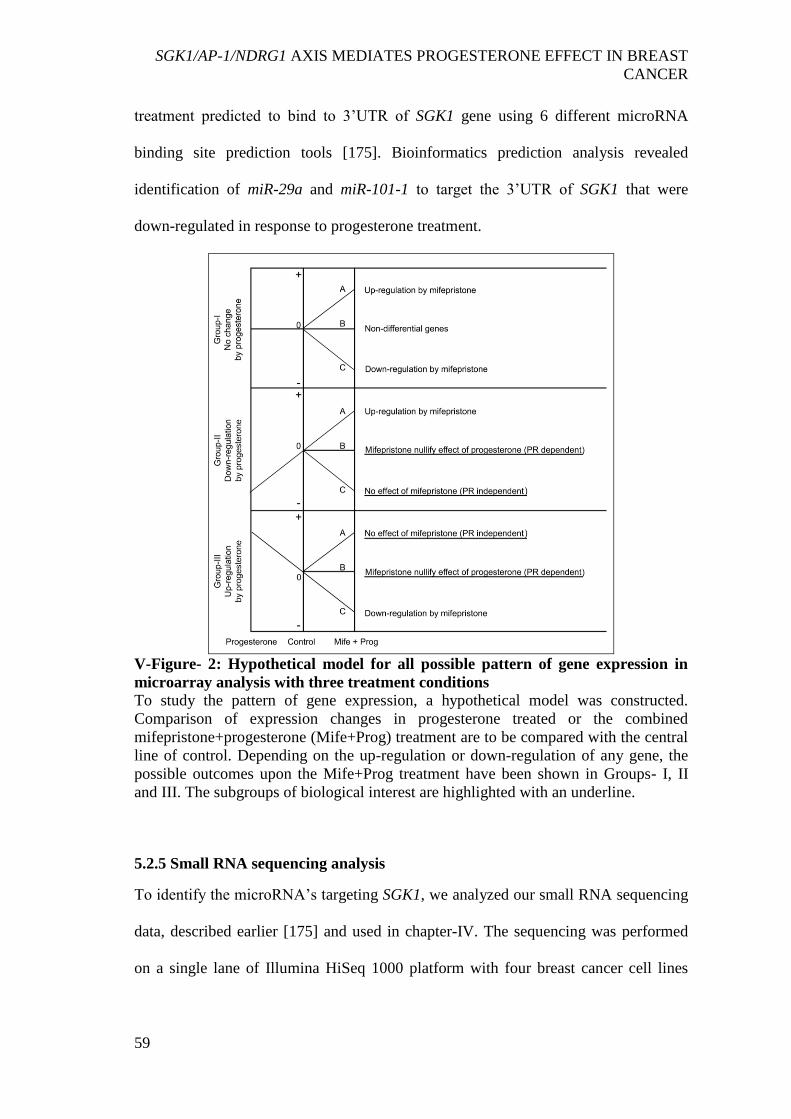
SGK1/AP-1/NDRG1 AXIS MEDIATES PROGESTERONE EFFECT IN BREAST
CANCER
59
treatment predicted to bind to 3‘UTR of SGK1 gene using 6 different microRNA
binding site prediction tools [175]. Bioinformatics prediction analysis revealed
identification of miR-29a and miR-101-1 to target the 3‘UTR of SGK1 that were
down-regulated in response to progesterone treatment.
V-Figure- 2: Hypothetical model for all possible pattern of gene expression in
microarray analysis with three treatment conditions
To study the pattern of gene expression, a hypothetical model was constructed.
Comparison of expression changes in progesterone treated or the combined
mifepristone+progesterone (Mife+Prog) treatment are to be compared with the central
line of control. Depending on the up-regulation or down-regulation of any gene, the
possible outcomes upon the Mife+Prog treatment have been shown in Groups- I, II
and III. The subgroups of biological interest are highlighted with an underline.
5.2.5 Small RNA sequencing analysis
To identify the microRNA‘s targeting SGK1, we analyzed our small RNA sequencing
data, described earlier [175] and used in chapter-IV. The sequencing was performed
on a single lane of Illumina HiSeq 1000 platform with four breast cancer cell lines
SGK1/AP-1/NDRG1 AXIS MEDIATES PROGESTERONE EFFECT IN BREAST
CANCER
60
(T47D, BT474, MCF7 and MDA-MB-231). For identifying microRNAs targeting
3‘UTR of SGK1, differentially expressed microRNAs in response to progesterone
were overlapped with microRNAs predicted to target SGK1 using the 6 algorithms
used in our earlier study [175].
5.2.6 Quantitative Real-time PCR
Transcript levels of candidate genes and microRNAs were analysed by quantitative
real time PCR as previously described for genes and microRNAs [174, 175]. Briefly,
cDNA from each cell line with the three conditions (control, progesterone and M+P)
were then subjected to qRT-PCR analysis. GAPDH gene was used as an internal
control and average of CT values from each condition was used to normalize the CT
values of candidate genes in each cell line. In case of microRNAs, U6 small RNA was
used as an internal control for normalizing the expression of microRNAs. Expression
change of candidate genes and microRNAs was calculated by the 2-ΔΔC
T method.
Primer sequences used for real-time PCR validation of genes and microRNAs have
been provided in V-Table-3.

SGK1/AP-1/NDRG1 AXIS MEDIATES PROGESTERONE EFFECT IN BREAST
CANCER
61
V-Table- 3: Primer sequences used for real-time PCR validation of genes and
microRNAs
5.2.7 Over-expression and knockdown studies
For over-expression of SGK1, two retrovirus-based constructs were used-- pBABE-
puro-SGK1 which expresses wild type SGK1 (WT-SGK1); while pWZL-Neo-Myr-
Sr.
No
OAD
Number Primer sequence
Gene/
microRNA
name
1. OAD 137 5‘ GGCGCTTCAGGCACTACAA F3
OAD 138 3‘ TTGATTGACGGGTTTGGGTTC
2. OAD 139 5‘ GCAGAGTCCGTGACAGAGG STAT5A
OAD 140 3‘ CCACAGGTAGGGACAGAGTCT
3. OAD 141 5‘ CCCACATGAAGCGACTTCCC EZF
OAD 142 3‘ CAGGTCCAGGAGATCGTTGAA
4. OAD 233 5‘ GCAGAAGAAGTGTTCTATGCAGT SGK1
OAD 234 3‘ CCGCTCCGACATAATATGCTT
5. OAD 457 5‘ GCCTCCTTCCCCGCAGGG NDRG1
OAD 458 3‘ GCCCAAACTGTTGAAGGACTCC
6. OAD 567 5‘ GCATTGGCAGGAGGGGCAAGG FOS
OAD 568 3‘ CAGCTCCCTCCTCCGGTTGCG
7. OAD 577 5‘ CCCAAGAACGTGACAGATGAG JUN
OAD 578 3‘ TGCCCCGTTGACCGGCTGC
8. OAD 573 5‘ GGCGAGCAGCCCTACGAGC EGR1
OAD 574 3‘ GTATAGGTGATGGGGGGCAGTC
9. OAD 571 5‘ CCTGCAGTACCCCACTCTACG DUSP1
OAD 572 3‘ CCCAAGGCATCCAGCATGTCC
10. OAD 328 5‘ AATCCCATCACCATCTTCCA GAPDH
OAD 329 3‘ GGACTCCACGACGTACTCA
11. OAD 552 5‘ TAGCACCATCTGAAATCGGTTA miR-29a
12. OAD 625 5‘ CAGTTATCACAGTGCTGATGCT miR-101-1
SGK1/AP-1/NDRG1 AXIS MEDIATES PROGESTERONE EFFECT IN BREAST
CANCER
62
Flag-SGK1 expresses myristoylated SGK1 (Myr-SGK1) (a kind gift from Dr. Shaida
Andrabi, University of Kashmir). Over-expression clones for each of the constructs
were selected in 1µg puromycin and 1600µg neomycin, respectively. Untransfected
T47D and MD-231 cells were used as control for over-expression of SGK1. For the
knockdown of SGK1, NDRG1 and EGR1 genes in T47D and MDA-MB-231 cells,
three lentiviral shRNA constructs (PLATINUM Select Human MLP lentiviral
shRNA-mir vector, Transomic technologies) each, against these genes, were used for
genetic depletion. sh-non-targeting (sh-NT) was used as vector/scrambled control.
Positive clones were selected using 1µg puromycin. Over-expression and knockdown
experiments were performed in T47D (PR-positive) and MDA-MB-231 (PR-negative)
breast cancer cells.
5.2.8 Protein sample preparation and Western blot analysis
Protein samples were prepared and western blots were developed as described earlier
[174]. Briefly, cells were serum starved and treated with progesterone for 8h or left
untreated. Cell lysates were prepared and equal amounts of lysate were resolved using
10% SDS-PAGE and transferred to a PVDF membrane by wet-transfer method. The
immunoblots were then incubated with primary antibodies against SGK1 (Cell
Signaling Technology, 1210S, Dilution 1:800); NDRG1 (Cell Signaling Technology,
9485S, Dilution 1:800); p-NDRG1 (Cell Signaling Technology, 5482S, Dilution
1:800); p-SGK1 (Abcam, ab55281, Dilution 1:500); EGR1 (Santa Cruz
Biotechnology, sc-515830, Dilution 1:1000); β-actin (I-19)-R (Santa Cruz
Biotechnology, sc-1616-R; Dilution 1:3000); p-EGFR (Y1068) (Cell Signaling,
3777S, Dilution 1:500), p-Akt (S473) (Cell Signaling, 4060S, Dilution 1:500); p-

SGK1/AP-1/NDRG1 AXIS MEDIATES PROGESTERONE EFFECT IN BREAST
CANCER
63
ERK1/2 (T202/Y204) (Cell Signaling, 9101S, Dilution 1:1000); EGFR (1005) (Santa
Cruz Biotechnology, sc-03, Dilution 1:1000); AKT (11E7) (Cell Signaling, 4685S,
Dilution 1:1000); and ERK2 (c-14) (Santa Cruz Biotechnology, sc-154, Dilution
1:1000). Goat anti-rabbit IgG-HRP (Santa Cruz Biotechnology, sc-2004, Dilution
1:3000); and goat anti-mouse IgG-HRP (Santa Cruz Biotechnology, sc-2005, Dilution
1:3000) were used as secondary antibodies.
5.2.9 Treatment with SGK1 inhibitor
Cells were grown till 70-80% confluence in a 6-well dish and then serum-starved
using low glucose DMEM medium (HiMedia) for 24hrs. 1.0µM concentration of
GSK650394A (SGK1-inhibitor) was added to respective wells for 4hrs, as discussed
in other studies [2]. After 4hrs, medium was removed and fresh low-glucose medium
was added to cells. Where indicated, cells were then treated with 10nM progesterone
for 6hrs and then used for RNA or protein isolation.
5.2.10 Invasion Assay
Cell invasion assay was performed as described earlier [174]. Briefly, 35000 cells
were allowed to invade matrigel in boyden chamber for 16-18h at 37ºC. Cells were
observed under upright microscope, ten random fields were chosen and number of
cells in each field were counted and plotted as percentage cell invasion.
SGK1/AP-1/NDRG1 AXIS MEDIATES PROGESTERONE EFFECT IN BREAST
CANCER
64
5.2.11 Wound healing assay
Wound scratch migration assay and analysis was performed as described earlier [174]
and section 3.2.8 of Chapter-III. Briefly, confluent cell monolayer in a 6-well plate
was serum-starved for 18hrs or treated with mitomycin-C for 2hrs and subjected to a
scratch manually with a sterile small pipette tip. Cell culture media was replaced with
low-glucose phenol-red free DMEM containing 10% charcoal-stripped FBS. Cell
migration was monitored for 20hrs and distance traversed by cells was quantified
using ImageJ.
5.2.12 Dual-luciferase assay with microRNAs/SGK1 3’UTR
Cloning of microRNA sequences and SGK1 3‘UTR was performed as described
earlier [175]. Briefly, a 400bp sequence of miR-29a and miR-101-1 containing the
seed sequence was PCR amplified using genomic DNA isolated from T47D.
Amplicons were cloned in a T/A cloning vector (Fermentas, USA) followed by sub-
cloning in BamHI and HindIII sites of pCDNA 3.1 (-) expression vector (Invitrogen).
SGK1-3′UTR of 1000bp was PCR amplified using T47D cDNA. Amplicons were
cloned in a T/A cloning vector followed by sub-cloning between XbaI sites in a
pGL3-promoter vector (Luciferase Expressing vector, Promega). For the dual-
luciferase assay, 293FT cells (50,000 cells/well) were transfected using lipofectamine
2000 reagent (Life Technologies) with combination of these constructs along with a
Renilla luciferase vector (for normalizing transfection efficiency) in separate wells.
5nM miR-inhibitors (anti-miR‘s) (SIGMA) were also transfected in combination to
expression vectors for specifically inhibiting the activity of both the microRNAs. 48
hours post-transfection, cells were lyzed and luciferase assay was performed to

SGK1/AP-1/NDRG1 AXIS MEDIATES PROGESTERONE EFFECT IN BREAST
CANCER
65
measure Firefly luciferase activity post normalization to Renilla luciferase values
(Berthold Luminometer, Germany). Experiment was performed in triplicates and
differences between group showing P-values <0.05 (calculated using an unpaired
student‘s t-test) were considered significant.
5.2.13 Transfection of microRNA inhibitor in breast cancer cells
T47D cells were grown up to 60% confluence and transfected with 25nM negative
control miR-inhibitor (anti-miR-129-2); anti-miR-29a; and anti-miR-101-1 (SIGMA).
Post-transfection, cells were incubated for 48h and then treated with progesterone for
6h as described above. Cell lysate was prepared and western blot analysis was
performed to study expression of SGK1.
5.2.14 Statistical analysis
The statistical analysis was performed using GraphPad Prism software (GraphPad,
version 5). Student‘s unpaired t-test was used to determine the statistical significance.
5.3 Results:
5.3.1 Gene expression analyses reveal a novel dual-phase regulation of SGK1 by
progesterone in breast cancer cells
An integrated analysis of micro array based mRNA expression profile and deep
sequencing of non-coding small RNA of breast cancer cells (as described in 5.2.4 and
5.2.5) led us to identify up-regulation of a Serum- and Glucocorticoid-regulated
Kinase gene, SGK1 and a N-Myc Downstream Regulated Gene 1, NDRG1, along with

SGK1/AP-1/NDRG1 AXIS MEDIATES PROGESTERONE EFFECT IN BREAST
CANCER
66
down-regulation of miR-29a and miR-101-1, predicted to bind the 3‘UTR region of
SGK1, independent of the hormonal receptor status of the cells (V-Figure-3; V-Table-
4 and V-Table-5).
V-Figure- 3: Identification and validation of candidate genes and standardization
of progesterone treatment in breast cancer cells
A) Representation of expression array results; left panel indicates the WGCNA results
with SGK1 being the top up-regulated gene in cells indicated, in response to
progesterone. Figure panel on the right indicates results of the recurrent gene
expression analysis. Genes de-regulated in more than three cell lines were considered
to be recurrently expressed in response to progesterone. Red colour in the figure
indicates up-regulation, blue colour indicates down-regulation, and no colour/blank
stands for no change in gene expression in response to progesterone treatment. B)
Standardization of progesterone treatment was based on expression changes of three
known progesterone regulated genes (STAT5A, EZF and F3), studied using
quantitative real time PCR analysis. Data has been plotted as expression change in
response to progesterone or mifepristone+progesterone treatment, with respect to
control (horizontal black line). Figure is representative of three independent
experiments performed in triplicates in T47D cells (PR-positive). C) Real time PCR
validation for expression of SGK1 and NDRG1 in T47D cells (PR-positive) in
response to progesterone and mifepristone+progesterone treatment. Data has been
plotted as fold change for individual gene with respect to expression in control cells
and normalized with respect to expression of GAPDH. Figure is representative of two
independent experiments performed in triplicates. P-value was calculated using
student‘s unpaired t-test. * indicates P<0.05; ** indicates P<0.005; *** indicates
P<0.0005; ns indicates not significant.

SGK1/AP-1/NDRG1 AXIS MEDIATES PROGESTERONE EFFECT IN BREAST
CANCER
67
The up-regulation of SGK1, known to harbor multiple progesterone response element
[177, 178], and NDRG1 were observed to be relatively higher among the PR-positive
cells, while miR-29a and miR-101-1 were lower in PR-negative cells in response to
progesterone (V-Figure-4A-D). Interestingly, SGK1 activation (induction of phospho-
SGK1 in response to progesterone) was found to be comparable in breast cancer cells,
regardless of their PR status (V-Figure-4C).
V-Figure- 4: Validation of expression of SGK1 and NDRG1, and miR-29a and
miR-101-1 expression in breast cell lines treated with progesterone
A) Quantitative real-time PCR analysis for validation of expression of SGK1 and
NDRG1 transcripts in breast cell lines in response to progesterone. Expression of both
the genes was normalized with respect to expression of GAPDH in each cell line. Data
has been plotted as fold change for each gene with respect to the expression in control
sample of each cell line. Horizontal black line indicates gene expression in control
cells. Figure is representative of three independent experiments performed in
triplicates. P-value was calculated using student‘s unpaired t-test. * indicates P<0.05,
** indicates P<0.005 and *** indicates P<0.0005. B) Transcript levels of miR-29a
and miR-101-1 were measured using real-time PCR analysis in T47D and MDA-MB-
231 cells treated with progesterone. Graph has been plotted as expression fold change
of the two microRNAs normalized to expression of U6 small RNA in progesterone-
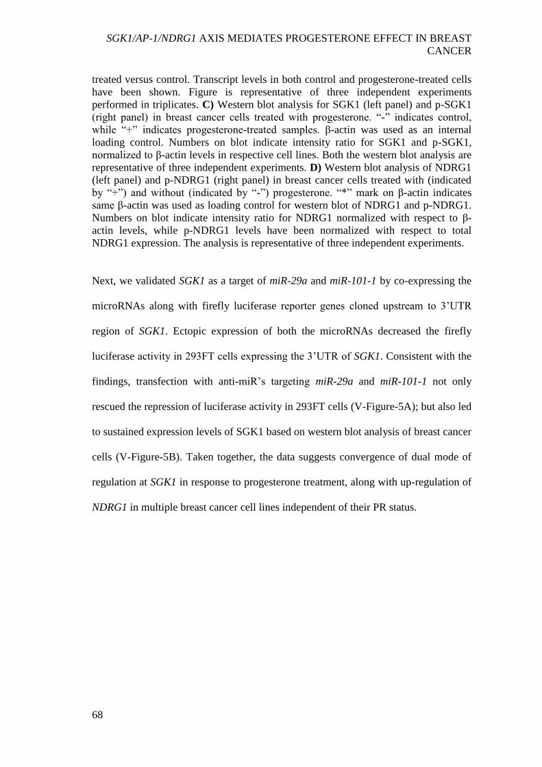
SGK1/AP-1/NDRG1 AXIS MEDIATES PROGESTERONE EFFECT IN BREAST
CANCER
68
treated versus control. Transcript levels in both control and progesterone-treated cells
have been shown. Figure is representative of three independent experiments
performed in triplicates. C) Western blot analysis for SGK1 (left panel) and p-SGK1
(right panel) in breast cancer cells treated with progesterone. ―-‖ indicates control,
while ―+‖ indicates progesterone-treated samples. β-actin was used as an internal
loading control. Numbers on blot indicate intensity ratio for SGK1 and p-SGK1,
normalized to β-actin levels in respective cell lines. Both the western blot analysis are
representative of three independent experiments. D) Western blot analysis of NDRG1
(left panel) and p-NDRG1 (right panel) in breast cancer cells treated with (indicated
by ―+‖) and without (indicated by ―-‖) progesterone. ―*‖ mark on β-actin indicates
same β-actin was used as loading control for western blot of NDRG1 and p-NDRG1.
Numbers on blot indicate intensity ratio for NDRG1 normalized with respect to β-
actin levels, while p-NDRG1 levels have been normalized with respect to total
NDRG1 expression. The analysis is representative of three independent experiments.
Next, we validated SGK1 as a target of miR-29a and miR-101-1 by co-expressing the
microRNAs along with firefly luciferase reporter genes cloned upstream to 3‘UTR
region of SGK1. Ectopic expression of both the microRNAs decreased the firefly
luciferase activity in 293FT cells expressing the 3‘UTR of SGK1. Consistent with the
findings, transfection with anti-miR‘s targeting miR-29a and miR-101-1 not only
rescued the repression of luciferase activity in 293FT cells (V-Figure-5A); but also led
to sustained expression levels of SGK1 based on western blot analysis of breast cancer
cells (V-Figure-5B). Taken together, the data suggests convergence of dual mode of
regulation at SGK1 in response to progesterone treatment, along with up-regulation of
NDRG1 in multiple breast cancer cell lines independent of their PR status.

SGK1/AP-1/NDRG1 AXIS MEDIATES PROGESTERONE EFFECT IN BREAST
CANCER
69
V-Figure- 5: Functional validation of miR-29a and miR-101-1 mediated
regulation of expression of SGK1
A) Quantification of luminescence units normalized to renilla luciferase activity has
been plotted for: pCDNA3.1-miR-29a or pCDNA3.1-miR-101-1 and pGL3-SGK1
3‘UTR in different combinations with anti-miR-29a or anti-miR-101-1 in 293FT cells.
Figure is representative of three independent experiments performed in 293FT cells.
P-value was calculated using student‘s unpaired t-test. * indicates P-value<0.05; **
indicates P-value <0.001; *** indicates P-value <0.0001. B) Western blot analysis of
SGK1 in T47D (PR-positive, top panel) and MDA-MB-231 (PR-negative, bottom
panel) treated with anti-miR-control, anti-miR-29a or anti-miR-101-1. As indicated in
the panel, cells were either treated with progesterone or untreated. β-actin was used as
an internal protein loading control. Numbers on the blot indicate intensity ratio of
expression of SGK1 with respect to the anti-miR-control lane and expression
normalized with respect to individual β-actin levels. The western blot analysis is
representative of three independent experiments.
V-Table- 4: Relative log-fold change of expression of genes-of-interest in
response to progesterone as identified in microarray gene expression analysis
and fold change from real-time PCR analysis
Gene name T47D BT474 MCF7 ZR-75-1 MDA-MB-231
Microarray SGK1 3.510 2.818 0.3748 0.6782 -0.0945
NDRG1 1.818 1.14 0.0890 0.5606 0.2172
Real-time
PCR
SGK1 22.9801 10.0952 1.7566 4.7698 2.1947
NDRG1 3.4690 2.1108 4.5726 3.5433 1.8818
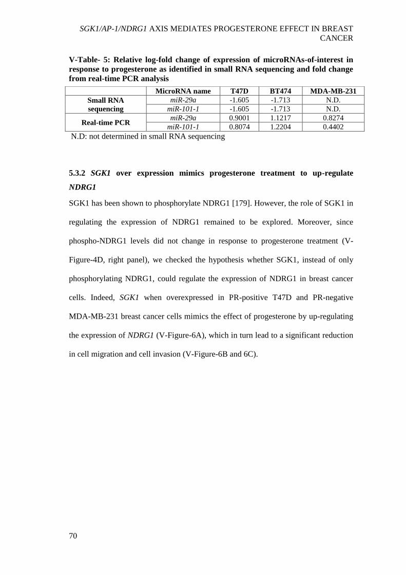
SGK1/AP-1/NDRG1 AXIS MEDIATES PROGESTERONE EFFECT IN BREAST
CANCER
70
V-Table- 5: Relative log-fold change of expression of microRNAs-of-interest in
response to progesterone as identified in small RNA sequencing and fold change
from real-time PCR analysis
N.D: not determined in small RNA sequencing
5.3.2 SGK1 over expression mimics progesterone treatment to up-regulate
NDRG1
SGK1 has been shown to phosphorylate NDRG1 [179]. However, the role of SGK1 in
regulating the expression of NDRG1 remained to be explored. Moreover, since
phospho-NDRG1 levels did not change in response to progesterone treatment (V-
Figure-4D, right panel), we checked the hypothesis whether SGK1, instead of only
phosphorylating NDRG1, could regulate the expression of NDRG1 in breast cancer
cells. Indeed, SGK1 when overexpressed in PR-positive T47D and PR-negative
MDA-MB-231 breast cancer cells mimics the effect of progesterone by up-regulating
the expression of NDRG1 (V-Figure-6A), which in turn lead to a significant reduction
in cell migration and cell invasion (V-Figure-6B and 6C).
MicroRNA name T47D BT474 MDA-MB-231
Small RNA
sequencing
miR-29a -1.605 -1.713 N.D.
miR-101-1 -1.605 -1.713 N.D.
Real-time PCR miR-29a 0.9001 1.1217 0.8274
miR-101-1 0.8074 1.2204 0.4402

SGK1/AP-1/NDRG1 AXIS MEDIATES PROGESTERONE EFFECT IN BREAST
CANCER
71
V-Figure- 6: Ectopic expression of SGK1 mimics the effect of progesterone in
breast cancer cells
A) Western blot analysis indicating expression of SGK1 and NDRG1 in T47D (PR-
positive, left panel) and MDA-MB-231 cells (PR-negative, right panel) over-
expressing SGK1. β-actin was used as an internal loading control. Numbers on blot
indicate intensity ratio for SGK1 and NDRG1, normalized to respective β-actin levels.
The analysis is representative of three independent experiments. B) Cell migration of
T47D (left panel) and MDA-MB-231 (right panel) cells over-expressing SGK1 was
compared to un-transfected parent cells in a wound scratch assay. Bar plots indicate
percentage cellular migration of the cells, with a direct comparison between
untransfected cells and cells over-expressing SGK1 and the analysis is representative
of three independent experiments performed in triplicates. C) In cells over-expressing
SGK1, cell invasion was studied in T47D (top panel) and MDA-MB-231 (bottom
panel) and percent cell invasion was compared with respective parent cells. Parent
cells treated with progesterone were also used to compare the level of cell invasion
upon SGK1-over-expression. Bar plot depicts percentage cell invasion and figure is
representative of three independent experiments, performed in triplicates. P-value was
calculated using student's unpaired t-test. ** indicates P <0.005; *** indicates P
<0.0005.
In a reciprocal approach, depletion of SGK1 in T47D and MDA-MB-231 cells lead to
decrease in expression of NDRG1 (V-Figure-7A) with inverse effect observed on
migration and invasion of the breast cancer cells (V-Figure-7B and 7C), regardless of
progesterone treatment (V-Figure8A and 8B). Furthermore, consistent with the
genetic perturbation, pharmacological inhibition of SGK1 with 1μM GSK650394A

SGK1/AP-1/NDRG1 AXIS MEDIATES PROGESTERONE EFFECT IN BREAST
CANCER
72
similarly block the effect of progesterone on breast cancer cell migration and cell
invasion, suggesting an essential role of SGK1/NDRG1-axis downstream to
progesterone in breast cancer cells independent of their hormonal receptor status (V-
Figure-9).
V-Figure- 7: Knockdown of SGK1 decreases expression of NDRG1 and increases
cell migration and invasion in breast cancer cells
A) Western blot analysis depicting expression of SGK1 and NDRG1 in T47D (PR-
positive, left panel) and MDA-MB-231 (PR-negative, right panel) upon depleting the
expression of SGK1. Expression of SGK1 and NDRG1 for each knockdown clone was
compared with expression in sh-Non-targeting (sh-NT) clone. β-actin was used as
loading control. Numbers on blot indicate intensity ratio for expression of SGK1 and
NDRG1, normalized to respective β-actin levels. The analysis is representative of
three independent experiments. B) Cell migration was studied upon knockdown of
SGK1 in T47D (left panel) and MDA-MB-231 (right panel) cells. The distance
traversed by migrating cells was calculated from the start point to the migrated point
over a period of 20h. Data has been plotted as representative percentage wound
closure observed in three independent experiments performed in triplicates. C)
Invasion assay upon depletion of SGK1 as compared to sh-NT clone of T47D (top
panel) and MDA-MB-231 (bottom panel) cells respectively. Bar plot represents
percentage cell invasion with respect to invasion in sh-NT clone. The analysis is
representative of three independent experiments performed in triplicates. P-value was
calculated using student's unpaired t-test. * indicates P<0.05; ** indicates P<0.005;
*** indicates P<0.0005.

SGK1/AP-1/NDRG1 AXIS MEDIATES PROGESTERONE EFFECT IN BREAST
CANCER
73
V-Figure- 8: Depletion of SGK1 renders breast cancer cells partially responsive
to progesterone
A) Cell migration assay upon depletion of SGK1 in MDA-MB-231 cells (PR-
negative), in the presence and absence of progesterone treatment. Cells were
monitored by time-lapse wound healing assay for 20h. Cell migration from 0h to 20h
time-point has been plotted as percentage wound closure and the comparison is with
respect to sh-NT clone. The analysis is representative of three independent
experiments performed in triplicates. B) Transcript levels of NDRG1 have been
analyzed in MDA-MB-231 cells (PR-negative) upon depletion of SGK1, in the
presence and absence of progesterone stimulation. Data has been plotted as fold
change of NDRG1 with respect to expression in untreated sh-NT cells and individual
SGK1 knockdown clones and NDRG1 expression normalized with respect to GAPDH.
The analysis is representative of three independent experiments performed in
triplicates. P-value was calculated using student's unpaired t-test. * indicates P<0.05;
** indicates P<0.001; *** indicates P<0.0001; ns indicates not significant.
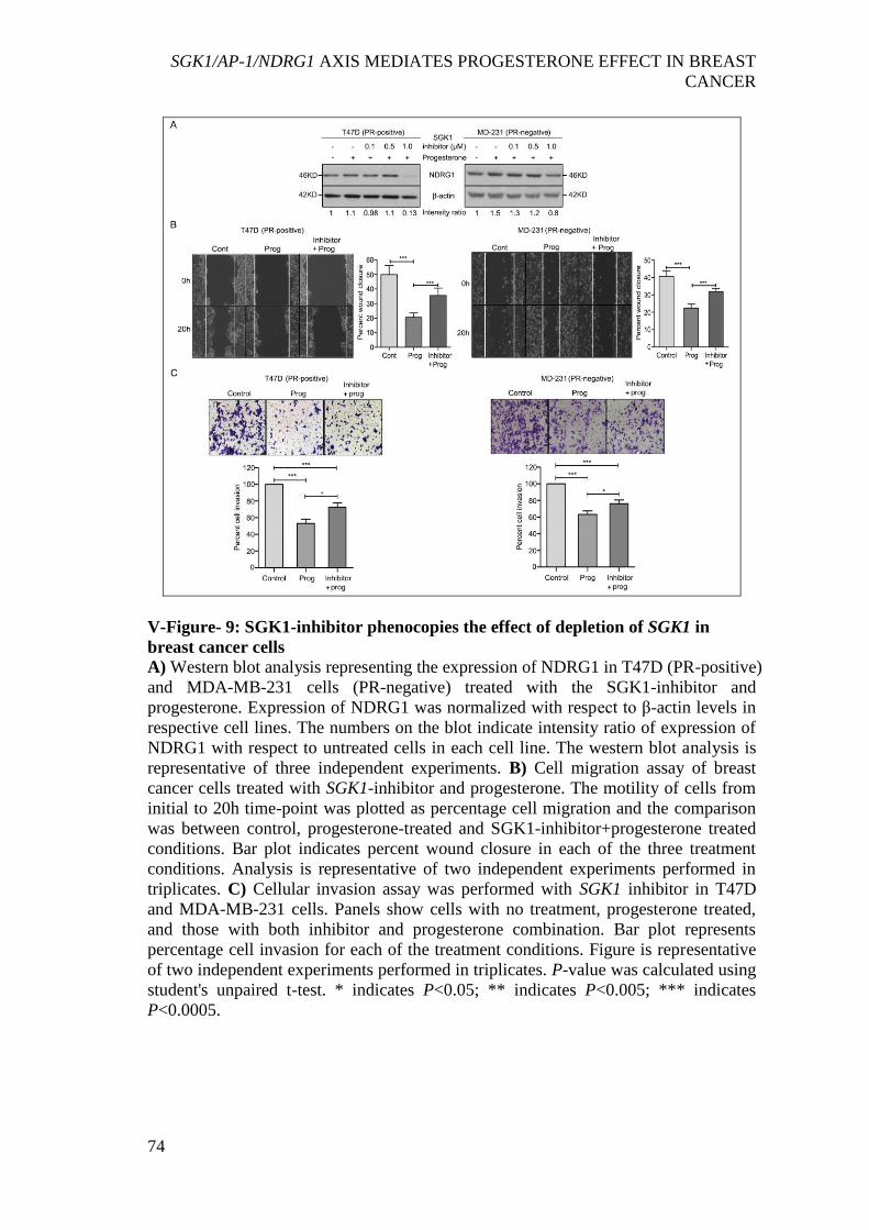
SGK1/AP-1/NDRG1 AXIS MEDIATES PROGESTERONE EFFECT IN BREAST
CANCER
74
V-Figure- 9: SGK1-inhibitor phenocopies the effect of depletion of SGK1 in
breast cancer cells
A) Western blot analysis representing the expression of NDRG1 in T47D (PR-positive)
and MDA-MB-231 cells (PR-negative) treated with the SGK1-inhibitor and
progesterone. Expression of NDRG1 was normalized with respect to β-actin levels in
respective cell lines. The numbers on the blot indicate intensity ratio of expression of
NDRG1 with respect to untreated cells in each cell line. The western blot analysis is
representative of three independent experiments. B) Cell migration assay of breast
cancer cells treated with SGK1-inhibitor and progesterone. The motility of cells from
initial to 20h time-point was plotted as percentage cell migration and the comparison
was between control, progesterone-treated and SGK1-inhibitor+progesterone treated
conditions. Bar plot indicates percent wound closure in each of the three treatment
conditions. Analysis is representative of two independent experiments performed in
triplicates. C) Cellular invasion assay was performed with SGK1 inhibitor in T47D
and MDA-MB-231 cells. Panels show cells with no treatment, progesterone treated,
and those with both inhibitor and progesterone combination. Bar plot represents
percentage cell invasion for each of the treatment conditions. Figure is representative
of two independent experiments performed in triplicates. P-value was calculated using
student's unpaired t-test. * indicates P<0.05; ** indicates P<0.005; *** indicates
P<0.0005.

SGK1/AP-1/NDRG1 AXIS MEDIATES PROGESTERONE EFFECT IN BREAST
CANCER
75
5.3.3 AP-1 transcription factors mediate up-regulation of NDRG1
NDRG1 is known to be regulated by AP-1 (FOS/JUN) and EGR1 in response to
stress-induced activation of kinases such p38, JNK and ERK [180-183]. We recently
showed that progesterone modulates the effect of surgical stress in primary breast
cancer patients [184]. Thus, we asked if NDRG1 could be regulated by AP-1 network
genes in response to progesterone-induced activation of SGK1, in a similar manner in
breast cancer cells. Indeed, treatment with progesterone or over-expression of SGK1
led to several fold over-expression of the AP-1 network genes in a panel of breast cell
lines irrespective of their PR status (V-Figure-10, V-Figure-11A and V-Figure-12A).
V-Figure- 10: Progesterone up-regulates expression of the AP-1 network genes in
breast cell lines
In the panel of breast cell lines representing different receptor status, transcript levels
of AP-1 network genes (EGR1, FOS, JUN and DUSP1) has been studied using
quantitative real-time PCR analysis. Expression of individual genes in the control
sample has been set as 1 and represented as a horizontal black line. Data has been
plotted as expression fold change upon progesterone treatment with respect to
expression in control. GAPDH has been used to normalize the gene expression. The

SGK1/AP-1/NDRG1 AXIS MEDIATES PROGESTERONE EFFECT IN BREAST
CANCER
76
figure is representative of three independent experiments performed in triplicates. P-
value was calculated using student's unpaired t-test. * indicates P<0.05; ** indicates
P<0.001; *** indicates P<0.0001; ns indicates not significant.
Consistent with this finding, knockdown of SGK1 significantly reduced the expression
of AP-1 network genes (V-Figure-11B and V-Figure-12B), and depleting the
expression of an AP-1 network gene EGR1 abrogated the expression of NDRG1 (V-
Figure-13A), a downstream component of the pathway, in T47D and MDA-MB-231.
Taken together, these results suggest that progesterone and SGK1 regulate the
expression of NDRG1 via the AP-1 network genes.
V-Figure- 11: SGK1 regulates expression of the AP-1 network genes in breast
cancer cells
Transcript levels of AP-1 network genes (EGR1, FOS, JUN and DUSP1) were studied
using quantitative real-time PCR, in T47D (PR-positive) cells A) over-expressing
SGK1, and B) upon knockdown of SGK1. For expression analysis upon over-
expression of SGK1, horizontal black line indicates transcript levels for the AP-1
network genes in untransfected cells. In case of analysis upon knockdown of SGK1,
transcript levels of AP-1 network genes were compared against sh-NT clone. Data has
been plotted as fold change for each individual gene with respect to expression in sh-

SGK1/AP-1/NDRG1 AXIS MEDIATES PROGESTERONE EFFECT IN BREAST
CANCER
77
NT clone and normalized with respect to GAPDH. Both the real-time PCR analyses
are representative of two independent experiments performed in triplicates. P-value
was calculated using student‘s unpaired t-test. * indicates P<0.05; ** indicates
P<0.005; *** indicates P<0.0005.
V-Figure- 12: SGK1 regulates expression of the AP-1 network genes in MDA-
MB-231 cells
Expression of AP-1 network genes was studied upon A) over-expression, and B)
knockdown of SGK1 in MDA-MB-231 cells (PR-negative) using quantitative real-
time PCR. For expression analysis upon SGK1 over-expression, data has been plotted
as fold change for each gene with respect to expression in untransfected parent cells.
The gene expression in untransfected cells has been represented using horizontal black
line. For expression analysis in SGK1 knockdown clones, data has been plotted as fold
change for each individual gene with respect to sh-NT clone. Expression of all genes
in both experiments has been normalized with respect to expression of GAPDH. Both
the real-time PCR analyses are representative of two independent experiments
performed in triplicates. The efficiency of over-expression and knockdown of SGK1
can be referred in Figure 2A and 3A, respectively. P-value was calculated using
student's unpaired t-test. * indicates P<0.05; ** indicates P<0.005; *** indicates
P<0.0005; ns indicates not significant.

SGK1/AP-1/NDRG1 AXIS MEDIATES PROGESTERONE EFFECT IN BREAST
CANCER
78
V-Figure- 13: Knockdown of EGR1 decreases expression of NDRG1 in breast
cancer cells
A) Western blot analysis of EGR1 and NDRG1 in T47D (PR-positive, left panel) and
MDA-MB-231 (PR-negative, right panel) cells upon genetic depletion of EGR1. sh-
NT was used as vector control. β-actin protein was used as loading control for western
blot. Numbers on blot indicate intensity ratio for expression of EGR1 and NDRG1,
normalized to respective β-actin levels. Western blot analysis is representative of three
independent experiments. B) Cell migration analysis upon depletion of EGR1 in T47D
(top panel) and MDA-MB-231 (bottom panel) breast cancer cells. Cells were
monitored by time-lapse wound healing assay for 20h. Cell migration from 0h to 20h
time-point was plotted as percentage wound closure and the comparison was with
respect to sh-NT clone. The analysis is representative of three independent
experiments performed in triplicates. P-value was calculated using student's unpaired
t-test. *** indicates P<0.0001; ns indicates not significant.
5.3.4 SGK1/NDRG1-axis inactivates EGFR- MAPK pathway to inhibit migration
and invasion of breast cancer cells
We recently showed that progesterone decreases the activation of multiple kinases like
EGFR, AKT1 and ERK1/2 in breast cancer cells, leading to suppression of cell
migration [174]. To test, if NDRG1 mediate inactivation of EGFR/AKT1/ERK1/2
SGK1/AP-1/NDRG1 AXIS MEDIATES PROGESTERONE EFFECT IN BREAST
CANCER
79
kinases in response to progesterone, we knocked down the expression of NDRG1 in
T47D and MDA-MB-231 cells (V-Figure-14A and V-Figure-15A).
V-Figure- 14: NDRG1 regulates the activation of multiple cellular kinases and
cell migration in T47D cells
A) Western blot analysis depicting knockdown of NDRG1 in T47D cells (PR-positive).
sh-NT was used as vector control for NDRG1 expression. β-actin protein was used as
loading control for western blot. Numbers on the blot indicate intensity ratio for
NDRG1 expression normalized to respective β-actin levels. Analysis is representative
of three independent experiments. B) Western blot analysis of p-EGFR (Y1086), p-
AKT (S473) and p-ERK1/2 (T202/Y204) in NDRG1 knockdown clones of T47D cells.
β-actin used as loading control for western blot. Numbers on blot indicate intensity
ratio for phosphorylation levels of EGFR, AKT and ERK1/2, normalized to respective
total protein levels (EGFR, AKT and ERK2). Figure is representative of three
independent experiments. C) Cell migration analysis upon depletion of NDRG1 in
T47D breast cancer cells has been shown. Cells were monitored by time-lapse wound
healing assay for 20h. Cell migration from initial to 20h time-point was plotted as
percentage wound closure and the comparison was with respect to sh-NT. Figure is
representative of three independent experiments performed in triplicates. P-value was
calculated using student's unpaired t-test. * indicates P<0.05; ns indicates not
significant.
Interestingly, 2 of 3 shRNA clones targeting NDRG1 significantly increased the
phosphorylation of EGFR (Y1068), AKT (S473) and ERK1/2 (T202/Y204) (V-
Figure-14B and V-Figure-15B). Furthermore, breast cancer cells expressing constructs
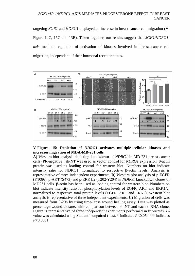
SGK1/AP-1/NDRG1 AXIS MEDIATES PROGESTERONE EFFECT IN BREAST
CANCER
80
targeting EGR1 and NDRG1 displayed an increase in breast cancer cell migration (V-
Figure-14C, 15C and 13B). Taken together, our results suggest that SGK1/NDRG1-
axis mediate regulation of activation of kinases involved in breast cancer cell
migration, independent of their hormonal receptor status.
V-Figure- 15: Depletion of NDRG1 activates multiple cellular kinases and
increases migration of MDA-MB-231 cells
A) Western blot analysis depicting knockdown of NDRG1 in MD-231 breast cancer
cells (PR-negative). sh-NT was used as vector control for NDRG1 expression. β-actin
protein was used as loading control for western blot. Numbers on blot indicate
intensity ratio for NDRG1, normalized to respective β-actin levels. Analysis is
representative of three independent experiments. B) Western blot analysis of p-EGFR
(Y1086), p-AKT (S473) and p-ERK1/2 (T202/Y204) in NDRG1 knockdown clones of
MD231 cells. β-actin has been used as loading control for western blot. Numbers on
blot indicate intensity ratio for phosphorylation levels of EGFR, AKT and ERK1/2,
normalized to respective total protein levels (EGFR, AKT and ERK2). Western blot
analysis is representative of three independent experiments. C) Migration of cells was
measured from 0-20h by using time-lapse wound healing assay. Data was plotted as
percentage wound closure, with comparison between sh-NT and each shRNA clone.
Figure is representative of three independent experiments performed in triplicates. P-
value was calculated using Student‘s unpaired t-test. * indicates P<0.05; *** indicates
P<0.0001.

SGK1/AP-1/NDRG1 AXIS MEDIATES PROGESTERONE EFFECT IN BREAST
CANCER
81
5.4 Discussion:
Preoperative endocrine therapies, in contrast to neoadjuvant chemotherapy, are much
simpler and economical to deliver. An understanding of the targets could thus be of
immense potential utility in monitoring the response of hormones in human cancer.
This study details the underlying molecular mechanism associated with benefits of
preoperative progesterone treatment as observed in our randomized trial [171]. We
present an intricate convergence model indicating a dual-phase regulation downstream
to progesterone treatment to regulate the expression of a Serum- and Glucocorticoid-
regulated Kinase gene 1, SGK1: predominantly driven as a direct transcriptional target,
consistent with earlier reports [177, 178], in PR-positive breast cancer cells; and,
down-regulation of miR-29a and miR-101-1 targeting SGK1 with relatively distinct
effect in PR-negative breast cells in response to progesterone. In addition, as described
earlier [174], glucocorticoid receptor GR or the membrane progesterone receptor
(PGRMC1 and SERBP1) likely mediates effect of progesterone in PR-negative breast
cancer cells that are uniformly expressed across breast cancer cells (V-Table-2). The
stringent up-regulation of SGK1 in response to progesterone lead to an activation of a
tumor metastasis suppressor gene, NDRG1, via a set of AP-1 network genes to
inactivate AKT1, ERK1/2 and EGFR kinases, impeding the invasion and migration of
breast cancer cells. As NDRG1 is known to be regulated by AP-1 network genes in
response to stress-induced activation of kinases [180-183], this model confirms and
extends our recent report that progesterone modulates the effect of surgical stress by
up-regulation of SGK1 in primary breast cancer patients [184], affecting the invasive
characteristics of breast cancer cells most likely by regulating their migration.
SGK1/AP-1/NDRG1 AXIS MEDIATES PROGESTERONE EFFECT IN BREAST
CANCER
82
Interestingly, SGK1 and NDRG1 are known to be down-regulated in human cancers as
compared to adjacent normal tissues and that increased expression of both these genes
has been associated with better survival of cancer patients [185-189]. Even the
recently described panel of 38 gene signature that predict favorable prognosis of
breast cancer patients include SGK1 [58]. Thus, enhanced expression of SGK1 and
NDRG1 could explain better survival of breast cancer patients [171]. Our study
suggests that SGK1 up-regulates the expression of NDRG1, with no significant change
in phosphorylation of NDRG1 in breast cancer cells. We describe that SGK1 regulates
the expression of NDRG1 via regulation of expression of EGR1, a transcription factor
from the AP-1 network genes, in breast cancer independent of the PR status of cells.
In summary, we propose a model for the mode of action of progesterone in breast
cancer deciphering the molecular basis of a randomized clinical trial studying the
effect of progesterone in breast cancer (VI-Figure-2). While there have been attempts
to understand the effect of progesterone as a physiological hormone [92], we provide
the first mechanistic insights into the role of progesterone in breast cancer, detailing
the genetic event leading to clinical observation of better survival of breast cancer
patients treated with pre-operative progesterone [171]. However, whether these
molecular targets of progesterone could help in stratification of breast cancer patients
and aid in better prognosis, remains to be studied.
DISCUSSION
83
VI. DISCUSSION:
Breast cancer is the most commonly occurring cancer in females across the world with
a mortality rate of about 50% [190]. Despite recent developments in diagnostic and
treatment methods, patients continue to develop metastatic tumors and relapse [21].
Hormones influence physiology and development of the normal breast tissue and can
also alter the outcome of breast cancer [191-193]. Early observational studies to
monitor the effect of hormones on outcome of breast cancer surgery performed in
different phases of menstrual cycle showed that progesterone improved the outcome
of pre-menopausal patients [123, 124, 194-196]. Taking leads from these studies, a
randomized clinical trial with pre-operative progesterone intervention was performed
in Tata Memorial Hospital to establish an association of progesterone with breast
cancer in a large cohort of operable breast cancer patients. It was observed that
progesterone improved the disease-free and overall survival of node-positive breast
cancer patients independent of their menopausal or PR status [128]. The survival
benefits obtained using a single injection of progesterone was an encouraging
outcome, however, a multicentre validation may help in the wider usability of
progesterone as a therapy for treatment of breast cancer patients [140]. This was a
hypothesis generating clinical outcome since in most other cases the effect of
endocrine therapy was purely based on the presence of corresponding hormone
receptors or the menopausal status of the patients [34, 40, 157]. However, the
mechanism of action of progesterone remained to be elucidated in breast cancer to
explain the outcome of this randomized clinical trial with pre-operative progesterone
treatment. This led to the origin of my thesis where I have focused on identifying the

DISCUSSION
84
underlying effects of progesterone on breast cancer, using a functional genomics and
proteomics approach performed in a panel of breast-derived cell lines. To understand
the PR-independent targets of progesterone, I have selected multiple breast cancer and
immortalized normal-like cell lines with different hormone receptor statuses and
breast cancer subtypes. I hypothesized that progesterone could induce transient
transcriptional and proteomic changes in breast cancer cells leading to phenotypic
alterations that can be captured using functional genomic and phenotypic analysis.
6.1 Phenotypic and proteomic changes induced by progesterone
Breast cancer poses a unique threat with capacity to metastasize and causes more
mortality compared to the primary tumor itself [46, 48, 197]. Surprisingly, as
observed in the clinical trial, breast cancer patients with adjacent lymphnode-
involvement showed a better survival upon pre-operative progesterone intervention.
This suggests that progesterone might have possibly targeted breast cancer cells
primed with metastatic property, leading to regression of tumor by controlling the
dissemination of cancer cells during surgery and their establishment in distant organs
[128]. While the effects of progesterone have been well studied for its context-
dependent proliferative and growth-inhibitory role in breast cells, functional studies
have not performed to study the role of progesterone in regulation of cell migration
and invasion independent of the PR status of cells [69, 91-93, 198]. It is well
established that over-expression or activation of kinases like EGFR or AKT1 provides
an opportunity for breast cancer cells to invade from the site of primary tumor and
escape to distant organs [146, 151]. To study whether progesterone affects the
activation of kinases in breast cancer cells, I performed an unbiased proteomic
analysis using a proteome profiling assay which accommodates a panel of 43 kinases
DISCUSSION
85
that are known to regulate migratory properties of cancer cells. I observed a
significant decrease in phosphorylation of 12 of 43 kinases like EGFR, AKT1,
ERK1/2, FAK and so on, in PR-positive and PR-negative breast cancer cells treated
with progesterone. Concomitantly, I observed a significant reduction in cell migration
and invasion in breast cancer cells independent of their PR statuses. Of note, I found
that PR plays a non-essential role in mediating this effect of progesterone, since
blocking the activity of PR using mifepristone did not hamper the inhibition of cell
invasion and migration of breast cancer cells. Thus, consistent with studies targeting
kinase activity using kinase-inhibitors [98, 142], I show that progesterone inhibits cell
migration and invasion of breast cancer cells by decreasing the phosphorylation of
multiple kinases. As a plausible explanation to the PR-independent mode of action, I
hypothesize that progesterone might be working in synergism with other steroid
hormone receptors like the GR or mPR which are known to associate with
progesterone [77, 83, 99]. Although the effect of progesterone on cell invasion and
migration of breast cancer cells has been studied in the context of PR isoforms or by
their over-expression in MDA-MB-231 cell line with varying outcome [153, 199, 200],
my study, using additional PR-negative breast cancer cells like ZR-75-1 and BT-549
that have not been studied before, suggests a consistent decrease in these cellular
phenotypes independent of the PR expression.
6.2 Genomics analysis of progesterone-induced changes in microRNAs and their
target genes
Endocrine therapy has helped in improving the survival of patients with hormone
receptor positive breast cancer. Moreover, studies have suggested that patients treated
with tamoxifen or aromatase inhibitors which target ER activity, show better response

DISCUSSION
86
in presence of PR and show reduced relapse as compared to patients with PR-negative
tumors [53, 59, 201]. Also, PR has been observed as a prognostic marker for lower
metastatic potential of breast cancer [52]. Though patients show an initial response to
endocrine therapy, a significant number of patients develop resistance, primarily due
to loss of PR expression over the period of progression of the disease. While over-
expression of growth factor receptors like Her2 or EGFR has been attributed to loss of
PR, few other pathways leading to degradation and silencing of PR have been studied
in breast cancer, with some of these pathways mediated in response to progesterone
[53, 62, 64, 202]. Hormones are known to decrease the expression of their receptors,
for instance, estrogen represses the expression of ER by up-regulating microRNAs in
breast cancer [61]. However, whether progesterone also suppresses expression of PR
in a similar way is not well studied. To identify microRNAs targeting PR in response
to progesterone, I performed small RNA sequencing of breast cancer cell lines treated
with progesterone and identified miR-129-2 as a direct target of progesterone which
decreases the expression of PR in breast cancer. I functionally validated the physical
association of miR-129-2 with PR and observed that their expression is inversely
correlated in breast cancer patients in the TCGA cohort. Interestingly, I observed that
inhibition of activity of miR-129-2 in breast cancer cells using anti-miR-129-2
inhibitor helped in rescuing the expression of PR even in the presence of progesterone.
Inhibiting the activity of such microRNAs in breast cancer patients expressing low PR
levels may help in stabilizing the expression of PR and concomitantly improve the
response to endocrine therapies. However, this is a preliminary observation and usage
of anti-miR-129-2 requires validation in breast cancer patients. Nevertheless, my study
has identified a novel microRNA that mediates the effect of progesterone by

DISCUSSION
87
suppressing the expression of PR. Knowledge of such microRNAs targeting PR may
provide an opportunity to reinstate the expression of PR in breast cancer.
6.3 Functional genomics analysis of progesterone-treated breast cancer cell lines
As discussed in the first part of my thesis, I observed a consistent decrease in cell
invasion and migration of breast cancer cells, possibly via inhibition of
phosphorylation of multiple kinases in response to progesterone. While this suggests
that progesterone may regulate the metastatic potential of breast cancer, the
underlying molecular mechanism of action of progesterone remained to be elucidated.
With this notion, I set to identify the transcriptional targets of progesterone by
performing a microarray gene expression analysis of breast cancer cells. Most of the
earlier studies have performed gene expression analysis in PR-positive cell lines and
identified isoform-specific targets of PR rather than genes regulated by progesterone
[58, 109, 203]. For my thesis work, I focused on identifying the targets of
progesterone independent of the PR expression of the cells. As a strategy to identify
true targets of progesterone, I depleted the culture medium of all growth factors and
hormone-like compounds prior to addition of progesterone to the cells. The expression
array analysis helped in the identification of Serum and glucocorticoid-regulated
kinase 1, SGK1 and N-Myc downstream regulated gene 1, NDRG1, as direct targets
up-regulated in response to progesterone in multiple breast cancer cells. Moreover, the
results suggest that progesterone increased the phosphorylation of SGK1 as well,
suggesting an increased activity of SGK1 in breast cancer cells. By performing
genetic perturbation experiments, I observed that SGK1 regulates the expression of
NDRG1 in breast cancer, rather than just phosphorylating it as reported earlier. I could
identify that progesterone and SGK1 mediate the increased expression of NDRG1 via

DISCUSSION
88
the AP-1 network genes viz. EGR1 and FOS-JUN. As a proof of concept, I show that
depletion of EGR1 decreases the expression of NDRG1 in breast cancer cells.
Furthermore, I observed that SGK1 mimics the activity of progesterone to suppress the
cell migration and invasion of breast cancer independent of the PR status, possibly via
the increased expression of NDRG1, a known tumor metastasis suppressor gene [189,
204]. Consistent with its known activity [205], I observed that depletion of NDRG1
gave cells an opportunity to activate EGFR, AKT1 and ERK1/2 kinases in breast
cancer cells irrespective of their PR status. Moreover, these cells showed an enhanced
migratory potential. While SGK1 has been shown to be a direct target of progesterone
[206], I observed an intricate regulatory network being regulated by SGK1, leading to
the suppression of cell invasion and migration of breast cancer cells.
In a parallel study, I observed that the expression of SGK1 is under a dual-regulatory
mode in response to progesterone. While expression of SGK1 is known to be up-
regulated by progesterone via PRE in the promoter region of SGK1, I show that
progesterone maintains sustained expression of SGK1 by suppressing the expression
of miR-29a and miR-101-1 in breast cancer. Using a biochemical approach, I observed
an association of miR-29a and miR-101-1 with the 3‘UTR of SGK1 and that inhibition
of these microRNAs stabilizes expression of SGK1 in breast cancer. Since
progesterone is able to up-regulate expression of SGK1 from the promoter and 3‘UTR
of SGK1, we propose a dual-regulatory model of regulation of expression of SGK1 in
response to progesterone in breast cancer, irrespective of the PR status of cells (VI-
Figure-1). Additional experiments like CRISPR-based knockout of SGK1, and over-
expression of a SGK1 kinase-dead mutant or dominant-negative form of SGK1 in
breast cancer cells would strengthen the interpretation made in my work This is the

DISCUSSION
89
first study to show that progesterone regulates expression of SGK1 in a dual-
regulatory manner in breast cancer and provides a proof-of-concept for presence of
such molecular targets of progesterone.
VI-Figure- 1: Proposed dual-regulatory model for regulation of expression of
SGK1
The model describes regulation of expression of SGK1 by progesterone at two sites.
Firstly, progesterone transcriptionally activates SGK1 via association of PR-PRE at
promoter. Secondly, progesterone decreases expression of miR-29a and miR-101-1
and thus stabilizes expression of SGK1 in breast cancer.
Though we could not correlate the expression of miR-29a and miR-101-1 with
expression of SGK1 in the TCGA breast cancer patient cohort, orthologous
methodologies can possibly be used to check the expression of these microRNAs and
SGK1 in patient samples. Moreover, I believe that an extended analysis of our
microarray gene expression and small RNA sequencing results should provide more
dual-regulated targets of progesterone. Also, the expression of microRNAs validated
in our study was found to be independent of the PR status of the cells. Since
microRNAs are known to be housed as ‗intronic‘ or ‗intergenic‘ [155, 207-209], a
thorough analysis needs to be performed for understanding their mode of regulation in
response to progesterone. Preliminary analysis suggests that the microRNAs could
either be expressed along with a gene responsive to progesterone (intronic, miR-29a

DISCUSSION
90
and miR-101-1) or could have their own promoter which responds to progesterone
(intergenic, miR-129-2) in breast cancer cells, which remains to be confirmed
experimentally.
6.4 Conclusion:
In overall, my thesis provides first leads to systematically model the effects of pre-
operative progesterone intervention in breast cancer patients, where the up-regulation
of SGK1 leads to sustained activity of progesterone to inhibit the activation of kinases
and suppress the cell migration and invasion of breast cancer cells via increased
expression of NDRG1, regulated by SGK1 and the AP-1 network genes, independent
of the PR status of cells. While on the other hand, I observe that of the several
microRNAs de-regulated in cell lines, progesterone fine-tunes the expression of miR-
129-2 and miR-29a and miR-101-1 that target PR and SGK1 in breast cancer cells. To
explain this tightly regulated pathway, I propose a working model explaining the
effect of progesterone on breast cancer (VI-Figure-2). Elevated expression of tumor
metastasis suppressor genes like NDRG1 could possibly explain the effect of pre-
operative progesterone intervention of breast cancer patients independent of their PR
status. Though my study focuses on the transient changes induced by progesterone in
breast cancer, I am optimistic that the targets identified in the study and the beneficial
role of increased expression of SGK1 and NDRG1 in survival of breast cancer patients
reported earlier may help in better prognosis or selection of breast cancer patients for
progesterone treatment [186, 210, 211].

DISCUSSION
91
VI-Figure- 2: Working model for progesterone-mediated regulation of coding
and non-coding genes leading to suppression of cell migration and invasion in
breast cancer
However, I do concur with the notion that progesterone could be regulating multiple
pathways in breast cancer, functioning along with the model described in my study.
Hence, I propose that the microarray gene expression and small RNA sequencing
analysis performed in my study should help in identifying additional targets of
progesterone and expand the model described herein. Moreover, details of how
progesterone acts in PR-negative breast cancer cells, by performing experiments with
PR/GR/mPR knockdown, along with in-depth study of SGK1-mediated regulation of
NDRG1 and downstream signaling, remains to be elucidated. And, the epigenetic
modifications induced by progesterone could be studied to better understand the
DISCUSSION
92
regulation of the candidate genes expressed or repressed in breast cancer cells.
Additionally, the mode of regulation of the microRNAs identified in this study
remains to be explored. Also, non-coding RNAs like lncRNAs are emerging as targets
of hormones like estrogen. These lncRNAs have been shown to mediate several
functional roles in cancer cells, including endocrine resistance and metastasis of cells
[212, 213]. Thus in extension to identifying the coding targets, lncRNAs and
epigenetic modifications de-regulated by progesterone can also be identified by
performing similar functional genomics approaches in breast cancer cells treated with
progesterone.

REFERENCES
93
VII. REFERENCES:
1. Bardon, S., et al., RU486, a progestin and glucocorticoid antagonist, inhibits the growth of breast cancer cells via the progesterone receptor. J Clin Endocrinol Metab, 1985. 60(4): p. 692-7.
2. Mansley, M.K. and S.M. Wilson, Effects of nominally selective inhibitors of the kinases PI3K, SGK1 and PKB on the insulin-dependent control of epithelial Na+ absorption. Br J Pharmacol, 2010. 161(3): p. 571-88.
3. Chen, N., et al., Inhibitory effect of mitomycin C on proliferation of primary cultured fibroblasts from human airway granulation tissues. Respiration, 2013. 85(6): p. 500-4.
4. Barral, Y. and J. Knoblich, Cell division, growth and death. Curr Opin Cell Biol, 2008. 20(6): p. 647-9.
5. Shen, Z., Genomic instability and cancer: an introduction. J Mol Cell Biol, 2011. 3(1): p. 1-3.
6. Stratton, M.R., P.J. Campbell, and P.A. Futreal, The cancer genome. Nature, 2009. 458(7239): p. 719-24.
7. Hanahan, D. and R.A. Weinberg, The Hallmarks of Cancer. Cell. 100(1): p. 57-70. 8. Jemal, A., et al., Global cancer statistics. CA Cancer J Clin, 2011. 61(2): p. 69-90. 9. Peto, J., Cancer epidemiology in the last century and the next decade. Nature, 2001.
411(6835): p. 390-5. 10. Chuffa, L.G., et al., The role of sex hormones and steroid receptors on female
reproductive cancers. Steroids, 2017. 118: p. 93-108. 11. Ferlay, J., et al., Cancer incidence and mortality worldwide: sources, methods and
major patterns in GLOBOCAN 2012. Int J Cancer, 2015. 136(5): p. E359-86. 12. Ghoncheh, M., Z. Pournamdar, and H. Salehiniya, Incidence and Mortality and
Epidemiology of Breast Cancer in the World. Asian Pac J Cancer Prev, 2016. 17(S3): p. 43-6.
13. Anderson, W.F., et al., How many etiological subtypes of breast cancer: two, three, four, or more? J Natl Cancer Inst, 2014. 106(8).
14. Nielsen, M., et al., Breast cancer and atypia among young and middle-aged women: a study of 110 medicolegal autopsies. Br J Cancer, 1987. 56(6): p. 814-9.
15. Benson, J.R. and I. Jatoi, The global breast cancer burden. Future Oncol, 2012. 8(6): p. 697-702.
16. Stuckey, A., Breast cancer: epidemiology and risk factors. Clin Obstet Gynecol, 2011. 54(1): p. 96-102.
17. Stephens, P.J., et al., The landscape of cancer genes and mutational processes in breast cancer. Nature, 2012. 486(7403): p. 400-4.
18. Comprehensive molecular portraits of human breast tumours. Nature, 2012. 490(7418): p. 61-70.
19. Torre, L.A., et al., Global Cancer Incidence and Mortality Rates and Trends--An Update. Cancer Epidemiol Biomarkers Prev, 2016. 25(1): p. 16-27.
20. Chlebowski, R.T., et al., Estrogen plus progestin and breast cancer incidence and mortality in the Women's Health Initiative Observational Study. J Natl Cancer Inst, 2013. 105(8): p. 526-35.
21. Cardoso, F., et al., Locally recurrent or metastatic breast cancer: ESMO Clinical Practice Guidelines for diagnosis, treatment and follow-up. Ann Oncol, 2010. 21 Suppl 5: p. v15-9.

REFERENCES
94
22. Killelea, B.K., et al., Evolution of breast cancer screening in the Medicare population: clinical and economic implications. J Natl Cancer Inst, 2014. 106(8).
23. Lannin, D.R. and S. Wang, Are Small Breast Cancers Good because They Are Small or Small because They Are Good? N Engl J Med, 2017. 376(23): p. 2286-91.
24. Reis-Filho, J.S. and L. Pusztai, Gene expression profiling in breast cancer: classification, prognostication, and prediction. Lancet, 2011. 378(9805): p. 1812-23.
25. Perou, C.M., et al., Molecular portraits of human breast tumours. Nature, 2000. 406(6797): p. 747-52.
26. Sørlie, T., et al., Gene expression patterns of breast carcinomas distinguish tumor subclasses with clinical implications. Proceedings of the National Academy of Sciences, 2001. 98(19): p. 10869-10874.
27. Prat, A., et al., Phenotypic and molecular characterization of the claudin-low intrinsic subtype of breast cancer. Breast Cancer Res, 2010. 12(5): p. R68.
28. Prat, A., et al., Clinical implications of the intrinsic molecular subtypes of breast cancer. Breast, 2015. 24 Suppl 2: p. S26-35.
29. Soni, A., et al., Breast cancer subtypes predispose the site of distant metastases. Am J Clin Pathol, 2015. 143(4): p. 471-8.
30. Goldhirsch, A., et al., Strategies for subtypes--dealing with the diversity of breast cancer: highlights of the St. Gallen International Expert Consensus on the Primary Therapy of Early Breast Cancer 2011. Ann Oncol, 2011. 22(8): p. 1736-47.
31. Burstein, H.J., et al., American Society of Clinical Oncology clinical practice guideline: update on adjuvant endocrine therapy for women with hormone receptor-positive breast cancer. J Clin Oncol, 2010. 28(23): p. 3784-96.
32. Burstein, H.J., et al., Adjuvant endocrine therapy for women with hormone receptor-positive breast cancer: american society of clinical oncology clinical practice guideline focused update. J Clin Oncol, 2014. 32(21): p. 2255-69.
33. Antolin-Novoa, S., et al., Adjuvant regimens with trastuzumab administered for small HER2-positive breast cancer in routine clinical practice. Clin Transl Oncol, 2015. 17(11): p. 862-9.
34. Cheang, M.C.U., et al., Ki67 Index, HER2 Status, and Prognosis of Patients With Luminal B Breast Cancer. J Natl Cancer Inst, 2009. 101(10): p. 736-50.
35. Ahmed, S., A. Sami, and J. Xiang, HER2-directed therapy: current treatment options for HER2-positive breast cancer. Breast Cancer, 2015. 22(2): p. 101-16.
36. Wilks, S.T., Potential of overcoming resistance to HER2-targeted therapies through the PI3K/Akt/mTOR pathway. Breast, 2015. 24(5): p. 548-55.
37. Moran, M.S., Radiation therapy in the locoregional treatment of triple-negative breast cancer. Lancet Oncol, 2015. 16(3): p. e113-22.
38. von Minckwitz, G. and M. Martin, Neoadjuvant treatments for triple-negative breast cancer (TNBC). Ann Oncol, 2012. 23 Suppl 6: p. vi35-9.
39. Ignatiadis, M. and C. Sotiriou, Luminal breast cancer: from biology to treatment. Nat Rev Clin Oncol, 2013. 10(9): p. 494-506.
40. Dowsett, M., et al., Meta-analysis of breast cancer outcomes in adjuvant trials of aromatase inhibitors versus tamoxifen. J Clin Oncol, 2010. 28(3): p. 509-18.
41. Esserman, L.J., I.M. Thompson, Jr., and B. Reid, Overdiagnosis and overtreatment in cancer: an opportunity for improvement. Jama, 2013. 310(8): p. 797-8.
42. Boccia, R.V., Translating Research into Practice: the Prosigna(R) (PAM50) Gene Signature Assay. Clin Adv Hematol Oncol, 2015. 13(6 Suppl 6): p. 3-13.
43. Weigelt, B., et al., Molecular portraits and 70-gene prognosis signature are preserved throughout the metastatic process of breast cancer. Cancer Res, 2005. 65(20): p.

REFERENCES
95
9155-8. 44. Slodkowska, E.A. and J.S. Ross, MammaPrint 70-gene signature: another milestone in
personalized medical care for breast cancer patients. Expert Rev Mol Diagn, 2009. 9(5): p. 417-22.
45. van 't Veer, L.J., et al., Gene expression profiling predicts clinical outcome of breast cancer. Nature, 2002. 415(6871): p. 530-6.
46. Lee, Y.T., Breast carcinoma: pattern of metastasis at autopsy. J Surg Oncol, 1983. 23(3): p. 175-80.
47. Bos, P.D., et al., Genes that mediate breast cancer metastasis to the brain. Nature, 2009. 459(7249): p. 1005-9.
48. Weigelt, B., J.L. Peterse, and L.J. van't Veer, Breast cancer metastasis: markers and models. Nat Rev Cancer, 2005. 5(8): p. 591-602.
49. Bernards, R. and R.A. Weinberg, A progression puzzle. Nature, 2002. 418(6900): p. 823.
50. Voduc, K.D., et al., Breast cancer subtypes and the risk of local and regional relapse. J Clin Oncol, 2010. 28(10): p. 1684-91.
51. Effects of chemotherapy and hormonal therapy for early breast cancer on recurrence and 15-year survival: an overview of the randomised trials. Lancet, 2005. 365(9472): p. 1687-717.
52. Pichon, M.F., et al., Relationship of presence of progesterone receptors to prognosis in early breast cancer. Cancer Res, 1980. 40(9): p. 3357-60.
53. Bardou, V.J., et al., Progesterone receptor status significantly improves outcome prediction over estrogen receptor status alone for adjuvant endocrine therapy in two large breast cancer databases. J Clin Oncol, 2003. 21(10): p. 1973-9.
54. Konecny, G., et al., Quantitative association between HER-2/neu and steroid hormone receptors in hormone receptor-positive primary breast cancer. J Natl Cancer Inst, 2003. 95.
55. Cui, X., et al., Biology of progesterone receptor loss in breast cancer and its implications for endocrine therapy. J Clin Oncol, 2005. 23.
56. Musgrove, E.A. and R.L. Sutherland, Biological determinants of endocrine resistance in breast cancer. Nat Rev Cancer, 2009. 9(9): p. 631-43.
57. Purdie, C.A., et al., Progesterone receptor expression is an independent prognostic variable in early breast cancer: a population-based study. Br J Cancer, 2014. 110(3): p. 565-72.
58. Mohammed, H., et al., Progesterone receptor modulates ERalpha action in breast cancer. Nature, 2015. 523(7560): p. 313-7.
59. Cui, X., et al., Biology of progesterone receptor loss in breast cancer and its implications for endocrine therapy. J Clin Oncol, 2005. 23(30): p. 7721-35.
60. Musgrove, E.A., et al., Cyclin D1 overexpression induces progestin resistance in T-47D breast cancer cells despite p27(Kip1) association with cyclin E-Cdk2. J Biol Chem, 2001. 276(50): p. 47675-83.
61. Castellano, L., et al., The estrogen receptor-alpha-induced microRNA signature regulates itself and its transcriptional response. Proc Natl Acad Sci U S A, 2009. 106(37): p. 15732-7.
62. Yang, S., et al., Systematic dissection of the mechanisms underlying progesterone receptor downregulation in endometrial cancer. Oncotarget, 2014. 5(20): p. 9783-97.
63. Lange, C.A., T. Shen, and K.B. Horwitz, Phosphorylation of human progesterone receptors at serine-294 by mitogen-activated protein kinase signals their degradation by the 26S proteasome. Proc Natl Acad Sci U S A, 2000. 97(3): p. 1032-7.

REFERENCES
96
64. Zhang, P.J., et al., CUE domain containing 2 regulates degradation of progesterone receptor by ubiquitin-proteasome. Embo j, 2007. 26(7): p. 1831-42.
65. Cochrane, D.R., D.M. Cittelly, and J.K. Richer, Steroid receptors and microRNAs: relationships revealed. Steroids, 2011. 76(1-2): p. 1-10.
66. Macias, H. and L. Hinck, Mammary gland development. Wiley Interdiscip Rev Dev Biol, 2012. 1(4): p. 533-57.
67. Stingl, J., Estrogen and progesterone in normal mammary gland development and in cancer. Horm Cancer, 2011. 2(2): p. 85-90.
68. Rajaram, R.D. and C. Brisken, Paracrine signaling by progesterone. Mol Cell Endocrinol, 2012. 357(1-2): p. 80-90.
69. Brisken, C., Progesterone signalling in breast cancer: a neglected hormone coming into the limelight. Nat Rev Cancer, 2013. 13(6): p. 385-96.
70. Chabbert-Buffet, N., et al., Selective progesterone receptor modulators and progesterone antagonists: mechanisms of action and clinical applications. Hum Reprod Update, 2005. 11(3): p. 293-307.
71. Kastner, P., et al., Two distinct estrogen-regulated promoters generate transcripts encoding the two functionally different human progesterone receptor forms A and B. Embo j, 1990. 9(5): p. 1603-14.
72. Law, M.L., et al., The progesterone receptor gene maps to human chromosome band 11q13, the site of the mammary oncogene int-2. Proc Natl Acad Sci U S A, 1987. 84(9): p. 2877-81.
73. Graham, J.D., et al., Characterization of progesterone receptor A and B expression in human breast cancer. Cancer Res, 1995. 55(21): p. 5063-8.
74. Obr, A.E. and D.P. Edwards, The biology of progesterone receptor in the normal mammary gland and in breast cancer. Mol Cell Endocrinol, 2012. 357(1-2): p. 4-17.
75. Grimm, S.L., S.M. Hartig, and D.P. Edwards, Progesterone Receptor Signaling Mechanisms. J Mol Biol, 2016. 428(19): p. 3831-49.
76. Bouchard, P., Progesterone and the progesterone receptor. J Reprod Med, 1999. 44(2 Suppl): p. 153-7.
77. Lei, K., et al., Progesterone acts via the nuclear glucocorticoid receptor to suppress IL-1beta-induced COX-2 expression in human term myometrial cells. PLoS One, 2012. 7(11): p. e50167.
78. Lieberman, B.A., et al., The constitution of a progesterone response element. Mol Endocrinol, 1993. 7(4): p. 515-27.
79. McNamara, K.M., A. Kannai, and H. Sasano, Possible roles for glucocorticoid signalling in breast cancer. Mol Cell Endocrinol, 2017. 466: p. 38-50.
80. Singhal, H., et al., Genomic agonism and phenotypic antagonism between estrogen and progesterone receptors in breast cancer. Sci Adv, 2016. 2(6): p. e1501924.
81. Boonyaratanakornkit, V., et al., The role of extranuclear signaling actions of progesterone receptor in mediating progesterone regulation of gene expression and the cell cycle. Mol Endocrinol, 2007. 21(2): p. 359-75.
82. Sen, A. and S.R. Hammes, Just when you thought it was safe to go into the membrane: the growing complexities of extra-nuclear progesterone signaling. Breast Cancer Res, 2010. 12(3): p. 109.
83. Fernandes, M.S., J.J. Brosens, and B. Gellersen, Honey, we need to talk about the membrane progestin receptors. Steroids, 2008. 73(9-10): p. 942-52.
84. Xie, M., et al., Membrane progesterone receptor alpha as a potential prognostic biomarker for breast cancer survival: a retrospective study. PLoS One, 2012. 7(4): p. e35198.

REFERENCES
97
85. Ruan, X., et al., Increased expression of progesterone receptor membrane component 1 is associated with aggressive phenotype and poor prognosis in ER-positive and negative breast cancer. Menopause, 2017. 24(2): p. 203-209.
86. Garg, D., et al., Progesterone-Mediated Non-Classical Signaling. Trends Endocrinol Metab, 2017. 28(9): p. 656-668.
87. Alkhalaf, M., A. El-Mowafy, and S. Karam, Growth inhibition of MCF-7 human breast cancer cells by progesterone is associated with cell differentiation and phosphorylation of Akt protein. Eur J Cancer Prev, 2002. 11(5): p. 481-8.
88. Musgrove, E.A. and R.L. Sutherland, Cell cycle control by steroid hormones. Semin Cancer Biol, 1994. 5(5): p. 381-9.
89. Lin, V.C., et al., Progesterone induces cellular differentiation in MDA-MB-231 breast cancer cells transfected with progesterone receptor complementary DNA. Am J Pathol, 2003. 162(6): p. 1781-7.
90. Lange, C.A., J.K. Richer, and K.B. Horwitz, Hypothesis: Progesterone primes breast cancer cells for cross-talk with proliferative or antiproliferative signals. Mol Endocrinol, 1999. 13(6): p. 829-36.
91. Chen, C.C., D.B. Hardy, and C.R. Mendelson, Progesterone Receptor Inhibits Proliferation of Human Breast Cancer Cells via Induction of MAPK Phosphatase 1 (MKP-1/DUSP1). J Biol Chem, 2011. 286(50): p. 43091-102.
92. Azeez, J.M., et al., Progesterone regulates the proliferation of breast cancer cells - in vitro evidence. Drug Des Devel Ther, 2015. 9: p. 5987-99.
93. Clarke, C.L. and R.L. Sutherland, Progestin regulation of cellular proliferation. Endocr Rev, 1990. 11(2): p. 266-301.
94. Gill, P.G., et al., Inhibition of T47D human breast cancer cell growth by the synthetic progestin R5020: effects of serum, estradiol, insulin, and EGF. Breast Cancer Res Treat, 1991. 20(1): p. 53-62.
95. Kester, H.A., et al., Novel progesterone target genes identified by an improved differential display technique suggest that progestin-induced growth inhibition of breast cancer cells coincides with enhancement of differentiation. J Biol Chem, 1997. 272(26): p. 16637-43.
96. Soares, R., S. Guerreiro, and M. Botelho, Elucidating progesterone effects in breast cancer: cross talk with PDGF signaling pathway in smooth muscle cell. J Cell Biochem, 2007. 100(1): p. 174-83.
97. Botelho, M.C., R. Soares, and H. Alves, Progesterone in Breast Cancer Angiogenesis. SM J Reprod Health Infertil, 2015. 1(1): p. 1001.
98. Xie, M., et al., Progesterone and Src family inhibitor PP1 synergistically inhibit cell migration and invasion of human basal phenotype breast cancer cells. Biomed Res Int, 2015. 2015: p. 426429.
99. Xie, M., et al., Progesterone inhibits the migration and invasion of A549 lung cancer cells through membrane progesterone receptor alpha-mediated mechanisms. Oncol Rep, 2013. 29(5): p. 1873-80.
100. Zuo, L., W. Li, and S. You, Progesterone reverses the mesenchymal phenotypes of basal phenotype breast cancer cells via a membrane progesterone receptor mediated pathway. Breast Cancer Res, 2010. 12(3): p. R34.
101. Lin, V.C., et al., Progesterone induces focal adhesion in breast cancer cells MDA-MB-231 transfected with progesterone receptor complementary DNA. Mol Endocrinol, 2000. 14(3): p. 348-58.
102. Wang, H.C. and W.S. Lee, Molecular mechanisms underlying progesterone-enhanced breast cancer cell migration. Sci Rep, 2016. 6: p. 31509.

REFERENCES
98
103. Seeger, H., D. Wallwiener, and A.O. Mueck, The effect of progesterone and synthetic progestins on serum- and estradiol-stimulated proliferation of human breast cancer cells. Horm Metab Res, 2003. 35(2): p. 76-80.
104. Bentel, J.M., et al., Androgen receptor agonist activity of the synthetic progestin, medroxyprogesterone acetate, in human breast cancer cells. Mol Cell Endocrinol, 1999. 154(1-2): p. 11-20.
105. Vicent, G.P., et al., Minireview: role of kinases and chromatin remodeling in progesterone signaling to chromatin. Mol Endocrinol, 2010. 24(11): p. 2088-98.
106. Beato, M. and G.P. Vicent, Impact of chromatin structure and dynamics on PR signaling. The initial steps in hormonal gene regulation. Mol Cell Endocrinol, 2012. 357(1-2): p. 37-42.
107. Pal, B., et al., Global changes in the mammary epigenome are induced by hormonal cues and coordinated by Ezh2. Cell Rep, 2013. 3(2): p. 411-26.
108. Le Dily, F., et al., Distinct structural transitions of chromatin topological domains correlate with coordinated hormone-induced gene regulation. Genes Dev, 2014. 28(19): p. 2151-62.
109. Richer, J.K., et al., Differential gene regulation by the two progesterone receptor isoforms in human breast cancer cells. J Biol Chem, 2002. 277(7): p. 5209-18.
110. Khan, J.A., et al., Differential regulation of breast cancer-associated genes by progesterone receptor isoforms PRA and PRB in a new bi-inducible breast cancer cell line. PLoS One, 2012. 7(9): p. e45993.
111. Faulkner, S., et al., Composition of the bovine uterine proteome is associated with stage of cycle and concentration of systemic progesterone. Proteomics, 2013. 13(22): p. 3333-53.
112. Lee, L.R., et al., Progesterone Enhances Calcitriol Antitumor Activity by Upregulating Vitamin D Receptor Expression and Promoting Apoptosis in Endometrial Cancer Cells. Cancer Prev Res, 2013. 6(7): p. 731-43.
113. Pang, H., et al., Epidermal growth factor suppresses induction by progestin of the adhesion protein desmoplakin in T47D breast cancer cells. Breast Cancer Res, 2004. 6(3): p. R239-45.
114. Fu, X.D., et al., Progesterone receptor enhances breast cancer cell motility and invasion via extranuclear activation of focal adhesion kinase. Endocr Relat Cancer, 2010. 17(2): p. 431-43.
115. Diaz, J., et al., Progesterone promotes focal adhesion formation and migration in breast cancer cells through induction of protease-activated receptor-1. J Endocrinol, 2012. 214(2): p. 165-75.
116. Key, T.J., et al., Sex hormones and risk of breast cancer in premenopausal women: a collaborative reanalysis of individual participant data from seven prospective studies. Lancet Oncol, 2013. 14(10): p. 1009-19.
117. Schernhammer, E.S., et al., Endogenous sex steroids in premenopausal women and risk of breast cancer: the ORDET cohort. Breast Cancer Res, 2013. 15(3): p. R46.
118. Zhao, S., et al., Sex hormone associations with breast cancer risk and the mediation of randomized trial postmenopausal hormone therapy effects. Breast Cancer Res, 2014. 16(2): p. R30.
119. Fournier, A., F. Berrino, and F. Clavel-Chapelon, Unequal risks for breast cancer associated with different hormone replacement therapies: results from the E3N cohort study. Breast Cancer Res Treat, 2008. 107(1): p. 103-11.
120. Fournier, A., et al., Breast cancer risk in relation to different types of hormone replacement therapy in the E3N-EPIC cohort. Int J Cancer, 2005. 114(3): p. 448-54.

REFERENCES
99
121. Campagnoli, C., et al., Progestins and progesterone in hormone replacement therapy and the risk of breast cancer. The Journal of Steroid Biochemistry and Molecular Biology, 2005. 96(2): p. 95-108.
122. Love, R.R., et al., Timing of adjuvant surgical oophorectomy in the menstrual cycle and disease-free and overall survival in premenopausal women with operable breast cancer. J Natl Cancer Inst, 2015. 107(6): p. djv064.
123. Badwe, R.A., et al., Timing of surgery during menstrual cycle and survival of premenopausal women with operable breast cancer. Lancet, 1991. 337(8752): p. 1261-4.
124. Badwe, R.A., I. Mittra, and R. Havaldar, Timing of surgery during the menstrual cycle and prognosis of breast cancer. J Biosci, 2000. 25(1): p. 113-20.
125. Mohr, P.E., et al., Serum progesterone and prognosis in operable breast cancer. Br J Cancer, 1996. 73(12): p. 1552-5.
126. Smith, I.E., et al., Neoadjuvant treatment of postmenopausal breast cancer with anastrozole, tamoxifen, or both in combination: the Immediate Preoperative Anastrozole, Tamoxifen, or Combined with Tamoxifen (IMPACT) multicenter double-blind randomized trial. J Clin Oncol, 2005. 23(22): p. 5108-16.
127. Eiermann, W., et al., Preoperative treatment of postmenopausal breast cancer patients with letrozole: A randomized double-blind multicenter study. Ann Oncol, 2001. 12(11): p. 1527-32.
128. Badwe, R., et al., Single-injection depot progesterone before surgery and survival in women with operable breast cancer: a randomized controlled trial. J. Clin. Oncol., 2011. 29(21): p. 2845-51.
129. Fentiman, I.S., Perioperative Progesterone for Obese Women with Breast Cancer May Improve Survival. Women's Health, 2016. 12(2): p. 175-180.
130. Dowsett, M. and I.E. Smith, Presurgical Progesterone in Early Breast Cancer: So Much for So Little? Journal of Clinical Oncology, 2011. 29(21): p. 2839-2841.
131. Clarke, C.L. and J.D. Graham, Non-overlapping progesterone receptor cistromes contribute to cell-specific transcriptional outcomes. PLoS One, 2012. 7(4): p. e35859.
132. Finn, R.S., et al., Dasatinib, an orally active small molecule inhibitor of both the src and abl kinases, selectively inhibits growth of basal-type/"triple-negative" breast cancer cell lines growing in vitro. Breast Cancer Res Treat, 2007. 105(3): p. 319-26.
133. Kao, J., et al., Molecular profiling of breast cancer cell lines defines relevant tumor models and provides a resource for cancer gene discovery. PLoS One, 2009. 4(7): p. e6146.
134. Neve, R.M., et al., A collection of breast cancer cell lines for the study of functionally distinct cancer subtypes. Cancer Cell, 2006. 10(6): p. 515-27.
135. Berthois, Y., J.A. Katzenellenbogen, and B.S. Katzenellenbogen, Phenol red in tissue culture media is a weak estrogen: implications concerning the study of estrogen-responsive cells in culture. Proc Natl Acad Sci U S A, 1986. 83(8): p. 2496-500.
136. Rajendran, K.G., T. Lopez, and I. Parikh, Estrogenic effect of phenol red in MCF-7 cells is achieved through activation of estrogen receptor by interacting with a site distinct from the steroid binding site. Biochem Biophys Res Commun, 1987. 142(3): p. 724-31.
137. Carlson, R.W. and I.C. Henderson, Sequential hormonal therapy for metastatic breast cancer after adjuvant tamoxifen or anastrozole. Breast Cancer Res Treat, 2003. 80 Suppl 1: p. S19-26; discussion S27-8.
138. Scully, O.J., et al., Breast cancer metastasis. Cancer Genomics-Proteomics, 2012. 9(5): p. 311-320.
REFERENCES
100
139. Wolczyk, D., et al., TNF-alpha promotes breast cancer cell migration and enhances the concentration of membrane-associated proteases in lipid rafts. Cell Oncol (Dordr), 2016. 39(4): p. 353-63.
140. Dowsett, M. and I.E. Smith, Presurgical progesterone in early breast cancer: so much for so little? J Clin Oncol, 2011. 29(21): p. 2839-41.
141. Steeg, P.S., Targeting metastasis. Nat Rev Cancer, 2016. 16(4): p. 201-218. 142. Yang, Z., et al., The epidermal growth factor receptor tyrosine kinase inhibitor
ZD1839 (Iressa) suppresses c-Src and Pak1 pathways and invasiveness of human cancer cells. Clin Cancer Res, 2004. 10(2): p. 658-67.
143. Li, W., et al., Binding of MMP-9-degraded fibronectin to beta6 integrin promotes invasion via the FAK-Src-related Erk1/2 and PI3K/Akt/Smad-1/5/8 pathways in breast cancer. Oncol Rep, 2015. 34(3): p. 1345-52.
144. Chen, Y.J., et al., Gallic acid abolishes the EGFR/Src/Akt/Erk-mediated expression of matrix metalloproteinase-9 in MCF-7 breast cancer cells. Chem Biol Interact, 2016. 252: p. 131-40.
145. Piperigkou, Z., et al., Estrogen receptor beta modulates breast cancer cells functional properties, signaling and expression of matrix molecules. Matrix Biol, 2016. 56: p. 4-23.
146. Whyte, J., et al., Key signalling nodes in mammary gland development and cancer. Mitogen-activated protein kinase signalling in experimental models of breast cancer progression and in mammary gland development. Breast Cancer Research, 2009. 11(5): p. 209.
147. Dutta, S., et al., Interactions between exosomes from breast cancer cells and primary mammary epithelial cells leads to generation of reactive oxygen species which induce DNA damage response, stabilization of p53 and autophagy in epithelial cells. PLoS One, 2014. 9(5): p. e97580.
148. El-Masry, O.S., B.L. Brown, and P.R. Dobson, Effects of activation of AMPK on human breast cancer cell lines with different genetic backgrounds. Oncol Lett, 2012. 3(1): p. 224-8.
149. Price, J.T., et al., Epidermal growth factor promotes MDA-MB-231 breast cancer cell migration through a phosphatidylinositol 3'-kinase and phospholipase C-dependent mechanism. Cancer Res, 1999. 59(21): p. 5475-8.
150. Wu, G.S., et al., Ganoderiol A-enriched extract suppresses migration and adhesion of MDA-MB-231 cells by inhibiting FAK-SRC-paxillin cascade pathway. PLoS One, 2013. 8(10): p. e76620.
151. Kim, D., et al., Akt/PKB promotes cancer cell invasion via increased motility and metalloproteinase production. Faseb j, 2001. 15(11): p. 1953-62.
152. Fietz, E.R., et al., Glucocorticoid resistance of migration and gene expression in a daughter MDA-MB-231 breast tumour cell line selected for high metastatic potential. Sci Rep, 2017. 7: p. 43774.
153. Bellance, C., et al., Progesterone receptor isoforms PRA and PRB differentially contribute to breast cancer cell migration through interaction with focal adhesion kinase complexes. Mol Biol Cell, 2013. 24(9): p. 1363-74.
154. Ren, Y., et al., Down-regulation of the progesterone receptor by the methylation of progesterone receptor gene in endometrial cancer cells. Cancer Genet Cytogenet, 2007. 175(2): p. 107-16.
155. Cochrane, D.R., et al., Progestin regulated miRNAs that mediate progesterone receptor action in breast cancer. Mol Cell Endocrinol, 2012. 355(1): p. 15-24.
156. Langmead, B. and S.L. Salzberg, Fast gapped-read alignment with Bowtie 2. Nat

REFERENCES
101
Methods, 2012. 9(4): p. 357-9. 157. Chlebowski, R.T., et al., Breast Cancer After Use of Estrogen Plus Progestin and
Estrogen Alone: Analyses of Data From 2 Women's Health Initiative Randomized Clinical Trials. JAMA Oncol, 2015. 1(3): p. 296-305.
158. Bartlett, J.M., D. Rea, and D.L. Rimm, Quantification of hormone receptors to guide adjuvant therapy choice in early breast cancer: better methods required for improved utility. J Clin Oncol, 2011. 29(27): p. 3715-6.
159. Bertoli, G., C. Cava, and I. Castiglioni, MicroRNAs: New Biomarkers for Diagnosis, Prognosis, Therapy Prediction and Therapeutic Tools for Breast Cancer. Theranostics, 2015. 5(10): p. 1122-43.
160. Stenvang, J., et al., Inhibition of microRNA function by antimiR oligonucleotides. Silence, 2012. 3(1): p. 1.
161. Griffiths-Jones, S., et al., miRBase: microRNA sequences, targets and gene nomenclature. Nucleic Acids Res, 2006. 34(Database issue): p. D140-4.
162. Hahn, M.W. and G.A. Wray, The g-value paradox. Evol Dev, 2002. 4(2): p. 73-5. 163. Mattick, J.S., RNA regulation: a new genetics? Nat Rev Genet, 2004. 5(4): p. 316-23. 164. Catalanotto, C., C. Cogoni, and G. Zardo, MicroRNA in Control of Gene Expression: An
Overview of Nuclear Functions. Int. J. Mol. Sci., 2016. 17(10). 165. Shalgi, R., et al., Global and local architecture of the mammalian microRNA-
transcription factor regulatory network. PLoS Comput Biol, 2007. 3(7): p. e131. 166. Tsang, J., J. Zhu, and A. van Oudenaarden, MicroRNA-mediated feedback and
feedforward loops are recurrent network motifs in mammals. Mol Cell, 2007. 26(5): p. 753-67.
167. Bethke, A., et al., Nuclear hormone receptor regulation of microRNAs controls developmental progression. Science, 2009. 324(5923): p. 95-8.
168. Maillot, G., et al., Widespread estrogen-dependent repression of micrornas involved in breast tumor cell growth, in Cancer Res. 2009. p. 8332-40.
169. Fisher, B., et al., Presence of a growth-stimulating factor in serum following primary tumor removal in mice. Cancer Res, 1989. 49(8): p. 1996-2001.
170. Fisher, B., et al., Effect of local or systemic treatment prior to primary tumor removal on the production and response to a serum growth-stimulating factor in mice. Cancer Res, 1989. 49(8): p. 2002-4.
171. Badwe, R., et al., Single-injection depot progesterone before surgery and survival in women with operable breast cancer: a randomized controlled trial. J Clin Oncol, 2011. 29(21): p. 2845-51.
172. Need, E.F., et al., The unique transcriptional response produced by concurrent estrogen and progesterone treatment in breast cancer cells results in upregulation of growth factor pathways and switching from a Luminal A to a Basal-like subtype. BMC Cancer, 2015. 15: p. 791.
173. Malet, C., et al., Progesterone effect on cell growth, ultrastructural aspect and estradiol receptors of normal human breast epithelial (HBE) cells in culture. J Steroid Biochem Mol Biol, 2000. 73(3-4): p. 171-81.
174. Godbole, M., et al., Progesterone suppresses the invasion and migration of breast cancer cells irrespective of their progesterone receptor status - a short report. Cell Oncol (Dordr), 2017. 40(4): p. 411-417.
175. Godbole, M., et al., miR-129-2 mediates down-regulation of progesterone receptor in response to progesterone in breast cancer cells. Cancer Biology & Therapy, 2017. 18(10): p. 1-5.
176. Langfelder, P. and S. Horvath, WGCNA: an R package for weighted correlation

REFERENCES
102
network analysis. BMC Bioinformatics, 2008. 9: p. 559. 177. Lang, F., et al., (Patho)physiological significance of the serum- and glucocorticoid-
inducible kinase isoforms. Physiol Rev, 2006. 86(4): p. 1151-78. 178. Firestone, G.L., J.R. Giampaolo, and B.A. O'Keeffe, Stimulus-dependent regulation of
serum and glucocorticoid inducible protein kinase (SGK) transcription, subcellular localization and enzymatic activity. Cell Physiol Biochem, 2003. 13(1): p. 1-12.
179. Murakami, Y., et al., Identification of sites subjected to serine/threonine phosphorylation by SGK1 affecting N-myc downstream-regulated gene 1 (NDRG1)/Cap43-dependent suppression of angiogenic CXC chemokine expression in human pancreatic cancer cells. Biochem Biophys Res Commun, 2010. 396(2): p. 376-81.
180. Zhang, P., K.M. Tchou-Wong, and M. Costa, Egr-1 mediates hypoxia-inducible transcription of the NDRG1 gene through an overlapping Egr-1/Sp1 binding site in the promoter. Cancer Res, 2007. 67(19): p. 9125-33.
181. Salnikow, K., et al., The Regulation of Hypoxic Genes by Calcium Involves c-Jun/AP-1, Which Cooperates with Hypoxia-Inducible Factor 1 in Response to Hypoxia. Mol Cell Biol, 2002. 22(6): p. 1734-41.
182. Bandyopadhyay, S., et al., PTEN up-regulates the tumor metastasis suppressor gene Drg-1 in prostate and breast cancer. Cancer Res, 2004. 64(21): p. 7655-60.
183. Lopez-Bergami, P., E. Lau, and Z. Ronai, Emerging roles of ATF2 and the dynamic AP1 network in cancer. Nat Rev Cancer, 2010. 10(1): p. 65-76.
184. Chatterjee, S., et al., Pre-operative progesterone benefits operable breast cancer patients by modulating
surgical stress. Breast Cancer Res Treat, 2018. In Press: p. . 185. Hosoya, N., et al., Proteomics identified nuclear N-myc downstream-regulated gene 1
as a prognostic tissue biomarker candidate in renal cell carcinoma. Biochim Biophys Acta, 2013. 1834(12): p. 2630-9.
186. Blaes, J., et al., NDRG1 prognosticates the natural course of disease in WHO grade II glioma. J Neurooncol, 2014. 117(1): p. 25-32.
187. Marzook, H., et al., Cytoplasmic translocation of MTA1 coregulator promotes de-repression of SGK1 transcription in hypoxic cancer cells. Oncogene, 2017.
188. Bartlett, J.M., et al., Mammostrat as a tool to stratify breast cancer patients at risk of recurrence during endocrine therapy. Breast Cancer Res, 2010. 12(4): p. R47.
189. Sun, J., et al., Metastasis suppressor, NDRG1, mediates its activity through signaling pathways and molecular motors. Carcinogenesis, 2013. 34(9): p. 1943-54.
190. Dikshit, R., et al., Cancer mortality in India: a nationally representative survey. Lancet, 2012. 379(9828): p. 1807-16.
191. Hilton, H.N., C.L. Clarke, and J.D. Graham, Estrogen and progesterone signalling in the normal breast and its implications for cancer development. Mol Cell Endocrinol, 2017. 466: p. 2-14.
192. Jerry, D.J., Roles for estrogen and progesterone in breast cancer prevention. Breast Cancer Res, 2007. 9(2): p. 102.
193. Missmer, S.A., et al., Endogenous estrogen, androgen, and progesterone concentrations and breast cancer risk among postmenopausal women. J Natl Cancer Inst, 2004. 96(24): p. 1856-65.
194. Saad, Z., et al., Timing of surgery in relation to the menstrual cycle in premenopausal women with operable breast cancer. Br J Surg, 1994. 81(2): p. 217-20.

REFERENCES
103
195. Hrushesky, W.J., et al., Menstrual influence on surgical cure of breast cancer. Lancet, 1989. 2(8669): p. 949-52.
196. Marques, L.A. and E.L. Franco, Association between timing of surgery during menstrual cycle and prognosis in pre-menopausal breast cancer. Int J Cancer, 1993. 53(4): p. 707-8.
197. Redig, A.J. and S.S. McAllister, Breast cancer as a systemic disease: a view of metastasis. J Intern Med, 2013. 274(2): p. 113-26.
198. Formby, B. and T.S. Wiley, Progesterone inhibits growth and induces apoptosis in breast cancer cells: inverse effects on Bcl-2 and p53. Ann Clin Lab Sci, 1998. 28(6): p. 360-9.
199. Lin, V.C.L., et al., Effect of Progesterone on the Invasive Properties and Tumor Growth of Progesterone Receptor-transfected Breast Cancer Cells MDA-MB-231. Clinical Cancer Research, 2001. 7(9): p. 2880-2886.
200. McGowan, E.M., et al., Effect of progesterone receptor a predominance on breast cancer cell migration into bone marrow fibroblasts. Breast Cancer Res Treat, 2004. 83(3): p. 211-20.
201. Balduzzi, A., et al., Survival Outcomes in Breast Cancer Patients With Low Estrogen/Progesterone Receptor Expression. Clinical Breast Cancer, 2014. 14(4): p. 258-264.
202. Castrellon, A.B., Novel Strategies to Improve the Endocrine Therapy of Breast Cancer. Oncol Rev, 2017. 11(1): p. 323.
203. Jacobsen, B.M., et al., Progesterone-independent effects of human progesterone receptors (PRs) in estrogen receptor-positive breast cancer: PR isoform-specific gene regulation and tumor biology. Mol Endocrinol, 2005. 19(3): p. 574-87.
204. Mi, L., et al., The metastatic suppressor NDRG1 inhibits EMT, migration and invasion through interaction and promotion of caveolin-1 ubiquitylation in human colorectal cancer cells. Oncogene, 2017. 36(30): p. 4323-4335.
205. Dixon, K.M., et al., Dp44mT targets the AKT, TGF-β and ERK pathways via the metastasis suppressor NDRG1 in normal prostate epithelial cells and prostate cancer cells. Br J Cancer, 2013. 108(2): p. 409-19.
206. Feroze-Zaidi, F., et al., Role and regulation of the serum- and glucocorticoid-regulated kinase 1 in fertile and infertile human endometrium. Endocrinology, 2007. 148(10): p. 5020-9.
207. Gulyaeva, L.F. and N.E. Kushlinskiy, Regulatory mechanisms of microRNA expression. Journal of Translational Medicine, 2016. 14: p. 143.
208. Macias, S., G. Michlewski, and J.F. Cáceres, Hormonal Regulation of MicroRNA Biogenesis. Molecular Cell, 2009. 36(2): p. 172-173.
209. Ramalingam, P., et al., Biogenesis of intronic miRNAs located in clusters by independent transcription and alternative splicing. Rna, 2014. 20(1): p. 76-87.
210. Ando, T., et al., Decreased expression of NDRG1 is correlated with tumor progression and poor prognosis in patients with esophageal squamous cell carcinoma. Dis Esophagus, 2006. 19(6): p. 454-8.
211. Tian, S., X. Wang, and C.G. Proud, Oncogenic MNK signalling regulates the metastasis suppressor NDRG1. Oncotarget, 2017.
212. Prensner, J.R. and A.M. Chinnaiyan, The emergence of lncRNAs in cancer biology. Cancer Discov, 2011. 1(5): p. 391-407.
213. Niknafs, Y.S., et al., The lncRNA landscape of breast cancer reveals a role for DSCAM-AS1 in breast cancer progression. Nat Commun, 2016. 7: p. 12791.

APPENDIX
104
VIII. APPENDIX
8.1 APPENDIX: List of differentially expressed microRNAs in breast cancer cells
in response to progesterone.
Numbers in bracket indicate log-fold change of gene expression in response to
progesterone treatment
BT474
(PR+/ER+/Her2+)
T47D
(PR+/ER+/Her2-)
MCF7
(PR+/ER+/Her2-)
MDA-MB-231
(PR-/ER-/Her2-)
UP-REGULATED
miR-1226-5p
(3.124)
miR-3908
(6.254)
miR-193b-5p
(5.946)
miR-4485-3p
(9.204)
miR-4485-3p
(3.124)
miR-6764-5p
(3.841)
miR-3065-5p
(5.225)
miR-200c-3p
(9.204)
miR-4756-3p
(2.763)
miR-1273e
(3.459)
miR-1268b
(5.225)
miR-153-5p
(9.204)
miR-518f-3p
(2.423)
miR-1304-5p
(2.938)
miR-199a-3p
(4.97)
miR-96-5p
(8.79)
miR-4451
(2.41)
miR-548t-5p
(2.938)
miR-3127-5p
(4.97)
miR-148b-3p
(8.79)
miR-5096
(2.281)
miR-6817-5p
(2.938)
miR-449c-5p
(4.97)
miR-25-5p
(8.79)
miR-1278
(2.281)
miR-6832-5p
(2.938)
miR-6880-5p
(4.659)
miR-7977
(8.207)
miR-5683
(2.281)
miR-3934-5p
(2.938)
miR-449a
(4.659)
miR-615-5p
(8.207)
miR-6798-3p
(2.281)
miR-6783-5p
(2.864)
miR-95-3p
(4.659)
miR-4682
(8.207)
miR-4741
(2.281)
miR-548d-3p
(2.864)
miR-424-5p
(4.659)
miR-924
(8.207)
DOWN-REGULATED
miR-569
(-3.684)
miR-3615
(-4.756)
miR-25-5p
(-5.669)
miR-887-3p
(-8.931)
miR-937-3p
(-3.578)
miR-612
(-4.314)
miR-7161-3p
(-5.264)
miR-365a-3p
(-8.517)
miR-196a-3p
(-3.402)
miR-558
(-3.671)
miR-4483
(-5.264)
miR-205-5p
(-8.517)
miR-744-3
(-3.052)
miR-625-3p
(-3.551)
miR-548t-3p
(-4.697)
miR-149-5p
(-8.517)
miR-6886-5p
(-2.97)
miR-4664-3p
(-3.551)
miR-570-5p
(-4.697)
miR-148a-5p
(-8.517)
miR-26a-1-3p
(-2.884)
miR-186-3p
(-3.419)
miR-3182
(-4.697)
miR-1908-5p
(-8.517)
miR-548e-5p
(-2.792)
miR-3677-3p
(-3.419)
miR-935
(-4.697)
miR-532-3p
(-7.934)
miR-4783-3p
(-2.792)
miR-3659
(-3.34)
miR-147b
(-4.697)
miR-4477b
(-7.934)
miR-218-5p
(-2.588)
miR-23b-5p
(-3.196)
miR-3940-3p
(-4.697)
miR-4661-5p
(-7.934)
miR-3613-5p
(-2.588)
miR-3619-5p
(-3.196)
miR-942-5p
(-4.697)
miR-4286
(-7.934)

APPENDIX
105
8.2 APPENDIX: List of differentially expressed genes upon progesterone
treatment of breast-derived cell lines.
Numbers in bracket indicate log-fold change of gene expression in response to
progesterone treatment
BT474
(PR+/ER+/Her2+)
T47D
(PR+/ER+/Her2-)
MCF7
(PR+/ER+/Her2-)
ZR-75-1
(PR-/ER+/Her2-)
MDA-MB-231
(PR-/ER-/Her2-)
184A1
(PR-/ER-/Her2-)
UP-REGULATED
GAPDHL6
(3.2186)
SGK
(3.6077)
KIAA1370
(1.2780)
ARHGAP26
(1.4063)
C7ORF54
(1.9061)
GAPDHL6
(3.2186)
LOC341230
(2.8178)
SGK1
(3.5101)
ATAD2
(1.1675)
SCNN1G
(1.3262)
HS.571887
(1.52969)
LOC341230
(2.8178)
LOC440311
(2.7937)
STAT5A
(2.45918)
PCM1
(1.1638)
HMGCS2
(1.3247)
HS.436134
(1.52152)
LOC440311
(2.79372)
CCDC85B
(2.71265)
GOLSYN
(2.3103)
PHF3
(1.1625)
LOC346887
(1.2952)
HS.572444
(1.43157)
CCDC85B
(2.71265)
KRT8P9
(2.5719)
ANKRD35
(2.1517)
MKLN1
(1.1501)
SUPT7L
(1.2732)
HS.528873
(1.39739)
KRT8P9
(2.5719)
LOC644584
(2.5712)
RANBP3L
(2.1146)
STXBP3
(1.1076)
C17ORF77
(1.2055)
HS.143018
(1.3908)
LOC644584
(2.57125)
LOC650298
(2.5155)
SLC25A18
(1.9226)
RBM25
(1.0859)
LOC100133242
(1.1250)
HS.157344
(1.26579)
LOC650298
(2.5155)
GPC1
(2.4914)
PLLP
(1.91349)
FAM102B
(1.0849)
ZNF689
(1.1154)
LRP5L
(1.2045)
GPC1
(2.4914)
LOC391019
(2.4827)
NDRG1
(1.8181)
HS.473191
(1.0695)
ALOXE3
(1.0496)
HS.481659
(1.2037)
LOC391019
(2.4827)
LOC644936
(2.3761)
CMTM7
(1.7547)
LOC729978
(1.0543)
LOC644681
(1.0016)
HS.184721
(1.19539)
LOC644936
(2.3761)
DOWN-REGULATED
DDX49
(-1.3715)
LOC645762
(-1.7277) PTMA (-1.1156)
LOC100132564
(-1.6473)
CEBPZ
(-1.9835)
ADO
(-3.1315)
CLOCK
(-1.3580)
LOC643308
(-1.5930)
KRT18P28
(-1.007) MSTO2P (-1.6147)
IL8
(-1.9794)
STX8
(-2.8209)
SNPH
(-1.230)
PTGR2
(-1.4984)
LOC388344
(-0.882)
LOC644655
(-1.4671)
YEATS4
(-1.8307)
FLJ44124
(-2.7283)
KRT8P9
(-1.211)
HEY2
(-1.4359)
LOC100134273
(-0.8700)
LOC100132561
(-1.239)
HNRNPL
(-1.7735)
LOC643357
(-2.5947)
LOC728441
(-1.1526)
LOC728572
(-1.3458)
LOC650276
(-0.7701)
HS.539123
(-1.2356)
TAF1B
(-1.7522)
KIAA1826
(-2.4668)
MAF1
(-1.1324)
KRT18P28
(-1.3258)
LOC100133697
(-0.7687)
C3ORF41
(-1.1430)
TMEM126B
(-1.6825)
KIAA1618
(-2.3665)
LOC72905
(-1.1320)
MYB
(-1.2410)
LOC441550
(-0.7682)
HS.564701
(-1.1060)
IFI44
(-1.6782)
IFI16
(-2.2911)
GPR68
(-1.1193)
C6ORF141
(-1.2135)
LOC100129028
(-0.7682)
FGFBP1
(-1.0936)
CCDC90B
(-1.6495)
LOC100132391
(-2.2611)
CISH
(-1.0615)
LOC402644
(-1.1966)
LOC728649
(-0.7676)
LOC645927
(-1.0579)
NDUFAF2
(-1.6427)
ZYG11B
(-2.2497)
GPC1
(-1.0569)
FAM65C
(-1.18813)
LOC100131205
(-0.7562)
HS.545044
(-1.0390)
IFIT1
(-1.6381)
DPYSL2
(-2.2309)

REPRINTS OF PUBLICATIONS
106
IX. REPRINTS OF PUBLICATIONS
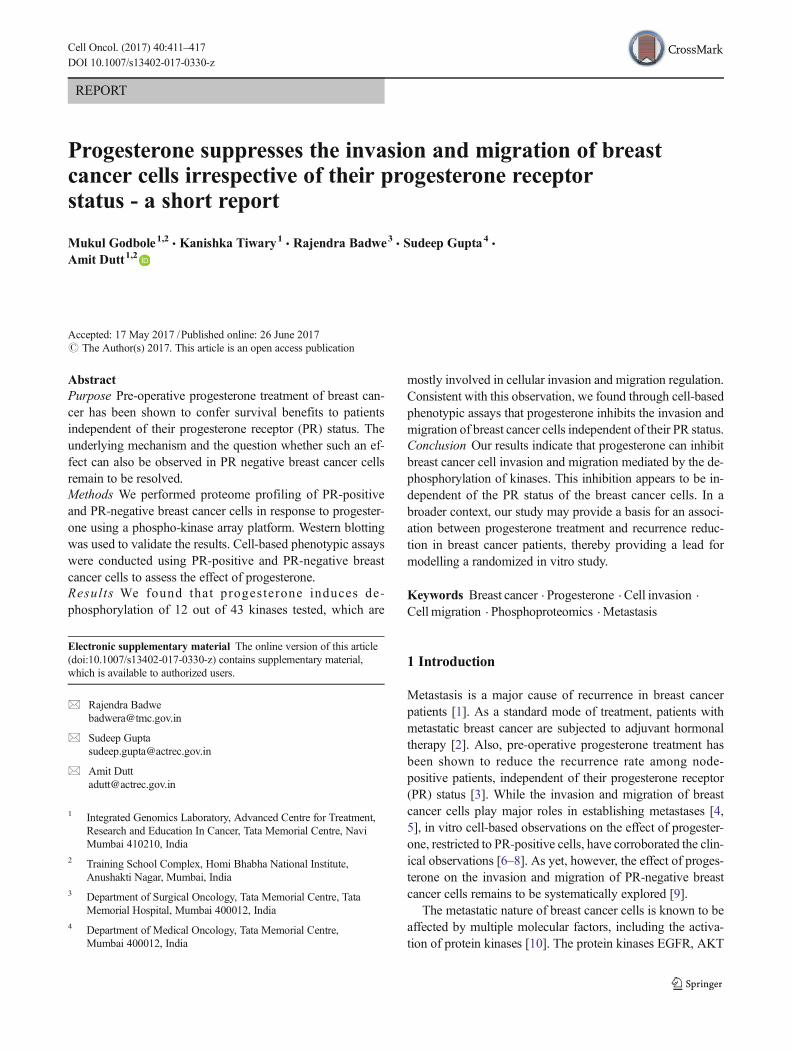
REPORT
Progesterone suppresses the invasion and migration of breastcancer cells irrespective of their progesterone receptorstatus - a short report
Mukul Godbole1,2 & Kanishka Tiwary1 & Rajendra Badwe3 & Sudeep Gupta4 &
Amit Dutt1,2
Accepted: 17 May 2017 /Published online: 26 June 2017# The Author(s) 2017. This article is an open access publication
AbstractPurpose Pre-operative progesterone treatment of breast can-cer has been shown to confer survival benefits to patientsindependent of their progesterone receptor (PR) status. Theunderlying mechanism and the question whether such an ef-fect can also be observed in PR negative breast cancer cellsremain to be resolved.Methods We performed proteome profiling of PR-positiveand PR-negative breast cancer cells in response to progester-one using a phospho-kinase array platform. Western blottingwas used to validate the results. Cell-based phenotypic assayswere conducted using PR-positive and PR-negative breastcancer cells to assess the effect of progesterone.Results We found that progesterone induces de-phosphorylation of 12 out of 43 kinases tested, which are
mostly involved in cellular invasion and migration regulation.Consistent with this observation, we found through cell-basedphenotypic assays that progesterone inhibits the invasion andmigration of breast cancer cells independent of their PR status.Conclusion Our results indicate that progesterone can inhibitbreast cancer cell invasion and migration mediated by the de-phosphorylation of kinases. This inhibition appears to be in-dependent of the PR status of the breast cancer cells. In abroader context, our study may provide a basis for an associ-ation between progesterone treatment and recurrence reduc-tion in breast cancer patients, thereby providing a lead formodelling a randomized in vitro study.
Keywords Breast cancer . Progesterone . Cell invasion .
Cell migration . Phosphoproteomics .Metastasis
1 Introduction
Metastasis is a major cause of recurrence in breast cancerpatients [1]. As a standard mode of treatment, patients withmetastatic breast cancer are subjected to adjuvant hormonaltherapy [2]. Also, pre-operative progesterone treatment hasbeen shown to reduce the recurrence rate among node-positive patients, independent of their progesterone receptor(PR) status [3]. While the invasion and migration of breastcancer cells play major roles in establishing metastases [4,5], in vitro cell-based observations on the effect of progester-one, restricted to PR-positive cells, have corroborated the clin-ical observations [6–8]. As yet, however, the effect of proges-terone on the invasion and migration of PR-negative breastcancer cells remains to be systematically explored [9].
The metastatic nature of breast cancer cells is known to beaffected by multiple molecular factors, including the activa-tion of protein kinases [10]. The protein kinases EGFR, AKT
Electronic supplementary material The online version of this article(doi:10.1007/s13402-017-0330-z) contains supplementary material,which is available to authorized users.
* Rajendra [email protected]
* Sudeep [email protected]
* Amit [email protected]
1 Integrated Genomics Laboratory, Advanced Centre for Treatment,Research and Education In Cancer, Tata Memorial Centre, NaviMumbai 410210, India
2 Training School Complex, Homi Bhabha National Institute,Anushakti Nagar, Mumbai, India
3 Department of Surgical Oncology, Tata Memorial Centre, TataMemorial Hospital, Mumbai 400012, India
4 Department of Medical Oncology, Tata Memorial Centre,Mumbai 400012, India
Cell Oncol. (2017) 40:411–417DOI 10.1007/s13402-017-0330-z
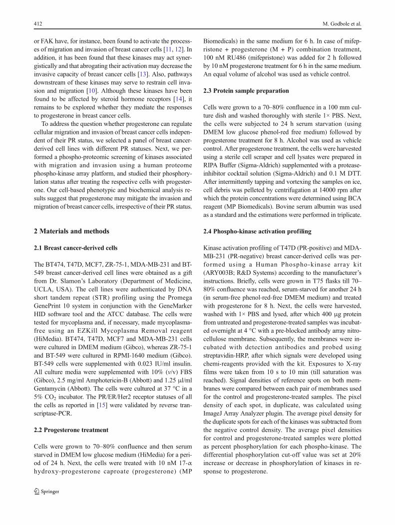
or FAK have, for instance, been found to activate the process-es of migration and invasion of breast cancer cells [11, 12]. Inaddition, it has been found that these kinases may act syner-gistically and that abrogating their activation may decrease theinvasive capacity of breast cancer cells [13]. Also, pathwaysdownstream of these kinases may serve to restrain cell inva-sion and migration [10]. Although these kinases have beenfound to be affected by steroid hormone receptors [14], itremains to be explored whether they mediate the responsesto progesterone in breast cancer cells.
To address the question whether progesterone can regulatecellular migration and invasion of breast cancer cells indepen-dent of their PR status, we selected a panel of breast cancer-derived cell lines with different PR statuses. Next, we per-formed a phospho-proteomic screening of kinases associatedwith migration and invasion using a human proteomephospho-kinase array platform, and studied their phosphory-lation status after treating the respective cells with progester-one. Our cell-based phenotypic and biochemical analysis re-sults suggest that progesterone may mitigate the invasion andmigration of breast cancer cells, irrespective of their PR status.
2 Materials and methods
2.1 Breast cancer-derived cells
The BT474, T47D, MCF7, ZR-75-1, MDA-MB-231 and BT-549 breast cancer-derived cell lines were obtained as a giftfrom Dr. Slamon’s Laboratory (Department of Medicine,UCLA, USA). The cell lines were authenticated by DNAshort tandem repeat (STR) profiling using the PromegaGenePrint 10 system in conjunction with the GeneMarkerHID software tool and the ATCC database. The cells weretested for mycoplasma and, if necessary, made mycoplasma-free using an EZKill Mycoplasma Removal reagent(HiMedia). BT474, T47D, MCF7 and MDA-MB-231 cellswere cultured in DMEM medium (Gibco), whereas ZR-75-1and BT-549 were cultured in RPMI-1640 medium (Gibco).BT-549 cells were supplemented with 0.023 IU/ml insulin.All culture media were supplemented with 10% (v/v) FBS(Gibco), 2.5 mg/ml Amphotericin-B (Abbott) and 1.25 μl/mlGentamycin (Abbott). The cells were cultured at 37 °C in a5% CO2 incubator. The PR/ER/Her2 receptor statuses of allthe cells as reported in [15] were validated by reverse tran-scriptase-PCR.
2.2 Progesterone treatment
Cells were grown to 70–80% confluence and then serumstarved in DMEM low glucose medium (HiMedia) for a peri-od of 24 h. Next, the cells were treated with 10 nM 17-αhydroxy-progesterone caproate (progesterone) (MP
Biomedicals) in the same medium for 6 h. In case of mifep-ristone + progesterone (M + P) combination treatment,100 nM RU486 (mifepristone) was added for 2 h followedby 10 nM progesterone treatment for 6 h in the same medium.An equal volume of alcohol was used as vehicle control.
2.3 Protein sample preparation
Cells were grown to a 70–80% confluence in a 100 mm cul-ture dish and washed thoroughly with sterile 1× PBS. Next,the cells were subjected to 24 h serum starvation (usingDMEM low glucose phenol-red free medium) followed byprogesterone treatment for 8 h. Alcohol was used as vehiclecontrol. After progesterone treatment, the cells were harvestedusing a sterile cell scraper and cell lysates were prepared inRIPA Buffer (Sigma-Aldrich) supplemented with a protease-inhibitor cocktail solution (Sigma-Aldrich) and 0.1 M DTT.After intermittently tapping and vortexing the samples on ice,cell debris was pelleted by centrifugation at 14000 rpm afterwhich the protein concentrations were determined using BCAreagent (MP Biomedicals). Bovine serum albumin was usedas a standard and the estimations were performed in triplicate.
2.4 Phospho-kinase activation profiling
Kinase activation profiling of T47D (PR-positive) and MDA-MB-231 (PR-negative) breast cancer-derived cells was per-formed using a Human Phospho-kinase array kit(ARY003B; R&D Systems) according to the manufacturer’sinstructions. Briefly, cells were grown in T75 flasks till 70–80% confluence was reached, serum-starved for another 24 h(in serum-free phenol-red-free DMEM medium) and treatedwith progesterone for 8 h. Next, the cells were harvested,washed with 1× PBS and lysed, after which 400 μg proteinfrom untreated and progesterone-treated samples was incubat-ed overnight at 4 °C with a pre-blocked antibody array nitro-cellulose membrane. Subsequently, the membranes were in-cubated with detection antibodies and probed usingstreptavidin-HRP, after which signals were developed usingchemi-reagents provided with the kit. Exposures to X-rayfilms were taken from 10 s to 10 min (till saturation wasreached). Signal densities of reference spots on both mem-branes were compared between each pair of membranes usedfor the control and progesterone-treated samples. The pixeldensity of each spot, in duplicate, was calculated usingImageJ Array Analyzer plugin. The average pixel density forthe duplicate spots for each of the kinases was subtracted fromthe negative control density. The average pixel densitiesfor control and progesterone-treated samples were plottedas percent phosphorylation for each phospho-kinase. Thedifferential phosphorylation cut-off value was set at 20%increase or decrease in phosphorylation of kinases in re-sponse to progesterone.
412 M. Godbole et al.

2.5 Western blotting
Equal amounts of cell lysate were resolved by 10% SDS-PAGE and transferred to PVDF membranes using a wet trans-fer method. Primary antibodies directed against p-EGFR(Y1068) (Cell Signaling, 3777S; Dilution 1:500), p-AKT(S473) (Cell Signaling, 4060S; Dilution 1:500), p-ERK1/2(T202/Y204) (Cell Signaling, 9101S; Dilution 1:1000), totalEGFR (1005) (Santa Cruz Biotechnology, sc-03; Dilution1:1000), total AKT (11E7) (Cell Signaling, 4685S; Dilution1:1000), total ERK2 (c-14) (Santa Cruz Biotechnology, sc-154; Dilution 1:1000) and β-actin (I-19)-R (Santa CruzBiotechnology, sc-1616-R; Dilution 1:3000) were dilutedin 3% BSA solution prepared in 1× TBST and incubatedover night at 4 °C. A goat anti-rabbit IgG-HRP secondaryantibody (Santa Cruz Biotechnology, sc-2004; Dilution1:3000) was used for the detection of primary antibodybinding. ECL Western Blotting Substrate (Pierce) andTakara Chemiluminescence substrate (ClonTech Takara)were used for visualization of the protein bands on X-ray films (Fuji Films).
2.6 RNA extraction and real-time PCR
For RNA extraction from alcohol and progesterone-treatedbreast cancer-derived cells, the respective cells were treatedwith progesterone for 6 h. Next, TRIzol reagent (Invitrogen)was used to lyse the cells after which RNA was isolated ac-cording to the manufacturer’s protocol. RNA concentrationswere measured using NanoDrop. For assessment of theDUSP1 transcript levels, cDNA was synthesized using aHigh capacity cDNA reverse transcription kit (AppliedBiosystems) and subjected to quantitative real-time PCRusing a Roche Light-Cycler-II 480 instrument in conjunctionwith a Roche real-time master mix (Roche). Expressionchanges were calculated using the 2-ΔΔCT method. GAPDHwas used as internal control for normalization. The primersequences used for DUSP1 were Forward primer OAD-571:CCTGCAGTACCCCACTCTACG; Reverse primer OAD-572: CCCAAGGCATCCAGCATGTCC and for GAPDHForward primer OAD-328: AATCCCATCACCATCTTCCA; Reverse primer OAD-329: TGGACTCCACGACGTACTCA.
2.7 Cell invasion assay
A Matrigel invasion assay was performed using 24-wellTranswell inserts (Corning) coated with 100 μg matrigel andallowed to settle for 24 h at 37 °C. Next, 35,000 cellssuspended in 350 μl serum-free medium were seeded intothe upper chamber and 600 μl of 10% serum-containing me-dium was added to the lower chamber. After this, the cellswere allowed to invade for 16-18 h at 37 °C, followed by
fixation of the invaded cells and staining by crystal violet.After mounting the membrane using DPX on a slide, the cellswere observed under an upright microscope. Ten randomfields were chosen after which the number of cells in eachfield was counted and plotted as percentage cell invasion.
2.8 Scratch wound healing assay
Confluent cell monolayers in 6-well plates were subjected to ascratch with a sterile pipette tip. After this, the cells werebriefly rinsed using 1× PBS to remove debris and subsequent-ly incubated with low-glucose phenol-red free DMEM medi-um containing 10% charcoal-stripped FBS (Gibco). The cellswere treated with 10 nM progesterone or 100 nMmifepristoneor a combination of both. Alcohol was used as a vehicle con-trol. Cell migration at the wound surface was measured duringa period of 20 h under an inverted microscope. Quantificationwas performed using the ImageJ wound healing plugin tool bymeasuring the distance of the wound edge of the migratingcells from the start point to the migrated point in three separatewounds in three independent experiments.
3 Results and discussion
The activation of kinases like EGFR and ERK1/2 has beenreported to play an important role in the de-regulation of cel-lular processes that are associated with the metastatic capacityof breast cancer cells [16]. Here, we set out to assess the effectof progesterone on the activation of kinases in breast cancercells using a human phospho-kinase array platform. To verifythe effect of progesterone independent of the progesteronereceptor (PR) status of the cells, we selected both PR-positive (T47D) and PR-negative (MDA-MB-231) breastcancer-derived cells for our study (Table 1). Untreated cellswere used as negative controls. As reported before, we ob-served a breast cancer cell-specific phosphorylation of p53(S392/S46/S15) andAMPK (T183), which were subsequentlyused as internal positive controls [17, 18]. Based on differen-tial phosphorylation analyses of the T47D andMDA-MB-231cells, 7 out of 43 kinases tested were found to be de-phosphorylated in the progesterone treated cells (Fig. 1a-gand Supplementary Fig. 1). Of these, p70 S6 kinase andSTAT3 showed the highest decrease in phosphorylation(30%) while FAK, AKT and RSK1/2/3 showed a 20% de-crease in both the cell lines in response to progesterone treat-ment. In addition, we observed a reduction in phosphorylationof the ERK1/2 (T202/Y204, T185/Y187), EGFR (Y1068),MSK1/2 (S376/S360), p38α (T180/Y182) and p27 (T198)kinases upon treatment with progesterone (SupplementaryFig. 1), as reported earlier [19], and validated the results byWestern blot analysis (Supplementary Fig. 2a). Consistentwith earlier reports [19], we also observed a significant up-
Progesterone suppresses the invasion and migration 413

regulation of a dual specificity phosphatase, DUSP1, upontreatment with progesterone in breast cancer cells that couldpossibly mediate the effect observed (Supplementary Fig. 2b).Taken together, our results indicate that progesterone can re-duce the phosphorylation of 12 out of 43 kinases tested in aPR-independent manner, which could affect cellular signalingpathways downstream to these kinases with a concomitantincrease in the expression of a dual-specificity phosphatase,DUSP1, that could mediate the de-phosphorylation of thesekinases [19].
Based on the known involvement of EGFR, AKT andERK1/2 in the invasion and migration of breast cancer cellsand the finding that their inhibition may block this phenotype[11, 12, 20] or vice versa, i.e., metastases inhibitors may in-hibit the phosphorylation of FAK in PR-negative MDA-MB-231 cells [21] or lung cancer cells [22], we set out to analyzethe in vitro effects of progesterone on breast cancer cells.Using aMatrigel chamber assay in conjunction with cells withvarying PR/ER/Her2 statuses (Table 1) we found that proges-terone could decrease the invasion capacity of different breast
cancer-derived cells (BT474, T47D, MCF7 and MDA-MB-231), irrespective of their hormone receptor status (Fig. 2a-d).This result suggests that targeted activation of kinases by pro-gesterone may bring about phenotypic changes in breastcancer-derived cells independent of their PR-status, potential-ly decreasing their metastatic capacity similar to combinatorialEGFR and AKT inhibition, which is known to affect the in-vasion of breast and other cancer cells mediated by matrixmetalloproteinases [13, 23]. Importantly, we found that PRblocking by mifepristone had no significant effect on the in-vasive capacities of the cells, again suggesting that progester-one may induce suppression of invasion in breast cancer cellsin a predominantly PR-independent manner (Fig. 2a-d).
Next we performed scratch wound healing assays to assesswhether breast cancer cell migration is affected by progester-one. Similar to the effect of progesterone on breast cancer cellinvasion, we observed a significant decrease in cellular migra-tion in response to progesterone over the period of 20 h in aPR-independent manner (Fig. 3a-d). The non-essential role ofPR that we observed in the inhibition of migration of breast
Table 1 Selection of breast celllines and validation of PR/ER/Her2hormone receptor status. A panel ofbreast cancer cell lines with varyingreceptor statuses, as reported in theliterature, was selected for studyingthe effects of progesterone. Thevalidation status of PR/ER/Her2transcript expression in the cell linesis indicated as B+^ (positive) or B-^(negative)
Sr. No. Cell Line Literature reported
receptor status
Validation of receptor status by RT-PCR
PR ER HER2 PR ER HER2
1. BT474 + + + + + +
2. T47D + + − + + −3. MCF7 + + − + + −4. ZR-75-1 − + − − + −5. MDA-MB-231 − − − − − −6. BT-549 − − − − − −
a b c d
e f g
Fig. 1 Kinase phosphorylation is modulated by progesterone in breastcancer cells. The percentage of reduction in phosphorylation in responseto progesterone was calculated relative to that in untreated cells and isplotted for each of the differentially phosphorylated kinases (panels a-g).
In the bar plot the light grey bar indicates phosphorylation reduction inT47D cells and the dark grey bar indicates phosphorylation reduction inMDA-MB-231 cells
414 M. Godbole et al.

cancer-derived cells in response to progesterone, specificallyin the PR-negative MDA-MB-231, ZR-75-1 and BT-549cells, may be mediated by interaction of progesterone with
the glucocorticoid receptor (GR) or the membrane progester-one receptor (mPR), as has been reported before [22, 24, 25].Of note, it has also been reported that treatment with
a b c d
Fig. 3 Migration of breast cancer cells decreases in response toprogesterone treatment. Scratch wounds were made in breast cancer-derived cell lines (a) T47D, (b) ZR-75-1, (c) MDA-MB-231 and (d)BT-549, with differing receptor statuses. Subsequently, the cells weretreated with alcohol (control) and progesterone for 20 h and followed in
time for migration. The bar plots indicate the percentages of cellularmigration, with direct comparisons between control and progesteronetreated cells. The figures are representative of three independentexperiments performed in triplicates. P-values were calculated usingstudent’s unpaired t-test. ** p < 0.001; *** p < 0.0001
Fig. 2 Progesterone inhibits breast cancer cell invasion. Invasion assayswere performed with breast cancer-derived cell lines (a) BT474, (b)T47D, (c) MCF7 and d) MDA-MB-231 treated with progesterone,mifepristone or a combination of mifepristone and progesterone (M +
P). The bar plot represents the percentage of cell invasion for eachpanel. The figure is representative of three independent experimentsperformed in triplicates. P-values were calculated using student’sunpaired t-test. ** p < 0.001; *** p < 0001; ns not significant
Progesterone suppresses the invasion and migration 415

glucocorticoids may similarly decrease the migration of PR-negativeMDA-MB-231 cells [26], which suggests that redun-dant pathways may underlie the progesterone response in aPR-independent manner [27]. Consistent with these observa-tions, we found that blocking PR by mifepristone prior toexposing the cells to progesterone did not rescue the effectof progesterone, suggesting that the progesterone-mediatedsuppression of migration in breast cancer cells is predominant-ly mediated in a PR-independent manner (SupplementaryFig. 3a and b). This result corroborates a clinical study inwhich it was found that progesterone may reduce the recur-rence of node-positive breast cancer in patients independent oftheir PR status [3]. A recent in vitro study, however, suggestedthat the PR status may play an essential role as no significanteffect was observed in PR-negative MDA-MB-231 cells inresponse to R5020, which is a synthetic progestin [28]. But,it has also been shown that the downstream effects of proges-terone and progestin may be variable [29] and this notion,together with possible variations that may occur duringcell line passage, could account for the phenotypic differ-ences observed in MDA-MB-231 cells. Moreover, wefound that the progesterone-mediated suppression of mi-gration and invasion also occurred in other PR-negativebreast cancer-derived cells, i.e., ZR-75-1 and BT-549,which has not been reported before.
In summary, we present a first lead to model a randomizedin vitro study to systematically elucidate the role of kinasesthat may underlie the clinical outcome of pre-operative pro-gesterone intervention in breast cancer patients.
Acknowledgements We thank all members of the Dutt laboratory forcritically reviewing the manuscript and Pawan Upadhyay for STR profil-ing. A.D. is supported by an Intermediate Fellowship from the WellcomeTrust/DBT India Alliance (IA/I/11/2500278), intramural grants [IRB pro-ject 2712], and by a grant from DBT (BT/MED/30/VNCI-Hr-RCA/2015). M.G is supported by a research fellowship from the HomiBhabha National Institute (HBNI), ACTREC-TMC. The funders hadno role in the study design, data collection and analysis, decision topublish, or preparation of the manuscript.
Compliance with ethical standards
Conflict of interest None declared
Open Access This article is distributed under the terms of the CreativeCommons At t r ibut ion 4 .0 In te rna t ional License (h t tp : / /creativecommons.org/licenses/by/4.0/), which permits unrestricted use,distribution, and reproduction in any medium, provided you giveappropriate credit to the original author(s) and the source, provide a linkto the Creative Commons license, and indicate if changes were made.
References
1. B. Weigelt, J.L. Peterse, L.J. van't Veer, Breast cancer metastasis:Markers and models. Nat Rev Cancer 5, 591–602 (2005)
2. R.W. Carlson and I.C. Henderson, Sequential hormonal therapy formetastatic breast cancer after adjuvant tamoxifen or anastrozole.Breast Cancer Res. Treat. 80 Suppl 1, S19–26; discussion S27–18(2003)
3. R. Badwe, R. Hawaldar, V. Parmar, M. Nadkarni, T. Shet, S. Desai,S. Gupta, R. Jalali, V. Vanmali, R. Dikshit, I. Mittra, Single-injection depot progesterone before surgery and survival in womenwith operable breast cancer: A randomized controlled trial. J ClinOncol 29, 2845–2851 (2011)
4. O.J. Scully, B.-H. Bay, G. Yip, Y. Yu, Breast cancer metastasis.Cancer Genomics-Proteomics 9, 311–320 (2012)
5. D. Wolczyk, M. Zaremba-Czogalla, A. Hryniewicz-Jankowska, R.Tabola, K. Grabowski, A.F. Sikorski, K. Augoff, TNF-alpha pro-motes breast cancer cell migration and enhances the concentrationof membrane-associated proteases in lipid rafts. Cell Oncol 39,353–363 (2016)
6. H. Mohammed, I.A. Russell, R. Stark, O.M. Rueda, T.E. Hickey,G.A. Tarulli, A.A. Serandour, S.N. Birrell, A. Bruna, A. Saadi, S.Menon, J. Hadfield, M. Pugh, G.V. Raj, G.D. Brown, C. D'Santos,J.L. Robinson, G. Silva, R. Launchbury, C.M. Perou, J. Stingl, C.Caldas, W.D. Tilley, J.S. Carroll, Progesterone receptor modulatesERalpha action in breast cancer. Nature 523, 313–317 (2015)
7. H. Singhal, M.E. Greene, G. Tarulli, A.L. Zarnke, R.J. Bourgo, M.Laine, Y.F. Chang, S. Ma, A.G. Dembo, G.V. Raj, T.E. Hickey,W.D. Tilley, G.L. Greene, Genomic agonism and phenotypic antag-onism between estrogen and progesterone receptors in breast can-cer. Sci Adv 2, e1501924 (2016)
8. H.C. Wang, W.S. Lee, Molecular mechanisms underlyingprogesterone-enhanced breast cancer cell migration. Sci Rep 6(2016)
9. M. Dowsett, I.E. Smith, Presurgical progesterone in early breastcancer: So much for so little? J Clin Oncol 29, 2839–2841 (2011)
10. P.S. Steeg, Targeting metastasis. Nat Rev Cancer 16, 201–218 (2016)
11. Z. Yang, R. Bagheri-Yarmand, R.A. Wang, L. Adam, V.V.Papadimitrakopoulou, G.L. Clayman, A. El-Naggar, R. Lotan,C.J. Barnes, W.K. Hong, R. Kumar, The epidermal growth factorreceptor tyrosine kinase inhibitor ZD1839 (Iressa) suppresses c-Srcand Pak1 pathways and invasiveness of human cancer cells. ClinCancer Res 10, 658–667 (2004)
12. W. Li, Z. Liu, C. Zhao, L. Zhai, Binding of MMP-9-degradedfibronectin to beta6 integrin promotes invasion via the FAK-Src-related Erk1/2 and PI3K/Akt/Smad-1/5/8 pathways in breast can-cer. Oncol Rep 34, 1345–1352 (2015)
13. Y.J. Chen, K.N. Lin, L.M. Jhang, C.H. Huang, Y.C. Lee, L.S.Chang, Gallic acid abolishes the EGFR/Src/Akt/Erk-mediated ex-pression of matrix metalloproteinase-9 in MCF-7 breast cancercells. Chem Biol Interact 252, 131–140 (2016)
14. Z. Piperigkou, P. Bouris, M. Onisto, M. Franchi, D. Kletsas, A.D.Theocharis, N.K. Karamanos, Estrogen receptor beta modulatesbreast cancer cells functional properties, signaling and expressionof matrix molecules. Matrix Biol 56, 4–23 (2016)
15. J. Kao, K. Salari, M. Bocanegra, Y.L. Choi, L. Girard, J. Gandhi,K.A. Kwei, T. Hernandez-Boussard, P. Wang, A.F. Gazdar, J.D.Minna, J.R. Pollack, Molecular profiling of breast cancer cell linesdefines relevant tumor models and provides a resource for cancergene discovery. PLoS One 4, e6146 (2009)
16. J. Whyte, O. Bergin, A. Bianchi, S. McNally and F. Martin, Keysignalling nodes in mammary gland development and cancer.Mitogen-activated protein kinase signalling in experimental modelsof breast cancer progression and in mammary gland development.Breast Cancer Res 11, 209 (2009)
17. S. Dutta, C. Warshall, C. Bandyopadhyay, D. Dutta, B. Chandran,Interactions between exosomes from breast cancer cells and prima-ry mammary epithelial cells leads to generation of reactive oxygen
416 M. Godbole et al.

species which induce DNA damage response, stabilization of p53and autophagy in epithelial cells. PLoS One 9, e97580 (2014)
18. O.S. El-Masry, B.L. Brown, P.R. Dobson, Effects of activation ofAMPK on human breast cancer cell lines with different geneticbackgrounds. Oncol Lett 3, 224–228 (2012)
19. C.C. Chen, D.B. Hardy, C.R. Mendelson, Progesterone receptorinhibits proliferation of human breast cancer cells via induction ofMAPK phosphatase 1 (MKP-1/DUSP1). J Biol Chem 286, 43091–43102 (2011)
20. J.T. Price, T. Tiganis, A. Agarwal, D. Djakiew, E.W. Thompson,Epidermal growth factor promotes MDA-MB-231 breast cancer cellmigration through a phosphatidylinositol 3′-kinase and phospholi-pase C-dependent mechanism. Cancer Res 59, 5475–5478 (1999)
21. G.S. Wu, Y.L. Song, Z.Q. Yin, J.J. Guo, S.P. Wang, W.W. Zhao,X.P. Chen, Q.W. Zhang, J.J. Lu, Y.T. Wang, Ganoderiol A-enrichedextract suppresses migration and adhesion of MDA-MB-231 cellsby inhibiting FAK-SRC-paxillin cascade pathway. PLoS One 8,e76620 (2013)
22. M. Xie, S. You, Q. Chen, X. Chen, C. Hu, Progesterone inhibits themigration and invasion of A549 lung cancer cells through mem-brane progesterone receptor alpha-mediated mechanisms. OncolRep 29, 1873–1880 (2013)
23. D.Kim, S. Kim,H.Koh, S.O.Yoon, A.S. Chung, K.S. Cho, J. Chung,Akt/PKB promotes cancer cell invasion via increased motility andmetalloproteinase production. FASEB J 15, 1953–1962 (2001)
24. K. Lei, L. Chen, E.X. Georgiou, S.R. Sooranna, S. Khanjani, J.J.Brosens, P.R. Bennett, M.R. Johnson, Progesterone acts via thenuclear glucocorticoid receptor to suppress IL-1beta-inducedCOX-2 expression in human term myometrial cells. PLoS One 7,e50167 (2012)
25. M. Xie, L. Zhou, X. Chen, L.O. Gainey, J. Xiao, M.S. Nanes, A.Hou, S. You, Q. Chen, Progesterone and Src family inhibitor PP1synergistically inhibit cell migration and invasion of human basalphenotype breast cancer cells. BiomedRes Int 2015, 426429 (2015)
26. E.R. Fietz, C.R. Keenan, G. Lopez-Campos, Y. Tu, C.N. Johnstone,T. Harris, A.G. Stewart, Glucocorticoid resistance of migration andgene expression in a daughter MDA-MB-231 breast tumour cellline selected for high metastatic potential. Sci Rep 7, 43774 (2017)
27. M.S. Fernandes, J.J. Brosens, B. Gellersen, Honey, we need to talkabout the membrane progestin receptors. Steroids 73, 942–952 (2008)
28. C. Bellance, J.A. Khan, G. Meduri, A. Guiochon-Mantel, M.Lombes, H. Loosfelt, Progesterone receptor isoforms PRA andPRB differentially contribute to breast cancer cell migrationthrough interaction with focal adhesion kinase complexes. MolBiol Cell 24, 1363–1374 (2013)
29. H. Seeger, D. Wallwiener, A.O. Mueck, The effect of progesteroneand synthetic progestins on serum- and estradiol-stimulated prolif-eration of human breast cancer cells. Horm Metab Res 35, 76–80(2003)
Progesterone suppresses the invasion and migration 417

Full Terms & Conditions of access and use can be found athttp://www.tandfonline.com/action/journalInformation?journalCode=kcbt20
Cancer Biology & Therapy
ISSN: 1538-4047 (Print) 1555-8576 (Online) Journal homepage: http://www.tandfonline.com/loi/kcbt20
miR-129-2 mediates down-regulation ofprogesterone receptor in response toprogesterone in breast cancer cells
Mukul Godbole, Pratik Chandrani, Nilesh Gardi, Hemant Dhamne, KuldeepPatel, Neelima Yadav, Sudeep Gupta, Rajendra Badwe & Amit Dutt
To cite this article: Mukul Godbole, Pratik Chandrani, Nilesh Gardi, Hemant Dhamne, KuldeepPatel, Neelima Yadav, Sudeep Gupta, Rajendra Badwe & Amit Dutt (2017) miR-129-2 mediatesdown-regulation of progesterone receptor in response to progesterone in breast cancer cells,Cancer Biology & Therapy, 18:10, 801-805, DOI: 10.1080/15384047.2017.1373216
To link to this article: https://doi.org/10.1080/15384047.2017.1373216
© 2017 The Author(s). Published withlicense by Taylor & Francis Group, LLC©Mukul Godbole, Pratik Chandrani, NileshGardi, Hemant Dhamne, Kuldeep Patel,Neelima Yadav, Sudeep Gupta, RajendraBadwe, and Amit Dutt
View supplementary material
Accepted author version posted online: 06Sep 2017.Published online: 26 Sep 2017.
Submit your article to this journal
Article views: 375 View related articles
View Crossmark data

BRIEF REPORT
miR-129-2mediates down-regulation of progesterone receptor in responseto progesterone in breast cancer cells
Mukul Godbolea,b, Pratik Chandrania,b, Nilesh Gardia,b, Hemant Dhamnea, Kuldeep Patela, Neelima Yadava,b,Sudeep Guptac, Rajendra Badwed, and Amit Dutt a,b
aIntegrated Genomics Laboratory, Advanced Centre for Treatment, Research and Education In Cancer, Tata Memorial Centre, Maharashtra, NaviMumbai, India; bIntegrated Cancer Genomics Laboratory, Homi Bhabha National Institute, Training School Complex, Anushakti Nagar, Maharashtra,Mumbai, India; cDepartment of Medical Oncology, Tata Memorial Centre, Maharashtra, Mumbai, India; dDepartment of Surgical Oncology, TataMemorial Hospital, Tata Memorial Centre, Maharashtra, Mumbai, India
ARTICLE HISTORYReceived 5 May 2017Revised 27 July 2017Accepted 24 August 2017
ABSTRACTObjective: Hormonal therapy is an important component of first line of treatment for breast cancer. Responseto hormonal therapy is influenced by the progesterone receptor (PR)-status of breast cancer patients.However as an early effect, exposure to progesterone decreases expression of PR in breast cancer cells.An understanding of the mechanism underlying down-regulation of PR could help improve response tohormonal therapy. Methods: We performed small RNA sequencing of breast cancer cells for identification ofmicroRNAs targeting PR in response to progesterone treatment. Biochemical approaches were used tovalidate the findings in breast cancer cells. Results: Analysis of small RNA sequencing of four breast cancercell lines treated with progesterone revealed an up-regulation of miR-129-2 independent of the PR status ofthe cells. We show that miR-129-2 targets 30UTR of PR to down-regulate its expression. Furthermore,inhibition of miR-129-2 expression rescues the down-regulation of PR in breast cancer cells. Also, theexpression levels ofmiR-129-2 was observed to be elevated in patients with low expression of PR in the TCGAcohort (nD 359). Conclusion:miR-129-2mediates down-regulation of PR in breast cancer cells in response toprogesterone, while anti-miR-129-2 could potentiate PR expression levels among patients with inadequate PRlevels. Thus, modulation of activity of miR-129-2 could stabilize PR expression and potentially improveresponse to hormonal therapy under adjuvant or neo-adjuvant settings.
KEYWORDSBreast cancer; hormonaltherapy; progesterone;progesterone receptor;microRNA; cancer genomics;small RNA sequencing
Introduction
Breast cancer is the most prevalent cancer among womenworldwide. Despite all advances in early diagnosis and treat-ment, nearly 30% node-negative and 70% node-positivepatients relapse with metastatic disease.1 Treatment of breastcancer patients is influenced by the presence of estrogenreceptor (ER) and progesterone receptor (PR). ER/PR posi-tive patients tend to respond better to hormonal therapy andhave a lower risk of relapse compared to ER-positive,PR-negative patients.2 The down-regulation of PR expressionin breast cancer cells is caused either by methylation atPR promoter3, or in response to progesterone by post transla-tional modification of the PR protein by CUEDC2 andMAPK.4,5 Growing evidence also suggest microRNAs torespond to steroid hormones and suppress the activity ofrespective hormone receptor.6 For instance, miR-18a,miR-19b and miR-20b (paralogous pri-microRNAs) down-regulate the expression of ER in response to estrogen inbreast cancer.7 Comparatively, similar regulation of micro-RNA expression in response to progesterone has been less
explored.8 In order to study the progesterone-regulatedmicroRNAs targeting PR, we performed small RNA sequenc-ing of breast cancer cell lines treated with progesterone. Thedifferentially expressed microRNAs were used to identifymicroRNAs that target 30UTR of PR. Our analysis revealsmiR-129-2 targets PR and is up-regulated in response to pro-gesterone. The association of miR-129-2 and PR was func-tionally validated by luciferase assay. Also western blotanalysis suggests that inhibition of miR-129-2 stabilizes PR inbreast cancer cells even in presence of progesterone. More-over, patients with high miR-129-2 levels had significantlylower expression of PR as compared to patients with nomiR-129-2 expression in The Cancer Genome Atlas cohort.
Results
Identification of progesterone responsive microRNAstargeting PR expression in breast cancer cells
Consistent with earlier reports, we observed down-regulation ofPR transcripts when T47D and BT474 cells were treated with
CONTACT Amit Dutt [email protected] Integrated Genomics Laboratory, Advanced Centre for Treatment, Research and Education In Cancer, Tata MemorialCentre, Navi Mumbai-410210, India; Rajendra Badwe [email protected] Department of Medical Surgical Oncology, Tata Memorial Centre, Mumbai-400012,India; Sudeep Gupta [email protected] Department of Medical Oncology, Tata Memorial Hospital, Tata Memorial Centre, Mumbai-400012, India.
Supplemental data for this article can be accessed on the publisher’s website.© 2017 Mukul Godbole, Pratik Chandrani, Nilesh Gardi, Hemant Dhamne, Kuldeep Patel, Neelima Yadav, Sudeep Gupta, Rajendra Badwe, and Amit Dutt. Published with license by Taylor & Fran-cis Group, LLCThis is an Open Access article distributed under the terms of the Creative Commons Attribution License (http://creativecommons.org/licenses/by/4.0/), which permits unrestricted use, distribu-tion, and reproduction in any medium, provided the original work is properly cited.
CANCER BIOLOGY & THERAPY2017, VOL. 18, NO. 10, 801–805https://doi.org/10.1080/15384047.2017.1373216
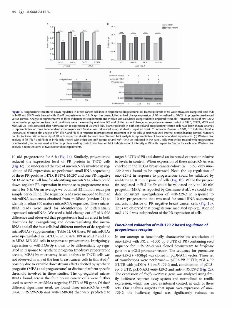
10 nM progesterone for 6 h (Fig. 1a). Similarly, progesteronereduced the expression level of PR protein in T47D cells(Fig. 1c). To understand the role of microRNA’s involved in reg-ulation of PR expression, we performed small RNA sequencingof three PR-positive T47D, BT474, MCF7 and one PR-negativeMDA-MB-231 cell line for identifying microRNAs which coulddown-regulate PR expression in response to progesterone treat-ment for 6 h. On an average we obtained 22 million reads persample per cell line. The sequence reads were mapped to humanmicroRNA sequences obtained from miRBase (version 21) toidentify median 800 mature microRNA sequences. These micro-RNA reads were used for identification of differentiallyexpressed microRNAs. We used a fold-change cut-off of 3-folddifference and observed that progesterone had an effect in bothdirections by up-regulating and down-regulating the micro-RNAs and all the four cells had different number of de-regulatedmicroRNAs (Supplementary Table 1). Of these, 98 microRNAswere up-regulated in T47D, 96 in BT474, 189 in MCF7 and 106in MDA-MB-231 cells in response to progesterone. Intriguingly,expression of miR-513a-5p shown to be differentially up-regu-lated in response to synthetic progestin (medroxy progesteroneacetate, MPA) by microarray-based analysis in T47D cells wasnot observed in any of the four breast cancer cells in this study8,possibly due to variable downstream effects elicited by syntheticprogestin (MPA) and progesterone9 or distinct platform specificthreshold involved in these studies. The up-regulated micro-RNAs found across the four breast cancer cells were furtherused to search microRNAs targeting 30UTR of PR gene. Of the 6different algorithms used, we found three microRNAs (miR-3908, miR-129-2-3p and miR-3140-3p) that were predicted to
target 30 UTR of PR and showed an increased expression relativeto levels in control. When expression of these microRNAs waschecked in the TCGA breast cancer cohort (nD 359), onlymiR-129-2 was found to be expressed. Next, the up-regulation ofmiR-129-2 in response to progesterone could be validated byreal-time PCR in our panel of cells (Fig. 1b). While the proges-tin-regulated miR-513a-5p could be validated only at 100 nMprogestin (MPA) as reported by Cochrane et al.8, we could vali-date consistent up-regulation of miR-129-2 in response to10 nM progesterone that was used for small RNA sequencinganalysis, inclusive of PR-negative breast cancer cells (Fig. 1b).Thus we observed that progesterone mediated up-regulation ofmiR-129-2was independent of the PR expression of cells.
Functional validation ofmiR-129-2 based regulation ofprogesterone receptor
In our attempt to functionally characterize the association ofmiR-129-2 with PR, a »1000 bp 30UTR of PR (containing seedsequence for miR-129-2) was cloned downstream to luciferasegene in a pGL3-promoter vector. The sequence for prematuremiR-129-2 (»400bp) was cloned in pcDNA3.1 vector. Three setof transfections were performed— pGL3-PR 30UTR; pGL3-PR30UTR with pcDNA-3.1-miR-129-2; and, combination of pGL3-PR 30UTR, pcDNA3.1-miR-129-2 and anti-miR-129-2 (Fig. 2a).The expression of firefly luciferase gene was analyzed using fire-fly luciferase reporter assay system and normalized to renillaexpression, which was used as internal control, in each of thesesets. Our analysis suggests that upon over-expression of miR-129-2, the luciferase signal was significantly reduced as
Figure 1. Progesterone receptor is down-regulated in breast cancer cell lines in response to progesterone. (a) Transcript levels of PR were measured using real-time PCRin T47D and BT474 cells treated with 10 nM progesterone for 6 h. Graph has been plotted as fold change expression of PR normalized to GAPDH in progesterone-treatedversus control. Analysis is representative of three independent experiments and P-value was calculated using student’s unpaired t-test. (b) Transcript levels of miR-129-2under similar progesterone treatment conditions were measured by real-time PCR and plotted as fold change in progesterone versus control of T47D, BT474, MCF7 andMDA-MB-231 cells obtained after normalization to expression of U6 small RNA. Transcript levels in both control and progesterone-treated cells have been shown. Analysisis representative of three independent experiments and P-value was calculated using student’s unpaired t-test. �� indicates P-value <0.001; ��� indicates P-value<0.0001. (c) Western blot analysis of PR (PR-A and PR-B) in response to progesterone treatment in T47D cells. b-actin was used internal protein loading control. Numberson blot indicate ratio of intensity of PR with respect to b-actin for each lane. Western blot analysis is representative of two independent experiments. (d) Western blotanalysis of PR (PR-A and PR-B) in T47D cells treated with either anti-miR-control or anti-miR-129-2. As indicated in the panel, cells were either treated with progesteroneor untreated. b-actin was used as internal protein loading control. Numbers on blot indicate ratio of intensity of PR with respect to b-actin for each lane. Western blotanalysis is representative of two independent experiments.
802 M. GODBOLE ET AL.

compared to signal in vector only cells. Addition of microRNAinhibitor against miR-129-2 (anti-miR-129-2), a double strandedRNA sequence which is complimentary to and specifically tar-gets miR-129-2, reversed the repression and showed an increasein luciferase signal (Fig. 2b). Next, we inhibited miR-129-2 inT47D cells and compared the PR expression in these cells withthe expression in cells transfected with negative control (target-ing miR-29a) inhibitor. Our western blot analysis suggests thatupon exposure to progesterone, T47D cells transfected with neg-ative control showed decrease in PR expression, while PRshowed stable expression in cells transfected with miR-129-2inhibitor even in the presence of progesterone treatment(Fig. 1d). Thus our results provide basis for direct interaction ofmiR-129-2 with PR, where in addition to previous findings, wedemonstrate that over-expression of miR-129-2 mimics theeffect of progesterone treatment to down-regulate PR and thatinhibition of miR-129-2 abrogates its interaction with PR inbreast cancer cells. Taken together, these studies emphasize theplurality in microRNA-mediated feedback regulation of PR.
Next, we analyzed the TCGA breast cancer cohort (n D 359)for studying expression of PR in breast cancer patients withhigh miR-129-2 expression and in absence of miR-129-2expression. When expression of PR was checked in patientswith high miR-129-2 expression (n D 134) versus patients withabsence of miR-129-2 expression (n D 225) we observed a sig-nificantly higher expression of PR in patients with absence ofmiR-129-2 expression as compared to patients with highexpression of miR-129-2 (P D 0.0002) as shown in Fig. 3. Thusa further in-depth analysis needs to be carried out to ascertainthe exact role of miR-129-2 in survival of breast cancer patients.
Discussion
Small RNA sequencing analysis of progesterone treated breastcancer cell lines led to the identification of a novel PR-targeting
microRNA miR-129-2. Since the increased expression ofmiR-129-2 was independent of the PR-status of breast cancercells, a possible role of other steroid hormone receptors like mem-brane progesterone receptor or glucocorticoid receptor as sug-gested in literature10,11 to mediate the role of progesterone inthese cells remains to be systematically analyzed. Consistent withour finding, analysis of TCGA breast cancer dataset suggests a sig-nificantly decreased expression of PR in patients with elevatedexpression of miR-129-2 as compared to patients with no miR-129-2 expression, indicating a possibility for the decreased expres-sion of PR in patients with low PR expression. It has beenobserved that factors like loss of PR or menopausal status ofwomen can alter the response to hormonal therapy.12 Some stud-ies have indicated that the absence of PR could underlie tumorsresistance to hormonal therapy2, or could potentially increase therisk of relapse13. Hence we propose that stabilization of PRexpression in patients with tumors expressing low PR levels byblocking activity of such microRNAs using specific microRNAinhibitors, along with other treatment modalities, could be poten-tially helpful in enhancing the response of patients to hormonaltherapies. In support of this notion, our in vitro luciferase assayand western blot results using miR-129-2 inhibitor suggest thatinhibition of miR-129-2 can increase the expression of PR. Thus,we validate under in vitro settings that addition of progesteroneleads to up-regulation of miR-129-2, which suppresses the expres-sion of PR in breast cancer cells; and, the inhibition of miR-129-2
Figure 2. Validation of miR-129-2-based regulation of PR. (a) pCDNA3.1-miR-129-2and pGL3- PR 30UTR in different combinations with anti-miR-129-2 were co-trans-fected in 293FT cells and luciferase signal in each condition was measured, asshown in the figure. (b) Quantified luminescence units normalized to renillaexpression was plotted for each of the sets mentioned above. Analysis is represen-tative of three independent experiments and the P-value was calculated using stu-dent’s t-test. �� indicates P-value<0.001; ��� indicates P-value<0.0001.
Figure 3. Expression of miR-129-2 in breast cancer patients in TCGA dataset.Expression plot for PR in breast cancer patients with high miR-129-2 expression(n D 134) and with absence of miR-129-2 expression (nD 225) in the TCGA cohort.The box-plot is overlaid with dot-plot wherein each point represents patient sam-ple. Y-axis indicates normalized read count (RSEM) values for PR in a total of 359breast cancer patients where expression of PR and miR-129-2 was available. P-value(P D 0.0002) was calculated using student’s unpaired t-test with Welch’scorrection.
CANCER BIOLOGY & THERAPY 803

reinstates the PR expression in these breast cancer cells even inpresence of progesterone. Also since microRNAs are beingassessed for their use in clinics14, strategies like microRNAsponges and chemically modified antisense oligonucleotides(inhibitors) hold promise as a promising line of treatment ofbreast cancer that need to be exhaustively explored with largerdatasets.15 Thus, our study suggests an underlying mechanism toa possible clinical consequence in response to progesterone treat-ment among patients with varying PR expression levels. Also, it issuggestive of treatment with anti-miR-129-2 among those patientsexpressing inadequate PR levels, under adjuvant and neo-adju-vant settings, before considering for hormonal therapy. Whethermodulation of activity of miR-129-2 could stabilize PR expressionand potentially improve response to hormonal therapy remainsto be validated as an immediate follow up to this pilot study.
Materials and methods
Breast cell lines
T47D, BT474, MDA-MB-231 and MCF7 breast cancer cell lineswere obtained from Dr. Slamon’s laboratory (Department ofMedicine, UCLA, USA). Human embryonic kidney 293FT cellswere obtained from Invitrogen. The cell lines were authenticatedby DNA Short Tandem Repeat (STR) profiling using the Prom-ega GenePrint 10 system and the analysis was performed usingthe GeneMarker HID software and the ATCC database. Cells inculture were tested for mycoplasma and were made myco-plasma-free using EZKill Mycoplasma Removal reagent (HiMe-dia). All the cell lines were grown in DMEM medium (Gibco)supplemented with 10% (v/v) FBS (Gibco), 2.5 mg/ml Ampho-tericin-B (Abbott) and 1.25 ml/ml Gentamycin (Abbott) andwere cultured at 37�C in a 5% CO2 incubator. The ER/PR/Her2receptor status of breast cancer cells was validated by RT-PCR.
Progesterone treatment, RNA isolation and protein samplepreparation
Cells were grown to 70–80% confluence and then serumstarved in a phenol-red free DMEM low glucose medium(HiMedia) for a period of 24h. In the same medium, cells weretreated with 10 nM concentration of 17-a hydroxy-progester-one caproate (progesterone) (MP Biomedicals) for 6 h. Equalamount of alcohol was used as vehicle control. After 6 h of pro-gesterone treatment, TRIzol reagent (Invitrogen) was used tolyze the cells. RNA was isolated according to the manufac-turer’s protocol. The RNA concentration was measured usingNanoDrop. For western blotting, proteins were isolated fromcells using RIPA buffer and separated on a 10% SDS-PAGEand transferred onto PVDF membrane for probing with pri-mary antibody. Primary antibodies used were PR-AB (sc-810,1:300 dilution) and b-actin (sc-1616-R, 1:4000 dilution). Sec-ondary antibody used was goat anti-mouse (sc-2005, 1:3000dilution) and goat anti-rabbit (sc-2004, 1:3000 dilution).
Small RNA sequencing analysis
Small RNA sequencing was performed on single lane of Illu-mina HiSeq 1000 with eight multiplex libraries from the four
breast cancer cell lines. The reads obtained from deep sequenc-ing of small RNAs were subjected to Illumina adaptor trim-ming using FastX tool kit and were size filtered to select forcandidate miRNA’s (14 to 24 bases) from a pool of small RNAsequences using in-house perl script. The size separated readswere then mapped onto human miRNA reads obtained frommiRBase (version 21) using Bowtie2 (version 2.1.0)16 with 0mismatches in the first 8 bases. MicroRNAs were quantifiedfollowed by normalisation by read per million using in-housescript. Deregulated miRNAs with > D 3 fold change wereretained for further analysis. For searching microRNAs target-ing PR 30UTR, differentially expressed microRNAs in responseto progesterone were compared to microRNAs predicted to tar-get PR using 6 algorithms (TargetScan, miRanda, miRWalk,miRMap, RNA22 and RNAhybrid).
Quantitative real-time PCR
Transcript levels of candidate microRNA’s were analyzed byquantitative real time PCR. 1 mg total RNA was used forcDNA synthesis using Mir-X miRNA First-Strand SynthesisKit (Clontech Takara). For analyzing transcript levels of de-regulated genes, cDNA was synthesized using High capacitycDNA reverse transcription kit (Applied Biosystems). cDNAfrom each cell line with the two treatment conditions werethen subjected to quantitative real-time PCR analysis usingRoche Light-Cycler-II 480 instrument using the Mir-XmiRNA qRT-PCR SYBR Kit (2X) Master Mix (ClontechTakara) for microRNAs and Roche real-time master mix(Roche) for genes. Expression change of candidate miRNAsand genes de-regulated by progesterone was calculated bythe 2¡DDC
T method. U6 small RNA (primers provided byClontech Takara) was used as an internal control for micro-RNAs and GAPDH was used for genes. Primer sequences foreach microRNA and gene used for validation purpose aregiven in Supplementary table 2.
Cloning of microRNA/PR 30UTR and luciferase assay
A 400bp sequence of miR-129-2 containing the seedsequence was PCR amplified using genomic DNA isolatedfrom T47D. Amplicons were cloned in a T/A cloning vector(Fermentas, USA) followed by sub-cloning in BamHI andHindIII sites of pCDNA 3.1 (¡) expression vector (In vitro-gen). PR-30UTR of 1000bp was PCR amplified using T47DcDNA. Amplicons were cloned in a T/A cloning vector fol-lowed by sub-cloning between XbaI sites in pGL3-promotervector (Luciferase Expressing vector, Promega). For theLuciferase assay, 293FT cells (50,000 cells/well) were trans-fected using lipofectamine 2000 reagent (Life Technologies)with the combination of these constructs along with Renillaluciferase vector (for normalizing transfection efficiency) and5 nM miR-inhibitors (SIGMA, HSTUD0162) in separatewells. 48 hours post-transfection cells were lyzed and lucifer-ase assay was performed to measure luminescence (BertholdLuminometer, Germany). Experiment was performed in trip-licates and differences between group showing p-values<0.05 (calculated using an unpaired student’s t-test) wereconsidered significant.
804 M. GODBOLE ET AL.

Transfection of microRNA inhibitor in breast cancer cells
T47D cells were grown up to 60% confluence and transfectedwith 25 nM negative control miRNA inhibitor (against miR-29a) and miR-129-2 inhibitor (SIGMA, HSTUD0162). Post-transfection, cells were incubated for 48 h and then treatedwith progesterone for 6 h. Cell lysate was prepared and westernblot analysis was performed.
Disclosure of potential conflict of interest
No potential conflict of interest was reported by the authors.
Acknowledgments
All members of the Dutt laboratory for critically reviewing the manuscriptand Pawan Upadhyay for STR profiling of breast cancer cells. Small RNAsequencing were performed at Genotypic Pvt Ltd, Bangalore, India.
Authors’ contributions
M.G. and A.D designed the research; M.G., P.C., N.G., H.D., K.P. and N.Y.performed the research; M.G., P.C. and A.D. analyzed the data; M.G., S.G.,R.B. and A.D. wrote the paper. All the authors have read and approved themanuscript.
Funding
A.D. is supported by an Intermediate Fellowship from the WellcomeTrust/DBT India Alliance (IA/I/11/2500278), intramural grants [IRB proj-ect 2712], and by a grant from DBT (BT/MED/30/VNCI-Hr-RCA/2015).M.G., P.C., N.G. and N.Y. are supported by research fellowship fromHomi Bhabha National Institute (HBNI), ACTREC-TMC. The fundershad no role in study design, data collection and analysis, decision to pub-lish, or preparation of the manuscript.
ORCID
Amit Dutt http://orcid.org/0000-0002-1119-4774
References
1. Cardoso F, Senkus-Konefka E, Fallowfield L, Costa A, Castiglione M.Locally recurrent or metastatic breast cancer: ESMO Clinical PracticeGuidelines for diagnosis, treatment and follow-up. Ann Oncol.2010;21(Suppl 5):v15-9. doi:10.1093/annonc/mdq160
2. Cui X, Schiff R, Arpino G, Osborne CK, Lee AV. Biology of pro-gesterone receptor loss in breast cancer and its implications forendocrine therapy. J Clin Oncol. 2005;23:7721-35. doi:10.1200/JCO.2005.09.004
3. Ren Y, Liu X, Ma D, Feng Y, Zhong N. Down-regulation of the pro-gesterone receptor by the methylation of progesterone receptor genein endometrial cancer cells. Cancer Genet Cytogenet. 2007;175:107-16. doi:10.1016/j.cancergencyto.2007.02.002
4. Zhang PJ, Zhao J, Li HY, Man JH, He K, Zhou T, Pan X, Li AL, GongWL, Jin BF, et al. CUE domain containing 2 regulates degradation ofprogesterone receptor by ubiquitin-proteasome. EMBO J.2007;26:1831-42. doi:10.1038/sj.emboj.7601602
5. Lange CA, Shen T, Horwitz KB. Phosphorylation of human progester-one receptors at serine-294 by mitogen-activated protein kinase sig-nals their degradation by the 26S proteasome. Proc Natl Acad Sci U SA. 2000;97:1032-7. doi:10.1073/pnas.97.3.1032
6. Cochrane DR, Cittelly DM, Richer JK. Steroid receptors and micro-RNAs: relationships revealed. Steroids. 2011;76:1-10. doi:10.1016/j.steroids.2010.11.003
7. Castellano L, Giamas G, Jacob J, Coombes RC, Lucchesi W,Thiruchelvam P, Barton G, Jiao LR, Wait R, Waxman J, et al. Theestrogen receptor-alpha-induced microRNA signature regulates itselfand its transcriptional response. Proc Natl Acad Sci U S A.2009;106:15732-7. doi:10.1073/pnas.0906947106
8. Cochrane DR, Jacobsen BM, Connaghan KD, Howe EN, Bain DL,Richer JK. Progestin regulated miRNAs that mediate progesteronereceptor action in breast cancer. Mol Cell Endocrinol. 2012;355:15-24.doi:10.1016/j.mce.2011.12.020
9. Seeger H, Wallwiener D, Mueck AO. The effect of progesterone andsynthetic progestins on serum- and estradiol-stimulated proliferationof human breast cancer cells. Horm Metab Res. 2003;35:76-80.doi:10.1055/s-2003-39061
10. Xie M, You S, Chen Q, Chen X, Hu C. Progesterone inhibits themigration and invasion of A549 lung cancer cells through membraneprogesterone receptor alpha-mediated mechanisms. Oncol Rep.2013;29:1873-80. doi:10.3892/or.2013.2336
11. Lei K, Chen L, Georgiou EX, Sooranna SR, Khanjani S, Brosens JJ,Bennett PR, Johnson MR. Progesterone acts via the nuclear gluco-corticoid receptor to suppress IL-1beta-induced COX-2 expressionin human term myometrial cells. PLoS One. 2012;7:e50167.doi:10.1371/journal.pone.0050167
12. Chlebowski RT, Rohan TE, Manson JE, Aragaki AK, Kaunitz A,Stefanick ML, Simon MS, Johnson KC, Wactawski-Wende J,O’Sullivan MJ, et al. Breast Cancer After Use of Estrogen Plus Proges-tin and Estrogen Alone: Analyses of Data From 2 Women’s HealthInitiative Randomized Clinical Trials. JAMA oncology. 2015;1:296-305. doi:10.1001/jamaoncol.2015.0494
13. Bartlett JM, Rea D, Rimm DL. Quantification of hormone receptors toguide adjuvant therapy choice in early breast cancer: better methodsrequired for improved utility. J Clin Oncol. 2011;29:3715-6.doi:10.1200/JCO.2011.37.3704
14. Bertoli G, Cava C, Castiglioni I. MicroRNAs: New biomarkers fordiagnosis, prognosis, therapy prediction and therapeutic tools forbreast cancer. Theranostics. 2015;5:1122-43. doi:10.7150/thno.11543
15. Stenvang J, Petri A, Lindow M, Obad S, Kauppinen S. Inhibition ofmicroRNA function by antimiR oligonucleotides. Silence. 2012;3:1.doi:10.1186/1758-907X-3-1
16. Langmead B, Salzberg SL. Fast gapped-read alignment with Bowtie 2.Nat Methods. 2012;9:357-9. doi:10.1038/nmeth.1923
CANCER BIOLOGY & THERAPY 805