ieee transactions on nanobioscience, vol. 6, no ... - fc.up.pt · 310 ieee transactions on...
TRANSCRIPT

IEEE TRANSACTIONS ON NANOBIOSCIENCE, VOL. 6, NO. 4, DECEMBER 2007 309
Glyconanoparticle–DNA Interactions: An AtomicForce Microscopy Study
Peter Eaton, Andrea Ragusa, Caroline Clavel, Cristina T. Rojas, Paul Graham, Raúl V. Durán, andSoledad Penadés*
Abstract—Glyconanoparticles which present carbohydrate andamino groups motifs at their surface were produced. These parti-cles were highly stable and soluble in aqueous solutions. The pres-ence of the carbohydrate groups also allowed the inclusion of morestrongly binding groups, without affecting solubility. The bindingof a model DNA, plasmid by these nanoparticles was studied byatomic force microscopy, transmission electron microscopy, andgel electrophoresis. Significant differences between the nanopar-ticles based on their affinities for the DNA were found, with impli-cations for their potential use as nonviral gene delivery agents.
Index Terms—Atomic force microscopy, DNA, glyconanoparti-cles, nucleic acid binders, transmission electron microscopy.
I. INTRODUCTION
THE BINDING of nanoparticles to DNA is currently a topicof great interest in a number of fields, and has been the
focus of many multidisciplinary research efforts [1], [2]. In thecontext of gene delivery, the aim of the interaction with nanopar-ticles is to create an alternative to viral gene delivery systems,which face a number of serious problems, such as antigenicity(that is, they commonly evoke a strong immune response) orinfectiousness, and safety concerns [3], [4]. On the other hand,nonviral gene delivery agents are often less efficient in transcrip-tion [5]–[7]. Such nonviral gene delivery agents aim to condense
Manuscript received April 2, 2007; revised August 11, 2007. This work wassupported in part by the EU under CARBONA and GlycoGold Marie-Curiegrants and in part by the Spanish Ministry of Education and Science underGrants NAN2004-09125 and CTQ2005-07993. Asterisk indicates corre-sponding author.
P. Eaton was with Laboratory of Glyconanotechnology, IIQ-CSIC, 41092Seville, Spain. He is now with REQUIMTE/Departamento de Química, Fac-uldade de Ciéncias, Universidade do Porto, 4169-007 Porto, Portugal (e-mail:[email protected]).
A. Ragusa, was with Laboratory of Glyconanotechnology, IIQ-CSIC,Americo Vespucio 49, 41092 Seville, Spain. He is now with the NationalNanotechnology Laboratory, CNR-INFM, Distretto Tecnologico ISUFI, ViaArnesano, 73100 Lecce, Italy.
C. Clavel was with Laboratory of Glyconanotechnology, IIQ-CSIC,41092 Seville, Spain and is now at Laboratoire de Chimie BiomoléculaireENSCM-UMR 5032-cc453, Montpellier, France
C. T. Rojas is with the Instituto de Ciencia de Materiales de Sevilla, CSIC-US,41092 Seville, Spain.
P. Graham was with Laboratory of Glyconanotechnology, IIQ-CSIC, 41092Seville, Spain.
R. V. Durán was with Instituto de Bioquímica Vegetal y Fotosíntesis,CSIC-US, 41092 Seville, Spain.
*S. Penadés was with Laboratory of Glyconanotechnology, IIQ-CSIC, 41092Seville, Spain. She is now with CICbiomaGUNE, Parque Tecnológico, Paseo deMiramon 182, 20009 San Sebastín, Spain (e-mail: [email protected]).
Digital Object Identifier 10.1109/TNB.2007.908998
the DNA so that it may more easily enter the cell, while also en-abling release of the DNA within the cell.
The interactions between carbohydrates and nucleic acidsare of interest because of their role in biological events andthe widespread presence of oligosaccharides in antibioticand anticancer drugs. However, studies on the role of sugarsin this interaction are rather scarce [8]. The use of carbohy-drate-bearing vectors in transfection has been, however, broadlyillustrated [9]–[16]. Galactosylated polyethylenimine [9], [11]and chitosan [10] have been prepared for gene delivery to hep-atocytes. Nanosized glycoclusters [12], polyglycoamidoamines[14], [15], and sugar dendritic hybrid polymers [13], [16] asgene delivery vectors were recently investigated. It was foundthat the nature of the carbohydrate motif had a significant effecton the glycocluster-DNA complex structure. It was also shownthat such transfection was highly dependent of the size of theglycocluster-DNA aggregates [12].
Gold nanoparticles represent an ideal scaffold for the cre-ation of DNA-binding systems. Cationic gold nanoparticlesthat present ammonium groups at the surface were able to bindDNA and prevent RNA transcription by RNA polymerase invitro [17]. Similarly, polyimine-based gene transfection hadbeen found to be enhanced when the polyimine was conjugatedto gold nanoparticles [18]. The authors concluded that this wasa result of greater effective molecular weight of the complexes.PEG-modified cationic gold nanoparticles have been used incombination with electroporation for in vivo gene delivery [19].Most of the nonviral delivery systems are based on the strongelectrostatic interactions between the positively charged aminogroups and the negatively charged DNA. However, additionalmolecules able to interact with DNA can modulate this inter-action. For example, light-regulated release of DNA has beenachieved by means of photolabile gold nanoparticles [20].
In order to elucidate further the characteristics that governDNA binding by carbohydrates, sugar-functionalized nanoparti-cles (glyconanoparticles) and amino/sugar hybrid nanoparticleswere prepared and their interaction with a linearized plasmidDNA were studied by means of atomic force microscopy (AFM)and transmission electron microscopy (TEM). Glyconanoparti-cles (GNPs) have been previously prepared and well character-ized in our laboratory [21]–[24]. It has been shown that theseparticles present a multivalent array of carbohydrates at the sur-face (typically between 60 and 100 molecules), and that theymay be used as tools to model carbohydrate-carbohydrate inter-actions [21], [24].
We report now on the interaction of hybrid glyconanoparti-cles functionalized with both glucose (Glc) or galactose (Gal)
1536-1241/$25.00 © 2007 IEEE

310 IEEE TRANSACTIONS ON NANOBIOSCIENCE, VOL. 6, NO. 4, DECEMBER 2007
monosaccharides and amino ending ethylene glycol chains anda linearized DNA as observed by AFM and TEM microscopy,and gel electrophoresis and the dependence of this interactionon sample preparation.
Preliminary results have shown by TEM and AFM imagingthat mixed amino/ -Gal (1 : 1) nanoparticles are highly efficientDNA-binders. Furthermore, they are able to condense DNA intoa compact globular shape, which is a desired property for genetransfection agents.
II. EXPERIMENTAL PROCEDURE
A. Chemicals
All chemical reagents were used without further purifica-tions. All water used was DNAse and RNAse free 18 Mwater (Sigma), which was further sterilized by autoclavingfor 30 min at 121 C before use. AFM images were obtainedwith a Topometrix Explorer atomic force microscope or aVeeco multimode IVa AFM (both from Veeco Metrology, SantaBarbara, CA), using silicon cantilevers (Mikromasch, Tallinn,Estonia) with a resonance frequency around 210 kHz using thecomputer controlled noncontact mode. No significant differ-ences were noticed between images from the two machines.In order to image isolated DNA molecules, it was necessary touse very dilute solutions (300 pM) due to the large size of themolecules. AFM images of the nanoparticles were obtained bydrying a 3 L droplet of a 10 nM to 10 M solution depositedon the mica. TEM imaging was carried out with a PhilipsCM200 microscope working at 200 kV with a LaB6 filament.Samples were deposited on standard carbon coated coppergrids and allowed to air-dry. Particle size was measured usingan automatic image analyzer.1
B. Preparation of the Molecules and Gold Glyconanoparticles
The 17-amino-1-thio-hexa(ethylene glycol) was synthesizedstarting from hexa(ethylene glycol) in a five-step synthesis (seesupplementary information). All thiol-conjugated saccharides(neoglycoconjugates) were synthesized by conventional gly-cosydation methods and isolated as the disulphide derivative(see supplementary information). The corresponding glyco-nanoparticles (GNPs) were prepared as described previously[22]. The nanoparticles as prepared were characterized byAFM, TEM, and H-NMR (see supplementary information).
C. Preparation of the Linearized Plasmid
pACC plasmid, a 5.1 kb pBluescript SK (+) (Stratagene)derivative was digested with Nsi1 restriction endonuclease(Roche) to prepare the linearized DNA fragment and purifiedusing standard procedures. Particular care was taken to removeproteins from the DNA sample, so as to avoid mistaking suchdebris for nanoparticles in the later analysis. All DNA solu-tions were 300 pM in concentration of DNA as estimated by260-280 nm absorption spectrophotometry [25].
D. Preparation of Glyconanoparticle-DNA Complexes
Method 1. A 300 pM solution of DNA in 1 mM tris-HClbuffer at pH 7.4 and a 4 M water solution of glyconanoparti-
1[Online]. Available: http://rsb.info.nih.gov/ij/
cles were mixed and allowed to interact for 10 min at room tem-perature. The solutions were mixed in 1 to 1 volume ratios, andthe total volume was 3-6 L for AFM analysis and 10-20 L forTEM analysis. For the AFM experiments, the buffer was 1 mMtris-HCl buffer, pH 7.4, 10 mM NiCl , as Ni is needed toaid binding of DNA onto mica. A droplet of 3-6 L of the so-lution was deposited onto freshly cleaved mica and allowed tostand for 2 min to fix the DNA to the surface. The sample wasthen further rinsed with water (5 mL H O) to remove salts andthen was dried with a stream of argon and further dried undervacuum before imaging. Samples for TEM analysis were simplydeposited from the mixed solution onto standard carbon coatedcopper grids and allowed to air-dry.
Method 2. 3-6 L from a 300 pM solution of DNA in1 mMtris-HCl buffer, pH 7.4, with 10 mM NiCl was deposited ontofreshly cleaved mica. The sample was allowed to stand for2 min, then washed copiously with water (5 mL H O), driedwith a stream of argon, and further dried under vacuum beforeimaging. An aqueous solution of nanoparticles 3-6 L of a4 M solution was then added and allowed to interact with thefixed DNA for 10 min followed by drying by argon and vacuumbefore imaging.
E. Gel Electrophoresis
GNP stock solutions at 1 mg/mL were diluted to the desiredconcentration and 10 L of each solution mixed with 10 L(20 ng) of pDNA. After resting for 15 min, the samples wereloaded into the wells of a 0.6% agarose gel and the elec-trophoresis performed in TAE buffer (90 mM Tris-acetate and2 mM EDTA, pH 8.0) at 125 V for 30 min. The gel was stainedwith ethidium bromide and illuminated with a UV illuminatorto observe the pDNA. Linearized pACC-Nsi1 plasmids consistof approximately 5100 base pairs, as confirmed by the corre-sponding line in the DNA molecular weight ladder.
III. RESULTS AND DISCUSSION
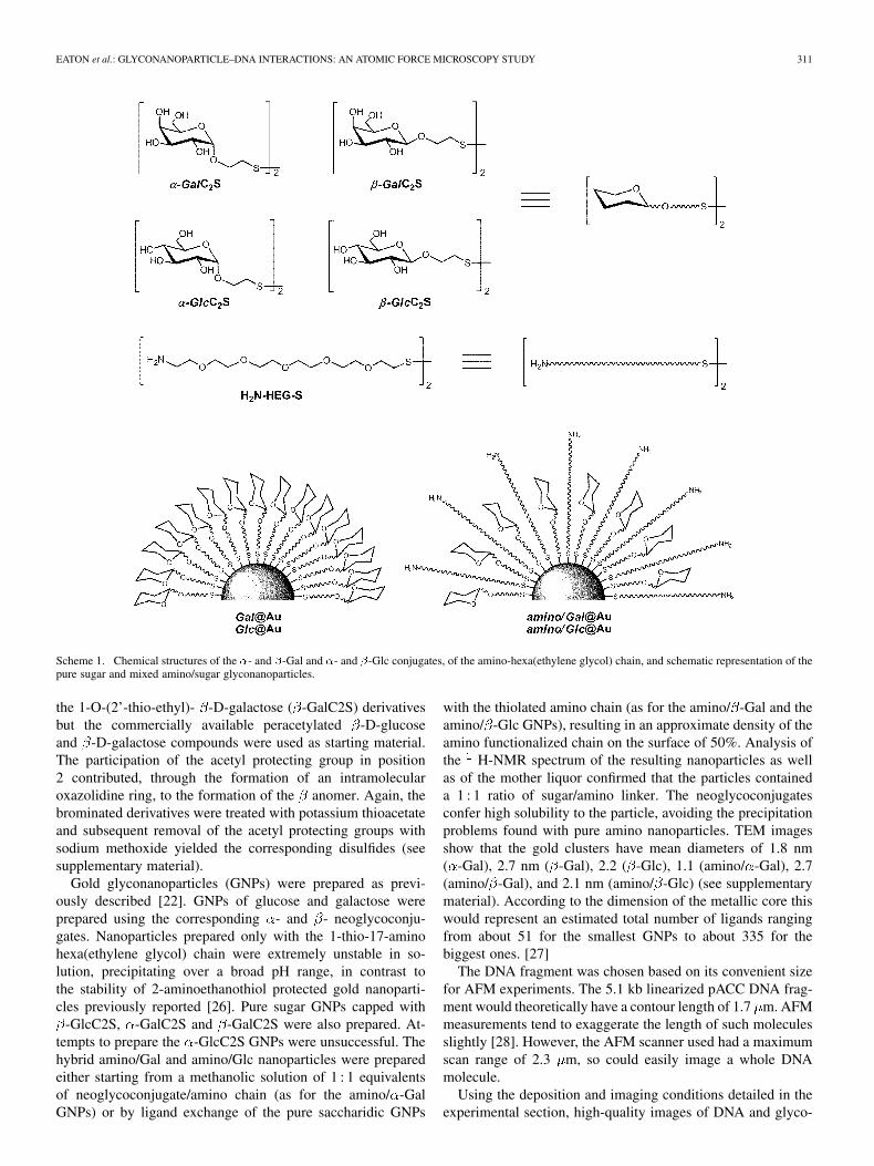
We used the monosaccharides glucose (Glc) and galactose(Gal) as the sugar motifs and the 17-amino-1-thio-hexa(ethyleneglycol) as the amino-bearing chain (Scheme 1). Synthesis ofthe 17-amino-1-thio-hexa(ethylene glycol) was accomplishedstarting from hexa(ethylene glycol) by modification of previ-ously reported reaction sequences [22].
The sugars were transformed into both - and -1-O-(2-thioethyl) glycosides to see the influence of the sugar anomericconfiguration in the interaction. The neoglycoconjugates1-O-(2-thioethyl) of - and -galactose ( -GalC2S and
-GalC2S), - and -glucose ( -GlcC2S and -GlcC2S) wereprepared by different glycosydation methods. Synthesis ofthe 1-O-(2-thio-ethyl)- -D-galactose ( -GalC2S) involvedinitially a Fischer glycosylation of D(+)-galactose with bro-moethanol. The resulting product was then acetylated usingacetic anhydride. Nucleophilic displacement of the bromogroup was then carried out using potassium thioacetate. Therequired product was obtained by deacetylation using sodiummethoxide in methanol. Synthesis of the 1-O-(2’-thio-ethyl)-
-D-glucose ( -GlcC2S) was carried out using the same proce-dure, but starting from D-(+)-glucose. A similar route was usedto obtain the 1-O-(2’-thio-ethyl)- -D-glucose ( -GlcC2S) and
EATON et al.: GLYCONANOPARTICLE–DNA INTERACTIONS: AN ATOMIC FORCE MICROSCOPY STUDY 311
Scheme 1. Chemical structures of the �- and �-Gal and �- and �-Glc conjugates, of the amino-hexa(ethylene glycol) chain, and schematic representation of thepure sugar and mixed amino/sugar glyconanoparticles.
the 1-O-(2’-thio-ethyl)- -D-galactose ( -GalC2S) derivativesbut the commercially available peracetylated -D-glucoseand -D-galactose compounds were used as starting material.The participation of the acetyl protecting group in position2 contributed, through the formation of an intramolecularoxazolidine ring, to the formation of the anomer. Again, thebrominated derivatives were treated with potassium thioacetateand subsequent removal of the acetyl protecting groups withsodium methoxide yielded the corresponding disulfides (seesupplementary material).
Gold glyconanoparticles (GNPs) were prepared as previ-ously described [22]. GNPs of glucose and galactose wereprepared using the corresponding - and - neoglycoconju-gates. Nanoparticles prepared only with the 1-thio-17-aminohexa(ethylene glycol) chain were extremely unstable in so-lution, precipitating over a broad pH range, in contrast tothe stability of 2-aminoethanothiol protected gold nanoparti-cles previously reported [26]. Pure sugar GNPs capped with
-GlcC2S, -GalC2S and -GalC2S were also prepared. At-tempts to prepare the -GlcC2S GNPs were unsuccessful. Thehybrid amino/Gal and amino/Glc nanoparticles were preparedeither starting from a methanolic solution of 1 : 1 equivalentsof neoglycoconjugate/amino chain (as for the amino/ -GalGNPs) or by ligand exchange of the pure saccharidic GNPs
with the thiolated amino chain (as for the amino/ -Gal and theamino/ -Glc GNPs), resulting in an approximate density of theamino functionalized chain on the surface of 50%. Analysis ofthe H-NMR spectrum of the resulting nanoparticles as wellas of the mother liquor confirmed that the particles containeda 1 : 1 ratio of sugar/amino linker. The neoglycoconjugatesconfer high solubility to the particle, avoiding the precipitationproblems found with pure amino nanoparticles. TEM imagesshow that the gold clusters have mean diameters of 1.8 nm( -Gal), 2.7 nm ( -Gal), 2.2 ( -Glc), 1.1 (amino/ -Gal), 2.7(amino/ -Gal), and 2.1 nm (amino/ -Glc) (see supplementarymaterial). According to the dimension of the metallic core thiswould represent an estimated total number of ligands rangingfrom about 51 for the smallest GNPs to about 335 for thebiggest ones. [27]
The DNA fragment was chosen based on its convenient sizefor AFM experiments. The 5.1 kb linearized pACC DNA frag-ment would theoretically have a contour length of 1.7 m. AFMmeasurements tend to exaggerate the length of such moleculesslightly [28]. However, the AFM scanner used had a maximumscan range of 2.3 m, so could easily image a whole DNAmolecule.
Using the deposition and imaging conditions detailed in theexperimental section, high-quality images of DNA and glyco-

312 IEEE TRANSACTIONS ON NANOBIOSCIENCE, VOL. 6, NO. 4, DECEMBER 2007
Fig. 1. AFM image of: (A) linearized pACC DNA fragments used in this work deposited from 300 pM solution and (B) �-Glc GNPs deposited from 60 nMsolution.
nanoparticles could be obtained. Fig. 1(A) and (B) shows typ-ical images of the plasmid DNA fragments and of the -GlcGNPs, respectively. It can be seen that the procedures for de-position resulted in clean images. All glyconanoparticles mea-sured had heights between 0.5 and 5 nm. The average particleheight was 2.47 nm ( -Glc), 0.95 nm ( -Gal), 2.1 nm ( -Gal),1.96 nm (amino/ -Gal), 3.3 nm (amino/ -Gal). Particle widthas measured by AFM is largely a function of the radius of theAFM tip used, due to convolution of tip shape with samplemorphology. According to previous results, all the particles areapproximately spherical, so real width and height should beidentical [24].
Interaction studies were performed in two ways. Plasmid andglyconanoparticles were mixed in solution and allowed to in-teract before deposition onto the surfaces (Method 1). Alterna-tively, the plasmid was deposited onto the mica surface and thefirst image recorded. Then glyconanoparticles were added fromaqueous solution and allowed to interact with the fixed DNAfor 10 min (Method 2). The first method employed allows thenanoparticles and DNA to interact in solution before depositingonto mica for imaging. Method 1 is more relevant to the inter-actions in, e.g., a test tube-based biosensor or gene delivery ap-plication, while the advantage of method 2 was that the fixationof the DNA to the surface allowed less ambiguous imaging byAFM.
When DNA and amino/ -Gal GNPs were allowed to in-teract in solution before depositing onto mica (Method 1),AFM images showed that the concentration of both free DNAand nanoparticles have been drastically reduced [Fig. 2(A)]and some very large features with globular morphology ap-peared on the surface. No individual DNA or GNPs couldbe observed. It was therefore suspected that these featuresrepresented aggregates formed between the nanoparticles andDNA. However, as this was not clear from the AFM imagesalone, the solution of amino/ -Gal GNPs-DNA was studied byTEM. Fig. 2(B) shows the corresponding TEM image whereit is clear that the globular features contain the nanoparticles[black dots in Fig. 2(B)]; moreover, close inspection showsthat all the nanoparticles were contained within the gray halos,
which indicate the presence of organic material. We interpretthis as indicating that all nanoparticles are associated with theDNA. Comparison of Fig. 2(A) and (B) shows that the samefeatures were seen by both AFM and TEM. Any differencesin the images can be accounted for by the different imagingtechniques; in both cases globular features of approximately50 to 200 nm diameter could be seen. The interaction betweenthe DNA molecules and the nanoparticles was also observedfollowing method 2. In this method DNA was first deposited onthe mica before the nanoparticles were added. This allowed theDNA to adopt an extended configuration on the mica surface,which was checked by AFM before addition of nanoparti-cles [as seen in Fig. 1(A)]. Addition of nanoparticles thenresulted in images showing the nanoparticles preferentiallyadopted positions along the DNA molecule. A typical imageof the resulting structures obtained by this method with hybridamino/ -Gal nanoparticles is shown in Fig. 2(C). In this figure,one may see that while there were some individual, unattachednanoparticles, the majority of them aggregated around the DNAmolecules on the surface. It is worth noting that this imageshows some “uncoated” DNA (the very thin white lines) as wellas the regions of DNA that have many nanoparticles attached.This suggests that initial binding of the hybrid amino/ -Galnanoparticles to DNA causes further nanoparticles to aggregatearound the DNA-nanoparticle complexes. The mechanism ofthis action is similar to that observed in the interaction betweenDNA and a great excess of lysine-modified gold nanoparticles[29] or in a plasmid deposited on poly-L-ornithine-coated micaafter mixing with polyethylenimine [30]. Fig. 2(D) shows atypical image of a sample further treated after initial imagingby washing with water. This process appears to have broken upthe initial aggregates leading to more unattached nanoparticles,but also to the formation of large compact globular aggregates,which may be the beginning of the compaction process, i.e.,the structures in Fig. 2(D) were intermediate between those inFig. 2(C) and (A). It seems that if the nanoparticles and DNAwere left for longer to interact (and the DNA was not partiallyfixed to the mica), large globular aggregates such as seen inFig. 2(A) would be seen, possibly with the eventual binding of

EATON et al.: GLYCONANOPARTICLE–DNA INTERACTIONS: AN ATOMIC FORCE MICROSCOPY STUDY 313
Fig. 2. Hybrid amino/�-Gal glyconanoparticles and DNA deposited from solution (method 1): (A) imaged by AFM and (B) by TEM, and AFM images of hybridamino/�-Gal nanoparticles deposited onto fixed DNA (method 2) before (C) and after (D) washing with water, and of hybrid amino/�-Gal nanoparticles depositedon to DNA fixed on mica before (E) and after (F) washing.
all unattached nanoparticles. In Fig. 2(E) and (F) are shown theimages of the -Gal nanoparticles on fixed mica, and the samesample after washing, respectively. It may be seen that in com-parison with the amino/ -Gal, the amino/ -Gal nanoparticles[Fig. 2(E)] exhibited much less DNA binding. Although parti-cles do appear to have bound the DNA chains, there are manymore unbound particles present than in Fig. 2(C). However,
after washing several large aggregates appeared [Fig. 2(F)],suggesting that over time compaction of the DNA also occursfor these glyconanoparticles.
The interactions between the pure sugar -Gal, -Gal and-Glc nanoparticles with DNA were also studied as for the
hybrid nanoparticles. Typical results are shown in Fig. 3.Fig. 3(A) and (B) correspond to -Glc nanoparticles on trapped

314 IEEE TRANSACTIONS ON NANOBIOSCIENCE, VOL. 6, NO. 4, DECEMBER 2007
Fig. 3. AFM images of �-Glc glyconanoparticles deposited onto fixed DNA before (A) and after (B) washing, of �-Gal nanoparticles deposited onto fixed DNAon mica before (C), and after (D) washing, and of �-Gal nanoparticles deposited onto fixed DNA on mica before (E) and after (F) washing.
DNA before washing (Method 2) and on trapped DNA afterwashing. Fig. 3(C) and (D) correspond to -Gal nanoparti-cles on trapped DNA before washing and on trapped DNAafter washing. Fig. 3(E) and (F) are of -Gal nanoparticleson trapped DNA before washing and on trapped DNA afterwashing. From Fig. 3(A), one may see both many unbound
-Glc nanoparticles, and some particles lying along the DNAstrands; however, selectivity to the DNA in this case seemslow. An image of the sample after washing with water is shownin Fig. 3(B). In this image, far fewer nanoparticles lie on themica, and close examination reveals that nanoparticles are
preferentially aligned along the DNA molecules. Furthermore,it may be seen that the DNA-nanoparticle complexes havebecome quite large (white features in the center of the image),with the DNA strands protruding from these features. It seemslikely that these globular features are the forerunners of evenlarger globular aggregates formed in solution (Fig. 2). Imagesof a sample of -Glc GNPs and DNA deposited from solution(Method 1) displayed also large aggregates, as well as both freenanoparticles and free DNA (see supplementary information).This contrasts with similar images from the hybrid nanoparti-cles-DNA complex deposited from solution [Fig. 2(A)], which
EATON et al.: GLYCONANOPARTICLE–DNA INTERACTIONS: AN ATOMIC FORCE MICROSCOPY STUDY 315
showed no free DNA fragments or nanoparticles. Fig. 3(A) and(B) show that the -Glc nanoparticles did interact with DNA;but much less strongly than the hybrid nanoparticles.
The image of the interaction of -Gal GNPs with DNA[Fig. 3(C)] shows the nanoparticles aligned along the DNAchains, though again, no formation of aggregates as in the caseof the amino/ -Gal nanoparticles (Method 1) was observed.However, after washing with water large aggregates can beobserved in the image [Fig. 3(D)]. Similarly to the case of
-Glc nanoparticles, the images obtained from the solution-de-posited GNPs-DNA complex, showed both free nanoparticlesand DNA in contrast to the results obtained from the hybridnanoparticles-DNA interaction in solution (see supplementaryinformation).
Fig. 3(E) and (F) show that for the -Gal glyconanoparti-cles, before washing a relatively large number of nanoparti-cles were not bound to the DNA chains, although each DNAmolecule had several nanoparticles bound to it. After washing,however, the percentage of unbound nanoparticles decreasedgreatly, and large features appeared in the images. This showedthat the nanoparticles bound the DNA relatively weakly butformed some larger aggregates after washing. Taken together,the data on the carbohydrate-only nanoparticles indicate that theform of the saccharide present was not important for sugar-onlynanoparticles.
Comparison of Figs. 2 and 3 reveals significant differencesin the behavior between the nanoparticles that had the aminochain, and those that did not. Although it appears that a higherproportion of the -Gal nanoparticles were bound preferen-tially to the DNA than the -Gal, the difference between thesetwo samples and the hybrid amino -Gal nanoparticles ismore remarkable. Under the conditions of method 2 and atthe same concentration of added GNPs, the carbohydrate-onlynanoparticles showed a slight preference for assembling alongthe DNA chains [Fig. 3(A), (C), and (E)], when compared withthe extremely high affinity for the DNA of hybrid nanoparticles[Fig. 2(C)]. This strong difference is not observed as comparingthe behavior of the pure sugar -Glc, -Gal, and -Gal GNPs.However, there were a higher percentage of nanoparticlesaligned along the DNA molecules in the images of -Galnanoparticles compared to the other two glyconanoparticles[see Fig. 3(A), (C), and (E)]. The effect was much morepronounced in the case of the hybrid amino/ -Gal GNPs,however, leading us to conclude that the combination of the
-Gal and amino groups gave a synergistic effect. Electrostaticinteractions between the amino groups of the GNPs and thephosphate groups along the DNA backbone may be the maindriving force for this association. However, sugar configurationseems to also play also a role, as the -Gal nanoparticles wereconsiderably stronger DNA binders than the -Gal particles.While it may be postulated that the immobilized DNA doesnot act like free DNA in solution, it has been shown that DNAwhich is bound to the surface by divalent cations is, while“fixed” to the surface, able to engage in complex biologicallyrelevant reactions, which also occur in solution, such as DNAcleavage by DNAse [31] and specific protein binding [32].In this context, it is feasible that similar reactions with thenanoparticles may occur on the surface and in solution.
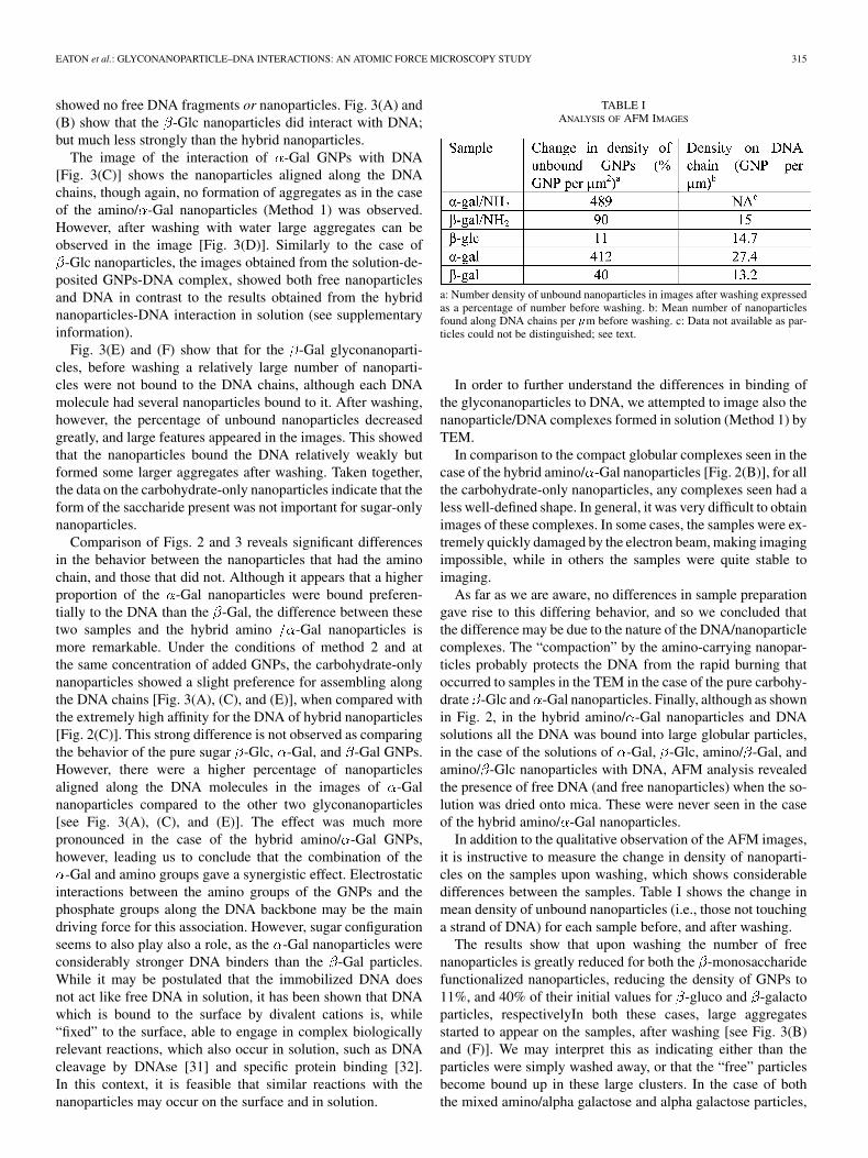
TABLE IANALYSIS OF AFM IMAGES
a: Number density of unbound nanoparticles in images after washing expressedas a percentage of number before washing. b: Mean number of nanoparticlesfound along DNA chains per �m before washing. c: Data not available as par-ticles could not be distinguished; see text.
In order to further understand the differences in binding ofthe glyconanoparticles to DNA, we attempted to image also thenanoparticle/DNA complexes formed in solution (Method 1) byTEM.
In comparison to the compact globular complexes seen in thecase of the hybrid amino/ -Gal nanoparticles [Fig. 2(B)], for allthe carbohydrate-only nanoparticles, any complexes seen had aless well-defined shape. In general, it was very difficult to obtainimages of these complexes. In some cases, the samples were ex-tremely quickly damaged by the electron beam, making imagingimpossible, while in others the samples were quite stable toimaging.
As far as we are aware, no differences in sample preparationgave rise to this differing behavior, and so we concluded thatthe difference may be due to the nature of the DNA/nanoparticlecomplexes. The “compaction” by the amino-carrying nanopar-ticles probably protects the DNA from the rapid burning thatoccurred to samples in the TEM in the case of the pure carbohy-drate -Glc and -Gal nanoparticles. Finally, although as shownin Fig. 2, in the hybrid amino/ -Gal nanoparticles and DNAsolutions all the DNA was bound into large globular particles,in the case of the solutions of -Gal, -Glc, amino/ -Gal, andamino/ -Glc nanoparticles with DNA, AFM analysis revealedthe presence of free DNA (and free nanoparticles) when the so-lution was dried onto mica. These were never seen in the caseof the hybrid amino/ -Gal nanoparticles.
In addition to the qualitative observation of the AFM images,it is instructive to measure the change in density of nanoparti-cles on the samples upon washing, which shows considerabledifferences between the samples. Table I shows the change inmean density of unbound nanoparticles (i.e., those not touchinga strand of DNA) for each sample before, and after washing.
The results show that upon washing the number of freenanoparticles is greatly reduced for both the -monosaccharidefunctionalized nanoparticles, reducing the density of GNPs to11%, and 40% of their initial values for -gluco and -galactoparticles, respectivelyIn both these cases, large aggregatesstarted to appear on the samples, after washing [see Fig. 3(B)and (F)]. We may interpret this as indicating either than theparticles were simply washed away, or that the “free” particlesbecome bound up in these large clusters. In the case of boththe mixed amino/alpha galactose and alpha galactose particles,

316 IEEE TRANSACTIONS ON NANOBIOSCIENCE, VOL. 6, NO. 4, DECEMBER 2007
there was actually an increase in free particle count uponwashing. These two samples were those that in the initialimages [Figs. 2(C) and 3(C)], showed the highest number ofnanoparticles bound to the DNA. Therefore, what happensupon washing is that the binding of some of the nanoparticlesto the DNA is broken up, and at least some of those “released”nanoparticles remain on the mica surface. This may be seenas surprising, as after 10 min in solution, the -galacto/aminoGNPs had formed very large clusters [see Fig. 2(A) and (B)].This may only mean, however, that the condensing into largecluster does not occur easily with the DNA trapped on thesurface in this case.
Finally, the -galacto/amino nanoparticles showed interme-diate behavior, as the number of free nanoparticles before andafter washing was very similar (only 10% loss, which may beattributed to “washing away”). This suggests than upon a shorttreatment time, these nanoparticles neither came unbound fromthe DNA and neither did they start to form large clusters.
Counting of the number of nanoparticles bound to eachDNA strand imaged (before washing) gave results that ingeneral varied little from one type of nanoparticle to another,most particles giving results around 15 3 nanoparticlesbound per m of exposed DNA. This density of binding israther low, considering the size of each nanoparticle, but isperhaps reduced due to the trapping of the DNA on the micasurface, reducing the number of available binding sites. Theexception to this was the alpha galacto nanoparticles whichbound almost double the number of nanoparticles 27 14 per
m). The amino/ -galacto NPs of course showed a completelydifferent type of binding with the nanoparticles clusteringtogether very tightly such that the AFM is unable to distinguishthem [Fig. 2(C)]. It was not possible to asses accurately thenumber of nanoparticles in these clusters around the DNAchains, but comparision of the volume of the clusters with thevolume of chains with bound -galacto GNPs suggests thereare many more nanoparticles bound in the case of the mixednanoparticles.
The influence of the carbohydrate and of the amine-chains incomplexing pACC DNA was also evaluated by gel shift elec-trophoresis in 0.6 % agarose gels using various weight/weight(w/w) ratios of GNP to pDNA. Each GNP was combined withthe DNA at w/w ratios 0, 2.5, 5, 10, 20, 40, 50, and 100 for15 min before being loaded into the gel and electrophoresed.The GNPs without amine-ending chain were used as negativecontrol in all experiments since they were not expected to beable to charge-neutralize, hence retain, the DNA. On the otherhand all the amino/sugar GNPs were able to inhibit the elec-trophoretic mobility of the plasmid upon addition of the cationicGNPs, although at different concentrations. However, the de-gree of retention strongly depended on the type of GNPs.
The ability of the GNPs to completely inhibit the DNA fromprogressing toward the positive electrode indicates either thatthe complexes formed were completely charge-neutralized orthat the resultant complexes were too large to enter the gel. How-ever, in some cases the formation of intermediate assemblies,due either to partial charge-neutralization or to the formationintermediate complexes with different molecular weights, wasnoticed. Fig. 4(A), (C), and (E) reveal that, as expected, none of
Fig. 4. Agarose gel electrophoresis of GNPs-pDNA complexes. Lineassignments are as follows: (A) control experiments with �-Gal GNPs;(B) experiments with mixed amino/�-Gal GNPs; (C) control experiments with�-Gal GNPs; (D) experiments with mixed amino/�-Gal GNPs; (E) controlexperiments with �-Glc GNPs; (F) experiments with mixed amino/�-Glc NPs.Column assignments are as follows: line 1) represents the DNA ladder; lines 2)to 8) represent GNPs-pACC-Nsi1 complexes at w/w ratios of 0, 2.5, 10, 20, 40,50, and 100, respectively.
the GNPs functionalized with only carbohydrates is able to bindand neutralize pDNA at any concentration used. Fig. 4(B), (D),and (F) refer to the binding ability of the mixed amino/sugarnanoparticles. As expected hybrid GNPs are better binders thanthe only sugar GNPs. The amino/ -Gal GNPs resulted to bethe less efficient in inhibiting the migration of the pDNA underthe condition used [Fig. 4(B)]. A light DNA fluorescence canstill be observed even at high w/w ratios, although basically allthe plasmid is retained (Fig. 4(B), line 8). On the other hand,the amino/ -Gal GNPs were able to retain the linear plasmid atw/w ratios between 50 and 100 (Fig. 4(D), lines 7–8). Of partic-ular interest are the gels obtained with the mixed amino/ -GlcGNPs [Fig. 4(F)]. Interaction between these GNPs and plas-mids gave rise to bands with a drop-like shape, which might becaused by the formation of intermediate species as observed inAFM which may undergo conformational changes or degrada-tion during migration. Still, at high concentration these GNPswere also able to inhibit migration of the pDNA (Fig. 4(F),line 8).
In conclusion, it was shown that AFM is a suitable tech-nique to image the interaction between DNA and glyconanopar-ticles, especially using the method of immobilizing the DNAfirst, before allowing interaction with nanoparticles. In this waydifferences in binding could clearly be seen between the carbo-hydrate-bearing nanoparticles, and those modified with aminogroups. It was also possible to distinguish different modes ofbinding when analyzing solutions containing DNA/nanoparticlemixtures, although in the case of the hybrid nanoparticles, therewas some ambiguity in the interpretation of the AFM imagesalone, as the nature of the large globular feature was not clear.In this case, TEM images allowed the determination that the

EATON et al.: GLYCONANOPARTICLE–DNA INTERACTIONS: AN ATOMIC FORCE MICROSCOPY STUDY 317
large features seen in the AFM were indeed large DNA/nanopar-ticle complexes, and the wide field imaging of the TEM showedthat these complexes are remarkably uniform in morphology,and that all the nanoparticles were contained within these com-plexes. Imaging of these samples by AFM also indicated thatall the DNA molecules were bound into these complexes. Fi-nally, as shown in Fig. 2, in the case of the mixed amino/ -Galnanoparticle and DNA solutions all the DNA was bound intolarge globular particles. In the case of the solutions of -Galand -Glc nanoparticles, AFM analysis revealed the presenceof free DNA (and free nanoparticles) when the solution wasdried onto mica. These were never seen in the case of the hy-brid amino/ -Gal nanoparticles.
It was shown, therefore that hybrid amino/ -Gal nanoparti-cles make highly efficient, highly water soluble DNA-bindersand condense the DNA into a compact globular shape, which is adesired property for gene-transfection agents [11]. Further workto prepare and characterize more effective amino/sugar GNPsfor binding and transfecting DNA was already begun.
ACKNOWLEDGMENT
The authors would like to thank Dr. F. Pinto (IIQ-CSIC) forhis discussions and help with gel electrophoresis experiments.
REFERENCES
[1] C. M. Niemeyer and M. Adler, “Nanoparticles, proteins, and nucleicacids: Biotechnology meets material science,” Angew. Chem. Int. Ed.,vol. 41, pp. 3779–3783, 2002.
[2] E. Katz and I. Willner, “Integrated nanoparticles-biomolecule hybridsystems: Synthesis, properties, and applications,” Angew. Chem. Int.Ed., vol. 43, pp. 6042–6108, 2004.
[3] D. A. Muruve, “The innate immune response to adenivirus vectors,”Hum. Gene Ther., vol. 15, pp. 1157–1166, Dec. 2004.
[4] K. Benihoud, P. Yeh, and M. Perricaudet, “Adenovirus vectors for genedelivery,” Curr. Opin. Biotechnol., vol. 10, pp. 440–447, Oct. 1999.
[5] T. Montier, P. Delepine, C. Pichon, C. Ferec, D. J. Porteous, and P.Midoux, “Non-viral vectors in cystic fibrosis gene therapy: Progressand challenges,” Trends Biotechnol., vol. 22, pp. 586–592, Nov. 2004.
[6] M. D. Brown, A. G. Schatzlein, and I. F. Uchegbu, “Gene delivery withsynthetic (non-viral) carriers,” Int. J. Pharm., vol. 229, pp. 1–21, Oct.2001.
[7] K. Kostarelos and A. D. Miller, “Synthetic, self-assembly ABCDnanoparticles; a structural paradigm for viable synthetic non-viralvectors,” Chem. Soc. Rev., vol. 34, pp. 970–994, Nov. 2005.
[8] J. Rojo, J. C. Morales, and S. Penadés, “Carbohydrate-carbohydrateinteractions in biological and model systems,” Top. Curr. Chem., vol.218, pp. 45–92, 2002.
[9] M. A. Zanta, O. Boussif, A. Adib, and J. P. Behr, “In vitro gene de-livery to hepatocytes with galactosylated polyethylenimine,” Biocon-jugate Chem., vol. 8, pp. 841–844, Nov.–Dec. 1997.
[10] J. Murata, Y. Ohya, and T. Ouchi, “Design of quaternary chitosanhaving antennary galactose residues as a gene delivery tool,” Carbohyd.Polym., vol. 32, pp. 105–109, Feb. 1997.
[11] T. Ren, G. S. Zhang, and D. X. Liu, “Synthesis of galactosyl com-pounds for targeted gene delivery,” Bioorg. Med. Chem., vol. 9, pp.2969–2978, Nov. 2001.
[12] Y. Aoyama, “Macrocyclic glycoclusters: From amphiphiles troughnanoparticles to glycovirus,” Chem.-Eur. J., vol. 10, pp. 588–593, Feb.2004.
[13] D. Wakebayashi, N. Nishiyama, Y. Yamasaki, K. Itaka, N. Kanayama,A. Harada, Y. Nagasaki, and K. Kataoka, “Lactose-conjugated polyioncomplex micelles incorporating plasmid DNSA as a targetable genevector system: Their preparation and gene transfecting efficiencyagainst cultured HepG2 cells,” J. Controlled Release, vol. 95, pp.653–664, Mar. 2004.
[14] Y. Liu and T. M. Reineke, “Poly(glycoamidoamine)s for gene delivery.Structural effects on cellular internalization, buffering capacity, andgene expression,” Bioconjugate Chem., vol. 18, pp. 19–30, 2006.
[15] Y. Liu and T. M. Reineke, “Hydroxyl stereochemistry and aminenumber within poly(glycoamidoamin)s affect intracellular DNAdelivery,” J. Amer. Chem. Soc., vol. 127, pp. 3004–3015, 2005.
[16] K. C. Wood, S. R. Little, R. Langer, and P. T. Hammond, “A familyof hierarchically self-assembling linear-dendritic hybrid polymers forhighly efficient targeted gene delivery,” Angew. Chem. Int. Ed., vol. 44,pp. 6704–6708, 2005.
[17] C. M. McIntosh, E. A. Esposito, A. K. Boal, J. M. Simard, C. T. Martin,and V. M. Rotello, “Inhibition of DNA transcription using cationicmixed monolayer protected gold clusters,” J. Amer. Chem. Soc., vol.123, pp. 7626–7629, 2001.
[18] M. Thomas and A. M. Klibanov, “Conjugation to gold nanoparticlesenhances polyethylenimine’s transfer of plasmid DNA into mam-malian cells,” Proc. Nat. Acad. Sci., vol. 100, pp. 9138–9143, Aug.2003.
[19] T. Kawano, M. Yamagata, H. Takahashi, Y. Niidome, S. Yamada, Y.Katayama, and T. Niidome, “Stabilizing of plasmid DNA in vivo byPEG-modifies cationic gold nanoparticles and the gene expressionassisted with electrical pulses,” J. Controlled Release, vol. 111, pp.382–389, Apr. 2006.
[20] G. Han, C. C. You, B. J. Kim, R. S. Turingan, N. S. Forbes, C. T. Martin,and V. M. Rotello, “Light-regulated release of DNA and its delivery tonuclei by means of photolabile gold nanoparticles,” Angew. Chem. Int.Ed., vol. 45, pp. 3165–3169, 2006.
[21] J. M. De La Fuente, A. G. Barrientos, T. C. Rojas, J. Rojo, J. Canada, A.Fernandez, and S. Penades, “Gold glyconanoparticles as water-solublepolyvalent models to study carbohydrate interactions,” Angew. Chem.Int. Ed., vol. 40, pp. 2258–2261, 2001.
[22] A. G. Barrientos, J. M. de la Fuente, T. C. Rojas, A. Fernandez, and S.Penades, “Gold glyconanoparticles: Synthetic polyvalent ligands mim-icking glycocalix-like surfaces as tools for glycobiological studies,”Chem.-Eur. J., vol. 9, pp. 1909–1921, May 2003.
[23] T. C. Rojas, J. M. De La Fuente, A. G. Barrientos, S. Penades, L. Pon-sonnet, and A. Fernandez, “Gold glyconanoparticles as building blocksfor nanomaterial design,” Adv. Mater., vol. 14, pp. 585–588, Apr. 2002.
[24] J. M. de la Fuente, P. Eaton, A. G. Barrientos, M. Menendez, and S.Penades, “Termodynamic evidence for Ca2+-mediated self-aggrega-tion of Lewis X gold glyconanoparticles. A model for cell adhesionvia carbohydrate-carbohydrate interaction,” J. Amer. Chem. Soc., vol.127, pp. 6192–6197, 2005.
[25] R. V. Duran, M. Hervas, B. De la Cerda, M. A. De la Rosa, and J. A.Navarro, “A laser flash-induced kinetic analysis of in vivo photosystemI reduction by site-directed mutants of plastocyanin and cytochromec ,” Synechocystis, vol. 45, pp. 1054–1060, 2006, sp. PCC 6803 Bio-chemistry.
[26] T. Niidome, K. Nakashima, H. Takahashi, and Y. Niidome, “Prepara-tion of primary amine-modified gold nanoparticles and their transfec-tion ability into cultivated cells,” Chem. Commun., 2004, 1978-1979.
[27] M. J. Hostetler, J. E. Wingate, C.-J. Zhong, J. E. Harris, R. W. Vachet,M. R. Clark, J. D. Londono, S. J. Green, J. J. Stokes, G. D. Wignall, G.L. Glish, M. D. Porter, N. D. Evans, and R. W. Murray, “Alkanethiolategold cluster molecules with core diameters from 1.5 to 5.2 nm: Coreand monolayer properties as a function of core size,” Langmuir, vol.14, no. 1, pp. 17–30, 1998.
[28] A. Sanchez-Sevilla, J. Thimonier, M. Marilley, J. Rocca-Serra, andJ. Barbet, “Accuracy of AFM measurments of the contour length ofDNA fragments adsorbed on mica in air and in aqueos buffer,” Ultra-microscopy, vol. 92, pp. 151–158, Aug. 2002.
[29] M. Ganguli, J. V. Babu, and S. Maiti, “Complex formation betweencationically modified gold nanoparticles and DNA: An atomic forcemicroscopic study,” Langmuir, vol. 20, pp. 5165–5170, Jun. 2004.
[30] D. D. Dunlap, A. Maggi, M. R. Soria, and L. Monaco, “Nanoscopicstructure of DNA condensed for gene delivery,” Nucleic Acids Res.,vol. 25, pp. 3095–3101, Aug. 1997.
[31] H. G. Abdelhady, S. Allen, M. C. Davies, C. J. Roberts, S. J. B. Tendler,and P. M. Williams, “Direct real-time molecular scale visualisation ofthe degradation of condensed DNA complexes exposed to DNase I,”Nucleic Acids Res., vol. 31, pp. 4001–4005, Jul. 2003.
[32] Y. K. Jiao, D. I. Cherny, G. Heim, T. M. Jovin, and T. E. Schaffer, “Dy-namic interactions of p53 with DNA in solution by time-lapse atomicforce microscopy,” J. Mol. Biol., vol. 314, pp. 233–243, Nov. 2001.
318 IEEE TRANSACTIONS ON NANOBIOSCIENCE, VOL. 6, NO. 4, DECEMBER 2007
Peter Eaton received the B.Sc. degree from the University of York, U.K., andthe Ph.D. degree from Sheffield Hallam University, U.K., in 1998.
From 1998 to 2001 he was at the University of Portsmouth, U.K., usingatomic force microscopy (AFM). From 2002 to 2004 he was with the IIQ,Seville, Spain, where he used AFM to study nanoparticle aggregation, andto study the interactions between nanoparticles and DNA. He is currentlywith REQUIMTE in Porto, Portugal, where he is working as an AssociateResearcher and is using the AFM to study DNA-nanoparticle interactions andin a variety of other applications.
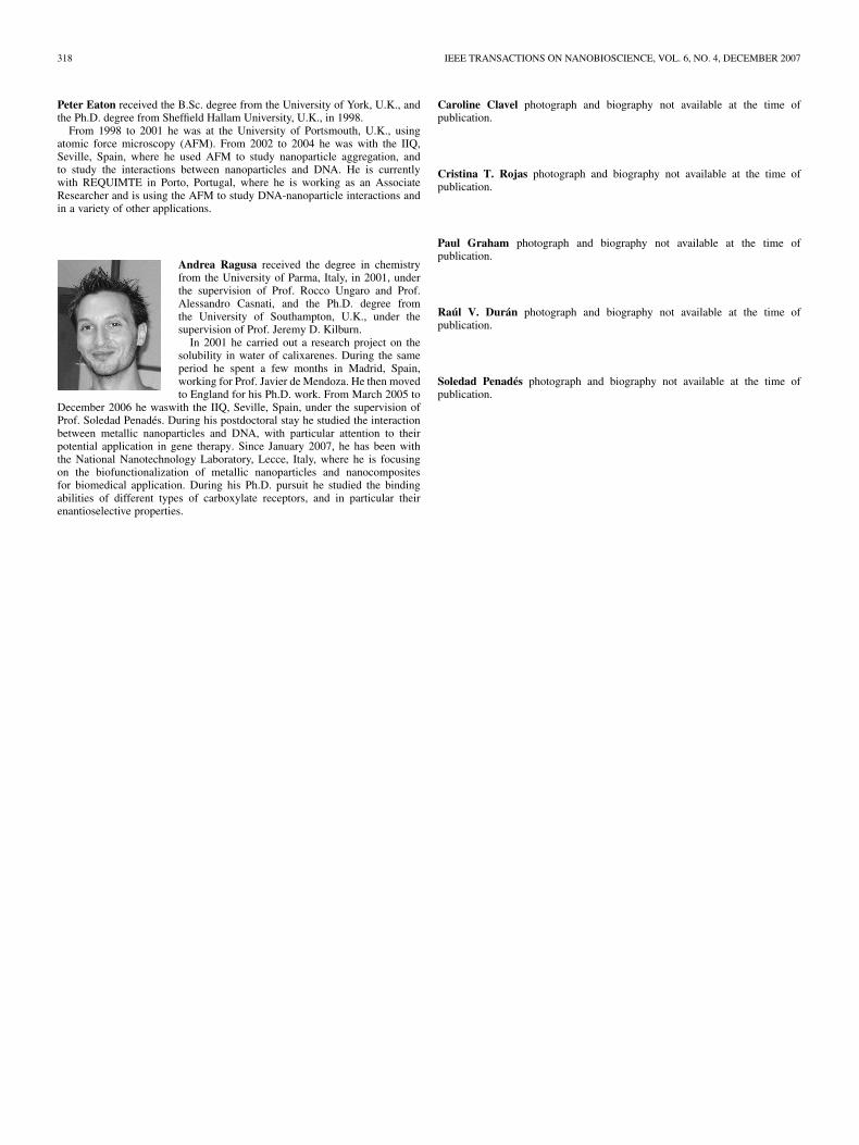
Andrea Ragusa received the degree in chemistryfrom the University of Parma, Italy, in 2001, underthe supervision of Prof. Rocco Ungaro and Prof.Alessandro Casnati, and the Ph.D. degree fromthe University of Southampton, U.K., under thesupervision of Prof. Jeremy D. Kilburn.
In 2001 he carried out a research project on thesolubility in water of calixarenes. During the sameperiod he spent a few months in Madrid, Spain,working for Prof. Javier de Mendoza. He then movedto England for his Ph.D. work. From March 2005 to
December 2006 he waswith the IIQ, Seville, Spain, under the supervision ofProf. Soledad Penadés. During his postdoctoral stay he studied the interactionbetween metallic nanoparticles and DNA, with particular attention to theirpotential application in gene therapy. Since January 2007, he has been withthe National Nanotechnology Laboratory, Lecce, Italy, where he is focusingon the biofunctionalization of metallic nanoparticles and nanocompositesfor biomedical application. During his Ph.D. pursuit he studied the bindingabilities of different types of carboxylate receptors, and in particular theirenantioselective properties.
Caroline Clavel photograph and biography not available at the time ofpublication.
Cristina T. Rojas photograph and biography not available at the time ofpublication.
Paul Graham photograph and biography not available at the time ofpublication.
Raúl V. Durán photograph and biography not available at the time ofpublication.
Soledad Penadés photograph and biography not available at the time ofpublication.