imaging grey matter abnormalities in multiple … filedysfunction. abnormal trans-membran ionic...
TRANSCRIPT

IMAGINGGREY MATTER
ABNORMALITIES INMULTIPLE SCLEROSIS
ORGANIZING COMMITTEE
Isabelle BerryGilles Edan
Jean-Philippe RanjevaAyman Tourbah
ParisBrain and Spine Institute - ICM
Salpetriere HospitalFebruary 4, 2011
6th annual ARSEP-MRI meeting

Programme of the day
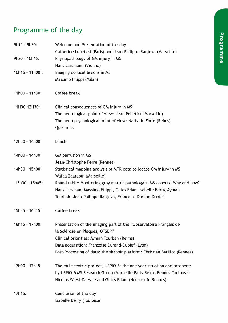
9h15 – 9h30: Welcome and Presentation of the day
Catherine Lubetzki (Paris) and Jean-Philippe Ranjeva (Marseille)
9h30 – 10h15: Physiopathology of GM injury in MS
Hans Lassmann (Vienne)
10h15 – 11h00 : Imaging cortical lesions in MS
Massimo Filippi (Milan)
11h00 – 11h30: Coffee break
11H30-12H30: Clinical consequences of GM injury in MS:
The neurological point of view: Jean Pelletier (Marseille)
The neuropsychological point of view: Nathalie Ehrlé (Reims)
Questions
12h30 – 14h00: Lunch
14h00 – 14h30: GM perfusion in MS
Jean-Christophe Ferre (Rennes)
14h30 – 15h00: Statistical mapping analysis of MTR data to locate GM injury in MS
Wafaa Zaaraoui (Marseille)
15h00 – 15h45: Round table: Monitoring gray matter pathology in MS cohorts. Why and how?
Hans Lassman, Massimo Filippi, Gilles Edan, Isabelle Berry, Ayman
Tourbah, Jean-Philippe Ranjeva, Françoise Durand-Dubief.
15h45 – 16h15: Coffee break
16h15 – 17h00: Presentation of the imaging part of the “Observatoire Français de
la Sclérose en Plaques, OFSEP”
Clinical priorities: Ayman Tourbah (Reims)
Data acquisition: Françoise Durand-Dubief (Lyon)
Post-Processing of data: the shanoir platform: Christian Barillot (Rennes)
17h00 – 17h15: The multicentric project, USPIO-6: the one year situation and prospects
by USPIO-6 MS Research Group (Marseille-Paris-Reims-Rennes-Toulouse)
Nicolas Wiest-Daessle and Gilles Edan (Neuro-info Rennes)
17h15: Conclusion of the day
Isabelle Berry (Toulouse)
Pro
gram
me


Intro
du
ction
Visualizing gray matter abnormalities in MS. Why and how ?
Jean-Phlippe RanjevaMedical School of Marseille
France*-*-*-*-*-*-*-*-*-*-*-*-*-*-*-*-*-*-*-*-*-*
Isabelle Berry,University Hospital of Toulouse
France
MS is the first cause of disability unrelated to traumatism in young adults. This disease is characterized by inflammation and demyelination inside white matter (WM) but also sustained inflammatory processes outside lesions in WM as well as in gray matter (GM).
Recent histological studies have evidenced axonal/neuronal degeneration, especially in heavily disabled patients. Numerous in vitro studies have also stressed out the role of cellular energetic failure, related to mitochondrial dysfunction, hypoxia or inflammation, leading to ionic pump dysfunction. Abnormal trans-membran ionic exchanges lead to intracellular sodium accumulation, followed by an increase in intracellular calcium accumulation that activates degrading enzymes (referred to the previous ARSEP MRI workshop 2010). Nevertheless, the onset of such phenomenon in the disease course is totally unknown.
To date, MRI has focused on the characterization of focal demyelinating and inflammatory processes. Nevertheless, progress of MRI methods does not yet provide predictive parameters of clinical deficit or progression rate of the disease. One major challenge is now to develop non invasive methods sensitive to the dynamics of processes underlying neuronal/axonal degeneration for the better understanding and monitoring of disease progression and for the better definition of the therapeutic windows of new neuroprotective drugs.
This sixth international ARSEP MRI workshop aims at offering to the French MS community an update of the latest scientific advances in the domain of MS pathophysiology and imaging techniques focused on GM damage through the lectures of the best specialists.
We hope this workshop will help you to answer some of your questions
and ours too….
The Organizing Committee

Pathophysiology of Grey Matter Injury in Multiple Sclerosis
Hans Lassmann Medical University of Vienna
Austria
While multiple sclerosis has for long been regarded a disease, which selectively affects the white matter, it became clear in recent years that grey matter pathology is prominent in particular in the progressive stage of the disease. On a structural basis grey matter alterations in MS brains have to be separated into two categories: grey matter demyelination and diffuse changes within the normal appearing grey matter. Demyelination affects the grey matter in different ways.
In early stages of RRMS small perivenous demyelinating lesions as well as cortico-subcortical lesions are seen, which are present in the cortex as well as in the deep grey matter nuclei. They arise in association with perivenous inflammation in a similar was as the white matter lesions, but inflam-mation, blood brain barrier disturbance and axonal injury are in general less severe than in the ad-jacent white matter. A different type of cortical demyelination is the formation of subpial lesions, which are mainly seen at late stages of MS and which are clearly associated with inflammation in the leptomeninges. These lesions develop predominantly in the sulci and the deep infoldings of the brain surface. They seem to be mediated by a one or more soluble factors, which are produced in meningeal inflammatory infiltrates and diffuse into the cortical parenchyma, where they activate microglia. Demyelination and neuronal injury is then driven by activated microglia. In addition to cortical demyelination there is also diffuse neuronal loss and atrophy in the MS cortex, mainly seen in patients with progressive disease. Cortical atrophy can be associated with destructive white matter lesions and in this condition neuronal loss in the cortex is mainly due to secondary retrograde degen-eration. In addition, however, there is neuronal injury in the absence of demyelination or retrograde degeneration, which is associated with meningeal inflammation.
Neuronal injury and demyelination in the cortex appear to be mediated through reactive oxygen and nitric oxide species (RONS), which damage neurons and glia cells through mitochondrial injury. Sub-threshold concentration of RONS may also induce functional deficits in the absence of structural damage. This may possibly explain disturbance of cognitive function in very early stages of RRMS, where structural damage in cortex and white matter is sparse or even absent.
Han
s La
ssm
ann

Imaging cortical lesions in MS
Massimo FilippiUniversity Ospedale San Raffaele of Milan
Italy
Recent pathologic and magnetic resonance imaging (MRI) studies have challenged the classic view of multiple sclerosis (MS) as a chronic inflammatory-demyelinating condition affecting solely the white matter (WM) of the central nervous system. Indeed, an involvement of the gray matter (GM), in terms of focal lesions, “diffuse” damage, and irreversible tissue loss, has been shown to occur from the early stages of the disease, to progress with time, and to be only moderately correlated with the extent of WM injury.
During the past few years, the introduction of double inversion recovery (DIR) sequences has improved the detection of GM lesions, in MS patients. Using such a sequence, cortical lesions (CL) have been demonstrated consistently not only in patients with the progressive phenotypes of the disease, but also in those with relapsing remitting (RR) MS, in individuals with clinically isolated syndromes (CIS) at presentation suggestive of this disease, and patients with a pediatric onset of the disease. The accuracy of MRI diagnostic criteria for MS is increased when considering the presence of CL on baseline scans from patients at presentation with CIS, thus suggesting that CL detection might be considered in future diagnostic algorithms for MS. Furthermore, RRMS and secondary progressive (SP) MS patients were found to accumulate new CL over a 3 year follow up at a virtually similar yearly rate. More interestingly, in both RRMS and SPMS, patients’ CL volume predicted disability progression over the follow up period, thus suggesting that the quantification of CL represents an additional useful paraclinical tool to monitor MS evolution.
High-resolution MR images and automated segmentation techniques have allowed the achievement of an accurate quantification of GM tissue volume and an improvement of our understanding of the dynamics of GM tissue loss in MS. Patients with CIS who developed MS during the subsequent 3 years had significant GM loss. A significant reduction with time of the GM fraction has also been observed in patients with early RRMS and PPMS. In a large-scale study of 597 patients with MS, significantly reduced WM and GM fractions were found in all phenotypes; the most severe tissue loss was detected in SPMS. GM atrophy is associated with MS clinical disability. Neocortical volume loss was found to occur in patients with RRMS with even mild cognitive disturbances. The application of quantitative MRI–based techniques, such as magnetization transfer and diffusion tensor MRI, has also shown that GM is diffusively damaged in all MS phenotypes since the earliest clinical stages of the disease, and is associated with the main clinical manifestations of MS. These abnormalities increase with time and are only partially associated with the extent of WM pathology. More recently, variable degrees of cortical plasticity with the potential to limit the functional consequences of tissue damage have been shown in patients with MS, suggesting that their disability is likely to result from the balance between structural damage and cortical reorganization, rather then being a mere reflection of tissue disruption.
Measuring GM MRI variables might, therefore, be a rewarding exercise for improving our understanding of MS pathobiology, which might result, in the future, in the identification of additional markers to monitor disease evolution, either natural or modified by treatment.
Massim
o Filip
pi

CLINICAL CONSEqUENCESOf GM INjURY IN MS

Clinical consequences of GM injury in MS
Imaging Grey Matter Abnormalities in Multiple Sclerosis
Jean PelletierUniversity hospital of Marseille
France
Multiple sclerosis (MS) is traditionally considered as a chronic inflammatory demyelinating disease affecting white matter (WM). However, several pathologic and MRI studies demonstrated involvement of gray matter (GM), in terms of both focal lesions (located in the cerebral cortex, the basal ganglia, the brain stem and the spinal cord) and irreversible tissue loss (evaluated by focal and global atrophy).
The clinical relevance of GM damage in MS can be approached on several points.
First, GM involvement has been clearly or potentially associated with clinical manifestations in MS. Cortical/juxtacortical MS lesions are correlated with seizures and cognitive impairment. In particular, GM atrophy has been associated with impairments in verbal memory, euphoria, and disinhibition, and neocortical atrophy has been related with impairment in verbal memory, verbal fluency, and attention/concentration. MS fatigue and depression has also been linked to subcortical GM pathology.
Secondly, physical disability has been related to GM involvement. Markers of GM damage correlate better with measures of physical disability than do WM lesion markers. Because of the frequency and extent of (intra)cortical and subpial demyelination in late stages of disease, cortical lesions and GM atrophy have been suggested to contribute to disease progression. In particular, the extensive cortical damage observed predominantly in progressive forms of MS suggests that GM involvement may be an important pathologic correlate of irreversible disability. However, recent studies also reported that GM involvement begin early in the course of the disease. These data suggest that GM damage may represent the primary and initial target, and the determinant of irreversible disability.
At least, GM involvement could have a crucial impact to increase the sensitivity of the MS diagnostic criteria. In this way, the detection and the visualization of cortical/juxtacortical lesions could obviously improve our ability to diagnose and to monitor the disease.
Jean Pelletier

Clinical consequences of GM injury in MS: The neuropsychological point of view
Nathalie EhrléHospital of Reims & university of Lille
France
The cognitive architecture of the human cortex was mainly studied from neuropsychological profile and cerebral imaging.
The first part of the talk will describe some of the problems related to traditional neuropsychological research, which attempted to provide accounts on cognitive performance at a neural as well as a functional level of description. The difficulties in making neural-level arguments from neuropsychological data include: choices of neurological pathologies to infer cerebral substrate, problems of associated deficits and determining factors of a neuropsychological assessment focused on location. We will then discuss how cognitive neuropsychology by-passed some of these problems.
Finally, we will consider distinguishing features of multiple sclerosis cognition: relationship between cognitive processes, fatigue and slowness, grey versus white-matter involvement, reorganization, longitudinal studies. Emerging trends and recommendations to improve imaging conclusions from neuropsychological data will be considered.
Nat
hal
ie E
hrl
é

Grey Matter perfusion in Multiple Sclerosis
Jean-Christophe FerréUniversity Hospital of Rennes
France
Recent pathologic and MRI studies have shown the damage of cortical and deep grey matter (GM) in multiple sclerosis (MS), even at the early stage of the disease. Conventional and quantitative MRI could show focal lesions, “diffuse” tissue abnormalities, and irreversible tissue loss (i.e., atrophy), consistent with pathologic observations.
Moreover, several immunopathological studies have suggested that microvascular factors may contribute to the pathogenesis of MS. Several imaging techniques could assess the cerebral hemodynamics. Positron emission tomography (PET) and single photon emission computed tomography (SPECT) have shown decreased of in cerebral blood flow (CBF) in areas of cortical GM and WM in patients with MS. MRI permits the assessment of cerebral hemodynamics in addition to morphologic study of the brain. The two main MRI methods to estimate CBF are dynamic susceptibility contrast (DSC)-enhanced T2*-weighted perfusion and Arterial Spin Labeling (ASL) techniques. DSC utilizes the signal changes that are due to the first passage of gadolinium through the cerebral vasculature and ASL technique uses magnetically labeled arterial blood water as an endogenous tracer. Applied to MS, DSC has demonstrated perfusion decrease in both chronic-lesional and normal-appearing (NA) white matter (WM), and perfusion increase in gadolinium-enhanced lesions.
Several studies with DSC or ASL have shown a decrease perfusion of GM of patients with MS. The cortical hypoperfusion seems to appear early in the disease progression: a decreased CBF was observed inside the precuneus in CIS patient, in central areas in patient with begin MS, and in more extended areas on frontal and parietal lobes in patients with progressive MS. The deep grey matter hypoperfusion is found in patients with Relapsing Remitting and Primary Progressive MS and is correlated with fatigue score and neuropsychological dysfunction. No correlation was found with EDSS or lesions volume.
A study suggests a beginning of perfusion decrease on NAWM before deep GM. GM decrease perfusion could be explained by the presence of lesions in GM, degeneration of axons running within deep GM, and neuronal loss secondary to demyelination, hypoperfusion or Wallerian degeneration.
Jean-C
hristo
ph
e Ferré

Statistical mapping analysis of MTR data to locate GM injury in Multiple Sclerosis
Wafaa ZaaraouiMedical university of Marseille
France
Recently, quantitative MRI methods have highlighted the existence of grey matter (GM) pathology in patients suffering from multiple sclerosis (MS) (Chard D et al, 2009).
However, these methods applied at the group level, have not been designed to quantify the extent of GM pathology at the individual level preventing any potential application for the clinical practice. In contrast, individual GM magnetization transfer ratio (MTR) mapping quantifies the GM pathology for each MS subject (Jure L et al, 2010) evidencing potential subtle GM MTR decrease (minimum 3%) at the individual level.
By applying an optimized statistical mapping analysis of MTR maps at the individual level in patients with a clinically isolated syndrome with a high risk developing multiple sclerosis (n=88) compared to healthy controls (n=44), we evidenced that individual analysis of GM MTR map is an accurate tool to detect and quantify GM pathology at the individual level. This method allowed demonstrating that GM pathology is highly heterogeneous across patients at the early stage of MS and partly underlies irreversible disability.
Waf
aa Z
aara
ou
i

PRESENTATION Of ThE IMAGING PARTOf ThE “OBSERvATOIRE fRANçAISdE SCLéROSE EN PLAqUES, OfSEP”

Presentation of the imaging part of the “Observatoire Français de Sclérose en Plaques, OFSEP”
Clinical prioritiesAyman TourbahReims Hospital
France
The general objective of the French Observatoire is long-term follow up of cohorts of patients with multiple sclerosis, integrating them to the existing procedures. Four groups/ axes have been proposed: clinical, biological, imaging, cognitive.
The Imaging part includes a general research objective, opened and accessible to all centers, and more specific research programs reserved to MS centers. The workpackage Neuroimaging group has a coordination committee, a steering committee and 3 Task groups for acquisition, data management, and image analysis.
The clinical priorities have been discussed and established as diagnosis, prognosis, and treatment. Diagnosis is based on international criteria. A specific effort will be driven for harmonization and standardization among centers. The sequences have been discussed and defined. This major step will open the possibility to reach the prognosis, in determining the real profile of the disease, and measuring the effect of therapeutical interventions. The third clinical priority will be to define the role and the use of Imaging in treatment decision and follow-up.
Specific research project will be conducted in specialized centers.
This project offers a great opportunity to the French community to contribute significantly to the advances of knowledge relative to multiple sclerosis.
Aym
an T
ou
rbah

Presentation of the imaging part of the “Observatoire Français de Sclérose en Plaques, OFSEP”
Data acquisition : The primary MRI-related objective of the Observatoire Français de la Sclérose en Plaques (OF-SEP) is to enable standardized measurements of disease activity and burden in patients of this cohort and to make them available to the medical and research community.
Françoise Durand-DubiefLyon
France*-*-*-*-*-*-*-*-*-*-*-*-*-*-*-*-*-*-*-*-*-*
The Neuro-Imaging Working Group (NIWG) ofthe Observatoire Français de la Sclérose en Plaques (OFSEP
For this purpose, the OFSEP Neuroimaging Working Group (NIWG) anticipates to define stand-ardized MRI acquisition protocols which can be performed on a large scale (over 30.000 persons with MS (PWMS) are currently registered in OFSEP), with multiple imaging machines at numer-ous centers. A minimal MRI protocol will be set up for being performed for any PWMS in any MRI center with a 1.5 or more Tesla MRI device and for allowing longitudinal assessments. Such brain MRI assessments will be advocated at least every 3 years for any patient. They will consist in DP/T2, FLAIR, T1 pre- and post-Gadolinium acquisitions. They will allow measurements of corpus callosum area, T2 lesion volume, enhancing lesions and of total brain volume. Whole spine MRI will be proposed once every 5 years. Besides, an advanced MRI protocol for 3T MRI platforms will be proposed to a subset of participating centers for pre-defined patients included in specific nested cohorts with a specific follow up.
The OFSEP NIWG also anticipates developing a network of Imaging Resource Centers (IRCs) dedi-cated to MS throughout France. These IRCs will allow storing, processing, and integrating crude and derived imaging data into a shared database, connected to clinical information within the EDMUS database. This network will interoperate using the Shanoir environment from INRIA. Fur-ther it will be complemented with solutions proposed by other national and international initia-tives, notably the SUMMIT initiative, an international MS working consortium (Amsterdam, Basel, Boston, San Francisco) recently funded.
The Neuro-Imaging Working Group (NIWG) of the Observatoire Français de la Sclérose en Plaques (OFSEP) : René ANXIONNAT (Nancy), Jean-Pierre ARMSPACH (Strasbourg), Bertrand AUDOIN (Marseille), Christian BARILLOT (Rennes), Isabelle BERRY (Toulouse), Fabrice BONNEVILLE (Toulouse), Bruno BROCHET (Bordeaux), Giovanni CASTELNOVO (Nîmes), Christophe COLLET (Strasbourg), Christian CONFAVREUX (Lyon), François COTTON (Lyon), Jérôme DE SEZE (Strasbourg), Vincent DOUSSET (Bordeaux), Françoise DURAND-DUBIEF (Lyon), Gille EDAN (Rennes), Jean-Christophe FERRE (Rennes), Damien GALANAUD (Paris), Charles GUTTMANN (Harvard), Stéphane KREMER (Strasbourg), Pierre LABAUGE (Nîmes), Christine LEBRUN-FRENAY (Nice), Grégoire MALANDAIN (Nice), Jean PELLETIER (Marseille), Jean-Philippe RANJEVA (Marseille), Dominique SAPPEY-MARINIER (Lyon), Bruno STANKOFF (Paris), Ayman TOURBAH (Reims), Thomas TOURDIAS (Bordeaux), Sandra VUKUSIC (Lyon).
Franço
ise Du
rand
-Du
bief

Presentation of the imaging part of the “Observatoire Français de Sclérose en Plaques, OFSEP”
Post-Processing of data: the shanoir platformChristian Barillot
Univsersity of RennesFrance
One key objective of the OFSEP project is to provide the access of the MS cohort to the community, a second objective of the Imaging work package will be to set up and maintain an information processing and data management infrastructure. In this context, open access solutions are emerging from either projects conducted in North America, and from Europe and especially in France were several national projects have already been financed in the last 7 years to elaborate new computer infrastructures for this purpose. Among these systems, a new software environment called Shanoir is emerging.
Shanoir (Sharing NeurOImaging Resources: http://www.shanoir.org) is an open source neuroinformatics platform designed to share, archive, search and visualize neuroimaging data. It provides a user-friendly secure web access and offers an intuitive workflow to facilitate the collecting and retrieving of neuroimaging data from multiple sources and a wizard to make the completion of metadata easy. Shanoir comes along many features such as anonymization of data, support for multi-centers clinical studies on subjects or group of subjects. Shanoir offers an ontology-based data organization (OntoNeuroLOG) to facilitate the reuse of data and metadata, the integration of processed data and provides traceability trough an evolutionary approach. Shanoir allows clinicians, researchers, and engineers to undertake quality research projects with an emphasis on remote collaboration. It allows you safely storing and archiving, with no more requirements than a computer with an Internet connection. Furthermore, Shanoir is not only a web application: it is also a complete neuroinformatics platform in which you can easily integrate your existing image processing tools or develop your own ones.
Ch
rist
ian
Bar
illo
t

The multicentric project, USPIO-6: One year situation and prospects by USPIO-6 MS Research Group
Nicolas Wiest-Daesslé & Gilles Edan
University hospital of RennesFrance
While Multiple Sclerosis (MS), a chronic autoimmune disease of the central nervous system, is usually diagnosed at its early stage using MR imaging and contrast enhanced MR imaging (as a complement of clinical criteria), the conventional contrast agent (Gadolinium) used in MRI is neither specific to MS, nor predictive of MS induced disability.
A new longitudinal clinical research study, called USPIO-6, relying on the use of Ultra-small Super-Paramagnetic Iron Oxide (USPIO) as a cellular biomarker and advanced MR imaging techniques, is currently ongoing. The aim of the USPIO-6 project is to analyze the predictive capacity and the specificity of USPIO in MS at onset. We present here the USPIO-6 study and some imaging and processing preliminary results.
Nico
las Wiest-D
aessléG
illes Edan

no
tes

no
tes

no
tes

no
tes


ww
w.arsep
.org

fondation pour l’aide à la recherche sur la sclérose en plaques14 rue Jules Vanzuppe94200 Ivry sur Seine
France00 33 (0)1 43 90 39 39 - www.arsep.org