imaging of abusive head trauma - pedrad of abusive head trauma v. michelle silvera md boston...
TRANSCRIPT

Imaging of Abusive Head Trauma
V. Michelle Silvera MD
Boston Children’s Hospital

No disclosures

Overview
• Imaging findings in AHT
• Dating of injury based on imaging
• Approaches to increase detection of abnormalities and
specificity
– Post process your CTs: reformats and 3D models
– Ct positive: follow with MRI
– Image serially
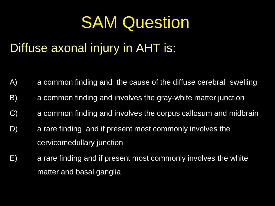
SAM Question
Diffuse axonal injury in AHT is:
A) a common finding and the cause of the diffuse cerebral swelling
B) a common finding and involves the gray-white matter junction
C) a common finding and involves the corpus callosum and midbrain
D) a rare finding and if present most commonly involves the
cervicomedullary junction
E) a rare finding and if present most commonly involves the white
matter and basal ganglia

Abusive Head Trauma in Infants Shaken Baby Syndrome
Whiplash Shaken Baby Syndrome
Shaken Impact Syndrome
Shaken-slam Syndrome
Battered Child Syndrome
Non-accidental Trauma
Non-accidental Injury
Intentional Injury
Trauma-X
Non-accidental Head Injury
Inflicted Head Injury
Abusive Head Injury
Abusive Head Trauma

Abusive Head Trauma
Triad: – subdural hematoma
– retinal hemorrhage
– encephalopathy (brain swelling/anoxic brain injury)
• Little or no external evidence for injury

Mechanism of Inflicted Head Injury
• Direct impact injury to head
• Asphyxiation, strangulation
• Shaking an infant held by the
arms or trunk ending with or
without impact
Abusive Head Trauma
Mostly children under the age of 2 – majority of cases in the first year of life
– peak incidence 6 months
Prospective study of children admitted for head injury <2 years
– 24% from inflicted trauma, 32% suspicious*
Duhaime AC et al. Head injury in very young children,: mechanisms, injury type and
opthalmologic findings in 100 hospitalized patients younger than 2 years of age. Pediatrics
90:179-185.

Abusive Head Trauma
• History:
– vague, changing, clinical findings incompatible with history or developmentally incompatible
• Symptoms:
– lethargy, decreased consciousness, irritability, vomiting, respiratory difficulties, apnea, seizures

Abusive Head Trauma
Clinical:
– retinal hemorrhage
– fractures
– bruising (patterned)
– burns

Abusive Head Trauma
Sequelae: – developmental delay
learning disabilities behavioral issues
– mental retardation cerebral palsy, blindness, death
National Center on Shaken Baby Syndrome

Mandatory Reporters of Child Abuse
• Teachers and other school personnel
• Child care providers
• Social Workers
• Physicians and other health-care workers
• Mental health professionals
• Law enforcement officers
• Medical examiners and coroners
• Clergy (some states)
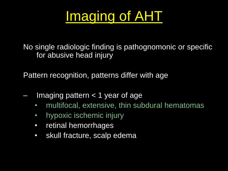
Imaging of AHT
No single radiologic finding is pathognomonic or specific for abusive head injury
Pattern recognition, patterns differ with age
– Imaging pattern < 1 year of age
• multifocal, extensive, thin subdural hematomas
• hypoxic ischemic injury
• retinal hemorrhages
• skull fracture, scalp edema

AHT: Skull Fractures
diastatic occipital comminuted

Fracture vs. Sutures 13 month old girl r/o occipital bone fracture
body bruises, inconsistent hx, multiple skeletal fxs various ages, in foster care

Fracture vs. Suture
Normal Anatomy

Occipital Bone Fractures 13 month old girl r/o occipital bone fracture
body bruises, inconsistent hx, multiple skeletal fxs various ages, in foster care
3D Surface Shaded Display

Fracture vs. Normal Variant 7 month old with growing head circumference
Axial CT

Further evaluation with plain films
AP plain film Lateral plain film

3D Surface Shaded Display

AHT: In-Plane Fractures 12 month old s/p fall from high chair

Cor and Sag Reformats
Coronal Reformat Sagittal Reformat

3 D Model

Digital-scout radiograph
AHT: In-Plane Fractures 12 month old s/p fall from high chair

Subdural Hematomas 3 month old with bradycardia and respiratory depression
In dad’s care, left unattended on floor, later found unresponsive, limp, apneic
multiple body bruises, SS: multiple old fractures

Subdural Hematomas 2 y.o. in mom’s boyfriend’s care, crawling, suddenly collapsed.
SS: multiple fractures varying ages
F/u: seizures, hemiparesis. Foster care

AHT : Parenchymal Injury
• Parenchymal hemorrhage
• White matter contusional clefts
• Diffuse axonal injury
• Hypoxic ischemic changes:
– diffuse or patchy hypodensity
– loss of gray-white matter differentiation
– sparing of basal ganglia and posterior fossa structures

AHT: Hypoxic Ischemic Injury 5 week old with respiratory distress, lethargy and seizures
SS: skull and rib fractures, liver laceration, duodenal hematoma

Follow up CT <24 hrs later
AHT: Hypoxic Ischemic Injury 5 week old with respiratory distress, lethargy and seizures
SS: skull and rib fractures, liver laceration, duodenal hematoma

MRI<24 hrs later
Axial T2 Axial MPGR Axial ADC
AHT: Hypoxic Ischemic Injury 5 week old with respiratory distress, lethargy and seizures
SS: skull and rib fractures, liver laceration, duodenal hematoma

8 months later father in jail, care facility, severe sz d.o., G-tube, stander, supportive chair
AHT: Hypoxic Ischemic Injury 5 week old with respiratory distress, lethargy and seizures
SS: skull and rib fractures, liver laceration, duodenal hematoma

AHT: Hypoxic Ischemic Injury 16 mos old in care of babysitter, 2-3 ft off couch,
tongue laceration, bruising chest, duodenal hematomas, baby died

AHT: Hypoxic Ischemic Injury 16 mos old in care of babysitter, 2-3 ft off couch,
tongue laceration, bruising chest, duodenal hematomas, baby died
1.5 hrs
6 hrs

Rel CBF ADC
SWI
7 hrs after insult
Axial T2

Small SDH with excessive midline shift

AHT: Unilateral HIE 2 year old girl cared for by mother’s boyfriend found unresponsive by brother
retinal hemorrhages, facial bruising

10 days later
AHT: Unilateral HIE 2 year old girl cared for by mother’s boyfriend found unresponsive by brother
retinal hemorrhages, facial bruising

6 months later
child lives with father, seizures, dense hemiparesis, wheelchair, communication board
AHT: Unilateral HIE 2 year old girl cared for by mother’s boyfriend found unresponsive by brother
retinal hemorrhages, facial bruising

Patterns of HIE in AHT
All cases of AHT

White Matter Contusional Tears
Credit F. Lonergan MD. et al.

White Matter Contusional Tears
Axial T2 Axial FLAIR
1 month old with AHT

White Matter Contusional Tears

AHT: Contusional Tear 1 month old with confessed shaking and impact
SS: multiple fractures
Cor T2 Cor T2

Sagttal
AHT: Contusional White Matter Tear
Coronal

Contusional White Matter Tear 5 week infant with vomiting, increased lethargy, possible seizure.
SHDs, skull fxs. SS: mult fxs. No RH

AHT : Parenchymal Contusions 3 month old with bradycardia and respiratory depression
In dad’s care, left unattended on floor, later found unresponsive, limp, apneic
multiple body bruises, SS: multiple old fractures
Day of Admission

AHT : Parenchymal Contusions
Day of
Admission
1 Day
later

Retinal Hemorrhages 16 mos baby girl in care of babysitter, s/p fall off couch
tongue laceration, bruising chest, pancreatic contusion, duodenal hematoma, pt died
Axial T2
Axial T2

Dating of Subdural Hematomas

AHT: Timing of SDHs
Quick dynamic changes in size and density in the first few days:
– ongoing bleeding and clotting
– acute on chronic sdh
– layering of blood products
– redistribution of blood products
– arachnoid tears
All Subdural collections referred to as
“ Subdural Hematomas”= hematohygromas, hygromas, subdural collections

Axial T1 Axial T2
AHT: Layering Subdural Hematomas

AHT: Layering Subdural Hematomas
Axial T1 Axial T2

AHT: Low Density SDH 1 month old with AHT
Day 3 after injury

AHT: Low Density SDHs 1 month old with AHT
Day 1 Day 3 Day 10
Same patient

SDH: Changes in Density AHT in 3 month old
Day 1 Day 3

Subdurals of Different Ages?
Axial T1 Axial T2

Day 2
Day 8
Axial T1 Axial T2
Day 1

Heterogeneous SDH: Acute on Chronic? both AHT cases
xanthochromic fluid and membranes
Acute on Chronic SDH
acute blood all one age, no membranes
Acute SDH
reported as ” “acute on chronic” reported as: “heterogeneous sdh”

Enhancing Membranes: Acute on chronic? 3 month old with AHT
8 days post trauma

SDHs: Enhancing Membranes 3 month old with AHT
8 days
post trauma
3 days
post trauma

Dating of subdural collections
Dating a SDH is challenging
Terminology:
• “acute”, “chronic”, “acute on chronic”
• hygroma, effusion, hematohygroma
• hyperdense, isodense, hypodense, uniform, mixed density

Spinal Subdural Hematoma 5 wk old male, respiratory distress and lethargy, szs
skull fracture, liver laceration, healing rib fx
Sag T1 Axial T2
Axial T1
Sag T1

Summary
• Pattern recognition
• CT reformats and 3D models
in evaluation of skull fractures
• Care in dating SDHs
• MRI and serial imaging
improves confidence and
specificity