imaging of the patient with tinnitus - whooshers.com 1 imaging of the patient with tinnitus mary...
TRANSCRIPT

12/31/2013
1
Imaging of the Patient with Tinnitus
Mary Beth Cunnane MD
Massachusetts Eye and Ear Infirmary
Goals
• To understand tinnitus and its causes
• To determine which patients with tinnitus will benefit from imaging
• To select the appropriate imaging modality for evaluating different forms of tinnitus
• To review the typical appearance of pathologies which cause tinnitus
What is tinnitus?
• The perception of a sound which is not present in the external world.
• Unlike auditory hallucinations, tends to be unformed and repetitive: buzzing, screeching, hissing, pulsing, whooshing.
How common is the problem?
• Roughly 15% of the population experiences tinnitus.
• Up to 4% have very intense tinnitus. – Can lead to chronic disability and suicide – “my ears whistle and buzz continually, day and night …
such a condition is truly frightful” Ludwig van Beethoven
• Most of us have experienced it at one time or another – Transient tinnitus – Noise induced tinnitus following noise exposure
Who is at risk?
• Three major risk factors: – Hearing impairment – Increasing age – Male gender
• Probably increasing in incidence due to: – Increased age of the population – War
• Many common military experiences (interior of a Chinook helicopter, interior of tank, deck of aircraft carrier) are over 100 dB.
• Blast injuries cause damage to both middle and inner ear. Soldiers reluctant to wear ear protection.
Tinnitus Classification
• Subjective
• Objective
– Pulsatile
– Non-pulsatile
12/31/2013
2
Subjective tinnitus
• Most common type of tinnitus
• Associated with hearing loss.
– Associated symptoms depend on cause
• Vertigo – SSCD, Meniere’s
• Conductive hearing loss – otosclerosis, SSCD
• SNHL – vestibular schwannoma, presbycusis, noise induced hearing loss
Objective Tinnitus
• An actual sound made by the human body.
• Can be due to vascular sounds
• Can be due to other physiologic sounds
• Muscular contractions (palatal myoclonus – clicking)
• Respiration (patulous eustachian tube)
• Venous hum (flow murmers)
• “normal” tinnitus
• Frequently can be perceived by an observer.
Pulsatile tinnitus
• Can be altered with compression of arterial or venous structures.
• Can be perceived by the examiner if stethoscope placed in the right location.
• Can be venous or arterial
• Tends to produce whooshing sound.
• Pulse synchronous
Pulsatile tinnitus - causes • Hypervascular states
– Graves disease
• Vascular tumors – Paragangliomas: glomus tympanicum or glomus jugulare
• Arteriovenous malformations and fistulas – Dural AVMs or AVFs frequently involving transverse sinus
• Congenital malformations – Aberrant carotid, high-riding jugular bulb, diverticula
• Narrowing of the transverse sinus – IIH
– Transverse sinus thrombosis
• Carotid dissection, carotid atherosclerosis
Congenital causes of pulsatile tinnitus
12/31/2013
3
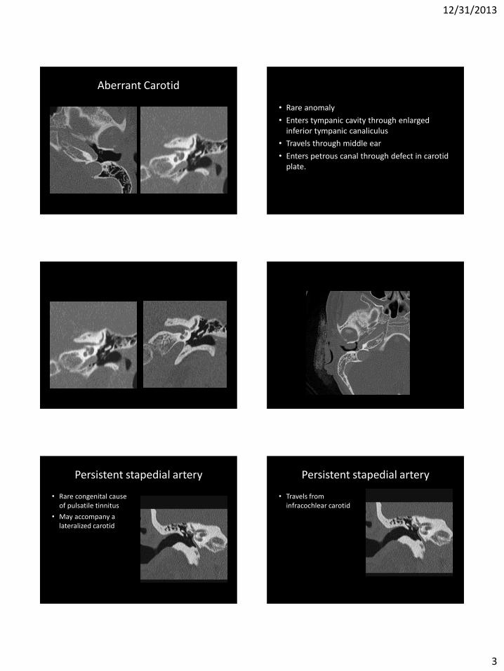
Aberrant Carotid
• Rare anomaly
• Enters tympanic cavity through enlarged inferior tympanic canaliculus
• Travels through middle ear
• Enters petrous canal through defect in carotid plate.
Persistent stapedial artery
• Rare congenital cause of pulsatile tinnitus
• May accompany a lateralized carotid
Persistent stapedial artery
• Travels from infracochlear carotid
12/31/2013
4
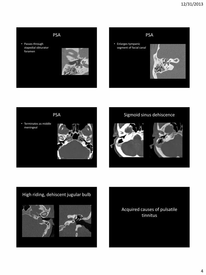
PSA
• Passes through stapedial obturator foramen
PSA
• Enlarges tympanic segment of facial canal
PSA
• Terminates as middle meningeal
Sigmoid sinus dehiscence
High riding, dehiscent jugular bulb
Acquired causes of pulsatile tinnitus

12/31/2013
5
Dural Arteriovenous Fistulas
• Idiopathic
• Related to trauma, prior craniotomy or dural sinus thrombosis.
• Classified according to direction of flow and presence or absence of cortical venous drainage
• In absence of CVD, very low risk of hemorrhage, do not require treatment
• Most common locations caverous, transverse, sigmoid sinuses.
MRI findings
• Flow related enhancement in venous structure on 3D TOF MRA.
• Prominent flow voids in transverse sinus
• Prominent meningeal arteries
• Prominent vascular channels across calvarium to transverse sinus.
CTA findings
• Asymmetric arterial feeding vessels
• Shaggy appearance to the dural sinus or tentorium
• Prominent transcalvarial venous channels
Paragangliomas
• Extra-adrenal pheochromocytomas
• Can be limited to the middle ear
– Along Jacobson’s nerve (tympanic branch of glossopharyngeal)
– Glomus tympanicum
• Can involve the jugular foramen
– Along Arnold’s nerve (auricular branch of vagus)
– Glomus jugulare or jugularetympanicum
12/31/2013
6
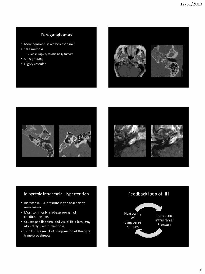
Paragangliomas
• More common in women than men
• 10% multiple
– Glomus vagale, carotid body tumors
• Slow growing
• Highly vascular
Idiopathic Intracranial Hypertension
• Increase in CSF pressure in the absence of mass lesion.
• Most commonly in obese women of childbearing age.
• Causes papilledema, and visual field loss, may ultimately lead to blindness.
• Tinnitus is a result of compression of the distal transverse sinuses.
Feedback loop of IIH
Increased Intracranial
Pressure
Narrowing of
transverse sinuses
12/31/2013
7
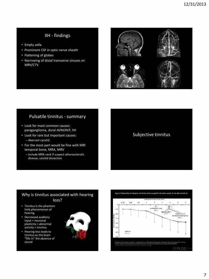
IIH - findings
• Empty sella
• Prominent CSF in optic nerve sheath
• Flattening of globes
• Narrowing of distal transverse sinuses on MRV/CTV
Pulsatile tinnitus - summary
• Look for most common causes: paraganglioma, dural AVM/AVF, IIH
• Look for rare but important causes:
– Aberrant carotid.
• For the most part would be fine with MRI temporal bone, MRA, MRV
– Include MRA neck if suspect atherosclerotic disease, carotid dissection
Subjective tinnitus
Why is tinnitus associated with hearing loss?
• Tinnitus is the phantom limb phenomenon of hearing.
• Decreased auditory input + neuronal plasticity = abnormal activity = tinnitus
• Hearing loss leads to tinnitus as the brain “fills in” the absence of sound
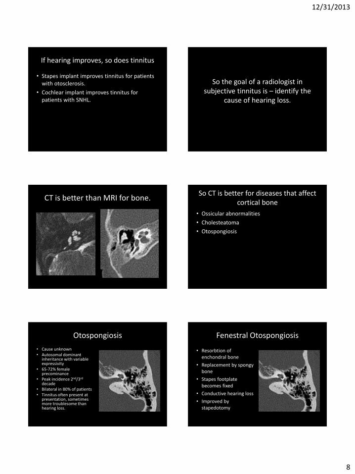
Figure 2. Relationship of audiogram and tinnitus pitch averaged for the whole sample, for the right and left ear.
Schecklmann M, Vielsmeier V, Steffens T, Landgrebe M, et al. (2012) Relationship between Audiometric Slope and Tinnitus Pitch in Tinnitus Patients: Insights into the Mechanisms of Tinnitus Generation. PLoS ONE 7(4): e34878. doi:10.1371/journal.pone.0034878 http://www.plosone.org/article/info:doi/10.1371/journal.pone.0034878
12/31/2013
8
If hearing improves, so does tinnitus
• Stapes implant improves tinnitus for patients with otosclerosis.
• Cochlear implant improves tinnitus for patients with SNHL.
So the goal of a radiologist in subjective tinnitus is – identify the
cause of hearing loss.
CT is better than MRI for bone. So CT is better for diseases that affect
cortical bone
• Ossicular abnormalities
• Cholesteatoma
• Otospongiosis
Otospongiosis
• Cause unknown • Autosomal dominant
inheritance with variable expressivity
• 65-72% female precominance
• Peak incidence 2nd/3rd decade
• Bilateral in 80% of patients • Tinnitus often present at
presentation, sometimes more troublesome than hearing loss.
Fenestral Otospongiosis
• Resorbtion of enchondral bone
• Replacement by spongy bone
• Stapes footplate becomes fixed
• Conductive hearing loss
• Improved by stapedotomy

12/31/2013
9
Retrofenestral or cochlear otospongiosis
• Involvement of bone surrounding cochlea
• Described as 4th turn
• Causes mixed hearing loss
Cholesteatoma
Cholesteatoma
• Collection of squamous debris which acts like a mass causing bony erosion
• Most are aquired – pars flaccida
• Minority are congenital – live anywhere
Congenital cholesteatoma
• Soft tissue mass in middle ear
• Non-enhancing
• May demonstrate restricted diffusion (if large enough)
• In the imaging DDX of glomus tympanicum
– But no red mass on exam
– Tinnitus is non-pulsatile
Acquired cholesteatoma
• Pars flaccida to epitympanum
• Erosion of scutum or ossicles
• Important landmarks to check
– Facial nerve
– Lateral SCC
Acquired cholesteatoma
12/31/2013
10
Acquired cholesteatoma Superior Semicircular Canal
Dehiscence
Superior Semicircular Canal Dehiscence
• Leads to conductive hearing loss
– Loss of pressure wave at dehiscence
• Associated with vertigo, particularly in the setting of loud noise (Tulio’s phenomenon)
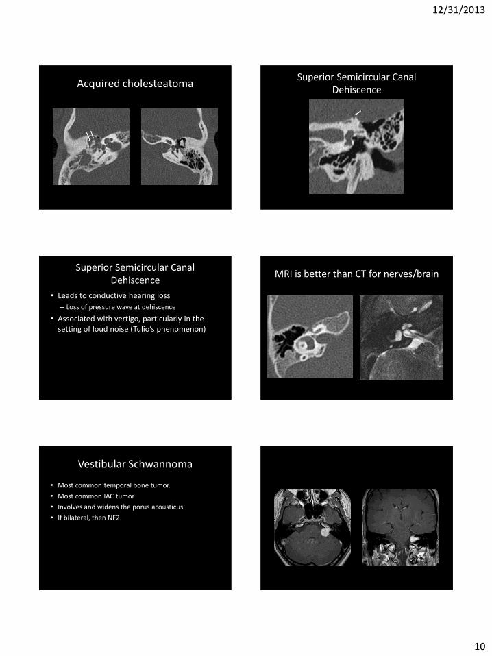
MRI is better than CT for nerves/brain
Vestibular Schwannoma
• Most common temporal bone tumor.
• Most common IAC tumor
• Involves and widens the porus acousticus
• If bilateral, then NF2
12/31/2013
11
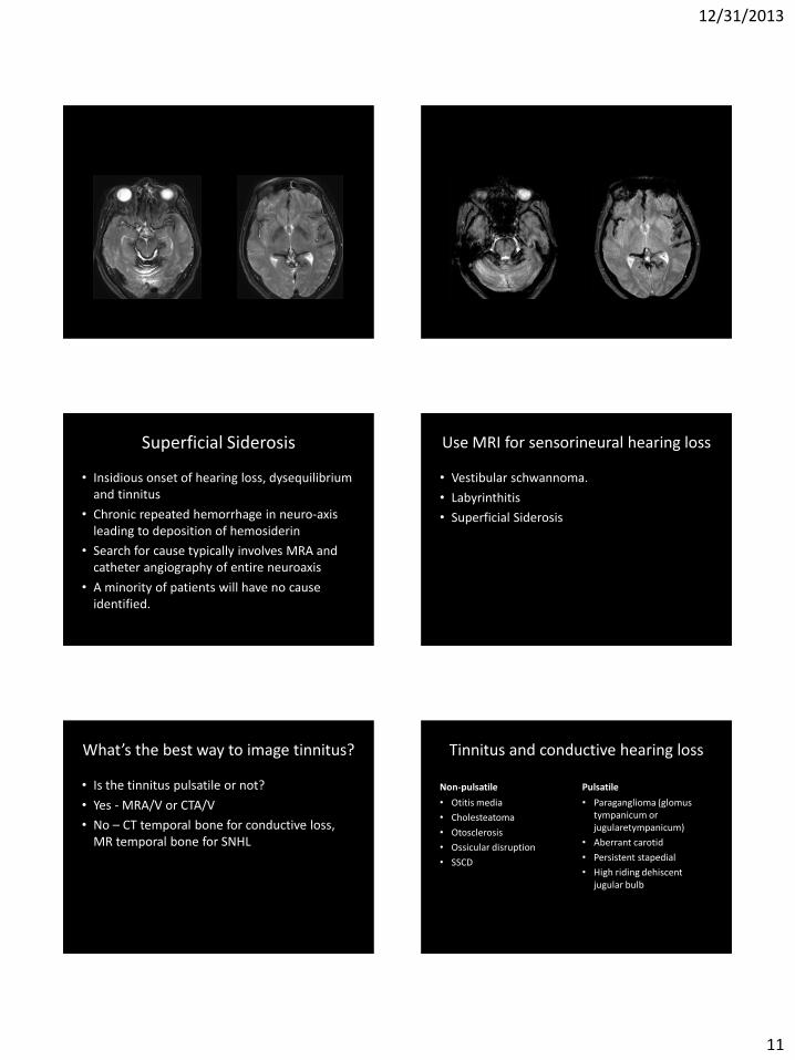
Superficial Siderosis
• Insidious onset of hearing loss, dysequilibrium and tinnitus
• Chronic repeated hemorrhage in neuro-axis leading to deposition of hemosiderin
• Search for cause typically involves MRA and catheter angiography of entire neuroaxis
• A minority of patients will have no cause identified.
Use MRI for sensorineural hearing loss
• Vestibular schwannoma.
• Labyrinthitis
• Superficial Siderosis
What’s the best way to image tinnitus?
• Is the tinnitus pulsatile or not?
• Yes - MRA/V or CTA/V
• No – CT temporal bone for conductive loss, MR temporal bone for SNHL
Tinnitus and conductive hearing loss
Non-pulsatile
• Otitis media
• Cholesteatoma
• Otosclerosis
• Ossicular disruption
• SSCD
Pulsatile
• Paraganglioma (glomus tympanicum or jugularetympanicum)
• Aberrant carotid
• Persistent stapedial
• High riding dehiscent jugular bulb

12/31/2013
12
Sensorineural hearing loss and tinnitus
• Labyrinthitis
• Vestibular schwannoma
• Presbycusis
• Noise induced hearing loss
Pulsatile tinnitus without hearing loss
• Paraganglioma
• Dural AV fistula
• IIH
• Dural venous thrombosis
SUMMARY: A simple approach to imaging in tinnitus
• If objective tinnitus – look for the cause of the sound.
• If subjective tinnitus – look for the cause of the hearing loss.