imaging of trauma to the spine - files.bridgeport.edu · imaging of trauma to the spine orthopedic...
TRANSCRIPT
Imaging of Trauma to the Spine
Orthopedic Diplomate ProgramUniversity of Bridgeport College of Chiropractic
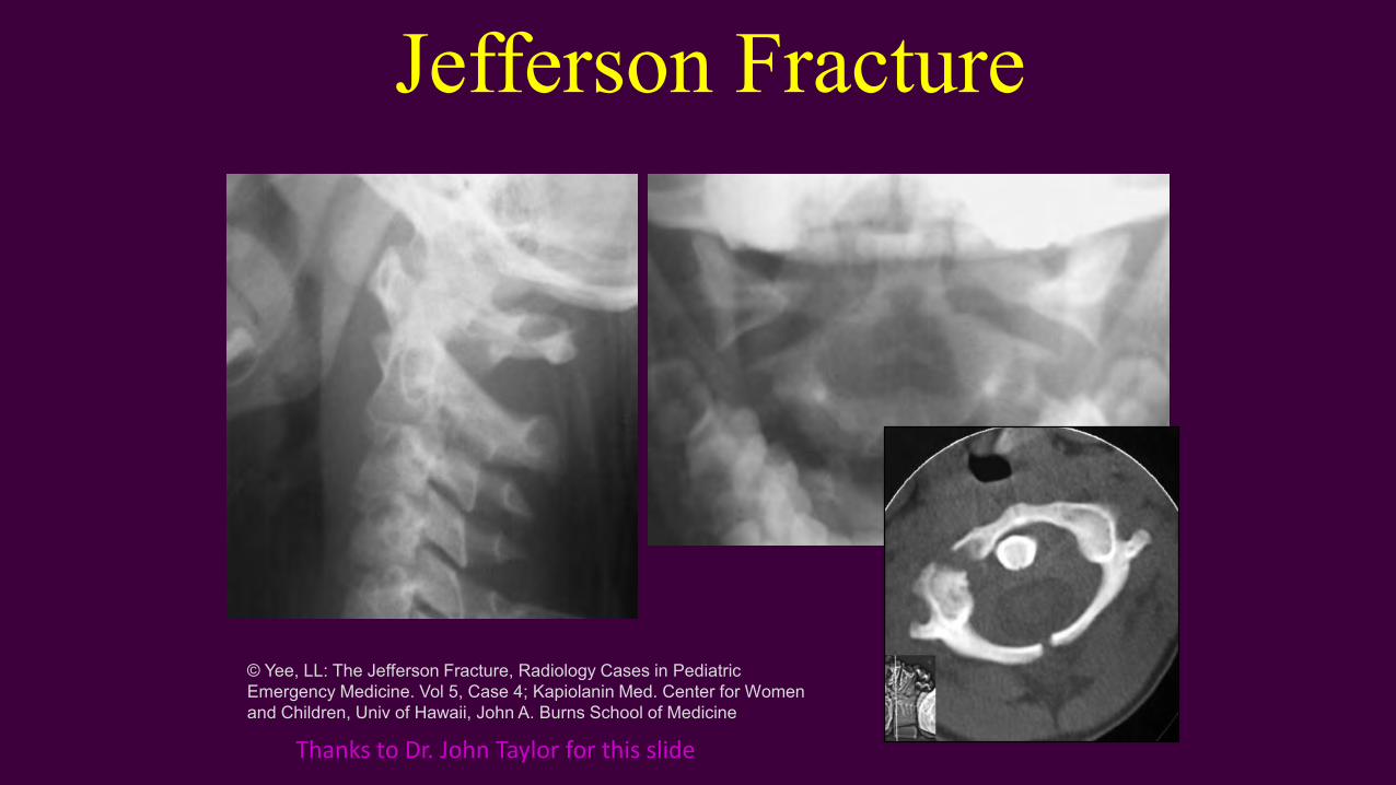
Jefferson Fracture
© Yee, LL: The Jefferson Fracture, Radiology Cases in Pediatric Emergency Medicine. Vol 5, Case 4; Kapiolanin Med. Center for Women and Children, Univ of Hawaii, John A. Burns School of Medicine
Thanks to Dr. John Taylor for this slide
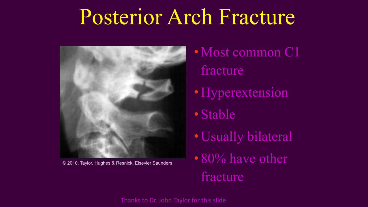
Posterior Arch Fracture• Most common C1
fracture• Hyperextension • Stable• Usually bilateral• 80% have other
fracture© 2010, Taylor, Hughes & Resnick. Elsevier Saunders
Thanks to Dr. John Taylor for this slide
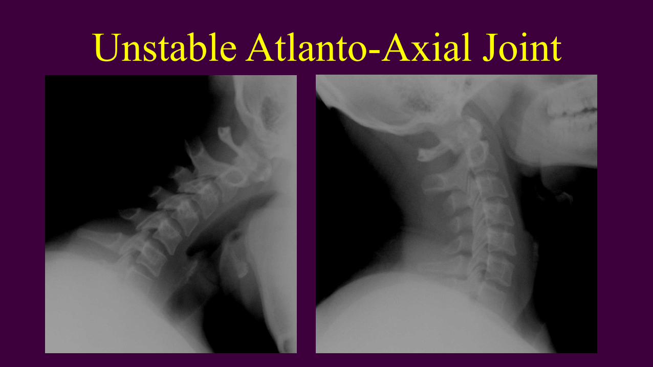
Unstable Atlanto-Axial Joint
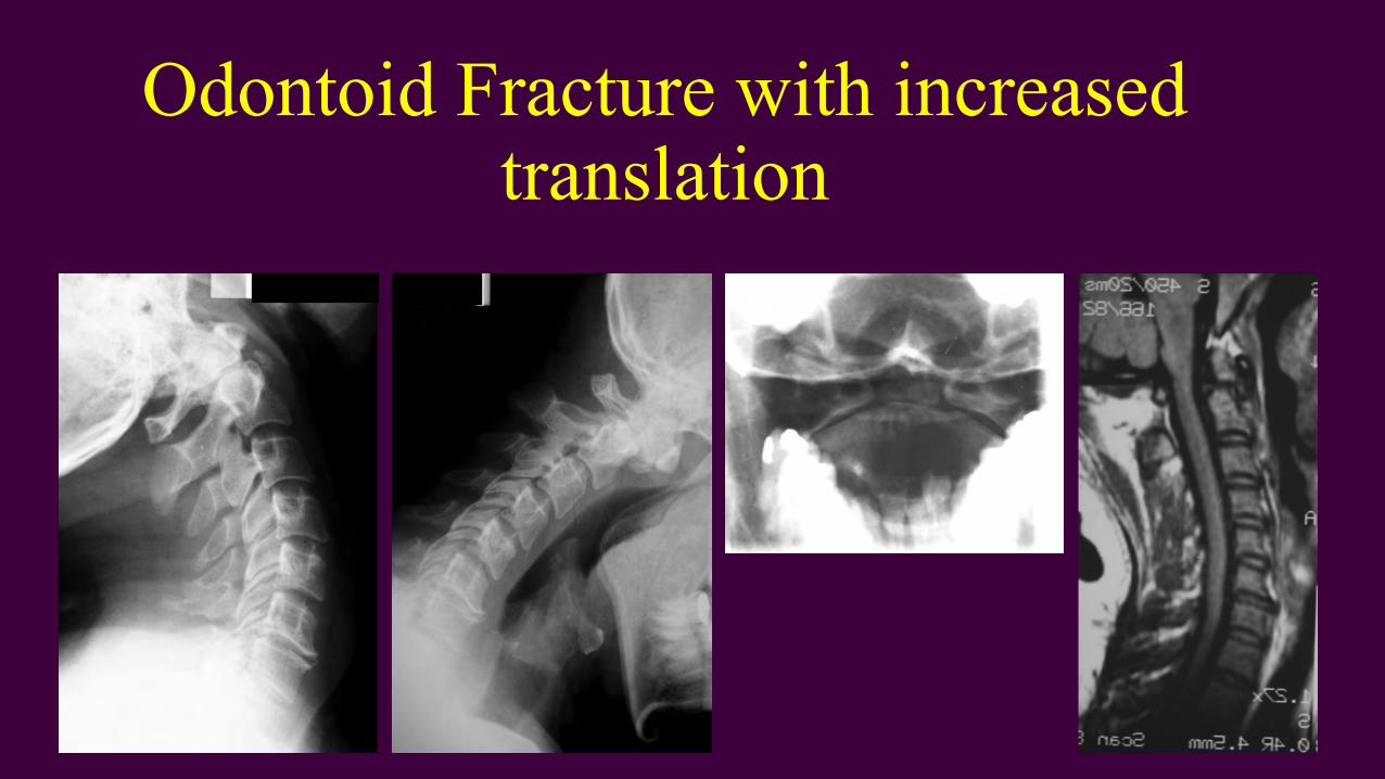
Odontoid Fracture with increased translation
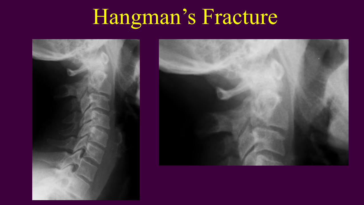
Hangman’s Fracture
28 y.o. male• Reportedly fell while chasing a puppy in the street a
night after a party
• Head hit the curb and was forced into hyperextension
• Significant pain (10/10) in neck & with all attempts at motion
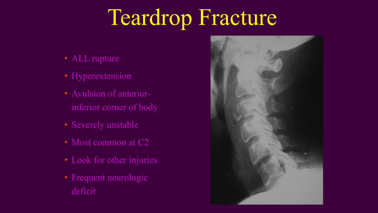
Teardrop Fracture• ALL rupture
• Hyperextension
• Avulsion of anterior-inferior corner of body
• Severely unstable
• Most common at C2
• Look for other injuries
• Frequent neurologic deficit
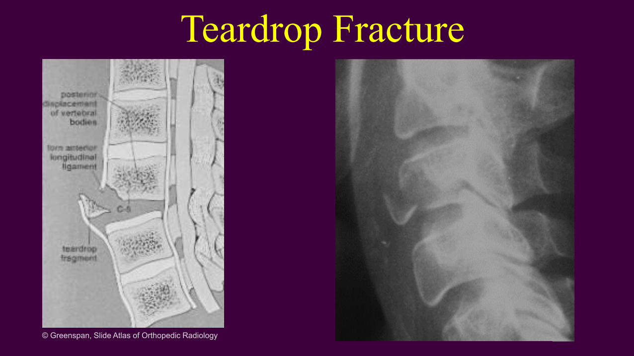
Teardrop Fracture
© Greenspan, Slide Atlas of Orthopedic Radiology
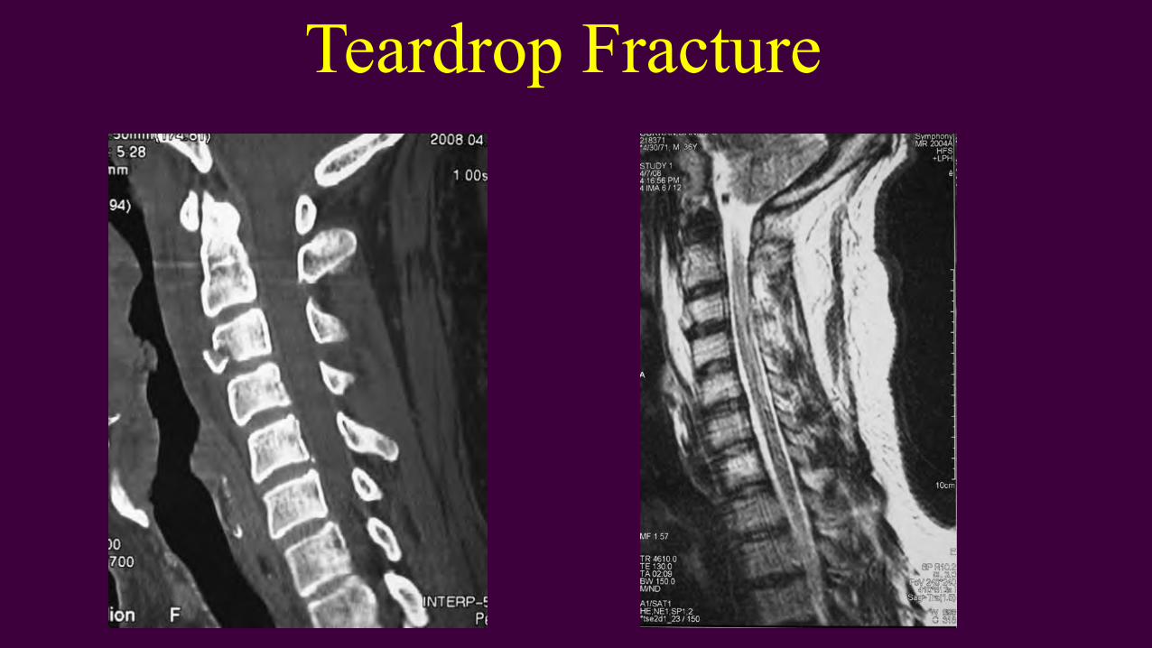
Teardrop Fracture
Burst Fracture
• Vertical compression combined with flexion
• Comminution of body by nucleus pulposus• Retropulsion• Kyphosis, spinous fanning, facet
dislocation• 85% neurologic deficit
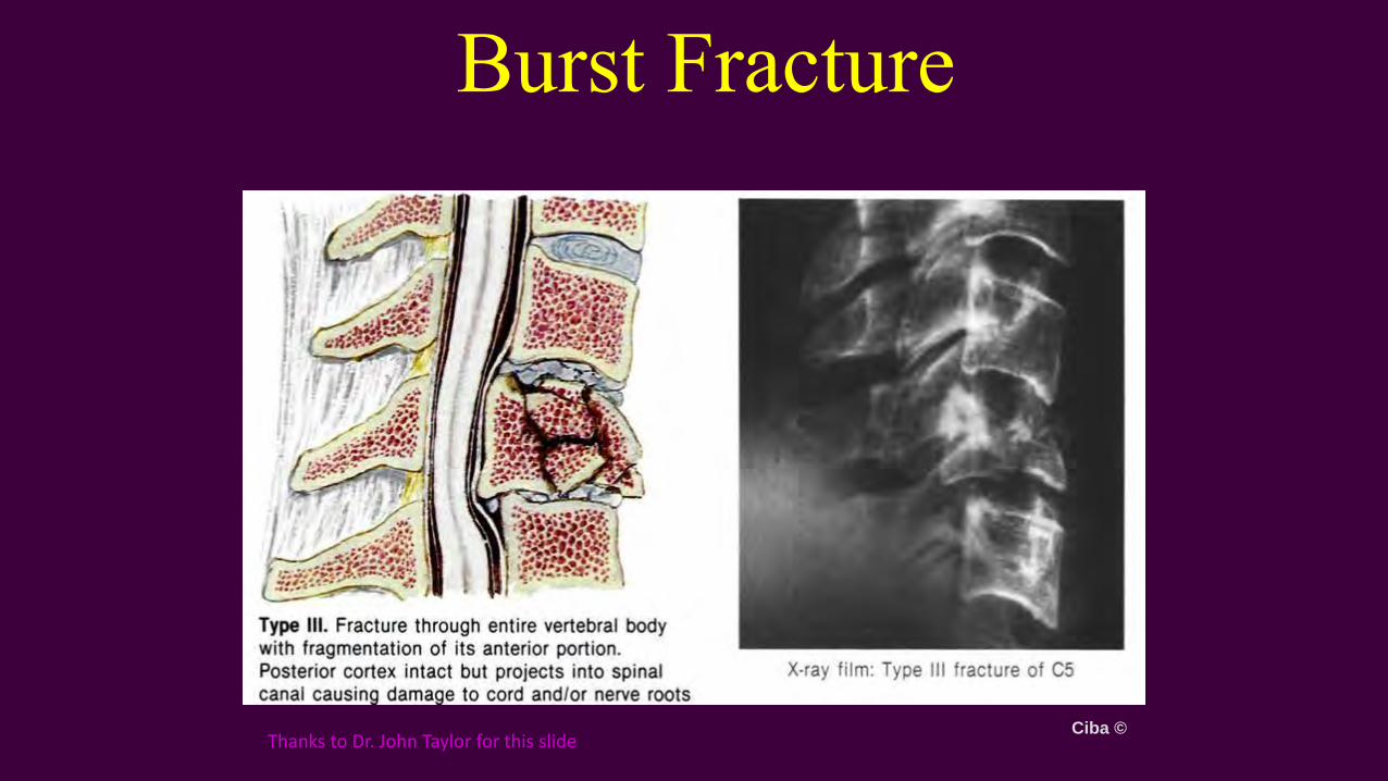
Burst Fracture
Ciba ©Thanks to Dr. John Taylor for this slide
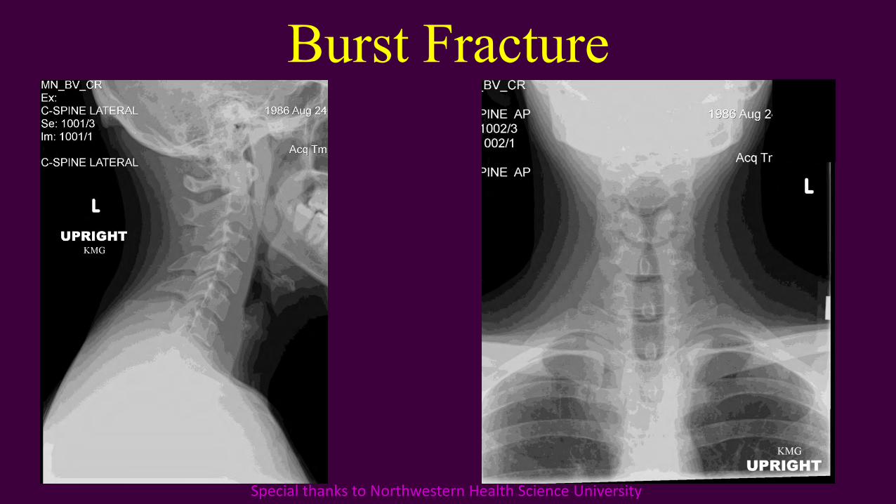
Burst Fracture
Special thanks to Northwestern Health Science University

Burst Fracture
Special thanks to Northwestern Health Science University for this case
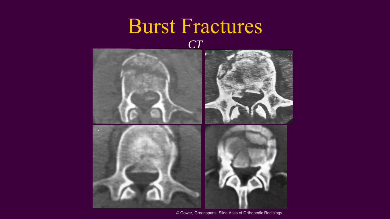
Burst FracturesCT
© Gower, Greenspans, Slide Atlas of Orthopedic Radiology
22 y.o Male
Attempting to ride his bike across the U.S.
Hit on a straight, flat stretch of road by motorist not paying attention
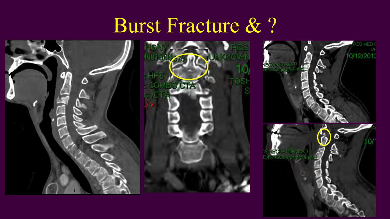
Burst Fracture & ?
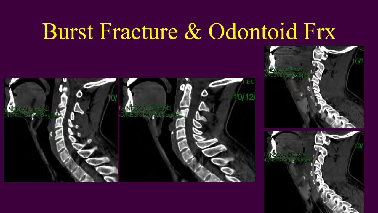
Burst Fracture & Odontoid Frx
Cervical Spine Dislocation
• Unilateral facet dislocation
• Bilateral facet dislocation
• Transverse ligament rupture
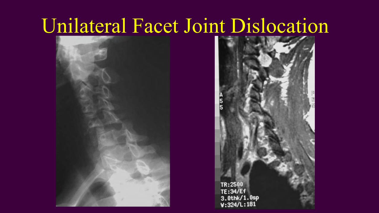
Unilateral Facet Joint Dislocation
35 y.o. UBCC student• Involved in MVA while in army approx. 12 yrs ago
• Neck injury (films?)• Has been getting adjusted weekly (2X times a week) by local
chiropractor since leaving the service
• For approx. 7 yrs with great relief• The results are what inspired him to attend UBCC
• Incoming student screening exam referred for cervical films
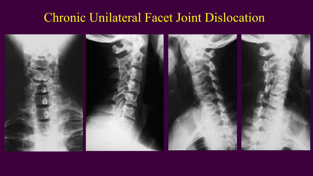
Chronic Unilateral Facet Joint Dislocation
Acute Compression FracturesRADIOGRAPHIC FINDINGS:
• Wedge deformity• Zone of impaction• Step defect• Paraspinal edema and hemorrhage• Abdominal ileus—excessive gas
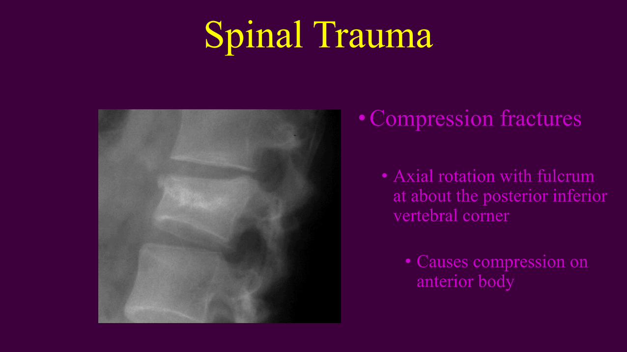
Spinal Trauma
• Compression fractures
• Axial rotation with fulcrum at about the posterior inferior vertebral corner
• Causes compression on anterior body
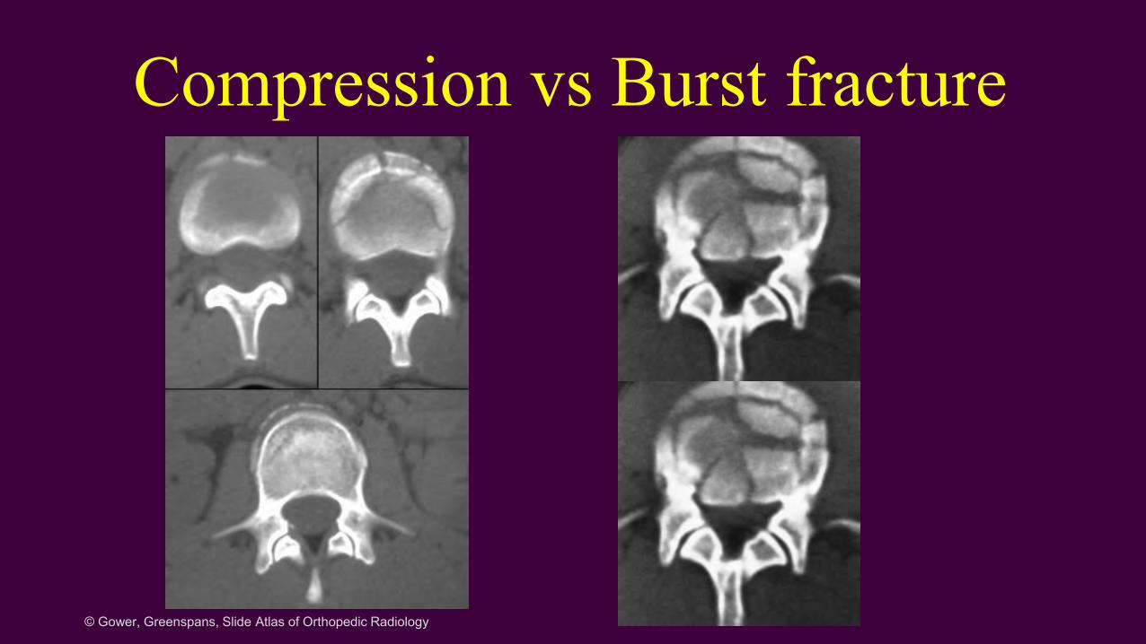
Compression vs Burst fracture
© Gower, Greenspans, Slide Atlas of Orthopedic Radiology
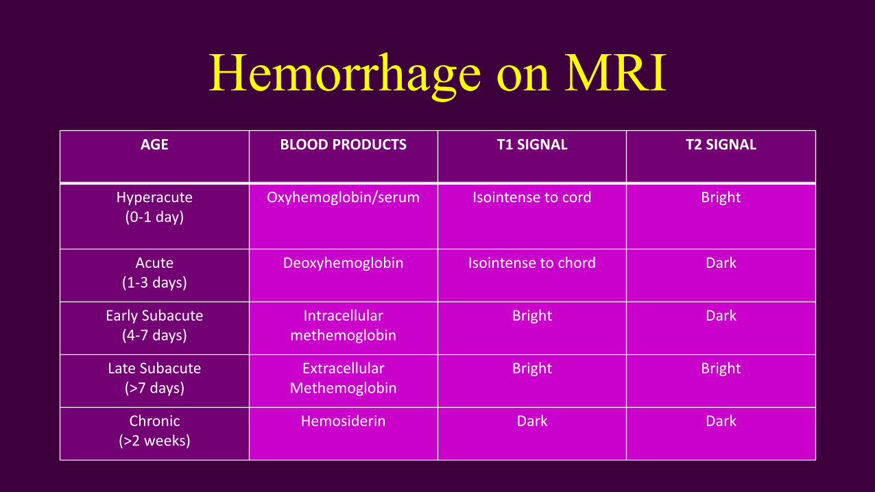
Hemorrhage on MRIAGE BLOOD PRODUCTS T1 SIGNAL T2 SIGNAL
Hyperacute(0-1 day)
Oxyhemoglobin/serum Isointense to cord Bright
Acute(1-3 days)
Deoxyhemoglobin Isointense to chord Dark
Early Subacute(4-7 days)
Intracellular methemoglobin
Bright Dark
Late Subacute(>7 days)
ExtracellularMethemoglobin
Bright Bright
Chronic(>2 weeks)
Hemosiderin Dark Dark
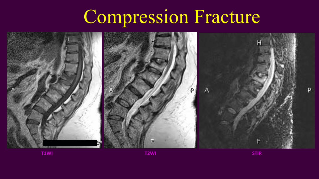
Compression Fracture
T1WI T2WI STIR
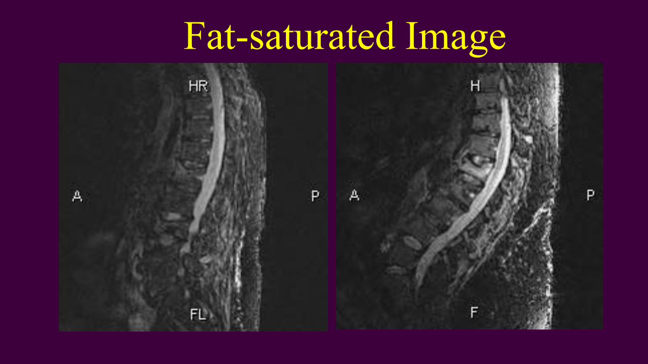
Fat-saturated Image
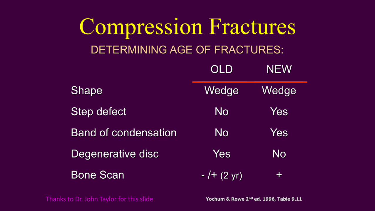
Compression FracturesDETERMINING AGE OF FRACTURES:
OLD NEW
Shape Wedge Wedge
Step defect No Yes
Band of condensation No Yes
Degenerative disc Yes No
Bone Scan -- /+ /+ (2 yr) +
Yochum & Rowe 2Yochum & Rowe 2ndndnd ed. 1996, Table 9.11Thanks to Dr. John Taylor for this slide
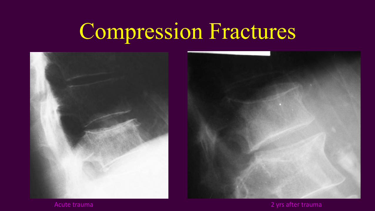
Compression Fractures
2 yrs after traumaAcute trauma
Pars Interarticulares
•Younger patients (under 30 yrs old) with low back pain
• Significant possibility of pars defects• Especially in athletes
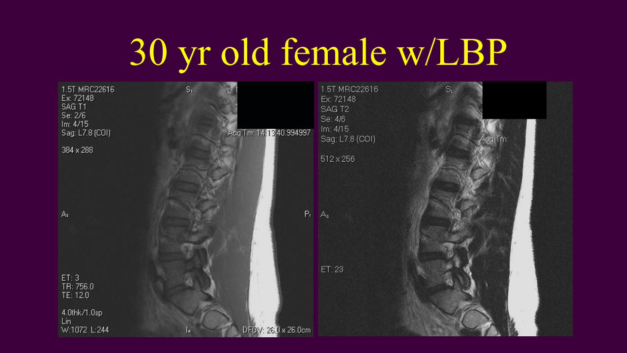
30 yr old female w/LBP
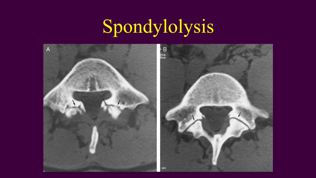
Spondylolysis
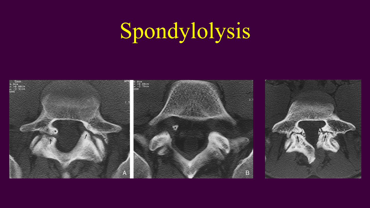
Spondylolysis
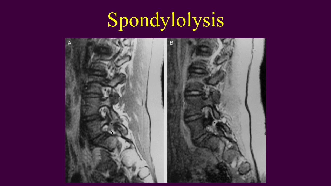
Spondylolysis
Stability of the Spine• Typically assumed no more than 3.5 mm. translation in
cervical spine
• Anything more considered excessive/instability
• Lumbar translation
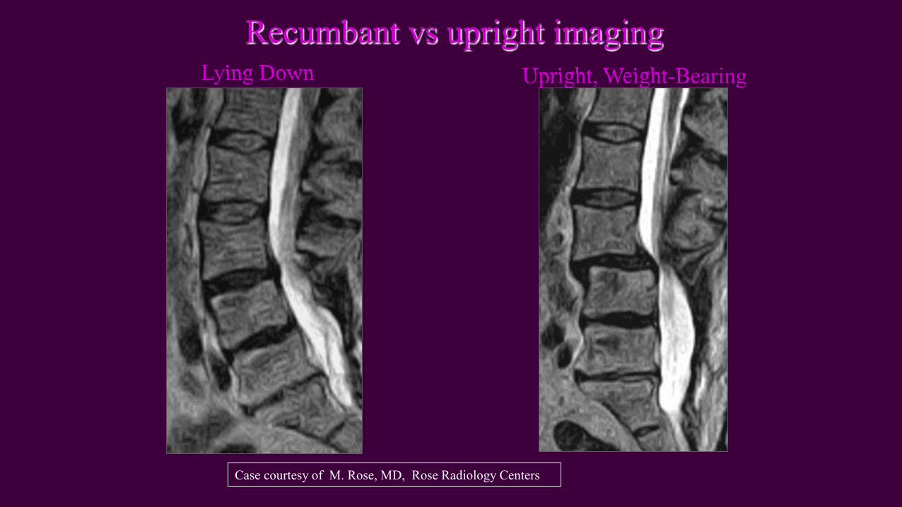
Lying Down Upright, Weight-BearingLying Down
RecumbantUpright, Weight Bearing
Recumbant vs upright imaging
Case courtesy of M. Rose, MD, Rose Radiology Centers
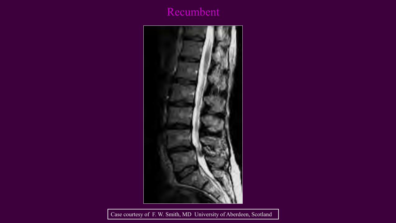
Case courtesy of F. W. Smith, MD University of Aberdeen, Scotland
Recumbent
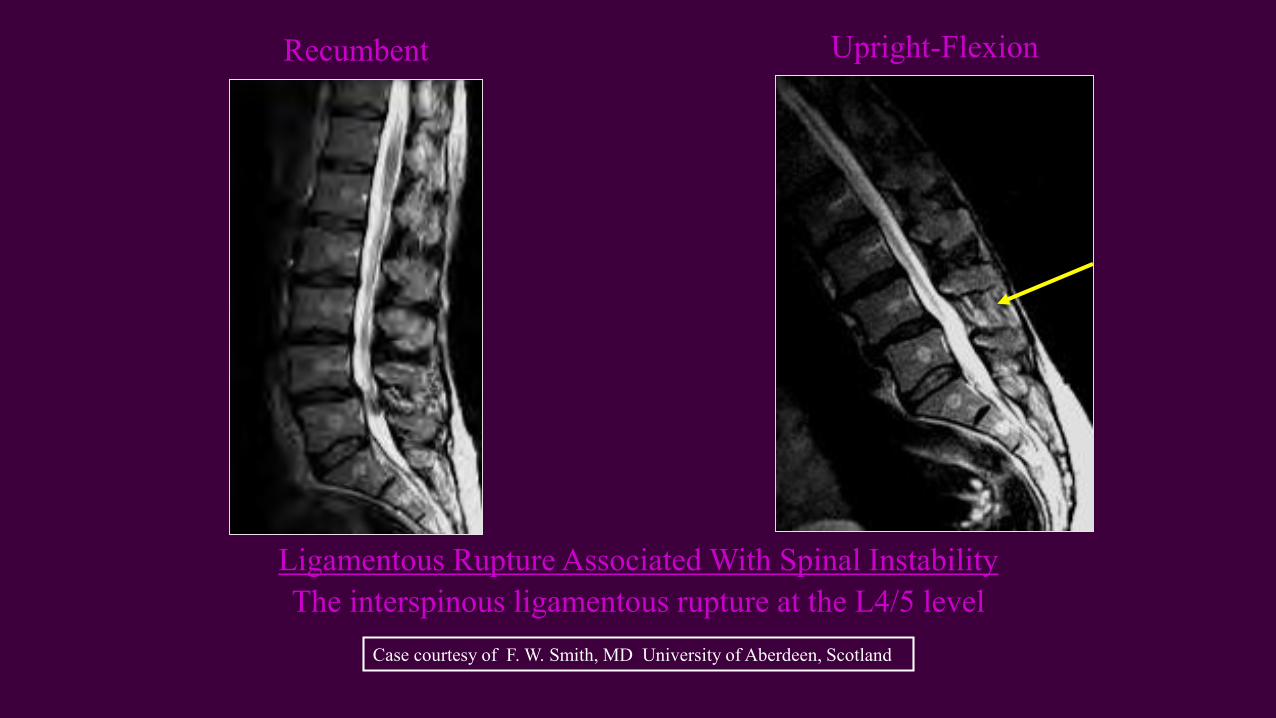
Ligamentous Rupture Associated With Spinal Instability
Upright-Flexion
The interspinous ligamentous rupture at the L4/5 level
Recumbent
Case courtesy of F. W. Smith, MD University of Aberdeen, Scotland
Arachnoiditis• Post-traumatic (post-surgical, post-pantopaque)
• Inflammatory process often d/t components being injected into subarachnoid space (i.e. contrast agents, anesthetics) or intrathecal hemorrhage forming adhesions
• Clumping of nerve roots instead of gently arching nerve roots• May adhere to the dura resulting in empty appearing thecal sac
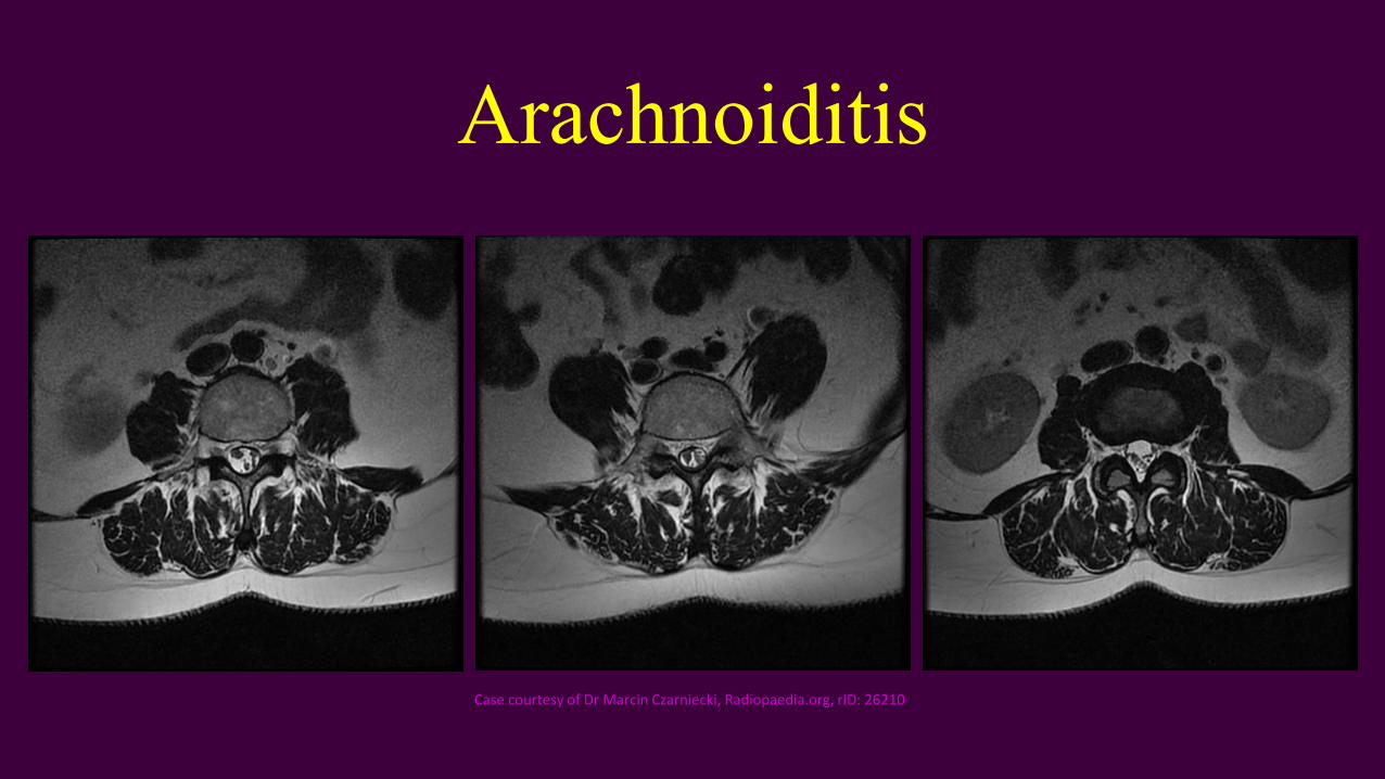
Arachnoiditis
Case courtesy of Dr Marcin Czarniecki, Radiopaedia.org, rID: 26210
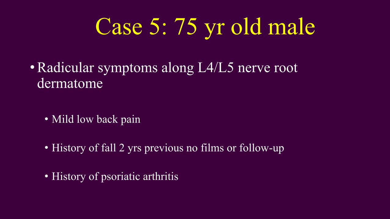
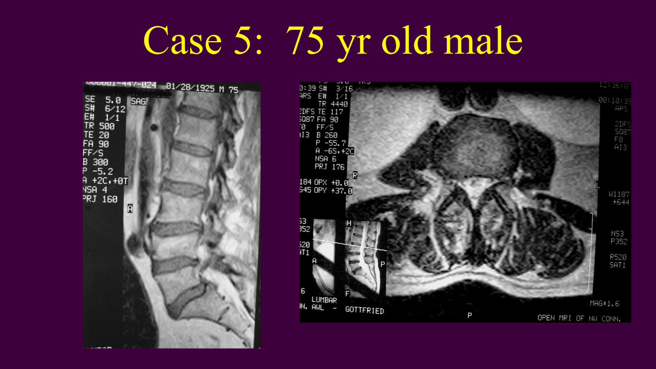
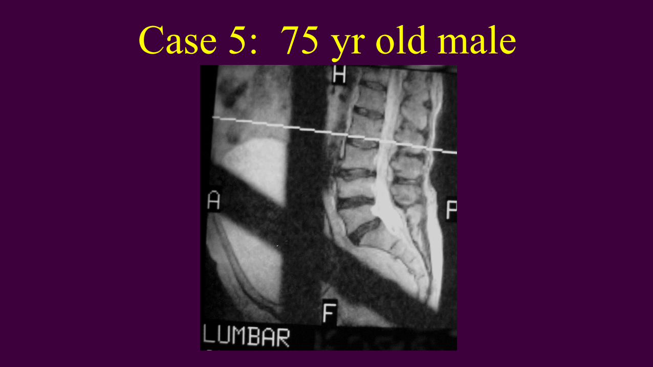
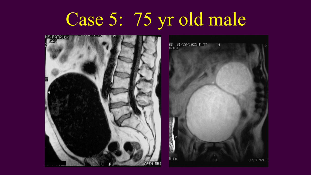
Case 5: 75 yr old male• Radicular symptoms along L4/L5 nerve root
dermatome
• Mild low back pain
• History of fall 2 yrs previous no films or follow-up
• History of psoriatic arthritis
Case 5: 75 yr old male
Case 5: 75 yr old male
Case 5: 75 yr old male
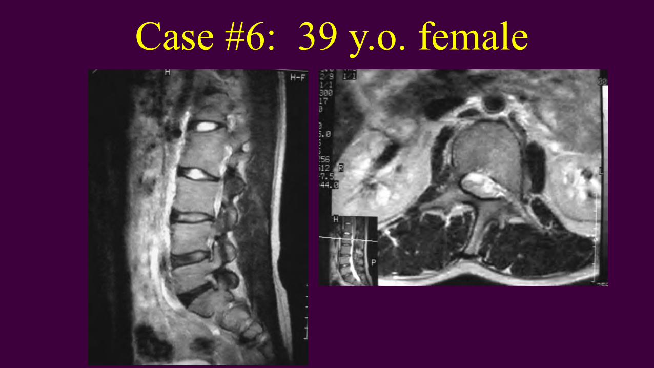
Case #6: 39 y.o. female
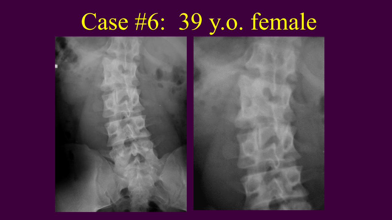
Case #6: 39 y.o. female
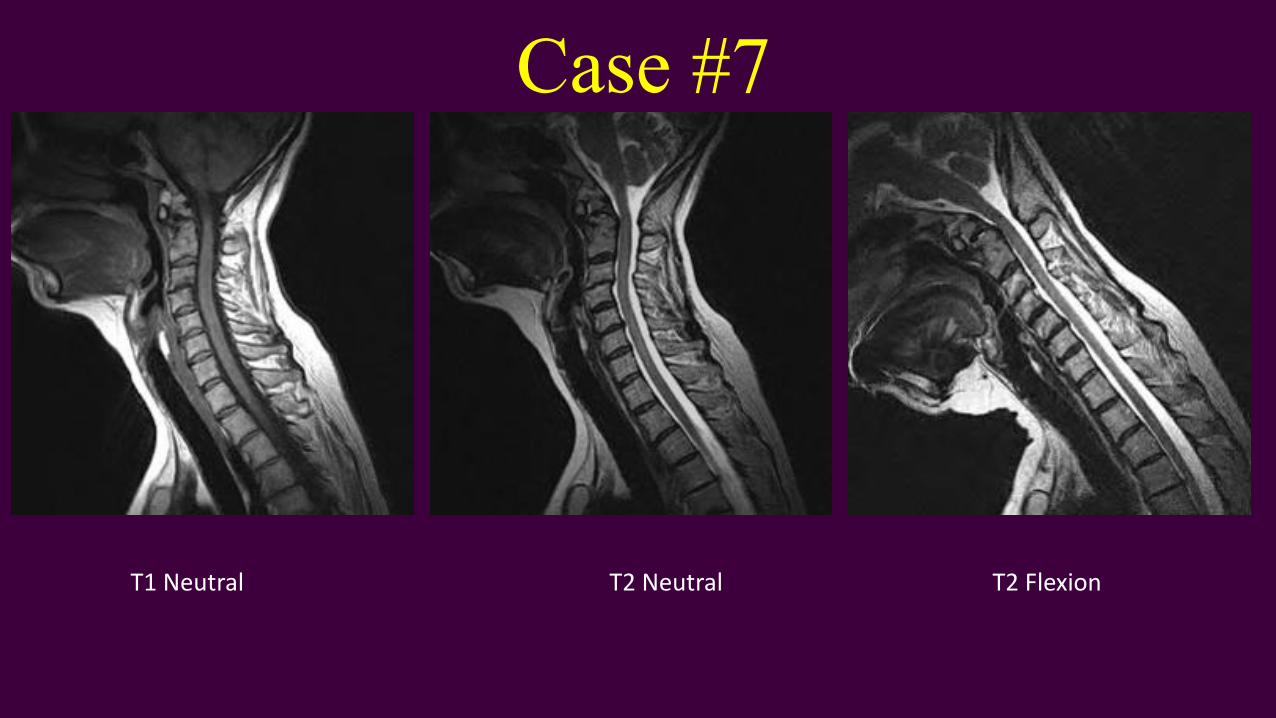
Case #7
T1 Neutral T2 Neutral T2 Flexion
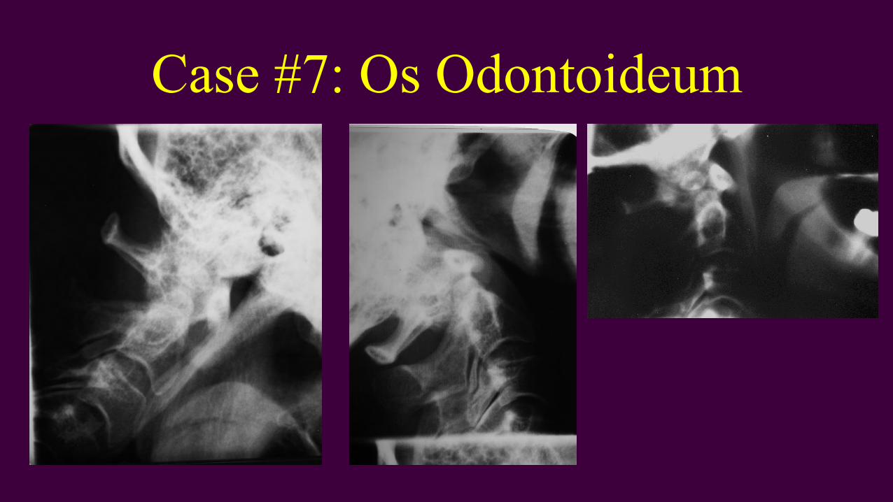
Case #7: Os Odontoideum
End of Spinal Trauma Section