immature oocyte quality and maturational competence … · immature oocyte quality and maturational...
TRANSCRIPT
Immature oocyte quality and maturational competence of porcinecumulus-oocyte complexes subpopulations
GABRIEL MARTIN ALVAREZ1*, GABRIEL CARLOS DALVIT1, MARÍA VERÓNICA ACHI1, MARCELO SERGIO MIGUEZ2
AND PABLO DANIEL CETICA1
1. Area of Biochemistry, Institute of Research and Technology in Animal Reproduction (INITRA), School of VeterinarySciences, University of Buenos Aires, Argentina.
2. Area of Porcine Production, School of Veterinary Sciences, University of Buenos Aires, Argentina.
Key words: In vitro maturation, nuclear maturation, cytoplasmic maturation, pig
ABSTRACT: Porcine immature oocyte quality (i.e., that of live oocytes at the germinal vesicle stage) wasevaluated according to features of the surrounding cumulus, aiming to establish maturational competence ofdifferent subpopulations of such cumulus-oocyte complexes. Six subpopulations were identified: A
1 (with a
dense cumulus), A2 (with a translucent cumulus), B
1 (with the corona radiata), B
2 (partly naked oocytes), C
(naked oocytes), D (with a dark cumulus). The percent incidence of live oocyte in these subpopulationschanged significantly as related to cumulus features, however the occurrence of oocytes in the germinalvesicle stage was lower in class D only. Similar metaphase II rates achieved in A
1, A
2, B
1 and B
2 classes after
in vitro maturation suggest that the nucleus may in fact mature in vitro, in spite of the different accompanyingcumulus features which are typical of these classes. In contrast, a higher cytoplasmic maturation rate ob-tained in class A
1 may indicate a stronger dependence of this variable upon cumulus features than that shown
by nuclear maturation. When different types of cumulus expansion after in vitro maturation were considered(i.e., fully expanded cumulus, partly expanded cumulus, and partly naked oocyte), no differences were foundin the percent of oocytes reaching metaphase II or cytoplasmic maturation. It is concluded that morphologicalfeatures of the collected porcine cumulus-oocyte complexes (rather than cumulus behavior during culture)may be useful for selection of potentially competent oocytes for in vitro fertilization and embryo production.
BIOCELL2009, 33(3): 167-177
ISSN 0327 - 9545PRINTED IN ARGENTINA
Introduction
In vitro embryo production techniques have beenapplied for many years in different livestock species,obtaining viable embryos and even offspring births(Brackett et al., 1982; Eyestone and First, 1989; Yoshidaet al., 1993; Thompson et al., 1995; Macháty et al., 1998;Kikuchi et al., 1999). Particularly, in vitro embryo pro-duction systems have reached higher blastocyst rateswhen applied to cattle and sheep (Sutton-McDowall et
al., 2006; Gutnisky et al., 2007; García-García et al.,2007; Zhu et al., 2007) rather than when applied to swine(Kikuchi et al., 2002; Schoevers et al., 2003).
Oocyte maturation is a most important step in por-cine in vitro embryo production systems. Even thoughoocyte in vitro maturation has been extensively stud-ied in swine (Abeydeera, 2002; Krisher, 2004), wethink that quality of immature oocytes has not beensufficiently evaluated. Oocyte quality is acquired pro-gressively as the gamete grows within the ovarian fol-licle, and it impacts on in vitro oocyte maturation, invitro embryo development, establishment and mainte-nance of pregnancy and fetal development (Krisher etal., 2007).
*Address correspondence to: Gabriel Martin Alvarez.E-mail: [email protected]: December 2, 2008. Revised version received:September 26, 2009. Acepted: September 30, 2009.
GABRIEL MARTIN ALVAREZ et al.168
Oocyte size has been much used to evaluate thequality of immature porcine oocytes for in vitro matu-ration systems (Motlik and Fulka, 1986; Homa et al.,1988; Ikeda and Takahashi, 2003). Also, follicle diam-eter has been associated to oocyte quality, and it is gen-erally accepted that follicle size affects both nuclear andcytoplasmic maturation as well as embryo developmen-tal potential (Motlik et al., 1984; Sun et al., 2001;Marchal et al., 2002). Although these measurable vari-ables correlate well with immature oocyte quality, theyare not commonly used in in vitro maturation programsbecause of their low practicality. Contrariwise, imma-ture oocytes selection according to the morphologicalfeatures of the surrounding cumulus may be more com-monly used before in vitro maturation because of itseasy and fast implementation.
Follicular cell assistance to the oocyte is essentialfor acquiring in vitro developmental competence. It isgenerally accepted that cumulus cells support oocytematuration to the metaphase II stage and are involved inthe cytoplasmic maturation needed for postfertilizationdevelopmental capability (Abeydeera, 2002). Usually,
oocytes with a multilayered cumulus are used for in vitromaturation protocols, but selection criteria of cumulus-oocyte complexes widely diverge among authors (Wanget al., 1997; Gandhi et al., 2001; Qian et al., 2003;Algriany et al., 2004; Stokes et al., 2005). Therefore,association between immature oocyte quality and cu-mulus-oocyte complex features varies according to theselection criteria used.
It has been reported that nuclear status of imma-ture oocyte in cattle is related to morphological char-acteristics of the cumulus-oocyte complex and to itsmaturational competence (Cetica et al., 1999). In thepresent report, the immature porcine oocyte quality(i.e., that of live oocytes at the germinal vesicle stage)was evaluated according to features of the surround-ing cumulus in order to establish the maturational be-havior of different subpopulations of porcine cumu-lus-oocyte complexes. This was made by analyzingcumulus expansion, nuclear maturation (first polarbody and metaphase II plate) and cytoplasmic matu-ration (sperm head decondensation and pronuclearformation).
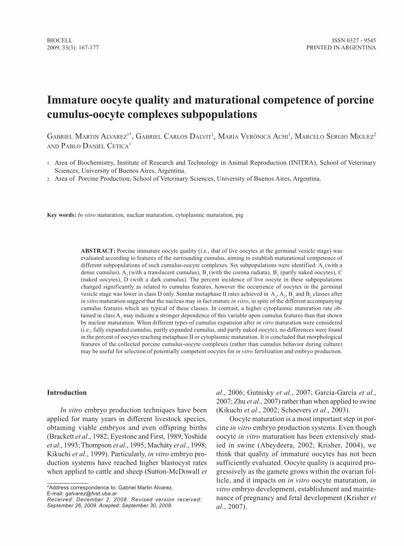
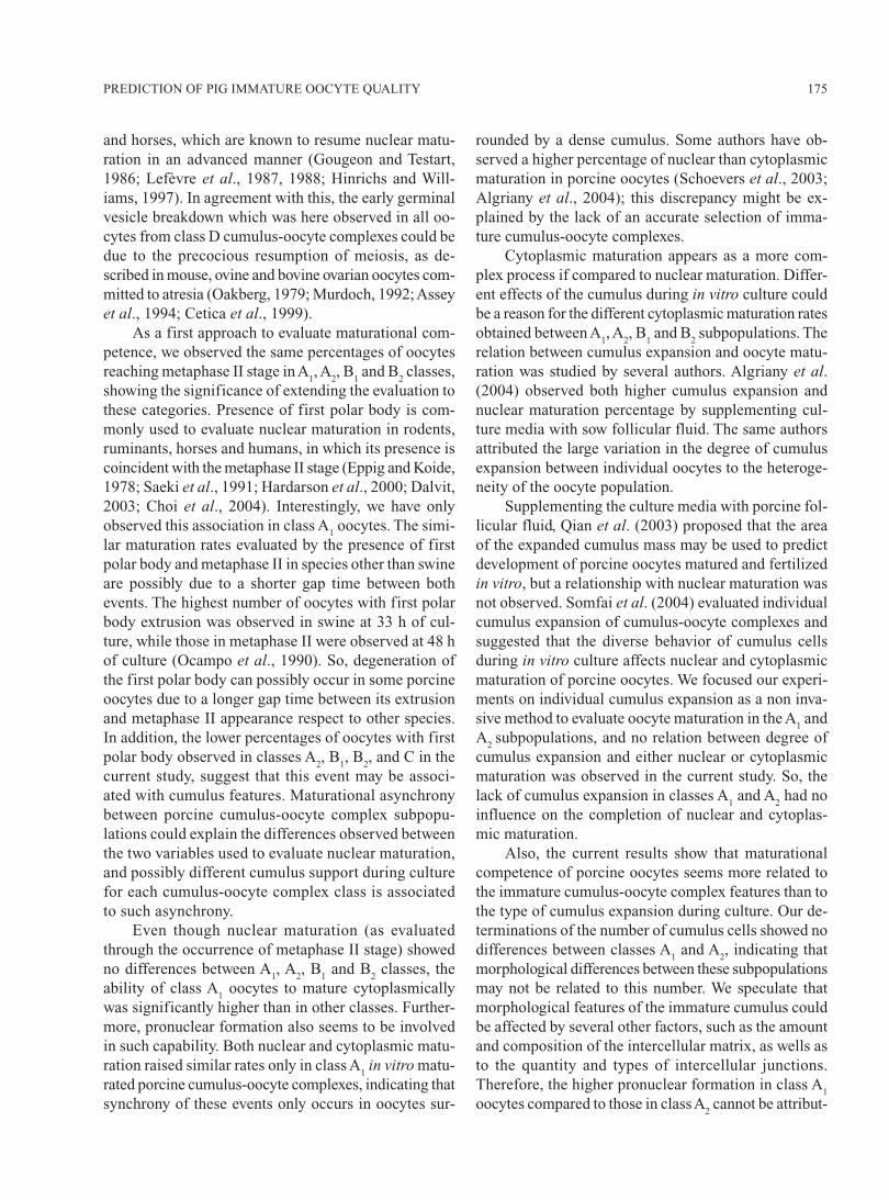
FIGURE 1. Morphological classes of immature cumulus-oocyte complexes recovered from gilt ovaries. A1:
oocytes surrounded by a dense cumulus. A2: oocytes surrounded by a translucent cumulus. B1: oocytes
surrounded by the corona radiata. B2: partly naked oocytes with some remaining cumulus cells. C: naked
oocytes. D: oocytes surrounded by a dark cumulus. Scale bar represents 50 μm.
169PREDICTION OF PIG IMMATURE OOCYTE QUALITY
Materials and Methods
Materials
Unless otherwise specified, all chemical used werepurchased from Sigma Chemical Company (St. Louis,MO, USA).
Recovery and classification of cumulus-oocyte com-plexes
Ovaries from slaughtered gilts were transported ina warm environment (28-33ºC) during the 2-3 h jour-ney to the laboratory. Ovaries were washed in 0.9% ClNacontaining 100000 IU/L penicillin and 100 mg/L strep-tomycin. Cumulus-oocyte complexes were aspiratedfrom 3-8 mm antral follicles using a 10 mL syringe andan 18-gauge needle, and were allotted to one of six dif-ferent classes according to morphological criteria un-der a stereoscopic microscope (Fig. 1): (A
1)
oocytes
surrounded by a dense cumulus; (A2)
oocytes surrounded
by a translucent cumulus; (B1)
oocytes surrounded by
the corona radiata; (B2) oocytes with some remaining
cumulus cells only; (C) naked oocytes; (D) oocytes sur-rounded by dark cumulus cells.
Oocyte denudation
Before staining, immature oocytes were denudedof somatic cells by vortex agitation during 1 min at 37ºC
in 3 g/L bovine serum albumin in a phosphate buffersalts medium (consisting of 136.9 mM NaCl, 2.7 mMKCl, 8.1 mM Na
2HPO
4, and 1.7 mM KH
2PO
4, pH 7.35-
7.65) and were then separated from the remaining cu-mulus cells with a Pasteur pipette.
In vitro matured cumulus-oocyte complexes wereincubated in 1 g/L hyaluronidase in the phosphate buffersalts medium for 5 min at 37ºC and then oocytes weredenuded by gentle pipetting.
Immature oocyte staining
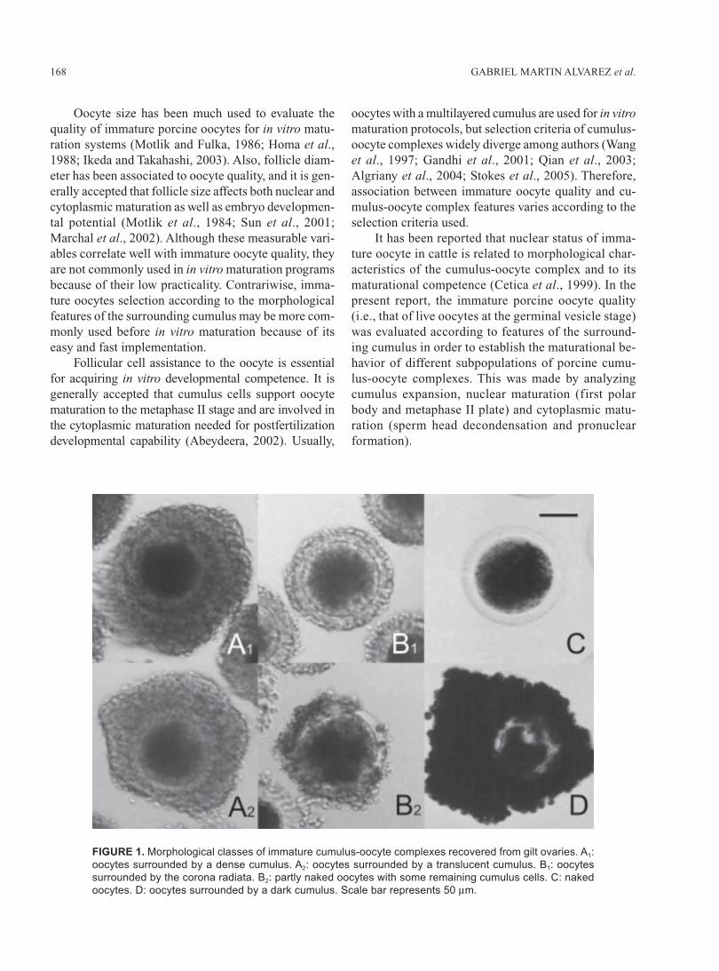
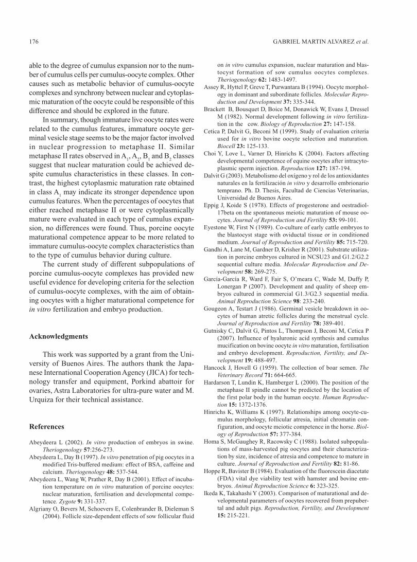
Once denuded, immature oocytes were divided intotwo groups to evaluate their vitality and nuclear stage.Oocyte vitality was assessed by incubation for 10 minat 37ºC in the phosphate buffer salts medium to which2.5 μg/L fluorescein diacetate fluorochrome and 2.5 g/L Trypan Blue were added. Oocytes were washed in thephosphate buffer salts medium before being observedin an epifluorescence microscope (Zeiss, Germany) us-ing a 510 nm filter at 100x magnification, live oocyteswere distinguished from dead ones based on their greenfluorescence (Hoppe and Bavister, 1984), while deadoocytes showed a characteristic blue staining underwhite light. The nuclear stage was determined as de-scribed by Cetica et al. (1999) with some modifications:briefly, denuded oocytes were centrifuged at 8200 g for30 min to polarize lipids and then incubated in 5 mg/LHoechst 33342 fluorochrome in the phosphate buffersalts medium for 30 min at 37ºC. After being washed in
FIGURE 2. A: Oocyte showing fluorescent nuclear material stained with Hoechst 33342 after lipid polariza-
tion. B: the same oocyte evaluated by Nomarsky differential-interferential contrast. Arrows indicate the
germinal vesicle. Scale bar represents 40 μm.
GABRIEL MARTIN ALVAREZ et al.170
the phosphate buffer salts medium, the oocytes wereobserved in an epifluorescence microscope using a 410nm filter (Luttmer and Longo, 1986) and in a Nomarskydifferential-interferential contrast microscope (Zeiss,Germany) at 100x and 400x magnification (Fig. 2).
Also, denuded oocytes were simultaneously incu-bated with fluorescein diacetate and Hoechst 33342fluorochromes for 30 min at 37ºC, and washed forstudying the association between oocyte viability andnuclear stage.
Number of cumulus cells in immature cumulus-oocytecomplexes
Immature cumulus-oocyte complexes were indi-vidually suspended in 2.5 g/L trypsine, 3.8 g/L EDTAand 3 g/L bovine serum albumin in phosphate buffermedium, and cumulus cells were separated by vortexagitation during 10 min at 37ºC. Cell concentration ofeach cumulus-oocyte complex was estimated using aNeubauer counting chamber.
In vitro oocyte maturation
Groups of 50 cumulus-oocyte complexes were cul-tured in 500 μL medium 199 (Earle’s salts, L-glutamine,2.2 mg/L sodium bicarbonate, GIBCO, Grand Island,NY, USA) supplemented with 10% (v/v) fetal bovineserum (GIBCO), 0.5 mg/L porcine follicle-stimulatinghormone (Folltropin-V, Bioniche, Belleville, Ontario,Canada), 0.5 mg/L porcine luteinizing hormone(Lutropin-V, Bioniche), 0.57 mM cysteine and 50 mg/Lgentamicin sulfate under mineral oil, at 39ºC for 48 hin a 5% CO
2 atmosphere (Abeydeera et al., 2001).
In vitro fertilization
It was carried out with fresh semen from a York-shire boar of proven fertility. Sperm rich fractions werecollected by the gloved hand method (Hancock andHovell, 1959). Sperm samples were washed twice inphosphate buffer salts medium with 3 g/L bovine se-rum albumin by centrifugation at 400 x g for 5 min andthen resuspended in fertilization modified Tris-bufferedmedium, consisted of 113.1 mM NaCl, 3 mM KCl, 10mM CaCl
2, 20 mM Tris, 11 mM glucose, 5 mM sodium
pyruvate, 4 g/L bovine serum albumin, 2.5 mM caffeineand 50 mg/L gentamicine sulfate (Abeydeera and Day,1997). Samples were filtered through a 20 mg glass woolcolumn (10 mm height, 4 mm diameter), previouslywashed with a modified Tris-buffered medium, in or-
der to obtain a live sperm rich fraction (Pereira et al.,2000). Matured cumulus-oocyte complexes were de-nuded by pipetting and inseminated to a final concen-tration of 5 x 10⁸/L spermatozoa, and coincubation wasperformed under mineral oil at 39ºC for 18 h in a 5%CO
2 atmosphere.One tenth of oocytes from each replicate were main-
tained throughout the fertilization procedure withoutexposure to sperm, to test for parthenogenesis.
Oocyte maturational criteria
For the evaluation of in vitro maturation, severalaspects were considered, such as degree of cumulusexpansion (in A
1 and A
2 classes only), nuclear matura-
tion (extrusion of first polar body and occurrence of ametaphase II plate), and cytoplasmic maturation (spermhead decondensation and pronuclear formation).
Cumulus-oocyte complexes in A1 and A
2 classes
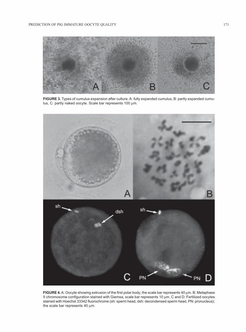
were classified according to their cumulus expansionas (1) fully expanded (widely expanded cumulus withloads of elastic intercellular matrix), (2) partly expanded(slightly expanded cumulus with scarce intercellularmatrix), and (3) partly naked ones (only some cumuluscells remained attached to the oocyte) (Fig. 3).
To assess the correlation between cumulus expan-sion and oocyte maturation, cumulus-oocyte complexeswere separated according to their cumulus expansionand then divided into two groups to evaluate meioticand cytoplasmic maturation.
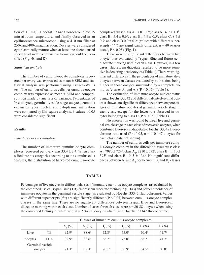
To evaluate the first polar body extrusion, in vitromatured oocytes were denuded, observed under a ste-reoscopic microscope at 40x magnification and con-firmed under an inverted microscope (Olympus IX70,Japan) at 400x magnification (Fig. 4A).
To determine the occurrence of metaphase II chro-mosome configuration, in vitro matured oocytes weredenuded, placed in a hypotonic medium of 10 g/L so-dium citrate at 37ºC for 15 min, fixed on a slide withCarnoy f ixing solution (3:1 ethanol:acetic acid)(Tarkowski, 1966), stained with 5% (v/v) Giemsa(Merck, Darmstadt, Germany) for 15 min and observedin a light microscope at 100x and 400x magnification(Fig. 4B). Only oocytes with condensed and well de-fined metaphase II chromosome configuration wereconsidered meiotically mature.
Cytoplasmic maturation was evaluated aftercoincubation of gametes for 18 h. Presumptively fertil-ized oocytes were freed from attached spermatozoa byrepetitive pipetting, fixed on a slide with Carnoy fixingsolution for at least 24 h, incubated in an aqueous solu-
171PREDICTION OF PIG IMMATURE OOCYTE QUALITY
FIGURE 3. Types of cumulus expansion after culture. A: fully expanded cumulus, B: partly expanded cumu-
lus, C: partly naked oocyte. Scale bar represents 100 μm.
FIGURE 4. A: Oocyte showing extrusion of the first polar body; the scale bar represents 40 μm. B: Metaphase
II chromosome configuration stained with Giemsa, scale bar represents 10 μm. C and D: Fertilized oocytes
stained with Hoechst 33342 fluorochrome (sh: sperm head, dsh: decondensed sperm head, PN: pronucleus);
the scale bar represents 40 μm.
GABRIEL MARTIN ALVAREZ et al.172
tion of 10 mg/L Hoechst 33342 fluorochrome for 15min at room temperature, and finally observed in anepifluorescence microscope using a 410 nm filter at250x and 400x magnification. Oocytes were consideredcytoplasmically mature when at least one decondensedsperm head and/or a pronuclear formation could be iden-tified (Fig. 4C and D).
Statistical analysis
The number of cumulus-oocyte complexes recov-ered per ovary was expressed as mean ± SEM and sta-tistical analysis was performed using Kruskal-Wallistest. The number of cumulus cells per cumulus-oocytecomplex was expressed as mean ± SEM and compari-son was made by analysis of variance. Percentages oflive oocytes, germinal vesicle stage oocytes, cumulusexpansion types, nuclear and cytoplasmic maturationwere compared by Chi-square analysis. P values < 0.05were considered significant.
Results
Immature oocyte evaluation
The number of immature cumulus-oocyte com-plexes recovered per ovary was 33.4 ± 2.4. When clas-sified into six categories according to the cumulus cellsfeatures, the distribution of harvested cumulus-oocyte
complexes was: class A1, 7.0 ± 1ab; class A
2, 6.7 ± 1.1b;
class B1, 5.4 ± 0.6b; class B
2, 4.9 ± 0.5b; class C, 8.7 ±
0.7a and class D 0.9 ± 0.2c (values with different super-scripts (a, b, c) are significantly different, n = 40 ovariestested, P < 0.05) (Fig. 1).
There were no significant differences between liveoocyte rates evaluated by Trypan Blue and fluoresceindiacetate marking within each class. However, in a fewcases, fluorescein diacetate resulted to be more sensi-tive in detecting dead oocytes (Table 1). There were sig-nificant differences in the percentages of immature aliveoocytes between classes evaluated by both stains, beinghigher in those oocytes surrounded by a complete cu-mulus (classes A
1 and A
2) (P < 0.05) (Table 1).
The evaluation of immature oocyte nuclear statususing Hoechst 33342 and differential-interferential con-trast showed no significant differences between percent-ages of immature oocytes at germinal vesicle stage ineach class, except for the lower rate observed in oo-cytes belonging to class D (P < 0.05) (Table 1).
No association was found between live and germi-nal vesicle stage in each class of recovered oocytes, whencombined fluorescein diacetate–Hoechst 33342 fluoro-chromes was used (P < 0.05, n = 110-147 oocytes foreach class, data not shown).
The number of cumulus cells per immature cumu-lus-oocyte complex in the different classes was: classA
1, 7080 ± 724a; class A
2, 7210 ± 572a; class B
1, 1110 ±
395b and class B2, 945 ± 136b. No significant differ-
ences between A1 and A
2 nor between B
1 and B
2 classes
TABLE 1.
Percentages of live oocytes in different classes of immature cumulus-oocyte complexes (as evaluated bythe combined use of Trypan Blue (TB)-fluorescein diacetate technique (FDA)) and percent incidence ofimmature oocytes in the germinal vesicle stage (as evaluated by Hoechst 33342 fluorochrome). Valueswith different superscripts (a,b,c) are significantly different (P < 0.05) between cumulus-oocyte complexclasses in the same line. There are no significant differences between Trypan Blue and fluoresceindiacetate marking within each class. Number of cases for each class were n = 80-88 oocytes when usingthe combined technique, while were n = 274-303 oocytes when using Hoechst 33342 fluorochrome.
Classes of immature cumulus-oocyte complexes
A1 (%) A
2 (%) B
1 (%) B
2 (%) C (%) D (%)
Live TB 92.9a 88.6a 72.8b 75.0b 70.4b 41.7c
oocytes FDA 92.9a 88.6a 66.7b 75.0b 66.7b 41.7c
Germinal vesicleoocytes 71.3a 68.3a 70.1a 66.9a 64.5a 50.0b
173PREDICTION OF PIG IMMATURE OOCYTE QUALITY
were observed, although A and B classes significantlydiffered in the number of cumulus cells (values withdifferent superscripts (a, b) are significantly different, n= 30 for each value, P < 0.05).
In vitro oocyte maturation
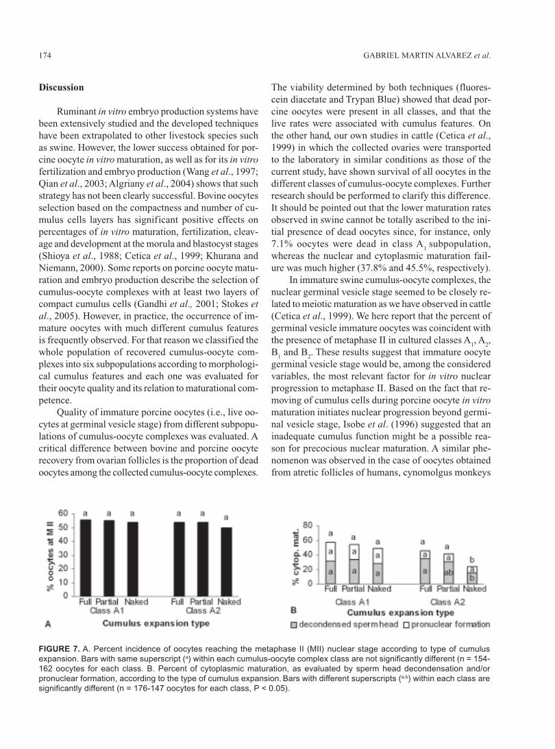
Two criteria were sequentially used to evaluatenuclear maturation in the same oocyte for each cumu-lus-oocyte complex class: extrusion of first polar bodyand development of the metaphase II chromosome con-figuration. The percent of oocytes that extruded theirfirst polar body was higher in class A
1 (P < 0.05). On
the other hand, the percent of oocytes that reachedmetaphase II were similar among the different classes,except for the lower rates obtained in classes C and D(P < 0.05). Nuclear maturation showed statistical asso-ciation between both nuclear maturation variables inclass A
1 only (P < 0.05) (Fig. 5).
decondensation rates were higher in both A1 and A
2
classes of cumulus-oocyte complexes (P < 0.05), pro-nuclear formation rates showed the highest value in cu-mulus-oocyte complexes belonging to class A
1 (P < 0.05)
(Fig. 6). The spontaneous parthenogenic activation ratesobserved at the pronuclear stage (18 h) were: 0% forclass A
1, 0% for class A
2, 0% for class B
1 and 5% for
class B2,
while those observed at the cleaved stage (48h), were: 0% for class A
1, 0% for class A
2, 10.5% for
class B1 and 5.3% for class B
2 (n = 18-20 for each value).
Cytoplasmic maturation analysis was performedonly in those classes of cumulus-oocyte complexes thatshowed significantly higher metaphase II rates (A
1, A
2,
B1 and B
2). The highest cytoplasmic maturation rate was
found in class A1 (54.5%, P < 0.05). While sperm head
Differential behavior of cumulus expansion duringmaturation could only be evaluated in cumulus-oocytecomplexes of classes A
1 and A
2 (Fig. 3). The distribu-
tion of matured cumulus-oocyte complexes among thethree different types of cumulus expansion was, for classA
1: 45.5% a fully expanded, 46.1% a partly expanded,
8.4% b partly naked, and for class A2: 46.9% a fully ex-
panded, 50.6% a partly expanded, 2.5% b partly naked(values with different superscripts (a, b) are significantlydifferent within each cumulus-oocyte complex class, n= 154-162 oocytes for each class, P < 0.05). When matu-ration was referred to the different types of cumulusexpansion, no differences were found in the percent-ages of oocytes that reached metaphase II or were cyto-plasmically mature, except in A
2 partly naked oocytes
in which cytoplasmic maturation was significantly lowerthan in the fully expanded A
2 cumulus-oocyte complexes
(P < 0.05) (Fig. 7).
FIGURE 5. Percentages of nuclear maturation in dif-
ferent cumulus-oocyte complex (COCs) classes, as
evaluated by extrusion of the 1st polar body (1st PB)
and the occurrence of metaphase II chromosome con-
figuration (M II). Different superscripts (a,b,c,d,e or A,B,C) on
bars indicate significant differences between cumulus-
oocyte complex classes evaluated by either extrusion
of 1st polar body or by metaphase II occurrence, re-
spectively (n = 84-100 oocytes for each class, P < 0.05).
Stars indicate significant differences in the incidence
of both phenomena within each cumulus-oocyte com-
FIGURE 6. Percentages of cytoplasmic maturation in
different cumulus-oocyte complex (COCs) classes, as
evaluated by sperm head decondensation and/or pro-
nuclear formation. Bars with different superscripts (a,b,c,d)
are significantly different (n = 176-187 oocytes for each
class, P < 0.05).
GABRIEL MARTIN ALVAREZ et al.174
Discussion
Ruminant in vitro embryo production systems havebeen extensively studied and the developed techniqueshave been extrapolated to other livestock species suchas swine. However, the lower success obtained for por-cine oocyte in vitro maturation, as well as for its in vitrofertilization and embryo production (Wang et al., 1997;Qian et al., 2003; Algriany et al., 2004) shows that suchstrategy has not been clearly successful. Bovine oocytesselection based on the compactness and number of cu-mulus cells layers has significant positive effects onpercentages of in vitro maturation, fertilization, cleav-age and development at the morula and blastocyst stages(Shioya et al., 1988; Cetica et al., 1999; Khurana andNiemann, 2000). Some reports on porcine oocyte matu-ration and embryo production describe the selection ofcumulus-oocyte complexes with at least two layers ofcompact cumulus cells (Gandhi et al., 2001; Stokes etal., 2005). However, in practice, the occurrence of im-mature oocytes with much different cumulus featuresis frequently observed. For that reason we classified thewhole population of recovered cumulus-oocyte com-plexes into six subpopulations according to morphologi-cal cumulus features and each one was evaluated fortheir oocyte quality and its relation to maturational com-petence.
Quality of immature porcine oocytes (i.e., live oo-cytes at germinal vesicle stage) from different subpopu-lations of cumulus-oocyte complexes was evaluated. Acritical difference between bovine and porcine oocyterecovery from ovarian follicles is the proportion of deadoocytes among the collected cumulus-oocyte complexes.
The viability determined by both techniques (fluores-cein diacetate and Trypan Blue) showed that dead por-cine oocytes were present in all classes, and that thelive rates were associated with cumulus features. Onthe other hand, our own studies in cattle (Cetica et al.,1999) in which the collected ovaries were transportedto the laboratory in similar conditions as those of thecurrent study, have shown survival of all oocytes in thedifferent classes of cumulus-oocyte complexes. Furtherresearch should be performed to clarify this difference.It should be pointed out that the lower maturation ratesobserved in swine cannot be totally ascribed to the ini-tial presence of dead oocytes since, for instance, only7.1% oocytes were dead in class A
1 subpopulation,
whereas the nuclear and cytoplasmic maturation fail-ure was much higher (37.8% and 45.5%, respectively).
In immature swine cumulus-oocyte complexes, thenuclear germinal vesicle stage seemed to be closely re-lated to meiotic maturation as we have observed in cattle(Cetica et al., 1999). We here report that the percent ofgerminal vesicle immature oocytes was coincident withthe presence of metaphase II in cultured classes A
1, A
2,
B1 and B
2. These results suggest that immature oocyte
germinal vesicle stage would be, among the consideredvariables, the most relevant factor for in vitro nuclearprogression to metaphase II. Based on the fact that re-moving of cumulus cells during porcine oocyte in vitromaturation initiates nuclear progression beyond germi-nal vesicle stage, Isobe et al. (1996) suggested that aninadequate cumulus function might be a possible rea-son for precocious nuclear maturation. A similar phe-nomenon was observed in the case of oocytes obtainedfrom atretic follicles of humans, cynomolgus monkeys
FIGURE 7. A. Percent incidence of oocytes reaching the metaphase II (MII) nuclear stage according to type of cumulus
expansion. Bars with same superscript (a) within each cumulus-oocyte complex class are not significantly different (n = 154-
162 oocytes for each class. B. Percent of cytoplasmic maturation, as evaluated by sperm head decondensation and/or
pronuclear formation, according to the type of cumulus expansion. Bars with different superscripts (a,b) within each class are
significantly different (n = 176-147 oocytes for each class, P < 0.05).
175PREDICTION OF PIG IMMATURE OOCYTE QUALITY
and horses, which are known to resume nuclear matu-ration in an advanced manner (Gougeon and Testart,1986; Lefèvre et al., 1987, 1988; Hinrichs and Will-iams, 1997). In agreement with this, the early germinalvesicle breakdown which was here observed in all oo-cytes from class D cumulus-oocyte complexes could bedue to the precocious resumption of meiosis, as de-scribed in mouse, ovine and bovine ovarian oocytes com-mitted to atresia (Oakberg, 1979; Murdoch, 1992; Asseyet al., 1994; Cetica et al., 1999).
As a first approach to evaluate maturational com-petence, we observed the same percentages of oocytesreaching metaphase II stage in A
1, A
2, B
1 and B
2 classes,
showing the significance of extending the evaluation tothese categories. Presence of first polar body is com-monly used to evaluate nuclear maturation in rodents,ruminants, horses and humans, in which its presence iscoincident with the metaphase II stage (Eppig and Koide,1978; Saeki et al., 1991; Hardarson et al., 2000; Dalvit,2003; Choi et al., 2004). Interestingly, we have onlyobserved this association in class A
1 oocytes. The simi-
lar maturation rates evaluated by the presence of firstpolar body and metaphase II in species other than swineare possibly due to a shorter gap time between bothevents. The highest number of oocytes with first polarbody extrusion was observed in swine at 33 h of cul-ture, while those in metaphase II were observed at 48 hof culture (Ocampo et al., 1990). So, degeneration ofthe first polar body can possibly occur in some porcineoocytes due to a longer gap time between its extrusionand metaphase II appearance respect to other species.In addition, the lower percentages of oocytes with firstpolar body observed in classes A
2, B
1, B
2, and C in the
current study, suggest that this event may be associ-ated with cumulus features. Maturational asynchronybetween porcine cumulus-oocyte complex subpopu-lations could explain the differences observed betweenthe two variables used to evaluate nuclear maturation,and possibly different cumulus support during culturefor each cumulus-oocyte complex class is associatedto such asynchrony.
Even though nuclear maturation (as evaluatedthrough the occurrence of metaphase II stage) showedno differences between A
1, A
2, B
1 and B
2 classes, the
ability of class A1 oocytes to mature cytoplasmically
was significantly higher than in other classes. Further-more, pronuclear formation also seems to be involvedin such capability. Both nuclear and cytoplasmic matu-ration raised similar rates only in class A
1 in vitro matu-
rated porcine cumulus-oocyte complexes, indicating thatsynchrony of these events only occurs in oocytes sur-
rounded by a dense cumulus. Some authors have ob-served a higher percentage of nuclear than cytoplasmicmaturation in porcine oocytes (Schoevers et al., 2003;Algriany et al., 2004); this discrepancy might be ex-plained by the lack of an accurate selection of imma-ture cumulus-oocyte complexes.
Cytoplasmic maturation appears as a more com-plex process if compared to nuclear maturation. Differ-ent effects of the cumulus during in vitro culture couldbe a reason for the different cytoplasmic maturation ratesobtained between A
1, A
2, B
1 and B
2 subpopulations. The
relation between cumulus expansion and oocyte matu-ration was studied by several authors. Algriany et al.(2004) observed both higher cumulus expansion andnuclear maturation percentage by supplementing cul-ture media with sow follicular fluid. The same authorsattributed the large variation in the degree of cumulusexpansion between individual oocytes to the heteroge-neity of the oocyte population.
Supplementing the culture media with porcine fol-licular fluid, Qian et al. (2003) proposed that the areaof the expanded cumulus mass may be used to predictdevelopment of porcine oocytes matured and fertilizedin vitro, but a relationship with nuclear maturation wasnot observed. Somfai et al. (2004) evaluated individualcumulus expansion of cumulus-oocyte complexes andsuggested that the diverse behavior of cumulus cellsduring in vitro culture affects nuclear and cytoplasmicmaturation of porcine oocytes. We focused our experi-ments on individual cumulus expansion as a non inva-sive method to evaluate oocyte maturation in the A
1 and
A2 subpopulations, and no relation between degree of
cumulus expansion and either nuclear or cytoplasmicmaturation was observed in the current study. So, thelack of cumulus expansion in classes A
1 and A
2 had no
influence on the completion of nuclear and cytoplas-mic maturation.
Also, the current results show that maturationalcompetence of porcine oocytes seems more related tothe immature cumulus-oocyte complex features than tothe type of cumulus expansion during culture. Our de-terminations of the number of cumulus cells showed nodifferences between classes A
1 and A
2, indicating that
morphological differences between these subpopulationsmay not be related to this number. We speculate thatmorphological features of the immature cumulus couldbe affected by several other factors, such as the amountand composition of the intercellular matrix, as wells asto the quantity and types of intercellular junctions.Therefore, the higher pronuclear formation in class A
1
oocytes compared to those in class A2 cannot be attribut-
GABRIEL MARTIN ALVAREZ et al.176
able to the degree of cumulus expansion nor to the num-ber of cumulus cells per cumulus-oocyte complex. Othercauses such as metabolic behavior of cumulus-oocytecomplexes and synchrony between nuclear and cytoplas-mic maturation of the oocyte could be responsible of thisdifference and should be explored in the future.
In summary, though immature live oocyte rates wererelated to the cumulus features, immature oocyte ger-minal vesicle stage seems to be the major factor involvedin nuclear progression to metaphase II. Similarmetaphase II rates observed in A
1, A
2, B
1 and B
2 classes
suggest that nuclear maturation could be achieved de-spite cumulus characteristics in these classes. In con-trast, the highest cytoplasmic maturation rate obtainedin class A
1 may indicate its stronger dependence upon
cumulus features. When the percentages of oocytes thateither reached metaphase II or were cytoplasmicallymature were evaluated in each type of cumulus expan-sion, no differences were found. Thus, porcine oocytematurational competence appear to be more related toimmature cumulus-oocyte complex characteristics thanto the type of cumulus behavior during culture.
The current study of different subpopulations ofporcine cumulus-oocyte complexes has provided newuseful evidence for developing criteria for the selectionof cumulus-oocyte complexes, with the aim of obtain-ing oocytes with a higher maturational competence forin vitro fertilization and embryo production.
Acknowledgments
This work was supported by a grant from the Uni-versity of Buenos Aires. The authors thank the Japa-nese International Cooperation Agency (JICA) for tech-nology transfer and equipment, Porkind abattoir forovaries, Astra Laboratories for ultra-pure water and M.Urquiza for their technical assistance.
References
Abeydeera L (2002). In vitro production of embryos in swine.Theriogenology 57:256-273.
Abeydeera L, Day B (1997). In vitro penetration of pig oocytes in amodified Tris-buffered medium: effect of BSA, caffeine andcalcium. Theriogenology 48: 537-544.
Abeydeera L, Wang W, Prather R, Day B (2001). Effect of incuba-tion temperature on in vitro maturation of porcine oocytes:nuclear maturation, fertilisation and developmental compe-tence. Zygote 9: 331-337.
Algriany O, Bevers M, Schoevers E, Colenbrander B, Dieleman S(2004). Follicle size-dependent effects of sow follicular fluid
on in vitro cumulus expansion, nuclear maturation and blas-tocyst formation of sow cumulus oocytes complexes.Theriogenology 62: 1483-1497.
Assey R, Hyttel P, Greve T, Purwantara B (1994). Oocyte morphol-ogy in dominant and subordinate follicles. Molecular Repro-duction and Development 37: 335-344.
Brackett B, Bousquet D, Boice M, Donawick W, Evans J, DresselM (1982). Normal development following in vitro fertiliza-tion in the cow. Biology of Reproduction 27: 147-158.
Cetica P, Dalvit G, Beconi M (1999). Study of evaluation criteriaused for in vitro bovine oocyte selection and maturation.Biocell 23: 125-133.
Choi Y, Love L, Varner D, Hinrichs K (2004). Factors affectingdevelopmental competence of equine oocytes after intracyto-plasmic sperm injection. Reproduction 127: 187-194.
Dalvit G (2003). Metabolismo del oxígeno y rol de los antioxidantesnaturales en la fertilización in vitro y desarrollo embrionariotemprano. Ph. D. Thesis, Facultad de Ciencias Veterinarias,Universidad de Buenos Aires.
Eppig J, Koide S (1978). Effects of progesterone and oestradiol-17beta on the spontaneous meiotic maturation of mouse oo-cytes. Journal of Reproduction and Fertility 53: 99-101.
Eyestone W, First N (1989). Co-culture of early cattle embryos tothe blastocyst stage with oviductal tissue or in conditionedmedium. Journal of Reproduction and Fertility 85: 715-720.
Gandhi A, Lane M, Gardner D, Krisher R (2001). Substrate utiliza-tion in porcine embryos cultured in NCSU23 and G1.2/G2.2sequential culture media. Molecular Reproduction and De-velopment 58: 269-275.
García-García R, Ward F, Fair S, O’meara C, Wade M, Duffy P,Lonergan P (2007). Development and quality of sheep em-bryos cultured in commercial G1.3/G2.3 sequential media.Animal Reproduction Science 98: 233-240.
Gougeon A, Testart J (1986). Germinal vesicle breakdown in oo-cytes of human atretic follicles during the menstrual cycle.Journal of Reproduction and Fertility 78: 389-401.
Gutnisky C, Dalvit G, Pintos L, Thompson J, Beconi M, Cetica P(2007). Influence of hyaluronic acid synthesis and cumulusmucification on bovine oocyte in vitro maturation, fertilisationand embryo development. Reproduction, Fertility, and De-velopment 19: 488-497.
Hancock J, Hovell G (1959). The collection of boar semen. TheVeterinary Record 71: 664-665.
Hardarson T, Lundin K, Hamberger L (2000). The position of themetaphase II spindle cannot be predicted by the location ofthe first polar body in the human oocyte. Human Reproduc-tion 15: 1372-1376.
Hinrichs K, Williams K (1997). Relationships among oocyte-cu-mulus morphology, follicular atresia, initial chromatin con-figuration, and oocyte meiotic competence in the horse. Biol-ogy of Reproduction 57: 377-384.
Homa S, McGaughey R, Racowsky C (1988). Isolated subpopula-tions of mass-harvested pig oocytes and their characteriza-tion by size, incidence of atresia and competence to mature inculture. Journal of Reproduction and Fertility 82: 81-86.
Hoppe R, Bavister B (1984). Evaluation of the fluorescein diacetate(FDA) vital dye viability test with hamster and bovine em-bryos. Animal Reproduction Science 6: 323-325.
Ikeda K, Takahashi Y (2003). Comparison of maturational and de-velopmental parameters of oocytes recovered from prepuber-tal and adult pigs. Reproduction, Fertility, and Development15: 215-221.
177PREDICTION OF PIG IMMATURE OOCYTE QUALITY
Isobe N, Fujihara M, Terada T (1996). Cumulus cells suppressmeiotic progression in pig oocytes cultured in vitro.Theriogenology 45: 1479-1489.
Khurana N, Niemann H (2000). Effects of oocyte quality, oxygentension, embryo density, cumulus cells and energy substrateson cleavage and morula/blastocyst formation of bovine em-bryos. Theriogenology 54: 741-756.
Kikuchi K, Kashiwazaki N, Noguchi J, Shimada A, Takahashi R,Hirabayashi M, Shino M, Ueda M, Kaneko H (1999). Devel-opmental competence, after transfer to recipients, of porcineoocytes matured, fertilized and cultured in vitro. Biology ofReproduction 60: 336-340.
Kikuchi K, Onishi A, Kashiwazaki N, Iwamoto M, Noguchi J,Kaneko H, Akita T, Nagai T (2002). Successful piglet pro-duction after transfer of blastocysts produced by a modifiedin vitro system. Biology of Reproduction 66: 1033-1041.
Krisher R (2004). The effect of oocyte quality on development. JAnim Sci 82 E-Suppl:E14-23.
Krisher R, Brad A, Herrick J, Sparman M, Swain J (2007). A com-parative analysis of metabolism and viability in porcine oo-cytes during in vitro maturation. Animal Reproduction Sci-ence 98: 72-96.
Lefèvre B, Gougeon A, Peronny H, Testart J (1988). Effect of cumu-lus cell mass and follicle quality on in-vitro maturation of cy-nomolgus monkey oocytes. Human Reproduction 3: 891-893.
Lefèvre B, Gougeon A, Testart J (1987). In-vitro oocyte matura-tion: some questions concerning the initiation and preventionof this process in humans. Human Reproduction 2: 495-497.
Luttmer S, Longo F (1986). Examination of living and fixed ga-metes and early embryos stained with supravital fluoro-chromes. Gamete Research 13: 267-283.
Macháty Z, Day B, Prather R (1998). Development of early por-cine embryos in vitro and in vivo. Biology of Reproduction59: 451-455.
Marchal R, Vigneron C, Perreau C, Bali-Papp A, Mermillod P (2002).Effect of follicular size on meiotic and developmental compe-tence of porcine oocytes. Theriogenology 57: 1523-1532.
Motlik J, Crozet N, Fulka J (1984). Meiotic competence in vitro ofpig oocytes isolated from early antral follicles. Journal ofReproduction and Fertility 72: 323-328.
Motlik J, Fulka J (1986). Factors affecting meiotic competence inpig oocytes. Theriogenology 25: 87-96.
Murdoch W (1992). Comparative morphometry and steroidogenicfunction of antral ovine follicles destined for ovulation or atre-sia. Domestic Animal Endocrinology 9: 219-224.
Oakberg E (1979). Follicular growth and atresia in the mouse. InVitro 15: 41-49.
Ocampo M, Ocampo L, Kanagawa H (1990). Timing of sequentialchanges in chromosome configurations during the 1st mei-otic division of pig oocytes cultured in vitro. The JapaneseJournal of Veterinary Research 38: 127-137.
Pereira R, Tuli R, Wallenhorst S, Holtz W (2000). The effect ofheparin, caffeine and calcium ionophore A23187 on in vitro
induction of the acrosome reaction in frozen-thawed bovineand caprine spermatozoa. Theriogenology 54: 185-192.
Qian Y, Shi W, Ding J, Sha J, Fan B (2003). Predictive value of thearea of expanded cumulus mass on development of porcineoocytes matured and fertilized in vitro. The Journal of Repro-duction and Development 49: 167-174.
Saeki K, Hoshi M, Leibfried-Rutledge M, First N (1991). In vitrofertilization and development of bovine oocytes matured inserum-free medium. Biology of Reproduction 44: 256-260.
Schoevers E, Kidson A, Verheijden J, Bevers M (2003). Effect offollicle-stimulating hormone on nuclear and cytoplasmic matu-ration of sow oocytes in vitro. Theriogenology 59: 2017-2028.
Shioya Y, Kuwayama M, Fukushima M, Iwasaki S, Hanada A(1988). In vitro fertilization and cleavage capability of bo-vine follicular oocytes classified by cumulus cells and ma-tured in vitro. Theriogenology 30: 489-496.
Somfai T, Kikuchi K, Onishi A, Iwamoto M, Fuchimoto D, Papp A,Sato E, Nagai T (2004). Relationship between the morpho-logical changes of somatic compartment and the kinetics ofnuclear and cytoplasmic maturation of oocytes during in vitromaturation of porcine follicular oocytes. Molecular Repro-duction and Development 68: 484-491.
Stokes P, Abeydeera L, Leese H (2005). Development of porcineembryos in vivo and vitro; evidence for embryo “cross talk”in vitro. Developmental Biology 284: 62-71.
Sun Q, Lai L, Bonk A, Prather R, Schatten H (2001). Cytoplas-mic changes in relation to nuclear maturation and early em-bryo developmental potential of porcine oocytes: effects ofgonadotropins, cumulus cells, follicular size, and protein syn-thesis inhibition. Molecular Reproduction and Development59: 192-198.
Sutton-Mcdowall M, Mitchell M, Cetica P, Dalvit G, Pantaleon M,Lane M, Gilchrist R, Thompson J (2006). Glucosamine supple-mentation during in vitro maturation inhibits subsequent em-bryo development: possible role of the hexosamine pathwayas a regulator of developmental competence. Biology of Re-production 74: 881-888.
Tarkowski A (1966). An air-drying method for chromosome prepa-rations from mouse eggs. Cytogenetics 5: 394-400.
Thompson J, Gardner D, Pugh P, Mcmillan W, Tervit H (1995).Lamb birth weight is affected by culture system utilized dur-ing in vitro pre-elongation development of ovine embryos.Biology of Reproduction 53: 1385-1391.
Wang W, Sun Q, Hosoe M, Shioya Y, Day B (1997). Quantifiedanalysis of cortical granule distribution and exocytosis ofporcine oocytes during meiotic maturation and activation.Biology of Reproduction 56: 1376-1382.
Yoshida M, Mizoguchi Y, Ishigaki K, Kogima T, Nagai T (1993).Birth of piglets derived from in vitro fertilization of pig oo-cytes matured in vitro. Theriogenology 39: 1303-1311.
Zhu S, Sun Z, Zhang J (2007). Ovine (Ovis aries) blastula from anin vitro production system and isolation of primary embry-onic stem cells. Zygote 15: 35-41.
