immature teeth with periradicular periodontitis or abscess ... · pdf fileimmature teeth with...
TRANSCRIPT

IAL
AFpssidaacwaao1
KAt
†
Se
MDaM0
Ed
Case Report/Clinical Techniques
J
mmature Teeth With Periradicular Periodontitis orbscess Undergoing Apexogenesis: A Paradigm Shift
ing-Huey Chueh, DDS, MS,* and George T.-J. Huang, DDS, MSD, DSc†
GbetpWCbdalcrpTaacebt
cwtiasm
C
thwTwpfhcmwwwr
bstractour clinical cases of immature teeth that developederiradicular periodontitis or abscess underwent a con-ervative treatment approach, i.e. without canal in-trumentation. Instead, only copious 2.5% NaOClrrigation was performed. All cases presented hereineveloped mature apices after 7 months to 5 yearsfter the initial treatment without complications,lthough narrowing canal space was observed. Ourlinical observations support a shifting paradigm to-ard a conservative approach by providing a favor-ble environment for tissue regeneration. The mech-nism of this continued development and formationf the root end is discussed. (J Endod 2006;32:205–1213)
ey Wordspexification, apexogenesis, calcium hydroxyde, imma-
ure teeth, NaOCl, open apex
From the *Elite Dental Clinic, Taipei, Taiwan; and theUniversity of Maryland, College of Dental Surgery, Dentalchool, Department of Endodontics, Prosthodontics and Op-rative Dentistry, Baltimore, Maryland.
Address requests for reprints to George T.-J. Huang, DDS,SD, DSc, University of Maryland, College of Dental Surgery,ental School, Department of Endodontics, Prosthodonticsnd Operative Dentistry, 666 West Baltimore St., Baltimore,D 21201. E-mail address: [email protected].
099-2399/$0 - see front matterCopyright © 2006 by the American Association of
ndodontists.oi:10.1016/j.joen.2006.07.010
p
OE — Volume 32, Number 12, December 2006
iven the right condition, many tissues are programmed for self-regeneration torestore the lost part. Pulp tissue in immature teeth with open apices has a rich
lood supply and contains a structure at developing stage that is more potent to regen-rate in response to damage. The general consensus for clinical treatment of immatureeeth with vital pulps is to preserve remaining normal vital tissue to allow continuedhysiological development and complete formation of the root end–apexogenesis.hereas for those teeth with nonvital pulps, it is to clean and fill the canals with
a(OH)2, the most commonly used material, to induce the formation of a calcifiedarrier at the open apex–apexification (1, 2). Teeth after successful apexogenesisevelop a normal thickness of dentin and root length. In contrast, those receivingpexification normally gain only an apical hard tissue bridge, not dentin, because of theoss of vital pulp tissues, odontoblasts, and Hertwig epithelial root sheath needed for theomplete root development. However, this paradigm has been challenged by recenteports showing convincingly that immature teeth clinically diagnosed with nonvitalulp and periradicular periodontitis or abscess can undergo apexogenesis (3, 4).hese reports stimulated a new perspective as to how we treat these cases. It has beendvocated that immature teeth should be treated as conservatively as practical to allowny possible apexogenesis to occur (5). Teeth inherit thin and weak roots after suc-essful apexification that are susceptible to fracture. Shifting apexification to apexogen-sis even for nonvital pulps with periradicular periodontitis or abscess is a clinicallyeneficial approach for patients if we gathered more clinical experience to help predict
he treatment outcome.Most interesting aspect of the reported cases (3, 4) is that those teeth showing
ontinual maturation of root and apex had developed extensive periradicular lesionsith sinus tract formation before the treatment; a condition normally resulting from
otal necrosis and infection of the pulp. Herein we further demonstrate four cases ofmmature teeth with periradicular periodontitis or abscess treated with conservativepproaches that led to continued development of normal apical morphology. The pos-ible mechanism underlying this clinical observation is discussed with published infor-ation regarding pulp healing after traumatic or experimentally induced injuries.
Case Reportase 1
A 10-year-old Asian girl, referred by a pediatric dentist, after presenting an asymp-omatic tooth #20 with a sinus tract on buccal gingiva (Fig. 1A). The general healthistory was noncontributory and periodontal probing was within normal limit. Thereas a fractured central cusp (dens evaginatus) that was likely the cause of the pathosis.he radiographic finding revealed periradicular radiolucency approximately 5 � 4 mmith a gutta-percha point tracing the sinus tract to tooth #20 (Fig. 1B). There was noercussion discomfort. Pulp vitality test was not performed as it gives unreliable results
rom immature teeth. Upon accessing the tooth without anesthesia, viable tissue withemorrhage was observed. The clinical diagnosis was partial pulp necrosis withhronic periradicular abscess. The pulp chamber was irrigated with approximately 20l of 2.5% NaOCl (Clorox, Oakland, CA), carefully dried with paper points and filledith Ca(OH)2 paste (powder mixed with saline, Merck, Frankfurt, Germany). Toothas sealed with Caviton (GC, Aichi, Japan) and IRM (Caulk Dentsply, Milford, DE). Twoeeks later, patient claimed that tooth was sensitive for 1 day after the first visit and
emained asymptomatic since. There was no hemorrhage upon re-entry and the same
rocedure was performed as in the previous appointment. Three months after the initialApexogenesis in Immature Teeth With Periradicular Periodontitis/Abscess 1205

taav(tpCiamm(
C
ugcwmgfnsEtdatpCat
tcss#appndtsccs
C
rpAdamCwsrcgw6s
Fo#
Case Report/Clinical Techniques
1
reatment, patient presented asymptomatic. Hard barrier was detectedfter removal of Ca(OH)2 paste. A new mixture of Ca(OH)2 was placednd the access closed with Caviton/IRM. Seven months after the firstisit, the tooth remained asymptomatic but still showed an open apexFig. 2A). The periapical radiograph made 11 months after the initialreatment (Fig. 2B) demonstrated a more narrowed root canal com-ared to that shown in Fig. 2A. Therefore, the IRM and part of theaviton were removed and replaced with amalgam. At 20 months after the
nitial treatment, the radiographic examination showed healing of the peri-pical bone and more reduction of the root canal space (Fig. 2C). At 35onths after the initial treatment, the radiographic examination revealed aarked reduction of the root canal space and maturation of the root apex
Fig. 2D).
ase 2
A 10-year-old Asian boy complained of toothache in the mandib-lar posterior region 4 days before the appointment and the conditionradually became worse with the development of swelling on the rightheek 2 days before. According to the patient’s statement, ice-packingas helpful in pain relief. The patient’s past medical history was unre-arkable. Intraoral examination revealed a swelling at the buccal gin-
iva and alveolar mucosa between teeth #28 and #29 and central cuspracture of tooth #29 (Fig. 3A). Tooth #29 showed a grade I mobility, aegative response to cold test and a sensitivity to palpation and percus-ion. Radiographic findings showed immature root with open apex.xtensive radiolucency was observed in the periradicular region ex-ending coronally on the mesial aspect of the root (Fig. 3B). The clinicaliagnosis was pulp necrosis with acute periradicular abscess. Withoutnesthesia, the tooth was accessed. Patient felt mild pain upon reachinghe pulp chamber and copious hemorrhage was noted (Fig. 3C). Aserformed in case 1, pulp chamber was irrigated with NaOCl, dried,a(OH)2 paste (Merck) placed into the pulp chamber and the canal,nd the access sealed with glass-ionomer cement. At the 4th week after
igure 1. Clinical photograph and periapical radiograph of case 1. A, Photographf a gutta-percha point into the sinus tract (arrow). The fractured central cusp20 with a wide open apex.
he initial visit and treatment, the tooth was asymptomatic and the soft 6
206 Chueh and Huang
issue had healed. Hemorrhage from the canal was observed upon ac-essing. The canal was again irrigated, Ca(OH)2 placed, and the accessealed as before. Periapical radiograph made after the second treatmenthowed partially healed periradicular bone with an open apex of tooth29 (Fig. 4A). At 8 weeks the patient returned asymptomatic. Afterccessing, calcified barrier was detected by probing with a periodontalrobe. Ca(OH)2 was refreshed and the tooth sealed. At 7 months theatient returned with no symptoms. Radiographic examination revealedearly complete maturation of the root apex and healing of the perira-icular bone except minor radiolucency around the apical bone, likely
he result of healed bone with less trabecular density. There was aignificant increase of hard tissue thickness of the root with reducedanal space. The coronal third of the canal appeared to be filled withalcified tissue (Fig. 4B). The tooth was then sealed with Caviton/Ketac-ilver. Patient did not return for subsequent follow-ups.
ase 3A 10-year-old Asian girl suffered from an acute toothache and was
eferred by a prosthodontist. The periapical radiograph brought by theatient showed a mesially tilted tooth #20 with an open apex (Fig. 5A).n emergency treatment, formocresol pulpotomy, was performed 9ays before at the referring clinic. Initial periapical radiograph madefter the emergency treatment revealed a radiolucent lesion (�3 � 4m) at the periradicular area of tooth #20 with an open apex (Fig. 5B).
linical diagnosis was chronic periradicular periodontitis of tooth #20ith possible partial necrosis of the pulp. Central cusp fracture was
uspected as the cause of pulp infection. After accessing, no hemor-hage was observed. The canal was irrigated with 2.5% NaOCl, medi-ated with Ca(OH)2 (Merck), and sealed with IRM. Periapical radio-raph taken after the initial treatment showed an open-apex tooth #20ith a radiolucent lesion at the periradicular area of tooth #20 (Fig.A). One month later, the periapical radiograph made before treatmenthowed a calcified barrier in the middle portion of the root canal (Fig.
ing a sinus tract at the alveolar mucosa between teeth #20 and #19 and insertionent. B, Radiograph showing a radiolucent lesion at the periapical area of tooth
showis evid
B). A firm mid-root stop was noted by probing. Fresh Ca(OH)2 was
JOE — Volume 32, Number 12, December 2006

pssmpdrNopmotttrac7tfit
C
ftfvdtcfamwsospmApt
FBbo
Case Report/Clinical Techniques
J
laced and the tooth was temporized with IRM. Another month later andubsequently every 2 to 3 months, Ca(OH)2 was refreshed and accessealed with Caviton/glass-ionomer cement. The periapical radiographade 4 months after the initial treatment showed healing of the peria-
ical bone (Fig. 6C) and that taken 10 months after the initial treatmentemonstrated a gradual hard-tissue deposition on the canal wall andeduction of root canal space at the apical half of the root (Fig. 6D).o endodontic procedure was made at this point (six treatments)nward. At the 11th month after the initial treatment, patient com-lained of sensitivity of the tooth while undertaking orthodontic treat-ent. Periapical radiograph made at that time showed a matured apex
f tooth #20 with a well-formed lamina dura (Fig. 7A). Further hard-issue deposition of canal wall and reduction of the root canal space athe apical half of the root was observed 13.5 months after the initialreatment (Fig. 7B). At 18.5 months after the initial treatment, there wasadiographic evidence of further deposition of calcified tissue at thepical half of the root canal (Fig. 7C), thus the coronal half of the rootanal and the coronal pulp chamber were filled with amalgam (Fig.D). At the 34th month after the initial treatment, the tooth was asymp-omatic with complete root formation and an almost completely calci-ied apical half of the root canal (Fig. 7E). Four and a half years after thenitial treatment, the tooth remained asymptomatic but the apical half of
igure 2. Follow-up periapical radiographs of case 1. A, Radiograph made 7 mo, Eleven months after the initial treatment demonstrating a slightly narrowed roone and more reduction of the root canal space. D, Thirty-five months after thef the root apex.
he root canal was completely calcified (Fig. 7F). a
OE — Volume 32, Number 12, December 2006
ase 4A 9-year-old Asian boy had a toothache in #29 for 5 days and went
or an emergency treatment, after which the tooth was left open. Pa-ient’s mother was told that an endodontic treatment was needed. At theirst endodontic appointment, periapical radiograph was made and re-ealed an open apex of tooth #29 without noticeable periradicular ra-iolucency (Fig. 8A). The tooth, without spontaneous pain, was sensi-
ive to percussion and the clinical diagnosis was pulp necrosis withhronic periradicular pathosis. The patient was apprehensive, there-ore, the test results may not reflect the actual tissue response. Withoutnesthesia, the rubber dam was placed and the canal irrigated with 40l 2.5% NaOCl. No instrumentation was performed, the canal was filledith Ca(OH)2, and the access sealed with IRM. Two weeks later, the
ame procedures were performed. A visit 5-weeks after the first end-dontic treatment, the same procedures were performed, and a firmtop at mid-root was detected. At 5 months, the same procedures wereerformed, and the periapical radiograph showed progressing apicalaturation of tooth #29 with an extension of the root length (Fig. 8B).
t 11, 17, 24 (Fig. 8C), and 32 months after the initial treatment, samerocedures were performed at each visit. Three years after the first
reatment, root formation was complete. The canal space was filled with
fter the initial treatment showing an asymptomatic tooth #20 with an open apex.al. C, Twenty months after the initial treatment showing healing of the periapicaltreatment revealing a market reduction of the root canal space and maturation
nths aot caninitial
malgam to the point of the calcified bridge (Fig. 8D). At 5 years there
Apexogenesis in Immature Teeth With Periradicular Periodontitis/Abscess 1207

w(d
vspcib
malfomw
mfrmBmmcrrtpBrc(ct
Fap rating
Case Report/Clinical Techniques
1
ere no symptoms, but evidence of severe narrowing of canal spaceFig. 8E). Intraoral examination revealed a sound tooth #29 withoutiscoloration (Fig. 8F).
DiscussionHere we reported four clinical cases, all of which have been con-
entionally considered as indications for apexification treatment. In-tead of utilizing endodontic files to clean and shape the infected andartially or totally necrotic pulp, these cases were treated with the mostonservative approach, namely no instrumentation of the canal besidesrrigation with 2.5% NaOCl. The treatment periods of these cases spanetween 1988 and 2000.
Iwaya et al. (3) reported an unconventional treatment of an im-ature permanent tooth (mandibular 2nd premolar) with periradicular
bscess and a sinus tract. The tooth was instrumented but not to its fullength because the patient felt the insertion of a smooth broach. In theirst visit the tooth was left open and at the following visits the canal wasnly irrigated with NaOCl and hydrogen peroxide without any instru-entation. Antimicrobial agents (metronidazol and ciprofloxacin)
igure 3. Clinical photographs and periapical radiograph of case 2. A, Photogrand #29 and central cusp fracture of tooth #29. B, Radiograph showing an imeriapical and mesial regions of the root of tooth #29. C, Photograph demonst
ere placed in the canal and at the next visit vital tissue was visualized 5 l
208 Chueh and Huang
m apical to the canal orifice. Thirty-five months later the root apexormation was complete along with thickened root structure as theesult of significantly reduced canal size compared to the adjacent nor-al first premolar. The tooth responded to electrical pulp test. Later,anchs and Trope (4) reported a case of similar condition but withore extensive periradicular bone loss. They applied a similar treat-ent procedure except some hemorrhage was induced to allow blood
lot formation in the canal. A remarkable result comparable to theeport by Iwaya et al. was also achieved. They also noted the positiveesponse to cold test at the recall. In both reports, the authors considerhat there was a regain of vascularization in the canal tissue, possiblyulp tissue. Although Iwaya et al. applied Ca(OH)2 at the 6th visit,anchs and Trope emphasized not to use Ca(OH)2 to preserve anyemaining viable pulp tissue and Hertwig epithelial root sheath. Ourlinical observations support this notion as demonstrated by cases 3Figs. 5–7) and 4 (Fig. 8) in which placement of Ca(OH)2 far down intoanals may have at least prevented the deposition of hard-tissue forma-ion in the coronal half of the canals.
In the traditional apexification treatment, apical barrier is estab-
vealing a swelling at the buccal gingiva and alveolar mucosa between teeth #28re root of tooth #29 with an open apex and an extensive radiolucency at thecopious hemorrhage upon accessing the pulp chamber.
ph rematu
ished by the formation of cementum-like tissue of various thicknesses.
JOE — Volume 32, Number 12, December 2006

Atsctfcdecrpradmt
c
bttrowt
muiwtccSbs
Fmr of the
Case Report/Clinical Techniques
J
recent review by Rafter (1) summarizes the histological characteris-ics reported in the literature. The hard tissue barrier has been de-cribed as a cap, bridge, or ingrown wedge that may be composed ofementum, dentin, bone, or so-called osteodentin that can deposit onhe inner walls of the canal (6 –11). Cementum formation can proceedrom the periphery of the apex towards the center in decreasing con-entric circles. In contrast to apexogenesis, apexification treatmentoes not generally lead to an additional formation of root dentin or anxtension of root length. The undesirable outcome is a weak root sus-eptible to fracture. Filling the canal from mid-root to coronal third withesin bonding to strengthen the root has been advocated after the com-letion of apexification (12–14). Conservative approach to reserve anyemaining vital pulp tissue may provide hope for a better outcome,lthough the control of root canal infection may be a difficult issue. Theuration of the infection, the involved microbial species, the host im-unity, and the size of the open apex all may theoretically play a role in
he outcome of this conservative treatment approach.In the present report, with an utmost conservative approach, all
igure 4. Periapical radiographs of case 2. A, After the second treatment showonths after the initial treatment showing complete maturation of the root apex
oot, a significant decrease of root canal space, and the calcified coronal third
ases showed noticeable apical maturation with increased root length d
OE — Volume 32, Number 12, December 2006
ut a significant narrowing of canal space. The question is, whether thehickened root was formed by pulp tissue from the remaining vital pulpissue at the apical region that was resistant to infection, capable ofegenerating the pulp tissue in the canal space and making new dentin;r the thickened root was formed by periodontal ligament (PDL) tissue,hich grew into the root canal from the apical foramen and deposited
he cementum onto the inner surface of the root dentin.Although lacking histological data from human specimens, one
ay speculate based on animal studies. Reports by Nevins et al. (15, 16)sing rhesus monkeys demonstrated that after total pulp tissue removal
n immature teeth, either treated with Ca(OH)2 or collagen gel, thereas cementum tissue formation at the apex and in the canal. PDL-like
issue can also be found in canal treated with collagen gel. PDL andementum tissues formed in the canal space can be verified histologi-ally by the presence of Sharpey’s fibers (Nevins, unpublished data).tudies by Ellis et al. (17) and Hitchcock et al., (18) revealed that whenlood supply is cut off in the middle of the root in monkeys, the canalpace is eventually replaced by cementum and PDL along the inner canal
artially healed periradicular bone and tooth #29 with an open apex. B, Sevenng of the periradicular bone, a significant increase of the calcified tissue in theroot canal.
ing p, heali
entinal wall, accompanied by some bone tissues. These tissues may
Apexogenesis in Immature Teeth With Periradicular Periodontitis/Abscess 1209

ecsdoc
ocTaod
FR on at
Ft4d
Case Report/Clinical Techniques
1
xtend into pulp chamber. Other animal studies focusing on thehanges in pulp tissue after replantation showed that various hard tis-ues including dentin, cementum, and bone may form in pulp spaceepending on the level of pulp recovery (11, 15, 19 –22). Therefore, ifne assumes the total loss of pulp tissue but remaining in a sterileondition, the outcome is the ingrowth of periodontal tissues. Evidence
igure 5. Periapical radiographs of case 3. A, Radiograph brought by the patieadiograph made after the formocresol pulpotomy revealing a radiolucent lesi
igure 6. Periapical radiographs of case 3. A, Radiograph taken right after threatment one month after initial treatment demonstrating a calcified barrier in
months after initial treatment showing a healing of the periapical bone. D
emonstrating a gradual deposition of calcified structure of the root and reduction o210 Chueh and Huang
f stem cells in PDL has recently been shown and the formation of newementum is characterized by the presence of Sharpey’s fibers (23).his possibility may explain the increased thickness of the canal wallnd the severe shrinkage of canal space. This deposition of cementumr bone in the canal may gradually and eventually obliterate the space asemonstrated in all four cases presented in the present report and those
the referring clinic showing a mesially tilted tooth #20 with an open apex. B,the periapical area of tooth #20 with an open apex.
l endodontic treatment of tooth #20. B, Radiograph made before the secondddle portion of the root canal. C, Radiograph made before the fourth treatmentiograph made before the sixth treatment 10 months after initial treatment
nt from
e initiathe mi
, Rad
f the root canal space at the apical half of the root.JOE — Volume 32, Number 12, December 2006
rTdmawat
s(Prcg
F#oarh
Case Report/Clinical Techniques
J
eported by Iwaya et al. (3) and Banchs and Trope (4). Tsukamoto-anaka et al. (24) observed hard tissue formation in rat dental pulpuring healing process after replantation. Using histochemical and im-unocytochemical approach, they identified specific cells and their
ctivities in the pulp and periodontal tissues. Tertiary dentin formationas observed by newly differentiated odontoblasts, whereas osteoclastsssociated with bone matrix in pulp were observed in cases where
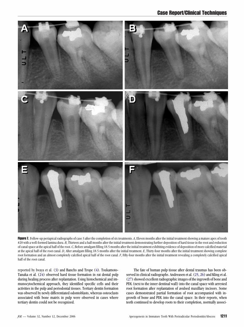
igure 7. Follow-up periapical radiographs of case 3 after the completion of six t20 with a well-formed lamina dura. B, Thirteen and a half months after the initif canal space at the apical half of the root. C, Before amalgam filling 18.5 montht the apical half of the root canal. D, After amalgam filling 18.5 months after thoot formation and an almost completely calcified apical half of the root canal.alf of the root canal.
ertiary dentin could not be recognized. t
OE — Volume 32, Number 12, December 2006
The fate of human pulp tissue after dental traumas has been ob-erved in clinical radiographs. Andreasen et al. (25, 26) and Kling et al.27) showed excellent radiographic images of the ingrowth of bone andDL (next to the inner dentinal wall) into the canal space with arrestedoot formation after replantation of avulsed maxillary incisors. Someases demonstrated partial formation of root accompanied with in-rowth of bone and PDL into the canal space. In their reports, when
ents. A, Eleven months after the initial treatment showing a mature apex of toothtment demonstrating further deposition of hard tissue in the root and reductionthe initial treatment exhibiting evidence of deposition of more calcified materiall treatment. E, Thirty-four months after the initial treatment showing complete
y-four months after the initial treatment revealing a completely calcified apical
reatmal treas aftere initiaF, Fift
eeth continued to develop roots to their completion, normally associ-
Apexogenesis in Immature Teeth With Periradicular Periodontitis/Abscess 1211
as
nefy
clpdnw
FnTft owing
Case Report/Clinical Techniques
1
ted with severe narrowing of canal space, the authors considered pulpurvival after the replantation.
Saad (28) reported a clinical case of a maxillary incisor withonvital pulp and with periradicular pathosis that underwent apexogen-sis after canal cleaning and shaping with NaOCl and instruments andilled with Ca(OH)2, although the canal appeared obliterated at 2.5
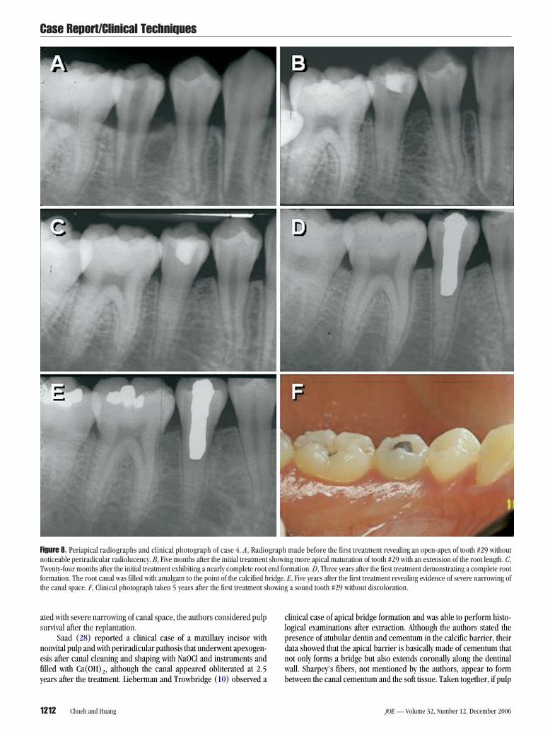
igure 8. Periapical radiographs and clinical photograph of case 4. A, Radiooticeable periradicular radiolucency. B, Five months after the initial treatmentwenty-four months after the initial treatment exhibiting a nearly complete rootormation. The root canal was filled with amalgam to the point of the calcified bhe canal space. F, Clinical photograph taken 5 years after the first treatment sh
ears after the treatment. Lieberman and Trowbridge (10) observed a b
212 Chueh and Huang
linical case of apical bridge formation and was able to perform histo-ogical examinations after extraction. Although the authors stated theresence of atubular dentin and cementum in the calcific barrier, theirata showed that the apical barrier is basically made of cementum thatot only forms a bridge but also extends coronally along the dentinalall. Sharpey’s fibers, not mentioned by the authors, appear to form
made before the first treatment revealing an open-apex of tooth #29 withoutng more apical maturation of tooth #29 with an extension of the root length. C,rmation. D, Three years after the first treatment demonstrating a complete rootE, Five years after the first treatment revealing evidence of severe narrowing ofa sound tooth #29 without discoloration.
graphshowiend foridge.
etween the canal cementum and the soft tissue. Taken together, if pulp
JOE — Volume 32, Number 12, December 2006

tPrt
dttlttttcsfpmOtwtoapttp(n
1
1
1
1
1
1
1
1
1
1
2
2
2
2
2
2
2
2
2
2
3
3
3
3
3
3
33
Case Report/Clinical Techniques
J
issue is totally lost, the canal space may be occupied by cementum,DL, and bone. In this situation, it is difficult to identify clinically viaadiographs because the canal space may well be PDL tissue and thehickened root structure be cementum.
On the other hand, if there was survived pulp tissue despite theevelopment of periradicular abscess because of the rich blood supplyhrough the wide open apex, apexogenesis could occur. Even in matureeeth, there may be remaining vital pulp tissues when a periradicularesion is developed (29, 30). Dental pulp stem cells have been identifiedo exist in permanent teeth (31–33). In the case of a developing tooth,he dental papilla at the apex may contain more stem cells than a matureooth and therefore possesses a greater potential to rebuild the lost pulpissue and continue the root maturation. If this is the case, using revas-ularization to describe this phenomenon does not encompass thecope of regeneration of pulp tissue in which genuine pulp containingunctional odontoblasts is capable of laying down new dentin to com-lete the root development. Pulp stem cells seeded onto existing dentinay differentiate into odontoblasts and may deposit new dentin (34, 35)r, the survived pulp tissue fragments may form dentin in the center of
he pulp and mixed with ingrown cementum (11, 19). In conclusion,hereas scientifically it is interesting to know the histological feature of
he tissues involved in the formation of the root after treatment, clinicallyur case reports shown here along with those by others strongly suggestparadigm shift in the clinical management of this type of cases, i.e.
rovide a favorable condition to allow natural tissue regeneration ratherhan replacement with artificial materials. Furthermore, a conservativereatment for mature teeth under this circumstance may even be aossibility in the future with advanced tissue engineering technologies35–37). Both basic and clinical research toward reaching this goal iseeded.
References1. Rafter M. Apexification: a review. Dent Traumatol 2005;21:1– 8.2. Goldstein S, Sedaghat-Zandi A, Greenberg M, Friedman S. Apexification & apexogen-
esis. N Y State Dent J 1999;65:23–5.3. Iwaya SI, Ikawa M, Kubota M. Revascularization of an immature permanent tooth
with apical periodontitis and sinus tract. Dent Traumatol 2001;17:185–7.4. Banchs F, Trope M. Revascularization of immature permanent teeth with apical
periodontitis: new treatment protocol? J Endod 2004;30:196 –200.5. Weisleder R, Benitez CR. Maturogenesis: is it a new concept? J Endod 2003;29:
776 – 8.6. Ghose LJ, Baghdady VS, Hikmat YM. Apexification of immature apices of pulpless
permanent anterior teeth with calcium hydroxide. J Endod 1987;13:285–90.7. Torneck CD, Smith JS, Grindall P. Biologic effects of endodontic procedures on
developing incisor teeth. IV. Effect of debridement procedures and calcium hydrox-ide-camphorated parachlorophenol paste in the treatment of experimentally in-duced pulp and periapical disease. Oral Surg Oral Med Oral Pathol 1973;35:541–54.
8. Steiner JC, Van Hassel HJ. Experimental root apexification in primates. Oral Surg OralMed Oral Pathol 1971;31:409 –15.
9. Walia T, Chawla HS, Gauba K. Management of wide open apices in non-vital perma-nent teeth with Ca(OH)2 paste. J Clin Pediatr Dent 2000;25:51– 6.
0. Lieberman J, Trowbridge H. Apical closure of nonvital permanent incisor teeth where
no treatment was performed: case report. J Endod 1983;9:257– 60.OE — Volume 32, Number 12, December 2006
1. Ritter AL, Ritter AV, Murrah V, Sigurdsson A, Trope M. Pulp revascularization ofreplanted immature dog teeth after treatment with minocycline and doxycyclineassessed by laser Doppler flowmetry, radiography, and histology. Dent Traumatol2004;20:75– 84.
2. Rabie G, Trope M, Garcia C, Tronstad L. Strengthening and restoration of immatureteeth with an acid-etch resin technique. Endod Dent Traumatol 1985;1:246 –56.
3. Pene JR, Nicholls JI, Harrington GW. Evaluation of fiber-composite laminate in the resto-ration of immature, nonvital maxillary central incisors. J Endod 2001;27:18–22.
4. Goldberg F, Kaplan A, Roitman M, Manfre S, Picca M. Reinforcing effect of a resinglass ionomer in the restoration of immature roots in vitro. Dent Traumatol2002;18:70 –2.
5. Nevins A, Finkelstein F, Laporta R, Borden BG. Induction of hard tissue into pulplessopen-apex teeth using collagen-calcium phosphate gel. J Endod 1978;4:76 – 81.
6. Nevins A, Wrobel W, Valachovic R, Finkelstein F. Hard tissue induction into pulplessopen-apex teeth using collagen-calcium phosphate gel. J Endod 1977;3:431–3.
7. Ellis E, 3rd, Cox CF, Hitchcock R, Baker J. Vital apicoectomy of the teeth: a 1– 4 weekhistopathological study in Macaca mulatta. J Oral Pathol 1985;14:718 –32.
8. Hitchcock R, Ellis E, 3rd, Cox CF, Intentional vital root transection: a 52-week his-topathologic study in Macaca mulatta. Oral Surg Oral Med Oral Pathol 1985;60:2–14.
9. Nevins AJ, Finkelstein F, Borden BG, Laporta R. Revitalization of pulpless open apex teethin rhesus monkeys, using collagen-calcium phosphate gel. J Endod 1976;2:159–65.
0. Skoglund A, Tronstad L. Pulpal changes in replanted and autotransplanted immatureteeth of dogs. J Endod 1981;7:309 –16.
1. Sheppard PR, Burich RL. Effects of extra-oral exposure and multiple avulsions onrevascularization of reimplanted teeth in dogs. J Dent Res 1980;59:140.
2. Kvinnsland I, Heyeraas KJ. Dentin and osteodentin matrix formation in apicoecto-mized replanted incisors in cats. Acta Odontol Scand 1989;47:41–52.
3. Seo BM, Miura M, Gronthos S, et al. Investigation of multipotent postnatal stem cellsfrom human periodontal ligament. Lancet 2004;364:149 –55.
4. Tsukamoto-Tanaka H, Ikegame M, Takagi R, Harada H, Ohshima H. Histochemicaland immunocytochemical study of hard tissue formation in dental pulp during thehealing process in rat molars after tooth replantation. Cell Tissue Res 2006;325:219 –29.
5. Andreasen JO, Borum MK, Jacobsen HL, Andreasen FM. Replantation of 400 avulsedpermanent incisors. 1. Diagnosis of healing complications. Endod Dent Traumatol1995;11:51– 8.
6. Andreasen JO, Borum MK, Jacobsen HL, Andreasen FM. Replantation of 400 avulsedpermanent incisors. 2. Factors related to pulpal healing. Endod Dent Traumatol1995;11:59 – 68.
7. Kling M, Cvek M, Mejare I. Rate and predictability of pulp revascularization in ther-apeutically reimplanted permanent incisors. Endod Dent Traumatol 1986;2:83–9.
8. Saad AY. Calcium hydroxide and apexogenesis. Oral Surg Oral Med Oral Pathol1988;66:499 –501.
9. Lin LM, Skribner J. Why teeth associated with inflammatory periapical lesions canhave a vital response. Clin Prev Dent 1990;12:3– 4.
0. Lin L, Shovlin F, Skribner J, Langeland K. Pulp biopsies from the teeth associated withperiapical radiolucency. J Endod 1984;10:436 – 48.
1. Gronthos S, Brahim J, Li W, et al. Stem cell properties of human dental pulp stemcells. J Dent Res 2002;81:531–5.
2. Shi S, Gronthos S. Perivascular niche of postnatal mesenchymal stem cells in humanbone marrow and dental pulp. J Bone Miner Res 2003;18:696 –704.
3. Gronthos S, Mankani M, Brahim J, Robey PG, Shi S. Postnatal human dental pulp stemcells (DPSCs) in vitro and in vivo. Proc Natl Acad Sci USA 2000;97:13625–30.
4. Batouli S, Miura M, Brahim J, et al. Comparison of stem-cell-mediated osteogenesisand dentinogenesis. J Dent Res 2003;82:976 – 81.
5. Huang GT, Sonoyama W, Chen J, Park SH. In vitro characterization of human dentalpulp cells: various isolation methods and culturing environments. Cell Tissue Res2006;324:225–36.
6. Nakashima M. Tissue engineering in endodontics. Aust Endod J 2005;31:111–3.7. Nakashima M, Akamine A. The application of tissue engineering to regeneration of
pulp and dentin in endodontics. J Endod 2005;31:711– 8.
Apexogenesis in Immature Teeth With Periradicular Periodontitis/Abscess 1213