immediate care of traumatic brain injury & subarachnoid ... · pdf fileimmediate care of...
TRANSCRIPT

Immediate Care of Traumatic Brain Injury & Immediate Care of Traumatic Brain Injury & Subarachnoid HaemorrhageSubarachnoid Haemorrhage
Paul HarrisonConsultant Anaesthetist
December 2011

Learning ObjectivesLearning Objectives
• Background information about TBI / SAH• Basic principles of immediate
management• Neurophysiology• Practical aspects of immediate
management• Further management of TBI / SAH

Traumatic Brain InjuryTraumatic Brain Injury
• Major cause of – Disability– Death– Economic cost

Traumatic Brain InjuryTraumatic Brain Injury
• Recent study in Scotland estimated an annual incidence of between 100 and 150 per 100,000 adults disabled a year after a head injury
• 47% of patients followed up for one year after discharge had survived with some form of restriction to lifestyle
• Proportion of patients experiencing the most serious sequelae did not vary according to the severity of the initial injury

Traumatic Brain InjuryTraumatic Brain Injury
• About 80% male
• About 15% elderly (≥ 65 years)
• About 40 – 50% children
• Alcohol may be involved in up to 65% of adult head injuries
Traumatic Brain InjuryTraumatic Brain Injury
• Common causes of minor head injury– Falls (22-43%) – Assaults (30-50%)– Road traffic accidents (~25%)
• RTA’s account for a far greater proportion of moderate to severe head injuries

InvestigationInvestigation
• Who should have a CT scan– The patient is eye opening only to pain or does not
converse (CGS 12/15 or less)– A deteriorating level of consciousness or progressive focal
neurological signs– Confusion or drowsiness (CGS 13 or 14/15) followed by
failure to improve within at most four hours of clinical observation
– Radiological/clinical evidence of a fracture, whatever the level of consciousness
– New focal neurological signs which are not getting worse– Full consciousness (GCS 15/15) with no fracture but other
features, e.g.:• Severe and persistent headache• Nausea and vomiting• Irritability or altered behaviour• A seizure.

Traumatic Brain InjuryTraumatic Brain Injury
• Types of injury– Impact injury
• Contusions/lacerations• Diffuse axonal injury
– Bleeding• Extradural• Subdural• Intracerebral (usually into contusions)
Traumatic Brain InjuryTraumatic Brain Injury
• Once injury has occurred, damage cannot be altered
• However, secondary brain damage starts to occur– Swelling– Ischaemia– (Infection)

Traumatic Brain InjuryTraumatic Brain Injury
• Once head injury has occurred, management is aimed at avoiding secondary injury
• GOOD SPECIFIC CARE IS ESSENTIAL.
Traumatic Brain InjuryTraumatic Brain Injury
• Outcome determined by – Severity of initial injury– Prevention / management of subsequent
complications
• Outcome poorer with increasing age
• Young children have greatest capacity for recovery
Subarachnoid HaemorrhageSubarachnoid Haemorrhage
• Traumatic
• Spontaneous– Rupture of intracranial aneurysm (70 – 75%)– Bleeding from arterio-venous malformation
(5%)– Others/undefined (20%)

Aneurysmal SAHAneurysmal SAH
• Incidence 5 – 30 per 100,000 population per year
• Peak age 40 – 60 years (rare in childhood)
• Female:male ratio 3:2

AneurysmalAneurysmal SAHSAH
• Predisposing factors– Hypertension– Smoking– Cocaine abuse– Alcohol
• Especially binge drinking– Race
• Black > white• High incidence in Finland & Japan, low in
South/Central America– Other conditions
• Autosomal dominant polycystic kidney disease• Sickle cell disease• Connective tissue disorders
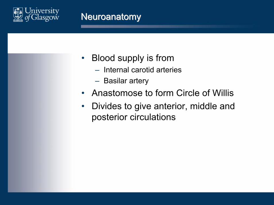
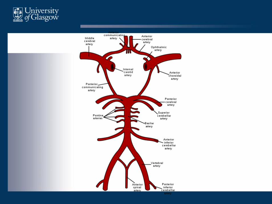
NeuroanatomyNeuroanatomy
• Blood supply is from– Internal carotid arteries– Basilar artery
• Anastomose to form Circle of Willis• Divides to give anterior, middle and
posterior circulations
AneurysmalAneurysmal SAHSAH
• Aneurysms are usually saccular and occur at the bifurcation of vessels
• Site of aneurysm– Anterior cerebral artery 35 – 40%– Middle cerebral artery 20 – 25%– Posterior circulation 10%– Internal carotid 30%
• Approximately 30% have > 1 aneurysm
• At post-mortem 1% of population have aneurysm
AneurysmalAneurysmal SAHSAH
• Presentation
• Classic– Instantaneous severe headache– “Thunderclap headache” or “Blow to the head”– Nausea/vomiting– Neck stiffness/photophobia– Seizure– Reduced conscious level– Focal signs e.g. IIIrd nerve palsy
• Presentation may not fit classical picture

AneurysmalAneurysmal SAHSAH
• 1/3 patients will die before reaching hospital
• Remaining patients have 30 – 50% mortality
• 1/3 of survivors remain dependent

AneurysmalAneurysmal SAHSAH
• Prognosis depends on– Severity of initial bleed– Prevention of rebleeding
• Success of procedure to secure aneurysm– Management/prevention of complications
• Secondary injury• Hydrocephalus• Vasospasm
AneurysmalAneurysmal SAHSAH
• Prone to cardiac problems
• SAH associated with release of large amounts of catecholamines
• Can lead to– Cardiac arrhythmias / ECG changes– Myocardial infarction– Neurogenic pulmonary oedema– Cardiac arrest
Immediate Care of Neurosurgical EmergencyImmediate Care of Neurosurgical Emergency
• Cannot alter primary event
• Good medical care essential to prevent secondary damage

Immediate Care of Neurosurgical EmergencyImmediate Care of Neurosurgical Emergency
• Initial management should be based on ATLS/ALS– Airway– Breathing– Circulation
• Not specific enough for TBI/SAH

Immediate Care of Neurosurgical EmergencyImmediate Care of Neurosurgical Emergency
• Some animal evidence of efficacy
• No clinical evidence in humans– Large randomised control trial (CRASH trial)
suggested corticosteroids worsen outcome
• No agents currently in use as neuroprotective agents
Immediate Care of Neurosurgical EmergencyImmediate Care of Neurosurgical Emergency
• Physiological neuroprotection– O2
– CO2
– Blood pressure– Intracranial pressure– Temperature– Blood glucose

Immediate Care of Neurosurgical EmergencyImmediate Care of Neurosurgical Emergency
• Need to know basic knowledge of neurophysiology!

NeurophysiologyNeurophysiology
• Brain weighs ~ 2% of body weight
• Receives ~ 15% of cardiac output
• 20% of basal O2 consumption
• CBF=50ml/100g/min
NeurophysiologyNeurophysiology
• Blood flow affected by several factors• Local metabolites
– H+, K+ , adenosine, NO• Other factors
– CO2 & O2– Cerebral perfusion pressure– Drugs– Temperature– Haematocrit

NeurophysiologyNeurophysiology
• Why does a small amount of swelling / bleeding in the brain cause such problems?

NeurophysiologyNeurophysiology
• Monro-Kellie Doctrine
– The skull is a rigid closed container
– Contents are incompressible (brain, blood, CSF)
– Any increase in the volume of one constituent results in a corresponding increase in ICP
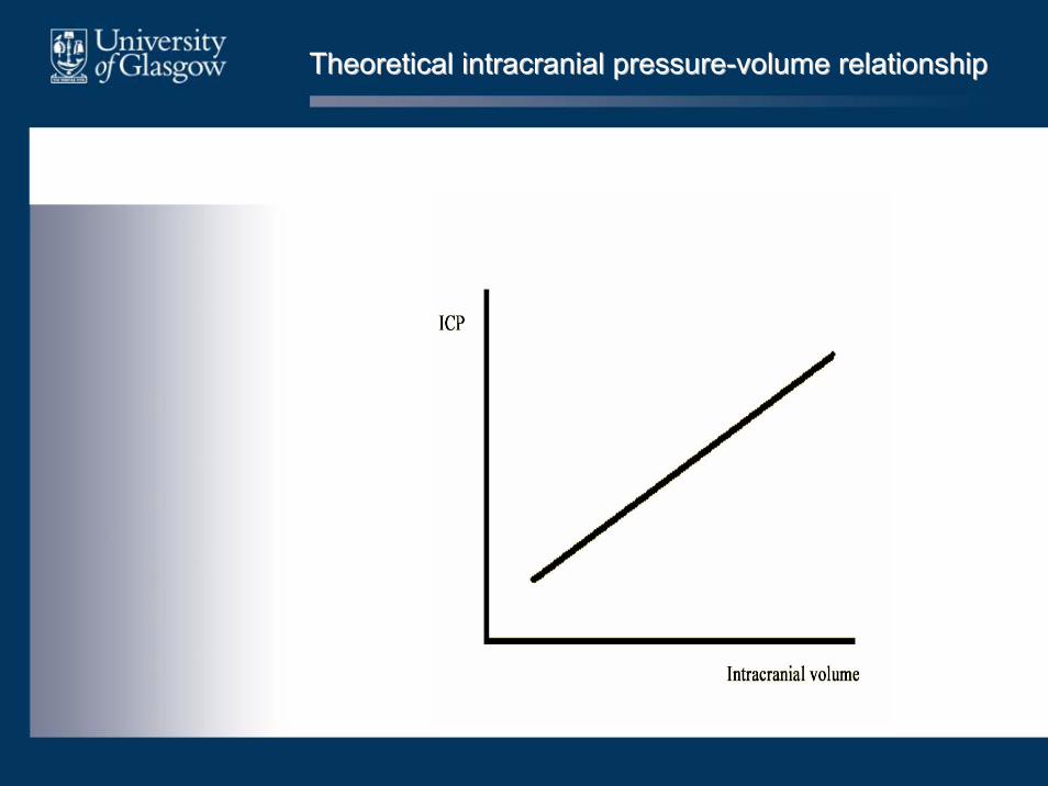
Theoretical intracranial pressureTheoretical intracranial pressure--volume relationshipvolume relationship

Actual intracranial pressureActual intracranial pressure--volume relationshipvolume relationship

Rising ICPRising ICP
• Some compensation occurs
• Veins, especially dural sinuses act as capacitance vessels
• Blood shunted to central circulation
• Also some movement of CSF

Rising ICPRising ICP
• If cerebral blood flow , cerebral blood volume
• If no compensation can occur this results in ICP
Rising ICPRising ICP
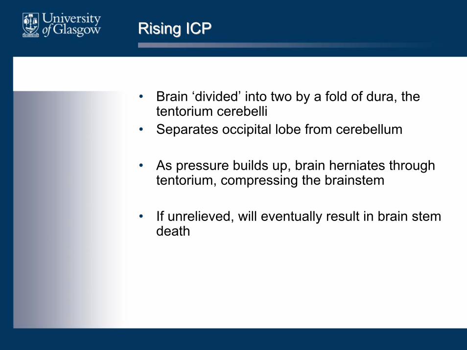
• Brain ‘divided’ into two by a fold of dura, the tentorium cerebelli
• Separates occipital lobe from cerebellum
• As pressure builds up, brain herniates through tentorium, compressing the brainstem
• If unrelieved, will eventually result in brain stem death
Rising ICPRising ICP
• As pressure builds up, the oculomotornerve is compressed, resulting in a fixed dilated pupil
Autoregulation
• Cerebral Blood Flow (CBF) normally constant despite changes in MAP

AutoregulationAutoregulation

AutoregulationAutoregulation
• Autoregulation maintains CBF constant despite changes in cerebral perfusion pressure
CPP = MAP CPP = MAP –– ICPICP

AutoregulationAutoregulation
• Occurs due to changes in cerebrovascularresistance
• Curve can be shifted to left or right e.g. hypertension, chronically raised ICP
• AUTOREGULATION IS IMPAIRED BY CNS PATHOLOGY e.g. Traumatic Brain Injury

Effect of OEffect of O22

Effect of OEffect of O22
• Little effect on CBF until pO2 falls to about 7 – 8 kPa
• Hyperoxia (>60 kPa) causes mild vasoconstriction

Effect of COEffect of CO22
Effect of COEffect of CO22
• Four fold increase in CBF between 2.7 kPa and 10.7 kPa
• Almost linear relationship in clinical range
Effect of COEffect of CO22
• Moderate hyperventilation may be useful to reduce ICP in the short term
• After about 24 – 48 hours cerebral blood flow returns to normal
• Return to normocapnia after hyperventilation results in overshoot in CBF
HaematocritHaematocrit
• O2 delivery is a compromise between – oxygen carrying capacity– flow characteristics of blood
• Haemodilution may improve cerebral blood flow in vasospasm associated with SAH

TemperatureTemperature
• Animal evidence that reducing body temperature after brain injury may improve outcome
• Evidence in humans that patients with normothermia after TBI may have a better outcome than those with hyperthermia
• No evidence that ‘therapeutic’ hypothermia improves outcome

TemperatureTemperature
• Need to treat hyperthermia aggressively
• Hypothermia associated with complications– Systemic vascular resistance / myocardial
work– Coagulopathy
• Aim for normal body temperature (36 – 37°C)

GlucoseGlucose
• Brain is almost totally dependent on exogenous glucose for its cellular energy requirements
• Prolonged hypoglycaemia causes neuronal damage
• Hyperglycaemia is associated with poorer outcome in TBI & SAH

GlucoseGlucose
• Mechanism unclear– In glycolysis, Pyruvate Lactate– Lactate is neurotoxic
– In areas where partial ischaemia exists, high glucose levels may allow lactate to build up to very high levels
• No evidence that treating hyperglycaemia improves outcome, but seems sensible

ManagementManagement
• Maintain normality!• Ensure optimal gas exchange
– May require intubation / ventilation– Avoid hypoxia, (PaO2 > 13kPa)– Keep C02 normal (PaCO2 4.5 – 5.0kPa)
• Avoid hypotension– Treat hypotension promptly
• Fluid resuscitation• Vasopressors/inotropes• Treat other sources of bleeding promptly e.g.
ruptured spleen• Aim MAP > 80mmHg
ManagementManagement
• Reduce ICP– Head up tilt– Avoid compression of neck veins– If intubated, keep sedated & paralysed– Consider mannitol / hypertonic saline
• Avoid hyperthermia
• Avoid hyperglycaemia

ManagementManagement
• Mannitol– Viscosity causes flow resulting in cerebral
blood volume causing ICP– Osmotic effect – decreased brain water
• Useful to ‘buy time’ until definitive treatment
Further ManagementFurther Management
• Traumatic Brain Injury– Evacuate haematomas causing increased ICP– Consider evacuation of contusions– If swelling likely, consider measuring ICP
• If ICP high with no surgical lesions– Medical management
• Sedation ( CMRO2, CBF, ICP), muscle relaxation, ventilation
– Decompressive craniotomy

Further ManagementFurther Management
• Subarachnoid haemorrhage– Prevent rebleeding
• Endovascular coiling• Craniotomy & clipping
– Prevent/treat vasospasm• Nimodipine 4 hourly for 21 days• Hypertension, hypervolaemia & haemodilution
– Treat hydrocephalus• External ventricular drain• Ventriculo-peritoneal shunt• Endoscopic IIIrd ventriculostomy
References / Further ReadingReferences / Further Reading
• Scottish Intercollegiate Guidelines Network (SIGN) Guideline 46: Early management of patients with a head injury. Edinburgh 2000. Available at http://www.sign.ac.uk/guidelines/fulltext/46/index.html
• Association of Anaesthetists of Great Britain and Ireland (AAGBI). Recommendations for the Safe Transfer of Patients with Brain Injury. London 2006. Available at http://www.aagbi.org/publications/guidelines/docs/braininjury.pdf
• National Institute for Health and Clinical Excellence (NICE) CG 56 Head Injury: Triage, assessment, investigation and early management of head injury in infants, children and adults. HMSO 2007. Available at http://www.nice.org.uk/Guidance/CG56/Guidance/pdf/English
• Brain Trauma Foundation. MR Bullock, JT Povlishock (editors). Guidelines for the Management of Severe Traumatic Brain Injury. Journal of Neurotrauma 24 (supplement 1) 2007
• Thornhill S, Teasdale GM, Murray GD, McEwen J, Roy CW, Penny KI. Disability in young people and adults one year after head injury: prospective cohort study. BMJ 2000, 320(7250):1631-5
• Haboubi NH, Long J, Koshy M, Ward AB. Short-term sequelae of minor head injury (6 years experience of minor head injury clinic). Disability and Rehabilitation 2001, 23(14):635-8
• Lindsay KW, Bone I. Neurology and Neurosurgery Illustrated (4th edition). London: Churchill Livingstone 2004• Matta BF, Menon DK, Turner JM. Textbook of Neuroanaesthesia and Critical Care. London: Greenwich Medical
Media 2000• Bendo AA, Kass IS, Hartung J Cottrell JE. Chapter 27 Anesthesia for Neurosurgery. In: Barash, Paul G.; Cullen,
Bruce F.; Stoelting, Robert K (editors). Clinical Anesthesia (5th Edition). Philadelphia: Lippincott Williams & Wilkins 2006. Available at Books@Ovid