immediate outcome and hospital universiti …eprints.usm.my/36357/1/dr._che_hadibiah_che_mohd... ·...
TRANSCRIPT

IMMEDIATE OUTCOME OF MECHANICALLY VENTILATED
TERM BABIES IN HOSPITAL KOTA BHARU AND HOSPITAL UNIVERSITI SAINS MALAYSIA
by
DR CHE HADIBIAH BT CHE MOHD RAZALI
Dissertation Submitted In Partial Fulfillment Of The
Requirements For The Degree Of Master Of Medicine
(Paediatrics)
UNIVERSITI SAINS MALAYSIA 2002

ACKNOWLEDGEMENTS
I would like to express my special thanks and deepest gratitude to my supervisor,
Dr.Nik Zainal Abidin Nik Ismail (Head of Paediatric department) for his advice,
correcting and giving me encouragement in the preparation of this dissertation.
I would also like to thank Dr Rus Anida Awang, Consultant Pediatrician of Hospital
Kota Bharu who was involved during initial preparation of this dissertation, Dr
Norazwany Yaacob and Dr Ariffin Nasir for helping me in statistical analysis.
My deepest gratitude also goes to my family who always give me their
encouragement through out the preparation of this dissertation.
My appreciation also goes to my friends who have helped me in various ways.
II

TABLE OF CONTENTS
CONTENTS PAGE
ACKNOWLEDGEMENT II
TABLE OF CONTENTS Ill
LIST OF TABLES IV
LIST OF FIGURES v
ABBREVIATIONS VI
ABSTRAK VII
ABS1RACT IX
INTRODUCTION 1
OBJECTIVES 21
METHODOLOGY 22
DEFINITIONS 26
RESULTS 28
DISCUSSION 41
CONCLUSION 56
LIMITATION 57
RECOMMENDATIONS 58
REFERENCES 59
III

LIST OF TABLES
TABLES TITLES PAGE
Table 1 Clinical characteristics of ventilated term babies 28-29 -
Table 2 Complications during ventilation in term babies 34
Table 3 The outcome of mechanically ventilated term babies 35-36
Table 4 Complications during ventilation and outcome 37
Table 5 Multiple logistic regression of clinical characteristics 40
and outcome
IV

LIST OF FIGURES
FIGURES
Figure 1
Figure 2
Figure 3
Figure 4
Figure 5
Figure 6
TITLE
Frequency of Apgar score at 1 minute
Percentage of Apgar score at 5 minutes among
ventilated cases
Causes for ventilation among term babies
Frequency of ventilated term babies with
length of ventilation
Outcome of ventilated babies with Apgar score
at one minute
Outcome of ventilated babies with Apgar score
at five minutes
v
PAGE
30
31
32
33
38
39

ABBREVIATIONS
AS
Cl
em
DIVC
g
HIE
HKB
HUSM
1: E ·
ICH
Kg
LSCS
MAS
MSAF
NEC
NICU
PPHN
SPSS
SVD
Apgar score
Confidence interval
Centimeter
Disseminated intravascular coagulopathy
Gram
Hypoxic ischemic encephalopathy
Hospital Kota Bharu
Hospital Universiti Sains Malaysia
Inspiratory to expiratory time ratio
Intracranial hemorrhage
Kilogram
Lower segment caesarean section
Meconium aspiration syndrome
Meconium stained amniotic fluid
Necrotizing enterocolitis
Neonatal Intensive Care Unit
Persistent pulmonary hypertension of the newborn
Statistical Package for the Social Science
Spontaneous vertex delivery
VI
ABSTRAK
OBJEKTIF: Mengenalpasti faktor demografi , ciri-ciri klinikal, penyebab serta
komplikasi dan faktor-faktor risiko kematian dikalangan bayi yang matang dan
mempunyai berat badan;:::: 2.50 Kg yang mendapat bantuan ventilasi.
METODOLOGI: Satu kajian prospektif telah dijalankan dari 1 hb. Julai-
31 hb.Disember 2000 di Hospital Kota Bharu dan dari 1 hb. Januari-31 hb.
Disember 2001 di Hospital Universiti Sains Malaysia .Bayi -bayi yang
memenuhi kriteria yand ditetapkan akan dimasukkan kedalam kajian.Segala
dater-data yang berkaitan seperti faktor demografi, ciri-ciri klinikal, komplikasi
dan kematian akan direkodkan. Analisa statistik akan dilakukan bagi
mendapatkan faktor-faktor risiko kematian di kalangan bayi-bayi matang yang
mendapat bantuan ventilasi.
KEPUTUSAN: Jumlah bayi lelaki yang memerlukan bantuan ventilasi adalah
lebih ramai (60.9%) berbanding dengan bayi perempuan (39.1 °/o). Lebih dari
50o/o bayi yang memerlukan bantuan ventilasi di kalangan golongan kategori
berat 3.00-3.49 Kg. Penyebab utama kepada perlunya bantuan ventilasi ini
ialah asfiksia (42°/o), sindrom aspirasi mekonium (28°/o), radang paru-paru
kongenital (21 °/o) dan sepsis (9°/o). Berbagai komplikasi berlaku kepada bayi
bayi ini termasuk tekanan darah yang rendah,sawan,PPHN dan lain-lain lagi.
Faktor risiko yang signifikan dalam menentukan kematian ialah skor Apgar
VII

kurang dari 4 pad a 1 min it (p=0.001 ), pendarahan dalam kepala (p=0.009),
PPHN (p=0.018) dan pneumothorax (p=0.036).
KESIMPULAN: Asfiksia,sindrom aspirasi mekonium , radang paru-paru
kongenital dan sepsis merupakan penyebab utama kepada bantuan ventilasi
dikalangan bayi yang matang . Disamping itu juga, bayi lelaki lebih ramai yang
memerlukan bantuan ventilasi. Kedua- dua keputusan ini setanding dengan
kajian-kajian yang dibuat di negara lain.Komplikasi yang berlaku dikalangan
bayi-bayi ini adalah berkait rapat dengan masalah asfiksia. Pengurangan kadar
asfiksia akan dapat mengurangkan kadar bantuan ventilasi dikalangan bayi
bayi yang matang ini.
VIII
ABSTRACT
Objectives:To determine the demographic profile, clinical characteristics and
common causes of ventilation and complications among ventilated term babies
weighing ~ 2.50 Kg and to determine the predictive factors for the outcome of
these ventilated term babies.
Methodology: A prospective study was conducted from 1st July 2000 until 31st
December 2000 in Hospital Kota Bharu and from 1st January 2001 to 31st
December 2001 in Hospital Universiti Sa ins Malaysia. All the term babies weighing
more than or equal to 2.50 kilogram and received assisted ventilation were
included in the study. Data regarding demographic and clinical characteristics were
collected and all the complications that occurred during ventilation and the final
outcome were documented. The predictors that determine the outcome were
analysed using multiple logistic regression.
Results: The proportion of male to female babies that required ventilation were
60.9°/o and 39.1 °/o respectively and more than 50°/o of babies weighed between
3.00 -3.49 Kg. The most common reasons for ventilation among term babies were
asphyxia (42o/o), MAS (28%,), congenital pneumonia (21 °/o) and sepsis {9°/o). Many
complications occurred while on ventilation like hypotension, seizures, PPHN and
others. The significant predictors that determined the outcome of ventilated babies
IX

Were Apgar score less than 4 at 1 minute (p=0.001 ), intracranial haemorrhage
(p=0.009), PPHN (p= 0.018) and pneumothorax (p=0.036).
Conclusions: Asphyxia, MAS, congenital pneumonia and sepsis were the most
common cause of ventilation and assisted ventilation was common among male
babies. These results corresponded well to other studies. The complications that
occured were mostly related to asphyxia and most causes of death were
preventable.
X
INTRODUCTION
The outcome of preterm babies has been well·documented and described but not
the "bigger" size babies. These bigger size term babies were considered low risk
babies but if they require admission to the neonatal intensive care unit, they must
be managed 9ggressively. It is important to identify those at ris~ among them, so
that we can anticipate the problems that might occur in high risk cases and prevent
further complications. However not many studies were conducted looking at this
problem specifically among those that received artificial ventilation. In view of these
discrepancies in the documented data concerning term babies who required
ventilatory support, we conducted this study to look at the clinical characteristics,
outcome and predictors for their outcome.
For all babies, birth weight is determined by two major factors that are duration of
gestation and intrauterine growth rate. (Kramer, 1987). The lowest risk of neonatal
mortality occurs among infants with a birth weight of 3000 to 4000 g whose
gestational age is between 38 and 42 weeks. Nevertheless, approximately 40°/o of
all perinatal deaths occur after 37 weeks of gestation in infants weighing 2500 g or
more. Many of these deaths occur in the period immediately after birth and are
more readily preventable than those of smaller and more immature infants.
(Dawodu and Effiong, 1985).
1

Neonatal care can be categorized into three levels of facilities:
(1) Levell facilities can provide care for normal, healthy newborns with
the capacity to stabilize sick infants for transport.
(2) Level II facilities have the capacity for an intermediate level of care of
more complicated cases including the administration of intravenous fluids
continuous positive airway pressure , short term ventilation and treatment
of pneumothoraces.
'
(3) Level Ill facilities contains a neonatal intensive care unit (NICU) capable of
complete care of the high risk foetus , neonates and mothers, also have
n_eonatologist and can provide specialized neonatal surgery.
(Berg, 1989).
The major determinants of NICU admission among normal birth weight infants
include congenital anomalies (21.6o/o), prematurity despite normal birth weight
(22%,) and acute complications during the neonatal period. Among those that were
admitted, only 59°/0 received active therapy, whereas the remainder received only
intensive monitoring. (Gray JE, 1996).
Assisted ventilation is a complex technique that has been responsible for much of
the improvement in neonatal morbidity and mortality during the last ten to fifteen
Years. In unskilled hands, it can be dangerous with complications as high as thirty
Percent (30o/o ). It requires a constantly available medical and nursing team that can
supervise the care of critically ill infants arou~d the clock. (Krauss AN, 1980).
2
A certain number of infants were able to survive without assisted ventilation.
Therefore, whenever the decision is made to begin assisted ventilation, the risk
must be carefully weighed against the benefits to be gained. Sometimes the
decision to begin assisted ventilation cannot be based on blood gases that are
abnormal. (Krauss AN, 1980).
The main function of assisted ventilation is to treat the respiratory failure. In any
patients, regardless of age, the respiratory failure can take two forms. In one form,
the infants are apnoeic and assisted ventilation is undertaken simply because the
infants cannot breathe without assistance and lungs are often normal. The second
group of infants requiring ventilation is those in whom failure of the pulmonary
exchanges mechanism has occurred. The infants manifest as hypoxemia,
hypercapnea and respiratory failure. (Kraus AN, 1980)
Assisted ventilation consists of supplying an additional volume of air to the lungs
during a given period of time sufficient to remove the carbon dioxide that is
produced by the metabolic process during that particular interval and to supply the
oxygen needed in order to allow these metabolic processes to continue without
excessive production of lactic acids. (Kraus AN 1980).
The earliest manifestation of impaired respiratory function is often hypoxemia
(partial pressure oxygen < 50mmHg) which is usually managed by the addition of
3

supplemental oxygen alone to ambient air. If hypoxemia progresses or respiratory
acidosis developes, artificial ventilation with a mechanical ventilator should be
initiated. (Kraus AN 1980).
In infants who had meconium aspiration syndrome, they were prone to develop
respiratory failure that can be recognized based on these parameters; partial
pressure oxygen (Pa02) less than 50mmHg in fraction inspired oxygen (fi02) 1.00,
partial pressure carbon dioxide (PaC02) more than 75mmHg or recurrent apnoea.
During the period of assisted ventilation, an attempt was made to keep partial
pressure oxygen (Pa02) between 50-90 mmHg and partial pressure carbon
dioxide (PaC02) less than 65 mmHg. (Vidyasagar, 1975).
The infant will be weaned down from the respirator once the clinical and
biochemical parameters were permissible, since ventilation is a known risk for
infection. Apart from that, its complications can compromise systemic circulation in
several ways. (Srimarthi G, 2000).
1. Causes of ventilation
Preterm infants with respiratory distress syndrome or term infants with various
causes usually require assisted ventilation
4

1.1 Perinatal asphyxia
Throughout the world, perinatal asphyxia remains an important cause of perinatal
acquired brain injury in full term infants and produces a huge burden of worldwide
disability. The incidence of death or severe neurological impairment following
perinatal asphyxia is 0.5 to 1.0 per 1000 live births in developed countries.
(Finer, 1981 ).
Mac Donald (1980) reported that the incidence of asphyxia was indirectly related to
the gestational age and weight. It was as low as 0.4°/o in infants > 38 weeks and
increased up to 62.3°/o in infants < 27 weeks. Concerning birth weight, the
incidence was 0.5o/o and 72.3°/o in infants >2500 gram and < 750 gram
respectively. Thornberg (1995) stated that the incidence of perinatal asphyxia in
Sweden was 2.9 per 1000 live births, but if premature babies were included, the
incidence went up as high as 17 per 1000 live births.
In developing countries, perinatal asphyxia appears to be more common. Boo NY
et al (1992) reported from their study at Maternity Hospital Kuala Lumpur that the
incidence of perinatal asphyxia was 18.7 per 1000 live birth. However, the
incidence was significantly more common in the neonates weighing less than
2500g ( 48.3 per 1000 live births) than those weighing 2500g or more (15.3 per
1000 live births with p<0.001 ). Meanwhile, AI Alfy (1990) from Kuwait reported that
the incidence of perinatal asphyxia was 9.4 per 1000 live birth. In Nigeria, the
5
incidence was higher with value of 26.5 per 1000 live births as stated by Airede
(1991 ). All the above studies using Apgar score as criteria for diagnosing perinatal
-asphyxia but Boo NY et al also included the intrapartum signs of abnormal foetal
heart rate.
The definition of asphyxia is still debatable. Avery (1981) defin~s asphyxia as
a condition that occurs when the organ of gas exchange fails. The failure of the
organ of gas exchange, whether it was the lungs or placenta is associated with
abnormal blood gas results; that is a rise in partial pressure carbon dioxide (Pa
C02) and a fall in partial pressure oxygen (Pa02), ultimately leading to a decrease
in pf-1 value.
The American Academy of Paediatrics (AAP) (1986) suggested that three criterias
should be met in order to diagnose asphyxia:
( 1) Apgar score of 0-3 at 1 0 minutes
(2) Early neonatal seizure
(3) Prolonged hypotonia.
The presence of metabolic acidosis in cord blood further helps to confirm
suspected hypoxia and multiple organ dysfunctions in the early neonatal period.
Jacobs MM (1989) defined asphyxia as combinations of hypoxemia, hypercarbia
and metabolic acidosis.
6
There is no single measurement for consistently qualifying asphyxia. The Apgar
score was the first attempt at a systematic assessment. It is useful but has
limitations because of maternal anaesthetic, sedatives, maternal drugs, foetal
sepsis and central nervous system pathology that can lower down the Apgar score.
(American Academy of Paediatrics , 1986).
The scoring of Apgar score should continue every 5 minutes until the score
increases to seven or above and the length of time taken to reach a score of seven
is a rough sign of severity of asphyxia. (Jacob MM, 1989) .
. Hypoxic ischemic encephalopathy (HIE) is a clinical syndrome when asphyxia is
followed by an abnormal neonatal behaviour. The severity of HIE can be classified
to mild, moderate, and severe according to criteria of Sarnat and Sarnat (1 976)
modified by Fenichel (1 983).
(1) Mild HIE is characterized by hyper alertness and irritability, normal muscle
tone, normal or hyperactive reflexes, ankle clonus and no seizure.
(2) Moderate HIE includes lethargy, decreased spontaneous movement, proximal
muscle weakness, depressed primitive reflexes and seizures.
(3) Severe HIE includes stupor or coma, markedly reduced muscle tone or
flaccidity and absent primitive reflexes. Seizures are often frequent and difficult
to control but may also be totally absent.
7

The hypoxic ischemic encephalopathy may result from impaired placental gas
exchange or blood flow from umbilical cord compression or may occur postnatally
as a result of neonatal respiratory or cardiac compromise. The postnatal insults.
usually account for only 1 0°/o of infants with evidence of hypoxic ischemic
encephalopathy. (Finer NN 1981 ).
The incidence of significant hypoxic ischemic encephalopathy was 3.6/1000 live
births and death of 1.3/1000 live birth in term infants. (Finer NN, 1981 ).
In a study by Finer NN (1981 ), ninety-five singleton term infants with hypoxic
ischemic encephalopathy were studied, 34.7°/o of them required ventilation with
18.9. 0/o had intracranial haemorrhage.
Mildly asphyxiated infant without underlying lung disease would respond quickly to
initial lung inflation and will need little or no assisted ventilation or oxygen therapy.
This transient post asphyxia event is probably due to ischemic lung injury. (Finer
NN, 1981 ).
There are four potential problems with the systemic circulation in the asphyxiated
infant. These occur at different times during the course of resuscitation. There is
cardiac arrest or severe myocardial failure at birth, hypovolemic shock, interference
with systemic circulation from complication of ventilation and postasphyxial
cardiomyopathy. (Jacobs MM , 1989).
8

An additional important factor in managing asphyxiated infants is hypoglycaemia,
which may occur quickly in infants during convalescing period. Repeated
screenings of blood glucose will detect this condition. Therefore, it is important to
check the blood sugar as soon as the initial resuscitation measures are completed.
(Jacob MM, 1989).
1.2 Meconium aspiration syndrome (MAS)
Meconium is a viscous green liquid that consists of gastrointestinal tract secretions:
bile, "bile acids, mucus, pancreatic juice, cellular debris, amniotic fluid, swallowed
vernix, lanugo and blood. It's present in the foetal gastrointestinal tract starting
from the 1oth to 16th week of gestation. (Holtzman et al, 1989).
Initially, the passage of meconium in utero was thought to be solely related to
foetal asphyxia. However, many studies done have failed to show a consistent
effect of asphyxia on the intrauterine passage of meconium. (Katz and Bowes,
1992).The intrauterine passage of meconium is not only thought to be related to
asphyxia alone, but also the result of physiological gastrointestinal maturity.
The passage of meconium in most cases is probably a physiological event related
to increasing maturity of the foetus; however, it is possible that it will at times occur
as the result of foetal hypoxia and acidosis. (Miller FC 1975). It is probable that
9

whenever meconium happens to be present in the amniotic fluids, foetal
hypoxemia plus acidosis can result in gasping and consequent in utero aspiration
of meconium. (Miller, 1975, Block, 1981 ).
The passage of meconium by the foetus is believed to be a maturational event. In
a study by Matthews and Warshaw (1979) the majority of meconium staining of
the amniotic fluid (MSAF) occurred in term newborns (98.4°/o) and none in those
less than 34 weeks of gestation. Myelination of the neural plexus of the
gastrointestinal tract progresses throughout gestation and as the nervous system
continues to mature in utero, parasympathetic stimuli are propagated to initiate
defecation, thus resulting in intrauterine passage of meconium. (Grand RJ, 1976).
The pathophysiology of meconium aspiration syndrome is related to mechanical
ob~truction of the airway and inactivation of the surfactant system by the
meconium leading to pulmonary atelectasis .This is followed by the development of
chemical pneumonitis that contributes to further pulmonary damage. (Lam BCC,
1999).
Meconium staining of amniotic fluid occurred in between 9 to 14 percent of all
pregnancies at the time of delivery. Indeed, the incidence can be as high as 44
percent in post date pregnancies. (Knox GE, 1979). Meconium aspiration
syndrome is a severe respiratory illness affecting mostly full term babies and often
requiring assisted ventilation and oxygen. (Swaminathan, 1989).
10

line results in increased activation of the transcription factor NF-B, with consequent
increased release of interleukin-8 in response to endotoxin and increased expression of
intercellular adhesion molecule I, providing a molecular mechanism for the amplification
during the inflammatory response (54).
Although smoking is the principal cause of COPD, quitting smoking does not appear to
result in resolution of the inflammatory response in the airways (55,56). This suggests
that there are perpetuating mechanisms that maintain the chronic inflammatory process
once it has become established. Such mechanisms may account for the presentation of
COPD in patients who have stopped smoking many years before their first symptoms
develop. The mechanisms of disease persistence are currently unknown.
f) Acute Exacerbations
Although acute exacerbations of COPD as defined by increased symptoms and worsening
lung function are a common cause of hospital admission, their mechanism is far from
clear. Acute exacerbations may be prolonged and may have a profound effect on the
quality of life (57). It was always assumed that the increased amount and purulence
sputum were due to bacterial infection of the respiratory tract. It is now evident that many
exacerbations in COPD, as in asthma, are due to upper respiratory tract viral infections
(such as rhinovirus infection) and to environmental factors, such as air pollution and
temperature (58,59). There is an increase in neutrophils and in the concentrations of
interleukin-6 and interleukin-8 in sputum during an exacerbation, and patients who have
frequent exacerbations have higher levels of interleukin-6, even when COPD is stable
11
I I I
I I
·I
(60). Bronchial biopsies show an increase in eosinophils during exacerbations in patients
with mild COPD (24) but there is no increase in sputum eosinophils during exacerbations
in patients with severe COPD (60). An increase in markers of oxidative stress and
exhaled nitric oxide, presumably reflecting increased airway inflammation, is observed
during exacerbations (39,61,62).
g) TreatmentofCOPD
Smoking cessation is the only measure that will slow the progression of COPD, as
confirmed in the large Lung Health Study (63). Nicotine-replacement therapy (by gum,
transdermal patch, or inhaler) provides help to patients in quitting smoking (64) but the
use of the recently introduced drug bupropion, a noradrenergic antidepressant, has proved
to be the most effective strategy to date. A recent controlled trial showed that after a 9-
week course of bupropion, abstinence rates were 30 percent at 12 months, as compared
with only 15 percent with placebo. The abstinence rate was slightly improved with the
addition of a nicotine patch (65).
Bronchodilators are the mainstay of current drug therapy for COPD. Bronchodilators
cause only a small (<10 percent) increase in FEV1 in patients with COPD, but these drugs
may improve symptoms by reducing hyperinflation and thus dyspnoea, and they may
improve exercise tolerance, despite the fact that there is little improvement in spirometric
measurements (66). Several studies have demonstrated the usefulness of the long-acting
inhaled 82-agonists salmeterol and formoterol in COPD (67,68,69). An additional benefit
12

of long-acting 62-agonists in COPD may be a reduction in infective exacerbations, since
these drugs reduce the adhesion of bacteria such as Haemophilus influenzae to airway
epithelial cells (70).
COPD appears to be more effectively treated by anticholinergic drugs than by 62-
agonists, in sharp contrast to asthma, for which 62-agonists are more effective (71). A
new anticholinergic drug, tiotropium bromide, which is not yet available for prescription,
has a prolonged duration of action and is suitable for once-daily inhalation in COPD (72).
Acute exacerbations of COPD are commonly assumed to be due to bacterial infection,
since they may be associated with increased volume and purulence of the sputum.
However, as noted above, it is increasingly recognized that exacerbations may be due to
viral infections of the upper respiratory tract or may be noninfective (58) so that antibiotic
treatment is not always warranted. A meta-analysis of controlled trials of antibiotics in
COPD showed a statistically significant but small benefit of antibiotics in terms of
clinical outcome and lung function (73). Although antibiotics are still widely used for
exacerbations of COPD, methods to diagnose bacterial infection reliably in the
respiratory tract are needed so that antibiotics are not used inappropriately. There is no
evidence that prophylactic antibiotics prevent acute exacerbations.
Inhaled corticosteroids are now the mainstay of therapy for chronic asthma, and the
recognition that chronic inflammation is also present in COPD provided a rationale for
their use in COPD. Indeed, inhaled corticosteroids are now widely prescribed for COPD,
13
and in North America they are used as frequently in patients with COPD as in those with
asthma. However, the inflammation in COPD is not suppressed by inhaled or oral
corticosteroids, even at high doses (34, 77). This lack of effect may be due to the fact that
corticosteroids prolong the survival of neutrophils (78) and do not suppress neutrophilic
inflammation in COPD.
Approximately I 0 percent of patients with stable COPD have some symptomatic and
objective improvement with oral corticosteroids (102). It is likely that these patients have
concomitant asthma, since both diseases are very common. Indeed, airway hyper
responsiveness, a characteristic of asthma, may predict an accelerated decline in FEV 1 in
patients with COPD (14). Recent studies had found no evidence that long-term treatment
with high doses of inhaled corticosteroids reduced the progression of COPD, even when
treatment was started before the disease became symptomatic.
Home oxygen therapy accounts for a large proportion of the costs of treating COPD (over
30 percent in the United States, where expensive liquid oxygen is widely used). Long
term oxygen therapy was justified by two large trials that showed reduced mortality and
improvement in quality of life in patients with severe COPD and chronic hypoxemia
(partial pressure of arterial oxygen, <55 mm Hg) (74). More recent studies have
demonstrated that oxygen does not increase survival in patients with less severe
hypoxemia, so that the selection of patients is important in prescribing this expensive
therapy (75). Similarly, in patients with COPD who have nocturnal hypoxemia, nocturnal
14

treatment with oxygen does not appear to increase survival or delay the prescription of
continuous oxygen therapy (76).
Inhaled corticosteroids may slightly reduce the severity of acute exacerbations (83) but it
is unlikely that their use can be justified in view of the risk of systemic side effects in
these susceptible patients and the expense of using high-dose inhaled corticosteroids for
several years.
By contrast, two recent studies have demonstrated a beneficial effect of systemic
corticosteroids in treating acute exacerbations of COPD, with improved clinical outcome
and reduced length of hospitalization (84,85). The reasons for this discrepancy between
the responses to corticosteroids in acute and chronic COPD may be related to differences
in the inflammatory response (such as increased numbers of eosinophils) or airway
oedema in exacerbations.
h) Nonpharmacologic Treatments
Noninvasive Ventilation
The use of noninvasive positive-pressure ventilation with a simple nasal mask, which
eliminates the necessity for endotracheal intubation, reduces the need for mechanical
ventilation in acute exacerbations of COPD in the hospital (86) although some studies
have shown little or no benefit (87). Uncontrolled studies have shown that noninvasive
15
positive-pressure ventilation used at home may improve oxygenation and reduce hospital
admissions in patients with severe COPD and hypercapnoea and may improve long-term
survival, although large, controlled trials are now needed (88). In one small, controlled
trial, noninvasive positive-pressure ventilation was not well tolerated and produced only
marginal benefits (89). The combination of noninvasive positive-pressure ventilation and
long-term oxygen therapy may be more effective (90) but larger trials are needed before
this combined therapy can be routinely recommended.
Pulmonary Rehabilitation
Pulmonary rehabilitation consisting of a structured program of education, exercise, and
physiotherapy has been shown in controlled trials to improve exercise capacity and
quality oflife among patients with severe COPD (91) and to reduce the amount of health
care needed (92).
Lung-Volume-Reduction Surgery
There has been considerable interest in the surgical removal of the most emphysematous
parts of the lung to improve ventilatory function (93,94). The reduction in hyperinflation
improves the mechanical efficiency of the inspiratory muscles. Careful selection of
patients after a period of pulmonary rehabilitation is essential. Patients with localized
upper-lobe emphysema appear to do best; relatively low lung resistance during inspiration
appears to be a good predictor of improved FEV 1 after surgery (95). Functional
16

improvements include increased FEY 1, reduced total lung capacity and functional
residual capacity, improved function of respiratory muscles, improved exercise capacity,
and improved quality of life (96,97). The benefits persist for at least a year in most
patients, but careful extended follow-up is needed to evaluate the long-term benefits of
this therapy. A large, multicenter study, the National Emphysema Treatment Trial, is now
under way in the United States to investigate the effectiveness and cost of lung-volume
reduction surgery in comparison with conventional therapy (98).
Randomised trials had been published on the efficacy of systemic corticosteroid
treatment in acute exacerbation of COPD using treatment that differed in initial dose
(prednisolone 30mg/day, prednisone 60mg/day, hydrocortisone 600mg/day;
methylprednisolone 2mg/kg/day to 500mg/day) and duration (3 to 56
days)(84,85,101,103,104). It is difficult to draw any conclusion from these data as to the
optimal dose and treatment duration of systemic steroid used in COPD exacerbations.
This study was undertaken to look at the effectiveness of oral prednisolone 20mg daily
for two weeks in acute exacerbation of COPD.
17

Objective
The main objective of this study is to evaluate the effectiveness of oral corticosteroid as a
treatment for acute exacerbation of chronic obstructive pulmonary disease (COPD)
Hypothesis
Oral corticosteroid is superior than placebo in improving airway obstruction in patients presenting with acute exacerbation of COPD
18

Methodology and Subjects
Settings in this study
Patients with the diagnosis of COPD presenting with acute exacerbation to casualty or
outpatient department in Hospital Alor Setar from 1st August 1999 to 1st April 2000 were
selected. Acute exacerbation was defined as increasing breathlessness and at least 2 of the
following symptoms:
I. increased cough frequency or severity
2. increased sputum volume or purulence
3. increased wheeze
Inclusion criteria were
Patients with
• acute exacerbation of COPD
• age 40 to 80 years
• smoking history of at least 20 pack-years
19

Exclusion criteria
The following patients were excluded from the study
• History of asthma or atopy
• Left ventricular failure
• Clinical or radiological evidence of pneumonia
• Had received oral corticosteroid within one month of presentation
• Arterial blood pH less than 7.30
Study Design
Full medical history and physical examination were taken on admission. Blood sample
was also taken for arterial blood gases. Sputum was collected for acid-fast bacilli direct
smear and also for culture and sensitivity.
Eligible patients received standard treatment with nebulised B2 agonist ( 5 mg
Salbutamol) and an anticholinergic ( 500 ug ipratropium bromide ) every 6 hours and
controlled oxygen therapy. Any patient who was receiving inhaled corticosteroid before
randomization was continued on this therapy.
20

Patients were randomised to receive prednisolone 20 mg everyday or similar-appearing
placebo (Vitamin B6) for two weeks. This particular dose of prednisolone was chosen
because of the relatively smaller size of our local population as compared to western
population. One study done in the United Kingdom for acute exacerbation of COPD used
30 mg of prednisolone to compare with placebo(85).
Randomisation was done by hospital pharmacy. The medication pack supplied by the
pharmacy were labeled A and B.
Spirometry was done daily before and 15 minutes after 5 mg nebulised salbutamol. A
daily symptom score was obtained, calculated from a questionnaire completed by the
patient. Questions were asked about breathlessness, sputum production, wheeze,
mobility, sleep quality, cough and general well being. Patients were asked to score each
of the symptoms from 0 (more than usual) to 5 (much worse than usual). The
questionnaire was piloted earlier among fifty patients admitted in medical ward.
Data were also collected about potential side effects of oral corticosteroids ( mood
swings, heart bum and overt gastrointestinal bleeding). Patients' urine was tested daily
for glucose. Daily morning capillary blood sugar was also done.
Study duration was from the time of admission to the time of discharge and included a
follow-up visit six weeks after admission.
21
The physician in charge of the patients were free to withdraw patients from this study at
any time if they felt clinical improvements were not satisfactory. Patients were also free
to withdraw at any time if they were not satisfied with their progress.
The physician incharge also decided the time at which patients were medically fit for
discharge. The patients were instructed to complete their treatment after discharge and to
bring their treatment pack during follow-up.
Six weeks after starting treatment, patients were recalled to repeat spirometry before and
after 5 mg nebulised salbutamol. Their medication pack was checked to make sure the
treatment was completed after discharge.
Statistical Analysis
We calculated (two means) that a sample size of 24 in each group would give us 80%
power to detect a difference of0.05L per day in mean FEV1 between the two groups, with
a standard deviation of 0. 0625.
We calculated means (standard error) and used Student's t test to compare normally
2 • distributed data. We used x test to compare proporttons.
Ethical Clearance
This study protocol was approved by local ethical committee in Hospital Alor Setar
22

Results
A total of seventy five patients were screened and admitted in Hospital Alor Setar during
the study. Sixty two patients met the inclusion criteria. Reasons for exclusion included
the use of oral corticosteroid in the past four weeks, evidence of pneumonia and
pulmonary tuberculosis. Two patients were confirmed to have pulmonary tuberculosis.
(Figure I)
Nine patients refused to sign consent of which three of them refused oral corticosteroid
after being told of the possible side effects. Six patients did not wish to participate in the
trial.
Twenty seven patients were randomly assigned to active treatment and twenty six were
assigned to placebo. Four patients were withdrawn from the study; two in the
corticosteroid group (day four and day five) and two in the placebo group (day three and
day five). Two patients in the corticosteroid group had severe gastrointestinal upset.
However, oesophagogastroduodenoscopy which was done for both patients were normal.
One patient in the placebo group died due to acute extensive myocardial infarction. The
other patient had asked to be withdrawn because he didn't feel better after treatment in
the ward.
23
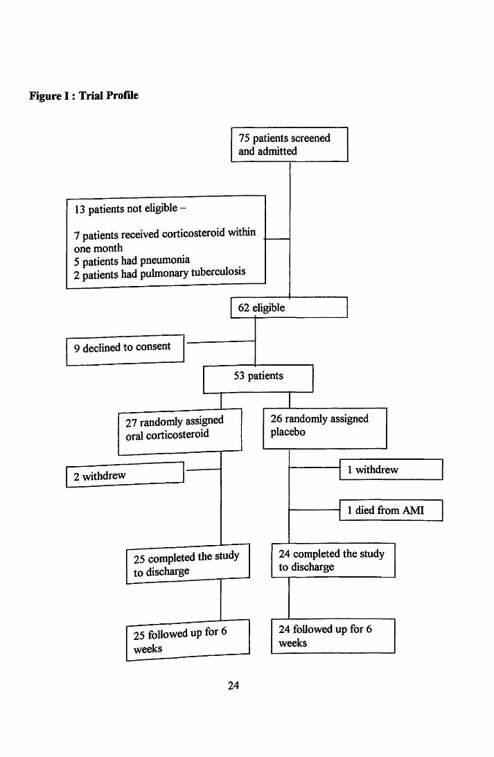
Figure I : Trial Profile
13 patients not eligible -
7 5 patients screened and admitted
7 patients received corticosteroid within one month 5 patients had pneumonia 2 patients had pulmonary tuberculosis
9 declined to consent
I 27 randomly assigned oral corticosteroid
2 withdrew I
I 62 eligible l
53 patients
I 26 randomly assigned placebo
J 1 withdrew
{ 1 died from AMI
25 completed the study 24 completed the study to discharge to discharge
r-25 followed up for 6
weeks
24
24 followed up for 6 weeks
l
l