immune responses in kala-azar - medind
TRANSCRIPT
Immune responses in kala-azar
Samiran Saha, Smriti Mondal, Antara Banerjee, Jayeeta Ghose, Sudipta Bhowmick & Nahid Ali
Infectious Diseases Group, Indian Institute of Chemical Biology, Kolkata, India
Received August 4, 2005
Human infection with Leishmania results in diverse clinical and immunopathological situations.The capacity of the parasites to cause this wide range of disease manifestations depends upon theirability to evade the immune defense mechanisms by performing a well-tuned orchestra of host-parasite interactions inside the macrophages. While updated knowledge focus on the key role ofcell-mediated immunity (CMI) in protection, the survival strategies of the parasites leads to thesuppression of CMI which can further be aggravated by the co-infections with HIV, tuberculosisetc. The present review describes the immune mechanisms in human leishmaniasis with a specialattention to visceral leishmaniasis or kala-azar, one of the most important epidemiological healthproblems in Indian subcontinent. Modulations of the both humoral and cell-mediated immuneresponses during asymptomatic infections, active disease and after successful chemotherapy arediscussed. The components responsible for the regulation of the critical balance of Th1/Th2 typeof responses are re-evaluated. Co-infection of HIV and visceral leishmaniasis and theirinterdependence has been addressed. Although the specific role of an elevated humoral responsein kala-azar is yet to be established, attempts for its application in diagnosis, precisely for thedevelopment of field diagnostic techniques, are presented. Also discussed are attempts to utilizethe immunogenic potentials of different leishmanial antigens in the development of anti-leishmanialvaccines.
Key words Cutaneous leishmaniasis - CD4+ T cell - CD8+ T cell - IFN-γ - IL-10 - IL-12 - immune response - macrophage -serodiagnosis - Th1 response - Th2 response - visceral leishmaniasis
245
Indian J Med Res 123, March 2006, pp 245-266
Leishmaniasis represents a spectrum of diseaseswith important clinical and epidemiologicaldiversity. There are three major forms ofleishmaniasis in human viz., cutaneous (CL),mucocutaneous (MCL) and visceral leishmaniasis(VL). The diseases are caused by the obligateintracellular protozoan parasites belonging to variousspecies of the genus Leishmania. Leishmaniasis isendemic in 88 countries, of which 72 are developingcountries, including 13 of the least developedcountries. Ninety per cent of CL occurs in seven
countries - Afghanistan, Algeria, Brazil, Iran, Peru,Saudi Arabia and Syria, while 90 per cent of VLoccurs in five countries - Bangladesh, India, Nepal,Sudan and Brazil1. CL is caused by a wide range ofspecies, including Leishmania major, L. aethiopicaand L. tropica in the Old World, and L. mexicana,L. brazil iensis, L. amazonensis, L. pifanoi,L. garnhami, L. venezuelensis, L. guyanensis,L. peruviana, and L. panamensis in the New World.The disease, which is usually localized and healsspontaneously, is characterized by skin lesions rich
Review Article
in parasites. MCL is most commonly caused by theNew World species, L. braziliensis2,3, thoughL. aethiopica has also been reported to cause thissyndrome. A persistent cutaneous lesion thateventually heals characterizes initial infection.Several years later, oral and respiratory mucosalinvolvements occur, causing inflammation andmutilation of the nose, mouth, oropharynx, andtrachea. Progressive disease is difficult to treat andoften recurs. With prolonged infection, death occursfrom respiratory compromise and malnutrition4. VL,commonly known as kala-azar, is caused byL. donovani and L. infantum in the Old World andL. chagasi in the New World. It is characterized byfever, cachexia, hepatosplenomegaly, and bloodcytopaenia, and is usually fatal without specificchemotherapy. VL is of higher priority than CL sinceanthroponotic VL foci are the origin of frequent anddeadly epidemics. The name ‘kala-azar’ hasoriginated from India, meaning ‘black-fever’, whichrefers to the hyperpigmentation of skin during thecourse of the disease. Alternatively, the term mightbe derived from the word ‘kal’ meaning ‘death’,which signifies the fatality of the disease5. Patients,cured of VL from Sudan and India, often developpost-kala-azar dermal leishmaniasis (PKDL), whichappears as a dermatotropic form of L. donovaniinfection. According to recent reports, there are1.0-1.5 million cases of CL and 500,000 cases of VLeach year, and a population of 350 million is at risk.Disability-adjusted life years (DALYs) lost due toleishmaniasis are close to 2.4 million all over theworld1,6.
Leishmania are protozoa belonging to the orderKinetoplastida and the family Trypanosomatidae.The parasites have a digenetic life cycle and exist intwo distinct morphologies, the promastigote in thesand fly vector, and the amastigote in the mammalianhost. The motile flagellated promastigotes exist,multiply and develop extracellularly in thealimentary tract of the blood sucking female sandfly vectors and are transmitted during the blood mealinto mammalian hosts. Inside the mammalian hoststhey infect macrophages of the reticuloendothelialtissue7,8 and differentiate into nonmotile amastigotesand multiply as such in the phagolysosomal vacuoles.Macrophages play a primary role in the host defense
and regulation of immune responses upon activation9.The parasites perform a complex host-parasiteinteraction inside the severe environment of thephagolysosomes and eventually evade this immunedefense mechanism10. Infection of macrophages withLeishmania results in impaired microbicidalmachinery as evidenced by decreased responsivenessto the lipopolysaccharide required for induction ofinterleukin (IL)-1 production11. Although most of theinformations on the immunologic mechanisms uponinfection and protection from the Leishmaniaparasites were accumulated from the studies in mice,some critical findings of murine CL have beenconfirmed in humans in recent years. However, theimmune response due to VL and the pathogenesis ofthe disease in human deviates considerably from themurine model.
Although it is evident that cell-mediated immuneresponse (CMI) plays a very important role in thesusceptibility or resistance and prophylaxis inresponse to chemotherapy, the role of humoralimmune response cannot be ruled out as VL is markedby high levels of Leishmania-specific antibodies12,13
which appear soon after infection and before thedevelopment of cellular immunologic abnormalities.The role of these elevated antibodies in resolutionof the disease and protective immunity is mostlyunknown. The occurrence of subclinical orasymptomatic infections among a large populationof individuals residing in the endemic areas withdevelopment of specific antibodies and/or T-cellresponse to leishmanial antigens, however, suggestsnaturally acquired immunity14-17.
Humoral responses in leishmaniasis
Infection of Leishmania in human is characterizedby the appearance of anti-leishmanial antibodies inthe sera of the patients. In CL, usually they arepresent at low levels during the active phase of thedisease18. However, in some studies the presence ofantibodies against L. braziliensis infection in the seraof infected patients has been critically monitored andutilized for the diagnosis and prognosis of thedisease19-23. Contrastingly, strong anti-leishmanialantibody titres are well documented in VL12,13.Although it is known that the Th1 cytokine
246 INDIAN J MED RES, MARCH 2006
interferon-gamma (IFN-γ) probably upregulatesisotypes IgG1 and IgG3, and the Th2 cytokines IL-4and IL-5 stimulates the production of IgG4 inhuman24,25, the role of the elevated anti-leishmanialantibodies in kala-azar patients towards protectionor pathogenesis is still unclear. Critical analysis ofLeishmania antigen-specific Ig isotypes from ourlaboratory and others has revealed the elevated levelsof IgG, IgM, IgE and IgG subclasses during disease26-
31. To establish a correlation of these isotypes withprogression and resolution of infection, anotherreport from our laboratory has shown that antimonialdrug resistance is associated with a reduction in IgG2and IgG3 antibodies, with no significant change inthe titres of IgG, IgM, IgA, IgE, and IgG4. However,a marked elevation of IgG1 was observed in all thepatients studied32. Another study attempted to utilizethe relative abundances of the anti-leishmanial IgGsubclasses to discriminate among the immunity ofthe active and cured patients as well as the endemichealthy individuals and showed that a low CMI, interms of delayed type hypersensitivity (DTH), iscorrelated with high IgG1, IgG3 and IgG4 and viceversa33. As it is stil l debatable whether theseantibodies have any role in the protection of thedisease, a recent experimental study postulated thatIgG not only fails to provide protection against thisintracellular pathogen, but it actually contributes todisease progression. Passive administration of anti-leishmanial IgG resulted in larger lesions inBALB/c mice with greater amount of IL-10production34. This result can be correlated with thehighly elevated titres of anti-leishmanial antibodiesduring the active phase of the disease and aconsecutive fall in the antibody titre after a successfulcure. The elevated antibody titres againstpromastigote or amastigote antigens, their fractionsor recombinant antigens have been extensivelyexploited for specific serodiagnosis in last twodecades in the form of direct agglutination test(DAT), ELISA, dot-ELISA, immunoblot, strip test,indirect immunofluorescence test (IFAT), indirecthaemagglutination antibody (IHA) etc. However,diagnosis of leishmaniasis remains problematic.While differential diagnosis of symptomatic kala-azar from other fever related diseases with splenicor hepatic disorders l ike malaria, typhoid,tuberculosis etc., is difficult, CL, MCL and PKDL
SAHA et al: IMMUNE RESPONSES IN KALA-AZAR 247
are often confused with leprosy and numerous otherprimary and secondary skin diseases like psoriasis,vitiligo, ringworm, lupus vulgaris, etc35,36. Althoughthe serological tests have been evaluated with variousdegree of sensitivity and specificity, a universalmethod of field diagnosis to replace the gold standardof kala-azar diagnosis by histological demonstrationof parasites from the splenic or bone marrow aspirateis still lacking35,37-39. A recent systematic analysisfrom our laboratory on the serodiagnostic potentialof L. donovani promastigote membrane antigen(LAg) has revealed differences in the recognitionpattern among VL and PKDL sera of Indian origin40.While the IgG- and IgG1-specific reactivity for bothVL and PKDL is 100 per cent, PKDL sera from thepatients with varied degree of disease manifestationshowed a wide range of anti-leishmanial IgG titres.IgE and IgG4, which were elevated in VL werenegligible or absent in PKDL. Moreover, Westernblot studies revealed that IgG specific recognitionof 67 kDa band of LAg is specific for PKDL whileIgG-specific recognition of 31 kDa band is specificfor VL 40. ELISA, immunoblot, IFAT and IHA aretoo sophisticated for field use. DAT with differentleishmanial antigens and strip test based on thedetection of serum-IgG against recombinant K39(rK39)41 antigen have so far been proved to be thebest options for serological field diagnosis.Determination of cut-off titre due to fluctuation ofsensitivity and specificity at different serumdilutions, relatively high cost (US$ 4.5 per test),storage of liquid antigen at 4°C, and a lengthy method(18 h) are the constraints for DAT42. Although fastagglutination screening test (FAST), a modifiedversion of DAT performed with freeze-dried antigen,has overcome the problem of antigen storage andassay time (3 h), it is costlier. Strip test using rK39,which is a 39 amino acid repeat within the 230 kDaLcKin protein of kinesin superfamily, can be used todiagnose VL caused by L. donovani, L. chagasi andL. infantum. It has been shown to produce very highsensitivity and specificity among the patients of theIndian subcontinent42-48. However, for African, LatinAmerican and Mediterranean VL patients, thesensitivity and specificity of this test are lower49-53.Appearance of anti-K39 IgG in the healthyindividuals of endemic regions and long persistenceof these antibodies in the patients after successful
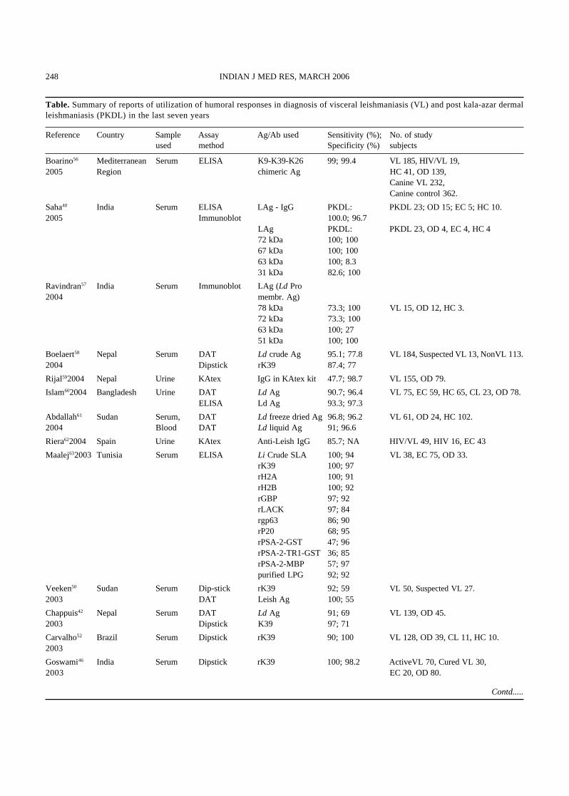
Table. Summary of reports of utilization of humoral responses in diagnosis of visceral leishmaniasis (VL) and post kala-azar dermalleishmaniasis (PKDL) in the last seven years
Reference Country Sample Assay Ag/Ab used Sensitivity (%); No. of studyused method Specificity (%) subjects
Boarino56 Mediterranean Serum ELISA K9-K39-K26 99; 99.4 VL 185, HIV/VL 19,2005 Region chimeric Ag HC 41, OD 139,
Canine VL 232,Canine control 362.
Saha40 India Serum ELISA LAg - IgG PKDL: PKDL 23; OD 15; EC 5; HC 10.2005 Immunoblot 100.0; 96.7
LAg PKDL: PKDL 23, OD 4, EC 4, HC 472 kDa 100; 10067 kDa 100; 10063 kDa 100; 8.331 kDa 82.6; 100
Ravindran57 India Serum Immunoblot LAg (Ld Pro2004 membr. Ag)
78 kDa 73.3; 100 VL 15, OD 12, HC 3.72 kDa 73.3; 10063 kDa 100; 2751 kDa 100; 100
Boelaert58 Nepal Serum DAT Ld crude Ag 95.1; 77.8 VL 184, Suspected VL 13, NonVL 113.2004 Dipstick rK39 87.4; 77
Rijal592004 Nepal Urine KAtex IgG in KAtex kit 47.7; 98.7 VL 155, OD 79.
Islam602004 Bangladesh Urine DAT Ld Ag 90.7; 96.4 VL 75, EC 59, HC 65, CL 23, OD 78.ELISA Ld Ag 93.3; 97.3
Abdallah61 Sudan Serum, DAT Ld freeze dried Ag 96.8; 96.2 VL 61, OD 24, HC 102.2004 Blood DAT Ld liquid Ag 91; 96.6
Riera622004 Spain Urine KAtex Anti-Leish IgG 85.7; NA HIV/VL 49, HIV 16, EC 43
Maalej632003 Tunisia Serum ELISA Li Crude SLA 100; 94 VL 38, EC 75, OD 33.rK39 100; 97rH2A 100; 91rH2B 100; 92rGBP 97; 92rLACK 97; 84rgp63 86; 90rP20 68; 95rPSA-2-GST 47; 96rPSA-2-TR1-GST 36; 85rPSA-2-MBP 57; 97purified LPG 92; 92
Veeken50 Sudan Serum Dip-stick rK39 92; 59 VL 50, Suspected VL 27.2003 DAT Leish Ag 100; 55
Chappuis42 Nepal Serum DAT Ld Ag 91; 69 VL 139, OD 45.2003 Dipstick K39 97; 71
Carvalho52 Brazil Serum Dipstick rK39 90; 100 VL 128, OD 39, CL 11, HC 10.2003
Goswami46 India Serum Dipstick rK39 100; 98.2 ActiveVL 70, Cured VL 30,2003 EC 20, OD 80.
Contd.....
248 INDIAN J MED RES, MARCH 2006
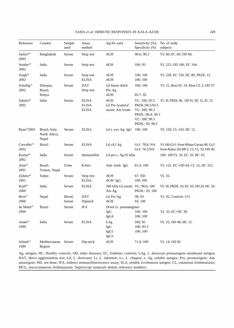
Sarker48 Bangladesh Serum Strip test rK39 96.6; 98.3 VL 60, EC 60, OD 60,2003
Sundar44 India Serum Strip test rK39 100; 93 VL 225, OD 100, EC 104.2002
Singh45 India Serum Strip test rK39 100; 100 VL 228, EC 150, HC 80, PKDL 12.2002 ELISA rK39 100; 100
Schallig51 Ethiopia, Serum DAT Ld freeze dried 100; 100 VL 21, Braz EC 19, Braz CL 5, OD 37.2002 Brazil, Strip test Pro Ag
Kenya rK39 85.7; 82
Salotra64 India Serum ELISA rK39 VL: 100; 93.5 VL 30, PKDL 88, OD 91, HC 32, EC 22.2002 ELISA Ld Pro lysateLd PKDL:94.5;93.5
ELISA axenic Am lysate VL: 100; 90.3PKDL: 86.4; 90.3VL: 100; 90.3PKDL: 92; 90.3
Ryan302002 Brazil, Italy, Serum ELISA Ld s. exo Ag- IgG 100; 100 VL 129, CL 143, HC 12.North Africa,Nepal
Carvalho65 Brazil Serum ELISA Ld rA2 Ag Gr1: 78.8; NA VL 68 (Gr1: from Minas Gerais 48, Gr2:2002 Gr2: 76.5;NA from Bahia 20) MCL 13, CL 33, OD 40.
Kumar66 India Serum Immunoblot Ld pro s. Ag 65 kDa 100; 100VL 10, EC 10, HC 10.2002
Attar67 Brazil, Urine KAtex Anti- leish IgG 81.4; 100 VL 132, EC+OD 64, CL 12, HC 312.2001 Yemen, Nepal
Zijlstra48 Sudan Serum Strip test rK39 67; ND VL 55.2001 ELISA rK39- IgG 100; ND
Kaul68 India Serum ELISA 200 kDa Ld axenic VL: 96.6; 100 VL 30, PKDL 10, EC 10, OD 20, HC 10.2000 Am Ag PKDL: 10; 100
Bern47 Nepal Blood, DAT Ld Pro Ag 98; 93 VL 92, Controls 113.2000 Serum Dipstick rK39 93; 100
da Matta29 Brazil Serum IFA Dried Lc promastigotes2000 IgG 100; 100 VL 33, EC+HC 39.
IgG4 100; 100
Anam26 India Serum ELISA LAg 100; 85 VL 25, OD 48, HC 12.1999 IgG 100; 83.3
IgG1 100; 100IgG3
Jelinek53 Mediterranean Serum Dip-stick rK39 71.4; 100 VL 14, OD 821999 Region
Ag, antigen; HC, Healthy controls; OD, other diseases; EC, Endemic controls; LAg, L. donovani promastigote membrane antigen;DAT, direct agglutination test; Ld, L. donovani; Li, L. infantum; Lc, L. chagasi; s. Ag, soluble antigen; Pro, promastigote; Am,amastigote; ND, not done; IFA, indirect immunofluorescence assay; SLA, soluble Leishmania antigen; CL, cutaneous leishmaniasis;MCL, mucocutaneous leishmaniasis. Superscript numerals denote reference numbers
SAHA et al: IMMUNE RESPONSES IN KALA-AZAR 249
Reference Country Sample Assay Ag/Ab used Sensitivity (%); No. of studyused method Specificity (%) subjects
treatment (up to 2 yr), are other constraints of usingthis method54,55. Another field diagnostic approachbased on latex agglutination test (KAtex), to detectthe leishmanial antigens in the urine of the patientshas shown very high sensitivity and specificity.Reports of the performances of different diagnosticmethods of VL based on the humoral responses inthe last seven years are summarized in the Table.
Cell mediated immune responses in humanvisceral leishmaniasis
Immune suppression and Th1/Th2 paradigm: Despitethe differences between CL and VL, resistance todisease in both the forms of leishmaniasis is markedby a dominant Th1 response. Severe manifestationof CL is associated with a strong Th2 compared to apredominant Th1 response in the mild manifestationof the disease69. On the other hand, it is welldocumented that VL is characterized by suppressionof CMI, which is proved from the unresponsivenessof the patients to the Leishmanin skin test (LST) orMontenegro test. This test measures a DTH reactionto an intradermal injection of leishmanial antigens70.Containment of the disease following a successfultreatment is associated with a strong cell mediatedDTH response71. The cell mediated immunesuppression is also evident from blastogenesis assayof the peripheral blood mononuclear cells (PBMCs)(lymphoproliferation) from untreated VL patientsfrom Brazil, Africa and India72-75. Reduction inproportion of the helper T cells and theimmunosuppression76 is rapidly reversible witheffective chemotherapy. The control and protectionof VL, in general, is dependent on the IFN-γ inducedinnate and adaptive cellular immune responses,which induce the intracellular killing by activatedmacrophages77. That this immunosuppression due toVL is antigen specific is evident from the positive invitro response as observed from lymphoproliferationor IFN-γ production from the PBMCs from both theinfected and cured patients stimulated withPhytohemaglutinin (PHA) or an unrelated antigen,purified protein derivative of tuberculin (PPD)17,74,78.It has long been strongly suspected that thisimmunosuppression in the active stage of the diseaseis regulated by a population of suppressor T cells,different from the conventional CD8+ T cells17,79,80.
It was suspected that this suppressor activity couldbe mediated by a subpopulation of the CD4+ T cellsand could be distinguished by the CD45RBlow
phenotype81,82.
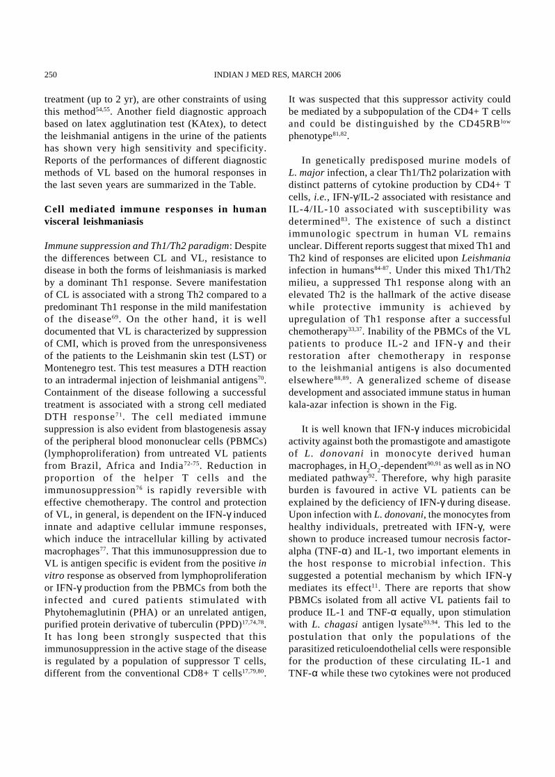
In genetically predisposed murine models ofL. major infection, a clear Th1/Th2 polarization withdistinct patterns of cytokine production by CD4+ Tcells, i.e., IFN-γ/IL-2 associated with resistance andIL-4/IL-10 associated with susceptibility wasdetermined83. The existence of such a distinctimmunologic spectrum in human VL remainsunclear. Different reports suggest that mixed Th1 andTh2 kind of responses are elicited upon Leishmaniainfection in humans84-87. Under this mixed Th1/Th2milieu, a suppressed Th1 response along with anelevated Th2 is the hallmark of the active diseasewhile protective immunity is achieved byupregulation of Th1 response after a successfulchemotherapy33,37. Inability of the PBMCs of the VLpatients to produce IL-2 and IFN-γ and theirrestoration after chemotherapy in responseto the leishmanial antigens is also documentedelsewhere88,89. A generalized scheme of diseasedevelopment and associated immune status in humankala-azar infection is shown in the Fig.
It is well known that IFN-γ induces microbicidalactivity against both the promastigote and amastigoteof L. donovani in monocyte derived humanmacrophages, in H
2O
2-dependent90,91 as well as in NO
mediated pathway92. Therefore, why high parasiteburden is favoured in active VL patients can beexplained by the deficiency of IFN-γ during disease.Upon infection with L. donovani, the monocytes fromhealthy individuals, pretreated with IFN-γ, wereshown to produce increased tumour necrosis factor-alpha (TNF-α) and IL-1, two important elements inthe host response to microbial infection. Thissuggested a potential mechanism by which IFN-γmediates its effect11. There are reports that showPBMCs isolated from all active VL patients fail toproduce IL-1 and TNF-α equally, upon stimulationwith L. chagasi antigen lysate93,94. This led to thepostulation that only the populations of theparasitized reticuloendothelial cells were responsiblefor the production of these circulating IL-1 andTNF-α while these two cytokines were not produced
250 INDIAN J MED RES, MARCH 2006
by the nonparasitized antigen stimulated PBMCs.Serum level of IL-4 was found to be high in theBrazilian VL patients compared to no or very lowlevel of IFN-γ86. However, considerable level of
IFN-γ is secreted at the very initial stages of theexposure to the parasites as observed in theseroconverted or subclinically infected individualsin the endemic area95. Although the reasons behind
Fig. Generalized scheme of disease development and associated immune status in kala-azar. Infection with Leishmania parasites causingvisceral disease leads to different pathological conditions. Most of the infected individuals develop symptomatic kala-azar. A considerablepercentage of the infected population undergoes subclinical infection and remains immune to symptomatic disease. Highly seropositiveasymptomatic individuals may convert to kala-azar. A small percentage of L. donovani infected individuals may directly result inPKDL, a dermatotropic manifestation, without occurrence of the visceral disease. Symptomatic kala-azar patients present characteristicantileishmanial serum antibodies and suppression of parasite specific CMI. Successful antimonial therapy leads to recovery of CMIwith mixed Th1/Th2 response. Despite a decline, antileishmanial antibodies persist in the sera of the cured individuals for many years.Many VL patients in India and Sudan, after successful cure of VL, develop PKDL. Although seropositive, PKDL patients differ fromVL in the absence of CMI suppression. A generalized mixed TH1/Th2 response in PKDL proceeds towards upregulation of Th2corresponding with the chronic disease. Thickness of the lines corresponds with the likelihood of the occurrence of the different events.LST, Leishmanin skin test; CMI, cell mediated immunity; IFN-γ, interferon-gamma; Leish-Ag. Leishmania antigen; IL-2, interleukin-2; TNF-α, tumor necrosis factor-alpha; IL-4, interleukin-4; IL-13, interleukin-13; IL-5, interleukin-5; TGF-β, transforming growthfactor-beta; IL-10, interleukin-10; MΦ, macrophage; PKDL, post kala-azar dermal leishmaniasis.
SAHA et al: IMMUNE RESPONSES IN KALA-AZAR 251
high levels of IFN-γ secretion at the initial phase ofinfection and the decline in its production at theactive stage of the disease is not clear, this IFN-γ isfound to come from the natural killer (NK) cells andmight be important as a component of innate immunemechanism for the macrophage activation96. Reportson the administration of IFN-γ in the VL patients fortherapy imply the functional importance of thiscytokine to establish the immune environment forthe clearance of the parasites97-105.
Importance of IL-10: IL-10 was initially characterizedas a Th2 cytokine but later on it was proved to be apleiotropic cytokine, secreted from different cell typesincluding the macrophages106. Experimental evidencesindicate that IL-10 plays an important regulatory rolein the progression of VL. The suppressive ability ofIL-10 on the IFN-γ mediated microbicidal activity ofmacrophages is well established for otherdiseases106,107. Recombinant IL-10 is shown to haveinhibitory effects towards NO mediated killing of L.infantum, L. major and L. braziliensis in humanmacrophages, derived from the monocytes of healthyindividuals92. Investigation on Brazilian patientsshowed that IL-10 production from L. chagasi antigenstimulated PBMC cultures of acute VL wassignificantly higher than in cured individuals, whereasasymptomatic leishmanin skin test (LST) positiveindividuals had no release of IL-10108. In active VLpatients of Sudan and India, production of high levelsof IL-10 mRNA is reported from the bone marrowaspirate, lymph nodes, PBMCs, and splenic aspirates,and the levels decrease after successfulchemotherapy109-111. These studies also showeddetectable levels of IFN-γ mRNA in the bone marrow,lymph node, PBMC or the splenic aspirate samplesfrom the infected individuals though their specificcellular sources were not determined. This isinteresting because, the production of IFN-γ at mRNAlevel is in contrast with the studies where evidencessuggest that there is suppression of the Th1 kind ofresponse, observed from the inability to produce IFN-γ by the PBMCs in active VL. Carvalho et al78 showedthat while a combination of exogenous IL-2 and IFN-γ were able to restore only the lymphoproliferativeresponse, a combination of anti-IL-4 and anti IL-10mAb were able to restore both the lymphoproliferativeresponse and the IFN-γ production of leishmanial
antigen stimulated cultures of PBMCs from VLpatients to a large extent. These observations clearlyindicate that the suppression of the Leishmania-specific Th1 kind of response in the patients is themain reason for the disease susceptibility and thissuppression is regulated by IL-1078. Decline in thelevel of IL-10 mRNA after successful chemotherapytherefore supports the fact that persistence of highlevels of IL-10 in the host cells is beneficial for theparasite survival and pathology. Besides its possiblerole in susceptibility and progressiveimmunopathology in VL infection, high levels ofendogenous IL-10 correlate with in the progressionof PKDL. Along with PBMCs, dermal keratinocytesof the African PKDL patients who eventuallydeveloped PKDL produced significantly higheramounts of IL-10 than the cured VL patients who didnot develop PKDL112.
Importance of IL-12: IL-12, known as an naturalkil ler (NK) cell stimulating factor, cytotoxiclymphocytes maturation factor, and a centralimmunoregulator of the initiation and maintenanceof the Th1 response, plays an important role in theinduction of IFN-γ production by T and NKcells113-117. IL-12 is shown to be a potent inducer ofthe Th1-type response and protective immunity inmurine L. major infection118,119. Along with IFN-γ,IL-12 was reported to be produced from the PBMCsof cured VL patients but not from the active VLpatients following stimulation with Leishmanialysate. That IL-12 plays a counter-regulatory effectagainst IL-10 in Leishmania infection, is proved fromthe observation that addition of recombinant IL-12or neutralizing anti-IL-10 mAb could restore theIFN-γ production as well as the lymphoproliferativeresponse in the Leishmania lysate stimulated PBMCsof active VL patients120,121. Conversely, neutralizinganti-IL-12 or rIL10 inhibits the production ofIFN-γ120. As there are strong evidences that IL-10inhibits IFN-γ production in human PBMCs bysuppressing IL-12 synthesis from the accessory cellslike macrophages, B cells or dendritic cells122,overproduction of IL-10 from different immune cellsis most likely the key driving force to decline theTh1 environment and therefore establishing aTh2 type response in the active human VL. Furtherstudies prove that this counter-regulatory activity of
252 INDIAN J MED RES, MARCH 2006
IL-10 and IL-12 plays a fundamental role inmodulating the immune responses of Leishmaniainfection in human towards Th2 or Th1 probably bymodulating the B7/CD28 costimulatory interactionrespectively123. Recent experimental observations onL. major revealed that these two counteractivefunctions, i.e., IL-12 and IFN-γ-dependent host-protective immune responses and IL-10-dependentprogressive Leishmania infection, are differentiallyregulated by CD40 signaling. While weak CD40signals induce extracellular stress-relatedkinase-1/2 (ERK-1/2)-dependent IL-10 expression,stronger signals induce p38 mitogen-activatedprotein kinase (p38MAPK)-dependent IL-12production. Induction of ERK-1/2 inhibits p38MAPKand vice versa. Upon Leishmania infection, CD40signaling is skewed towards ERK-1/2, thereforeinducing IL-10 and inhibiting IL-12 and iNOS-2expression. Conversely, ERK-1/2 inhibition or IL-10 neutralization restores CD40-induced p38MAPKactivation and parasite killing124.
Involvement of different cells in immune regulationof human VL: Studies on the involvement of thedifferent immune cells in the maintenance of Th1/Th2environment through the secretion of differentcytokines help to understand the nature ofimmunomodulation in VL. Most of the T cell clonesderived from the PBMCs of the active VL patientsand asymptomatic subjects (seropositive and DTHpositive) were CD8+ with negligible CD4+, while thesituation is reversed in the cured individuals with morenumbers of CD4+ than CD8+ cells. CD8+ T cellsisolated from the asymptomatic subjects producedhigh amounts of IFN-γ, which strongly suggests a roleof CD8+ cells in human resistance to Leishmaniainfection. However, a unique popuplation of CD4+cells producing both IFN-γ and IL-5 is found tocontribute in the control of infection in asymptomaticsubjects125. An earlier study demonstrated highernumber of CD4+ clones than CD8+ clones in theasymptomatic DTH positive individuals, althoughclones isolated from active VL were mostly CD8+112.Addition of the CD8+ clones isolated from acute kala-azar patients, augmented IL-10 production from thePBMC cultures of same patients after recovery,suggesting that CD8+ T cells may have a role inmediating IL-10 production during active disease112.
Further observations suggested that CD8+ cells arelikely to mediate the endogenous IL-10 secretionrather than antigen-specific IL-10, and thus theimmunodominance of CD8+ T cells could probablyplay an important role in progressive VL126. An earlierstudy by the same author postulated that CD8+suppressor T cell (Ts) population and Th1 CD4+populations have counter-regulatory effects forsusceptibility and resistance, and predicted that thebasic immunoregulatory mechanism during infectionis the prevention of endogenous IL-10 secretionmediated by CD8+ Ts cells by the activity ofleishmanial antigen-specific Th1 CD4+ cells.Symptomatic infections occur when this immuneregulatory cycle fails due to antagonistic demands onthe immune system by, malnutrition, stress or othercauses127. Epidemiological and clinical investigationssuggested association of undernutrition with thedevelopment of clinically apparent VL and that thedisease itself has a profound effect on nutritionalstatus128. There are evidences that PBMCs fromhealthy individuals, not exposed to Leishmania, showsome natural reactivity to the leishmanial antigens byproduction of IFN-γ and IL-4 as well as proliferationof the PBMCs. This might be caused as a consequenceof cross-activation by other micro-organisms or someevolutionarily conserved crosseracting antigens likeheat shock proteins (HSP)129-134. As mentioned earlier,Th1/Th2 dichotomy is much debated in human VLmostly at the cured stage135. Flowcytometricinvestigation for the specific T cells producing thesecytokines in the culture of PBMCs of the cured VLpatients revealed that these cytokines were producedmainly from the CD4+ cells, while a very lowpercentage of CD8+ cells were also involved incytokine production136. Examination of the co-expression of the cytokines revealed three types ofcytokine producing cells viz. (i) IFN-γ producers,( ii ) IL-4 producers, and (iii ) IFN-γ and IL-10producers; IFN-γ and IL-4 were never co-expressed136.This study demonstrated that besides conventional Th1and Th2 subsets there is existence of a T cellpopulation producing both IFN-γ and IL-10, whichcould be functionally important as the regulatorysubset that allows a balance between Th1 and Th2 cellsin the cured VL patients. Comparative flowcytometricanalysis on the PBMCs of healthy individuals and CLpatients infected with L. aethiopica, in response to
SAHA et al: IMMUNE RESPONSES IN KALA-AZAR 253

L. aethiopica antigen reveal that the CD16+/CD56+NK cells in the healthy individuals are the mainresponder cells rather than the T cells in the CLpatients while the few T cells which responded in thehealthy individuals were identified to be CD8+137.Elevated levels of IFN-γ were produced in similarcultures of PBMCs from healthy individuals. Furtherstudies show that live Leishmania promastigotes areable to stimulate human NK cells to secrete IFN-γ asan early response96,138. This stimulation is contributedlargely by the Leishmania homologue of receptors foractivated C-kinase (LACK)139.
Involvement of other cytokines in immuneregulationof human VL: IL-4 is considered to be the signaturecytokine of Th-2 response. Although known tocorrelate with disease in murine CL but not murineVL 3, IL4 is reported to be present in the sera, PBMCsupernatant86,140 or as mRNA in human VL78,110,111.But there are reports that IL-4 is not alwaysproduced in VL patients78,81,141 and that it has noimmunomodulatory effect in downregulating the Th1response during disease78,123. However, theseinconsistent detections of IL-4 in VL patients mightbe due to the fact that there are disease specificsoluble IL-4 receptors in the serum of VL patients,which can neutralize both the bioactivity andimmunologic detection of this cytokine142. OtherTh-2 cytokines like IL-13143 and transforming growthfactor-beta (TGF-β)144-146 have been reported to beproduced in VL though their biological role inmodulating the Leishmania specific immuneresponses is not well defined. Studies on regulatoryT cells (CD4+ CD25+), which function throughTGF-β and IL-10 production could help to understandthe role of TGF-β. However, it is evident that parasitesurvival is favoured by the conversion of latentTGF-β of the host to active TGF-β by some parasite-derived factors, which help to create its immediatemicroenvironment to its own survival advantage146.
Implications of immunogenecity of differentleishmanial antigens towards human cells
The development of vaccines is the essential aimof studies on leishmaniasis. In the endemic areas ofvisceral leishmaniasis, a significant population ofindividuals does not manifest any clinical symptoms
of the disease, but show elevated antileishmanialantibodies and/or a T cell response to leishmanialantigens15-17. Moreover, patients recovered from kala-azar are usually immune to re-infection70,74. Theseobservations provide the idea that these individualshave somehow gathered life long protection from VL,possibly through the appropriate stimulation ofprotective host response by the Leishmania antigens.As discussed previously, it is known that theimmunosuppression observed in VL is antigen-specific. The in vitro stimulation of immune cells fromactive or successfully treated VL patients by differentleishmanial antigen preparations or purified antigenfractions provides idea about the potentialimmunogenicity of those antigens as possible vaccinecandidates. While there is still no effective form ofimmunoprophylaxis against this disease, extensiveinvestigations in this field include human vaccine trialswith killed promastigotes and immunization of micewith attenuated, killed, and crude parasite fractions,as well as purified and recombinant antigens and theirDNA147-150. The impressive recent advances in this areamay soon result in the development of a safe andeffective vaccine. Search for the effective vaccinecandidates basically involves screening of the differentimmunodominent antigens, studies on the inherentmechanisms of host immune responses and evaluationof selected antigens as vaccine candidates either innative or recombinant forms.
To identify some potential vaccine candidateantigens, screening of immunogenicity of L. donovanipromastigote antigen fractions, isolated by electro-elution, revealed that different antigen fractions of14-80 kDa were almost equally potent to stimulatethe PBMCs of the Kenyan cured VL patients toproliferate and produce IFN-γ87. A more recent studyfrom our laboratory has revealed that sevenpolypeptides of L. donovani promastigote membraneantigen preparation (LAg) of approximate MW 31,34, 51, 63, 72, 91, and 120 kDa were recognized by100 per cent kala-azar patient sera57. The promisingpotentiality of these antigens as vaccine candidatesagainst VL is under investigation. LPG(lipophosphoglycan) and glycoprotein (gp63), whichare involved in the attachment of the parasite to thehost cells, are two abundant molecules on the surfaceof all species of Leishmania151-155. Observations on CL
254 INDIAN J MED RES, MARCH 2006
patients caused by L. braziliensis and L. mexicanashowed that LPG is a potent stimulator forlymphoproliferation of the PBMCs of these patientswhile gp63 is unable to stimulate similar cultures156,157.Supportive results were found from the studies onKenyan VL patients where LPG isolated from L. majorwere able to specifically proliferate the T cells of theVL patients158. On the other hand, gp63 could not evenstimulate the PBMCs of cured VL or healthyindividuals while crude L. donovani sonicate couldstimulate both. This study demonstrated that the clonalexpansion of LPG-specific T cells might contributeto the defense against infection158. However,information regarding the immunogenicity of gp63 isconflicting, since cells from spontaneously healedSudanese CL patients were reported to proliferate inresponse to purified native gp63 from L. major141.Moreover, gp63 stimulated T cell clones isolated fromthe cured CL patients, infected with L. major, werefound to be of Th1 type and culture supernatants ofthese cells inhibited the L. major infection in humanmacrophages159.
Analysis of the host immune responses towards theparasite antigens is important for vaccine strategies.Evidences from the studies of CL caused byL. aethiopica suggested that there are differences inresponsiveness towards L. aethiopica antigensbetween T cells from local cutaneous leishmaniasis(LCL) and diffused cutaneous leishmaniasis (DCL)patients. While the progressive DCL is characterizedby nonresponsiveness, LCL, which is self-healing,shows antigen specific cellular immune function133.In case of VL, though mixed Th1/Th2 is characteristicof cure, an essential Th1 bias at this stage has to beachieved by an effective vaccine. This could beconfirmed from the observations that nonspecificantigens like PPD presents a positive stimulation inthe PBMCs of cured VL patients while tetanous toxoid(TT) could not stimulate the similar cultures87,160,161. Itis known that PPD generally induces Th1-like and TTinduces Th2-like responses in human162. A recent studyon human trial of vaccination with alum-precipitatedautoclaved L. major (Alum/ALM) ± bacille Calmette-Guerin (BCG), has postulated that cellular immuneresponse to human leishmaniasis is dichotomous163.An early production of IFN-γ precedes over a positiveLST and it declines with time while LST positivity
persists. These indicate that IFN-γ production andelevated LST, two markers of protective Th1 response,probably measure two different facets of cellularimmunity. One is mediated by IFN-γ productionthrough less differentiated T cells or NK cells, whilethe other is mediated by more specialized anddifferentiated T cells capable of induction of a variedinflammatory reaction through chemotactic mediators.
There are several reports in the literature, whichfocus on the immunogenic potential of differentsynthetic or recombinant peptides. Among these,kinetoplast membrane protein-11 (KMP-11) ofmolecular mass 11 kDa, is physically associated withLPG (therefore previously known as LPGAP) inL. donovani. It was found to stimulate the productionof both Th1 and Th2 type of T cell clones from thecured African VL patients with vigorouslymphoproliferation160. Three 38-mer syntheticpeptides KMP-11-1 (a.a. 1-38), KMP-11-2 (a.a. 28-65) and KMP-11-3 (a.a. 55-92) had been shown to bepotent immunogen with variable antigenicity for B andT cells as observed through humoral and cell mediatedimmune response during disease. However, thesesynthetic peptides presented weaker proliferativeresponses than the native KMP-11, which might bethe result of either different processing andpresentation of native protein and peptide by APCs,or the lack of association with the glycolipid LPG,thereby losing a natural adjuvant164. Promastigotesurface antigen 2 (PSA-2), which is basically acomplex of three polypeptides of molecular mass 96,80 and 50 kDa and tethered to the promastigotemembrane with glycoinositol phospholipid anchors165
was tested for its immunogenic properties on the Tcells isolated from the cured Sudanese CL patients.While responses towards native PSA-2 isolated fromL. major as well as L. donovani was a clear Th1 typeas compared to the parallel TT stimulated cultures,there was a lack of response to the recombinantEscherichia coli derived PSA-2. This might be due toimportance of glycosylation in defining the T cellepitopes of the antigen which probably determine theglycosylated antigens as better vaccine candidates forleishmaniasis in human161. In contrast to crude parasitelysate, which induced a mixed Th1/Th2 response,recombinant LeIF was shown to induce a dominantTh1 type of immune response, which is largely IL-12mediated, in the PBMCs of CL patients infected with
SAHA et al: IMMUNE RESPONSES IN KALA-AZAR 255
L. braziliensis166. This is important in relation to theobservation that crude L. amazonensis lystate was ableto give rise to Leishmania-specific T cell lines withpotent Th1 response from the PBMCs of healthyindividuals in presence of IL-12. Moreover, these celllines contained high percentage of CD8+ populations,which could lyse autologous Leishmania-infected butnot uninfected macrophages167. Other recombinantleishmanial antigens rHSP-70, Leishmania type 2Cserine/threonine protein phosphatase (rLcPP2C) andrgp63 were found to induce Th1 type of response inasymptomatic and cured VL infected with L. chagasi,with elevated IFN-γ production and very low IL-10168.Lymphoproliferation in response to all these threeantigens were comparatively low than the crude LAg.Interestingly, response in the PBMCs of the healthycontrols in this study is contrasting to those as reportedearlier129-131. Recombinant papLe22, a 22 kDapotentially disease-aggravating protein ofL. infantum has been found to induce the productionof IL-10 in the PBMCs of active VL patients169.Leishmania homologue of receptors of activatedprotein kinase C (LACK), a 36kDa protein, is knownto induce an early induction of IL-4 secreting cells inL. major susceptible BALB/c mice and drive theimmune response towards Th2 phenotype170. LACKwas found to stimulate the proliferative response ofCD8+ and NK cells in healthy individuals as well asactive CL patients139,171. Both IFN-γ and IL-10 weresecreted in the supernatants of the LACK-inducedPBMC cultures of these individuals. Therefore, thevaccine strategies with this antigen in human CL mightbe through directing the LACK response away fromTh2 towards Th1171. However, the absence of anyresponse towards LACK in cured VL patients171
suggests that LACK might not be the right candidatefor vaccination against VL. Further studies onphenotyping of LACK stimulated PBMCs of healthyindividuals revealed that stimulation with recombinantLACK induced production of IFN-γ from CD8+ naïve(CD45RA+) cells and IL-10 from the CD4+ memory(CD45RA-, CD45RO+) cells172.
Immune responses in kala azar patients with HIVco-infection
Among all types of leishmaniasis, VL is the mostfrequent potential opportunistic disease associatedwith HIV-1 since the mid 1980s. Although ninety
per cent of the reported cases are from southwesternEurope, incidences of co-infection are increasingin eastern Africa and the Indian subcontinent173,174.VL is found to promote the clinical progression ofAIDS175 and it was suggested that Leishmaniaparasites could be seen as potential co-factor inHIV-1 pathogenesis176. Studies on latently HIV-1infected U1 moncytoid cell line, treated with liveL. donovani or LPG, showed very significantelevation of reverse trancriptase activity as well asproduction of TNF-α, a potent inducer of HIV-1expression, in the culture supernatants177.L. donovani-induced immunosuppression is alsoenhanced by HIV-1. When PBMCs from healthyindividuals are costimulated with both the antigens,addition of HIV antigens did suppress L. donovani-induced proliferation in a dose-dependent fashioncompared to only L. donovani antigen stimulation178.On the other hand, increased production of IL-6 andTNF-α, potent inducers for HIV-1, was found to beassociated with L. donovani- induced viralreplication. Further, L. donovani promastigotes andLPG were proved to mediate CD4+ T cell activationinduced HIV-1 replication. Leishmanial antigen-induced TNF-α produced in the culture wasimportant for the replication178,179. The mutualinterdependence as the agents for co-infectionbetween these two pathogens is also reported. Co-infection in THP-1 macrophage cell line increasesthe multiplication of L. donovani amastigotes in themacrophages. Again, killed HIV preparation wasshown to abrogate the proliferative response as wellas IFN-γ production from the L. donovani antigeninduced PBMCs of healthy individuals. Moreover,anti-IL-10 could not enhance IFN-γ production inHIV-VL co-infected patients as is generally foundin only VL patients180,181.
Role of regulatory T cells
Evidences from experimental murine models ofL. major infection suggest that endogenous CD4+CD25+ T cells (Treg) play an important role in theinfectivity of this parasite. These cells are unique inexpressing CD25 molecule, which is the IL-2receptor α-chain, on their surface and characterizedby their ability to control the excessive or misdirectedimmune response to microbial or self-antigens182-184.
256 INDIAN J MED RES, MARCH 2006
Constituting 5-10 per cent of the peripheral T cellpopulations in both human and mice at the steadystate, CD4+CD25+ cells execute their suppressiveor regulatory function either by cell contactdependent manner or by secretion of regulatorycytokines IL-10 and TGF-β185,186. Activation of thesecells is also triggered to some extent by thestimulation of toll-like receptor-4 (TLR-4) with itligand187. Production of high levels of IL-10 andTGF-β in the patients with acute infection ofLeishmania and the resultant immunosuppression inthem led to the investigation of the function of thesecells in leishmaniasis and is in fact shown toaccumulate in huge number in the cutaneous lesionsof mice infected with L. major and therefore suppressthe CD4+ CD25- effector cell functions to eliminateparasites188. Another report tends to impart that CD4+CD25+ cells in murine infection functions inexclusively IL-10 independent manner renderingtheir suppressive effect both on Th1 and Th2 cellsand thereby playing important role in suppressingdisease development of L. major infection in SCIDmice reconstituted with naïve CD4+ CD25- cells.This study also postulates that Th2 cells are moresusceptible to the inhibitory effect of CD4+ CD25+cells than the Th1 cells189. These informationsstrongly indicate that CD4+ CD25+ T cells may havesimilar immunomodulatory role in human VL, whichneeds to be investigated.
Immune response in PKDL
As a sequel to kala-azar, PKDL, first described byBrahmachari190 appears in a dermatotropic form ofL. donovani infection in >50 per cent patients inSudan and 10-20 per cent patients in India. Theimmunopathogenesis of the disease is still il lunderstood. Although patients of PKDL often bear thedifferent forms of PKDL lesions for years and henceremain as reservoir for L. donovani191,192, there arelimited attempts to reveal the underlying immunemechanisms for the appearance of the disease192.Studies, which have so far been made, indicate thatunlike VL there is positive cell mediated immuneresponse against leishmanial antigen in PKDL in termsof DTH (LST), lymphoproliferation and productionof IFN-γ by the PBMCs74,193-195. PKDL patientsmanifest an aggravation of the disease with time in
form of nodular lesions or erythematous plaqueformation. Again, PKDL patients with early infectionshow better CMI than the chronic patients. Theseindicate that there are different domains of PKDLpatients according to the immunopathogenesis of thedisease. Though there is generalized prominence ofTh1 in PKDL, as found from IFN-γ production, highlevels of IL-10 in the lesions as well as plasma of thepatients112 indicate the importance of Th2 response inchronic PKDL. Preponderance of the CD8+ cells overCD4+ cells in PKDL lesions of Indian patients196,197 issuggestive of persistence of parasites as CD8+ cellsmight block the IL-2 production locally and in turninhibit the IFN-γ production198 favouring disease.
Conclusion
Till date, the studies of the immunology on humaninfection of Leishmania emphasize the regulatory roleof the cell-mediated immune response. Although anti-leishmanial antibodies are abundant in the sera of thepatients during disease, a correlation between theelevation of Th1/Th2 response and the stimulation ofthese antibodies could not be established. As there istremendous ecological and genetic diversity amongthe different human populations exposed to theparasite, conclusive understanding of the parametersof resistance versus control in human is difficult.Moreover, the immunological data available are stillscarce. There is also an urgent need for a betterexperimental model mimicking human infections. Itis evident that there is a marked occurrence of bothTh1 and Th2 components of CMI response during VLas documented through the detection of serum andtissue cytokines. Recent reports suggest theinvolvement of immune cells other than the Th1 andTh2 subsets of CD4+ T cells. Among these, CD8+cells macrophages and NK cells play major roles. Inaddition, recent experimental data obtained withstudies on CD4+ CD25+ Treg cells point to a probableregulatory function of these cells in maintaining theimmune homeostasis in human leishmaniasis. Incontrast to the earlier ideas that antagonistic functionsof IFN-γ and IL-4 determine the outcome of protectionor pathogenesis of the disease, recent studiesemphasize the importance of the balance of the tworegulatory cytokines IL-12 and IL-10, critical for theregulation of the immune modulation during infection,
SAHA et al: IMMUNE RESPONSES IN KALA-AZAR 257
pathogenesis, and chemotherapy. To understand thenature of human infection with these parasites and todevelop better chemotherapeutic and vaccinestrategies, further in depth studies focused on theimmune modulation in the subclinical andasymptomatic individuals needs attention. Immunestatus of the patients demonstrating antimonyresistance and those suffereing from relapse ofinfection should also be a major area of investigationas these patients might have developed someimpairment towards protective immunity. Moreover,immunopathogenesis in patients with PKDL, so far aneglected field of research, deserve attention. As thepatients with PKDL come up with varied degrees ofdisease manifestation and there are also considerabledifferences in the PKDL patients of Indian and Africanorigin, systematic analysis of immune responses ofthis sole human reservoir of the disease will definitelyhelp to clear our vision of leishmaniasis as a whole.
References
1. Desjeux P. Leishmaniasis. Nat Rev Microbiol 2004; 2 :692-3.
2. Renee H, Wang NE, Halpern J. Leishmaniasis.www.emedicine.com/emerg/topic296.htm. Last Updated:September 13, 2005. eMedicine. Accessed on March 26,2006.
3. Wilson ME, Jeronimo SM, Pearson RD.Immunopathogenesis of infection with the visceralizingLeishmania species. Microb Pathog 2005; 38 :147-60.
4. Stark CG, Wortmann G. Leishmaniasis.www.emedicine.com/med/topic1275.htm. Last Updated:January 6, 2006. eMedicine. Accessed on March 26, 2006.
5. Brahmachari UN. A tretise on Kala-azar. London: John Bale,Sons & Danielsson; 1928 p. 1-3.
6. Desjeux P. Leishmaniasis: current situation and newperspectives. Comp Immunol Microbiol Infect Dis 2004;27 : 305-18.
7. Peters W, Killick-kendrick R, editors. The leishmaniasis inbiology and medicine. London: Academic Press; 1987.
8. Sacks D, Kamhawi S. Molecular aspects of parasite-vectorand vector-host interactions in leishmaniasis. Annu RevMicrobiol 2001; 55 : 453-83.
9. Unanue ER, Allen PM. The basis for the immunoregulatoryrole of macrophages and other accessory cells. Science 1987;236 : 551-7.
10. Alexander J, Russell DG. The interaction of Leishmaniaspecies with macrophages. Adv Parasitol 1992; 31 :175-254.
11. Reiner NE, Ng W, Wilson CB, McMaster WR, Burchett SK.Modulation of in vitro monocyte cytokine responses toLeishmania donovani. Interferon-gamma prevents parasite-induced inhibition of interleukin 1 production and primesmonocytes to respond to Leishmania by producing bothtumor necrosis factor-alpha and interleukin 1. J Clin Invest1990; 85 : 1914-24.
12. Bray RS. Immunodiagnosis of leishmaniasis. In: Cohen S,Sadun EH, editors. Immunology of parasite infections.Oxford, United Kingdom: Blackwell Scientific Publications;1976 p. 65-76.
13. Neogy AB, Nandy A, Ghosh Dastidar B, Chowdhury AB.Antibody kinetics in kala-azar in response to treatment.Ann Trop Med Parasitol 1987; 81 : 727-9.
14. Badaro R, Jones TC, Carvalho EM, Sampaio D, Reed SG,Barral A, et al. New perspectives on a subclinical form ofvisceral leishmaniasis. Lancet 1986; 1 : 647-8.
15. Desjeux, P. Human leishmaniasis: epidemiology and publichealth aspects. World Health Stat Q 1992; 45 : 267-75.
16. Kurtzhals JA, Hey AS, Theander TG, Odera E,Christensen CB, Githure JI, et al. Cellular and humoralimmune responses in a population from the Baringo District,Kenya, to Leishmania promastigote lipophosphoglycan.Am J Trop Med Hyg 1992; 46 : 480-8.
17. Sacks DL, Lal SL, Shrivastava SN, Blackwell J, Neva FA.An analysis of T cell responsiveness in Indian kala-azar.J Immunol 1987; 138 : 908-13.
18. Behin R, Jacques L. Immune response to Leishmania.Crit Rev Trop Med 1989; 2 :141-88.
19. Montoya Y, Leon C, Talledo M, Nolasco O, Padilla C,Munoz-Najar U, et al. Recombinant antigens for specific andsensitive serodiagnosis of Latin American tegumentaryleishmaniasis. Trans R Soc Trop Med Hyg 1997; 91 : 674-6.
20. Valli LC, Passos VM, Dietze R, Callahan HL, Berman JD,Grogl M. Humoral immune responses among mucosal andcutaneous leishmaniasis patients caused by Leishmaniabraziliensis. J Parasitol 1999; 85 : 1076-83.
21. Brito ME, Mendonca MG, Gomes YM, Jardim ML,Abath FG. Identif ication of potential ly diagnosticLeishmania braziliensis antigens in human cutaneousleishmaniasis by immunoblot analysis. Clin Diagn LabImmunol 2000; 7 : 318-21.
22. Brito ME, Mendonca MG, Gomes YM, Jardim ML,Abath FG. Dynamics of the antibody response in patients with
258 INDIAN J MED RES, MARCH 2006
therapeutic or spontaneous cure of American cutaneousleishmaniasis. Trans R Soc Trop Med Hyg 2001; 95 : 203-6.
23. Romero GA, de la Gloria Orge Orge M, de Farias Guerra MV,Paes MG, de Oliveira Macedo V, de Carvalho EM. Antibodyresponse in patients with cutaneous leishmaniasis infected byLeishmania (Viannia) braziliensis or Leishmania (Viannia)guyanensis in Brazil. Acta Trop 2005; 93 : 49-56.
24. Abbas AK, Murphy KM, Sher A. Functional diversity ofhelper T lymphocytes. Nature 1996; 383 : 787-93.
25. Rothman P, Coffman RL. Immunoglobulin heavy chain class-switching. In: Herzenberg LA, editor. Weir’s handbook ofexperimental immunology. 5th ed. Oxford, United Kingdom:Blackwell Scientific Publications; 1996 p. 19.1-4.
26. Anam K, Afrin F, Banerjee D, Pramanik N, Guha SK,Goswami RP, et al. Immunoglobulin subclass distributionand diagnostic value of Leishmania donovani antigen-specific immunoglobulin G3 in Indian kala-azar patients.Clin Diagn Lab Immunol 1999; 6 : 231-5.
27. Ghosh AK, Dasgupta S, Ghose AC. Immunoglobulin Gsubclass-specific antileishmanial antibody responses inIndian kala-azar and post-kala-azar dermal leishmaniasis.Clin Diagn Lab Immunol 1995; 2 : 291-6.
28. Atta AM, D’Oliveira, Correa J, Atta ML, Almeida RP,Carvalho EM. Anti-leishmanial IgE antibodies: a marker ofactive disease in visceral leishmaniasis. Am J Trop Med Hyg1998; 59 : 426-30.
29. da Matta VL, Hoshino-Shimizu S, Dietze R, Corbett CE.Detection of specific antibody isotypes and subtypes beforeand after treatment of American visceral leishmaniasis.J Clin Lab Anal 2000; 14 : 5-12.
30. Ryan JR, Smithyman AM, Rajasekariah GH, Hochberg L,Stiteler JM, Martin SK. Enzyme-linked immunosorbentassay based on soluble promastigote antigen detectsimmunoglobulin M (IgM) and IgG antibodies in sera fromcases of visceral and cutaneous leishmaniasis. J ClinMicrobiol 2002; 40 : 1037-43.
31. Atta AM, Colossi R, Sousa-Atta ML, Jeronimo SM,Nascimento MD, Bezerra GF, et al. Antileishmanial IgGand IgE antibodies recognize predominantly carbohydrateepitopes of glycosylated antigens in visceral leishmaniasis.Mem Inst Oswaldo Cruz 2004; 99 : 525-30.
32. Anam K, Afrin F, Banerjee D, Pramanik N, Guha SK,Goswami RP, et al. Differential decline in Leishmaniamembrane antigen-specific immunoglobulin G (IgG), IgM,IgE, and IgG subclass antibodies in Indian kala-azar patientsafter chemotherapy. Infect Immun 1999; 67 : 6663-9.
33. Hailu A, Menon JN, Berhe N, Gedamu L, Hassard TH,Kager PA, et al. Distinct immunity in patients with visceral
leishmaniasis from that in subclinically infected and drug-cured people: implications for the mechanism underlyingdrug cure. J Infect Dis 2001; 184 : 112-5.
34. Miles SA, Conrad SM, Alves RG, Jeronimo SM,Mosser DM. A role for IgG immune complexes duringinfection with the intracellular pathogen Leishmania. J ExpMed 2005; 201 : 747-54.
35. Singh S, Sivakumar R. Recent advances in the diagnosis ofleishmaniasis. J Postgrad Med 2003; 49 : 55-60.
36. Zijlstra EE, Khalil EAG, Kager PA, el-Hassan AM.Post-Kala-azar dermal leishmaniasis in the Sudan: clinicalpresentation and differential diagnosis. Br J Dermatol 2000;143 : 136-43.
37. Kubar J, Fragaki K. Recombinant DNA-derived Leishmaniaproteins: from the laboratory to the field. Lancet Infect Dis2005; 5 : 107-14.
38. Sundar S. Diagnosis of kala-azar - an important stride.J Assoc Physicians India 2003; 51 : 753-5.
39. Sundar S, Rai M. Laboratory diagnosis of visceralleishmaniasis. Clin Diagn Lab Immunol 2002; 9 : 951-8.
40. Saha S, Mazumdar T, Anam K, Ravindran R, Bairagi B,Saha B, et al. Leishmania promastigote membrane antigen-basedenzyme-linked immunosorbent assay and immunoblotting fordifferential diagnosis of Indian post-kala-azar dermalleishmaniasis. J Clin Microbiol 2005; 43 : 1269-77.
41. Burns JM Jr, Shreffler WG, Benson DR, Ghalib HW, BadaroR, Reed SG. Molecular characterization of a Kinesin-relatedantigen of Leishmania Chagasi that detects specific antibodyin African and American visceral leishmaniasis. Proc NatlAcad Sci USA 1993; 90 : 775-9.
42. Chappuis F, Rijal S, Singh R, Acharya P, Karki BM,Das ML, et al. Prospective evaluation and comparisonof the direct agglutination test and an rK39-antigen-based dipstick test for the diagnosis of suspected kala-azar in Nepal. Trop Med Int Health 2003; 8 : 277-85.
43. Sundar S, Reed SG, Singh VP, Kumar PC, Murray HW.Rapid accurate f ield diagnosis of Indian visceralleishmaniasis. Lancet 1998; 351 : 563-5.
44. Sundar S, Pai K, Sahu M, Kumar V, Murray HW.Immunochromatographic strip-test detection of anti-K39antibody in Indian visceral leishmaniasis. Ann Trop MedParasitol 2002; 96 : 19-23.
45. Singh S, Kumari V, Singh N. Predicting kala-azar diseasemanifestations in asymptomatic patients with latentLeishmania donovani infection by detection of antibodyagainst recombinant K39 antigen. Clin Diagn Lab Immunol2002; 9 : 568-72.
SAHA et al: IMMUNE RESPONSES IN KALA-AZAR 259
46. Goswami RP, Bairagi B, Kundu PK. K39 strip test - easy,reliable and cost-effective field diagnosis for visceralleishmaniasis in India. J Assoc Physicians India 2003;51 : 759-61.
47. Bern C, Jha SN, Joshi AB, Thakur GD, Bista MB. Use ofthe recombinant K39 dipstick test and the directagglutination test in a setting endemic for visceralleishmaniasis in Nepal. Am J Trop Med Hyg 2000; 63 :153-7.
48. Sarker CB, Momen A, Jamal MF, Siddiqui NI, Siddiqui FM,Chowdhury KS, et al. Immunochromatographic (rK39) striptest in the diagnosis of visceral leishmaniasis in Bangladesh.Mymensingh Med J 2003; 12 : 93-7.
49. Zijlstra EE, Nur Y, Desjeux P, Khalil EA, El-Hassan AM,Groen J. Diagnosing visceral leishmaniasis with therecombinant K39 strip test: experience from the Sudan.Trop Med Int Health 2001; 6 : 108-13.
50. Veeken H, Ritmeijer K, Seaman J, Davidson R. Comparisonof an rK39 dipstick rapid test with direct agglutination testand splenic aspiration for the diagnosis of kala-azar in Sudan.Trop Med Int Health 2003; 8 : 164-7.
51. Schallig HD, Canto-Cavalheiro M, da Silva ES. Evaluationof the direct agglutination test and the rK39 dipstick testfor the sero-diagnosis of visceral leishmaniasis. Mem InstOswaldo Cruz 2002; 97 : 1015-8.
52. Carvalho SF, Lemos EM, Corey R, Dietze R. Performance ofrecombinant K39 antigen in the diagnosis of Brazilian visceralleishmaniasis. Am J Trop Med Hyg 2003; 68 : 321-4.
53. Jelinek T, Eichenlaub S, Loscher T. Sensitivity andspecificity of a rapid immunochromatographic test fordiagnosis of visceral leishmaniasis. Eur J Clin MicrobiolInfect Dis 1999; 18 : 669-70.
54. Zij lstra EE, Daifal la NS, Kager PA, Khali l EA,el-Hassan AM, Reed SG, et al. rK39 enzyme-linkedimmunosorbent assay for diagnosis of Leishmania donovaniinfection. Clin Diagn Lab Immunol 1998; 5 : 717-20.
55. Badaro R, Benson D, Eulalio MC, Freire M, Cunha S,Netto EM, et al. rK39: a cloned antigen of Leishmaniachagasi that predicts active visceral leishmaniasis.J Infect Dis 1996; 173 : 758-61.
56. Boarino A, Scalone A, Gradoni L, Ferroglio E, Vitale F,Zanatta R, et al. Development of recombinant chimericantigen expressing immunodominant B epitopes ofLeishmania infantum for serodiagnosis of visceralleishmaniasis. Clin Diagn Lab Immunol 2005; 12 : 647-53.
57. Ravindran R, Anam K, Bairagi BC, Saha B, Pramanik N,Guha SK, et al. Characterization of immunoglobulin G andits subclass response to Indian kala-azar infection beforeand after chemotherapy. Infect Immun 2004; 72 : 863-70.
58. Boelaert M, Rijal S, Regmi S, Singh R, Karki B,Jacquet D, et al. A comparative study of the effectivenessof diagnostic tests for visceral leishmaniasis. Am J Trop MedHyg 2004; 70 : 72-7.
59. Rijal S, Boelaert M, Regmi S, Karki BM, Jacquet D,Singh R, et al. Evaluation of a urinary antigen-based latexagglutination test in the diagnosis of kala-azar in easternNepal. Trop Med Int Health 2004; 9 : 724-9.
60. Islam MZ, Itoh M, Mirza R, Ahmed I, Ekram AR,Sarder AH, et al. Direct agglutination test with urinesamples for the diagnosis of visceral leishmaniasis.Am J Trop Med Hyg 2004; 70 : 78-82.
61. Abdallah KA, Nour BY, Schallig HD, Mergani A, Hamid Z,Elkarim AA, et al. Evaluation of the direct agglutination testbased on freeze-dried Leishmania donovani promastigotes forthe serodiagnosis of visceral leishmaniasis in Sudanesepatients. Trop Med Int Health 2004; 9 : 1127-31.
62. Riera C, Fisa R, Lopez P, Ribera E, Carrio J, Falco V, et al.Evaluation of a latex agglutination test (KAtex) for detectionof Leishmania antigen in urine of patients with HIV-Leishmania coinfection: value in diagnosis and post-treatment follow-up. Eur J Clin Microbiol Infect Dis 2004;23 : 899-904.
63. Maalej IA, Chenik M, Louzir H, Ben Salah A, Bahloul C,Amri F, et al. Comparative evaluation of ELISAs based onten recombinant or purified Leishmania antigens for theserodiagnosis of Mediterranean visceral leishmaniasis.Am J Trop Med Hyg 2003; 68 : 312-20.
64. Salotra P, Sreenivas G, Nasim AA, Subba Raju BV,Ramesh V. Evaluation of enzyme-linked immunosorbentassay for diagnosis of post-kala-azar dermal leishmaniasiswith crude or recombinant k39 antigen. Clin Diagn LabImmunol 2002; 9 : 370-3.
65. Carvalho FA, Charest H, Tavares CA, Matlashewski G,Valente EP, Rabello A, et al. Diagnosis of American visceralleishmaniasis in humans and dogs using the recombinantLeishmania donovani A2 antigen. Diagn Microbiol InfectDis 2002; 43 : 289-95.
66. Kumar P, Pai K, Tripathi K, Pandey HP, Sundar S.Immunoblot analysis of the humoral immune response toLeishmania donovani polypeptides in cases of humanvisceral leishmaniasis: i ts usefulness in prognosis.Clin Diagn Lab Immunol 2002; 9 : 1119-23.
67. Attar ZJ, Chance ML, el-Safi S, Carney J, Azazy A,El-Hadi M, et al. Latex agglutination test for the detectionof urinary antigens in visceral leishmaniasis. Acta Trop2001; 78 : 11-6.
68. Kaul P, Malla N, Kaur S, Mahajan RC, Ganguly NK.Evaluation of a 200-kDa amastigote-specific antigen of
260 INDIAN J MED RES, MARCH 2006
L. donovani by enzyme-linked immunosorbent assay(ELISA) for the diagnosis of visceral leishmaniasis. TransR Soc Trop Med Hyg 2000; 94 : 173-5.
69. Gaafar A, Kharazmi A, Ismail A, Kemp M, Hey A,Christensen CB, et al. Dichotomy of the T cell response toLeishmania antigens in patients suffering from cutaneousleishmaniasis; absence or scarcity of Th1 activity isassociated with severe infections. Clin Exp Immunol 1995;100 : 239-45.
70. Manson-Bahr PE. Immunity in kala-azar. Trans R Soc TropMed Hyg 1961; 55 : 550-5.
71. Reiner SL, Locksley RM. The regulation of immunity toLeishmania major. Annu Rev Immunol 1995; 13 : 151-77.
72. Carvalho EM, Teixeira RS, Johnson WD Jr. Cell-mediatedimmunity in American visceral leishmaniasis: reversibleimmunosuppression during acute infection. Infect Immun1981; 33 : 498-500.
73. Ho M, Koech DK, Iha DW, Bryceson AD.Immunosuppression in Kenyan visceral leishmaniasis.Clin Exp Immunol 1983; 51 : 207-14.
74. Haldar JP, Ghose S, Saha KC, Ghose AC. Cell-mediatedimmune response in Indian kala-azar and post-kala-azardermal leishmaniasis. Infect Immun 1983; 42 : 702-7.
75. Neogy AB, Nandy A, Ghosh Dastidar B, Chowdhury AB.Modulation of the cell-mediated immune response in kala-azar and post-kala-azar dermal leishmaniasis in relation tochemotherapy. Ann Trop Med Parasitol 1988; 82 : 27-34.
76. Koech DK. Subpopulations of T lymphocytes in Kenyanpatients with visceral leishmaniasis. Am J Trop Med Hyg1987; 36 : 497-500.
77. Ribeiro-de-Jesus A, Almeida RP, Lessa H, Bacellar O,Carvalho EM. Cytokine profile and pathology in humanleishmaniasis. Braz J Med Biol Res 1998; 31 : 143-8.
78. Carvalho EM, Bacellar O, Brownell C, Regis T, Coffman RL,Reed SG. Restoration of IFN-gamma production andlymphocyte proliferation in visceral leishmaniasis. J Immunol1994; 152 : 5949-56.
79. Carvalho EM, Bacellar O, Barral A, Badaro R, Johnson WDJr. Antigen-specific immunosuppression in visceralleishmaniasis is cell mediated. J Clin Invest 1989; 83 : 860-4.
80. Blackwel l JM, Ulczak OM. Immunoregulat ion ofgenetically controlled acquired responses to Leishmaniadonovani infect ion in mice: demonstrat ion andcharacterization of suppressor T cells in noncure mice.Infect Immun 1984; 44 : 97-102.
81. Cillari E, Liew FY, Lo Campo P, Milano S, Mansueto S,Salerno A. Suppression of IL-2 product ion bycryopreserved peripheral blood mononuclear cells frompatients with active visceral leishmaniasis in Sicily.J Immunol 1988; 140 : 2721-6.
82. Powrie F, Correa-Oliveira R, Mauze S, Coffman RL.Regulatory interactions between CD45RBhigh andCD45RBlow CD4+ T cells are important for the balancebetween protective and pathogenic cell-mediated immunity.J Exp Med 1994; 179 : 589-600.
83. Heinzel FP, Sadick MD, Holaday BJ, Coffman RL,Locksley RM. Reciprocal expression of interferon gammaor interleukin 4 during the resolution or progression ofmurine leishmaniasis. Evidence for expansion of distincthelper T cell subsets. J Exp Med 1989; 169 : 59-72.
84. Kemp M, Kurtzhals JA, Bendtzen K, Poulsen LK,Hansen MB, Koech DK, et al. Leishmania donovani-react ive Th1- and Th2- l ike T-ce l l c lones f romind iv idua ls who have recovered f rom v iscera lleishmaniasis. Infect Immun 1993; 61 : 1069-73.
85. D’Oliveira Junior A, Costa SR, Barbosa AB, Orge M de laG, Carvalho EM. Asymptomatic Leishmania chagasi infectionin relatives and neighbors of patients with visceralleishmaniasis. Mem Inst Oswaldo Cruz 1997; 92 : 15-20.
86. Zwingenberger K, Harms G, Pedrosa C, Omena S,Sandkamp B, Neifer S. Determinants of the immuneresponse in visceral leishmaniasis: evidence forpredominance of endogenous interleukin 4 over interferon-gamma production. Clin Immunol Immunopathol 1990;57 : 242-9.
87. Bahrenscheer J, Kemp M, Kurtzhals JA, Gachihi GS,Kharazmi A, Theander TG. Interferon-gamma andinterleukin-4 production by human T cells recognizingLeishmania donovani antigens separated by SDS-PAGE.APMIS 1995; 103 : 131-9.
88. Carvalho EM, Badaro R, Reed SG, Jones TC, Johnson WDJr. Absence of gamma interferon and interleukin 2production during active visceral leishmaniasis. J Clin Invest1985; 76 : 2066-9.
89. Holaday BJ, Pompeu MM, Evans T, Braga DN, Texeira MJ,Sousa Ade Q, et al. Correlates of Leishmania-specificimmunity in the clinical spectrum of infection withLeishmania chagasi. J Infect Dis 1993; 167 : 411-7.
90. Murray HW, Rubin BY, Rothermel CD. Kill ing ofintracellular Leishmania donovani by lymphokine-stimulated human mononuclear phagocytes. Evidence thatinterferon-gamma is the act ivat ing lymphokine.J Clin Invest 1983; 72 : 1506-10.
SAHA et al: IMMUNE RESPONSES IN KALA-AZAR 261
91. Murray HW, Cartell i DM. Kil l ing of intracellularLeishmania donovani by human mononuclear phagocytes.Evidence for oxygen-dependent and -independentleishmanicidal activity. J Clin Invest 1983; 72 : 32-44.
92. Vouldoukis I, Becherel PA, Riveros-Moreno V, Arock M,da Silva O, Debre P, et al. nterleukin-10 and interleukin-4inhibit intracellular killing of Leishmania infantum andLeishmania major by human macrophages by decreasingnitric oxide generation. Eur J Immunol 1997; 27 : 860-5.
93. Ho JL, Badaro R, Schwartz A, Dinarello CA, Gelfand JA,Sobel J, et al. Diminished in vitro production ofinterleukin-1 and tumor necrosis factor-alpha during acutevisceral leishmaniasis and recovery after therapy. J InfectDis 1992; 165 : 1094-102.
94. Barral-Netto M, Badaro R, Barral A, Almeida RP, SantosSB, Badaro F, et al. Tumor necrosis factor (cachectin) inhuman visceral leishmaniasis. J Infect Dis 1991; 163 : 853-7.
95. Bacellar O, Barral-Netto M, Badaro R, Carvalho EM.Gamma interferon production by lymphocytes fromchildren infected with L. chagasi. Braz J Med Biol Res1991; 24 : 791-5.
96. Nylen S, Maasho K, Soderstrom K, Ilg T, Akuffo H. LiveLeishmania promastigotes can directly activate primaryhuman natural killer cells to produce interferon-gamma.Clin Exp Immunol 2003; 131 : 457-67.
97. Badaro R, Falcoff E, Badaro FS, Carvalho EM,Pedral-Sampaio D, Barral A, et al. Treatment of visceralleishmaniasis with pentavalent antimony and interferongamma. N Engl J Med 1990; 322 : 16-21.
98. Harms G, Zwingenberger K, Sandkamp B, Omena S,Pedrosa C, Richter J, et al. Immunochemotherapy ofvisceral leishmaniasis: a pilot trial of sequential treatmentwith recombinant interferon-gamma and pentavalentantimony. J Interferon Res 1993; 13 : 39-41.
99. Badaro R, Johnson WD Jr. The role of interferon-gammain the treatment of visceral and diffuse cutaneousleishmaniasis. J Infect Dis 1993; 167 (Suppl 1) : S13-7.
100. Squires KE, Rosenkaimer F, Sherwood JA, Forni AL,Were JB, Murray HW. Immunochemotherapy for visceralleishmaniasis: a controlled pilot trial of antimony versusantimony plus interferon-gamma. Am J Trop Med Hyg1993; 48 : 666-9.
101. van Lunzen J, Kern P, Schmitz J, Brzoska J,Flessenkamper S, Dietrich M. Short term treatment ofvisceral leishmaniasis of the Old World with low doseinterferon gamma and pentavalent antimony. Infection1993; 21 : 362-6.
102. Sundar S, Rosenkaimer F, Murray HW. Successfultreatment of refractory visceral leishmaniasis in India usingantimony plus interferon-gamma. J Infect Dis 1994; 170 :659-62.
103. Sundar S, Rosenkaimer F, Lesser ML, Murray HW.Immunochemotherapy for a systemic intracellularinfection: accelerated response using interferon-gamma invisceral leishmaniasis. J Infect Dis 1995; 171 : 992-6.
104. Sundar S, Murray HW. Effect of treatment with interferon-gamma alone in visceral leishman iasis. J Infect Dis 1995;172 : 1627-9.
105. Sundar S, Singh VP, Sharma S, Makharia MK, MurrayHW. Response to interferon-gamma plus pentavalentantimony in Indian visceral leishmaniasis. J Infect Dis1997; 176 : 1117-9.
106. de Waal Malefyt R, Abrams J, Bennett B, Figdor CG,de Vries JE. Interleukin 10(IL-10) inhibits cytokinesynthesis by human monocytes: an autoregulatory role ofIL-10 produced by monocytes. J Exp Med 1991; 174 :1209-20.
107. Oswald IP, Wynn TA, Sher A, James SL. Interleukin 10inhibits macrophage microbicidal activity by blocking theendogenous production of tumor necrosis factor alpharequired as a costimulatory factor for interferon gamma-induced activation. Proc Natl Acad Sci 1992; 89 : 8676-80.
108. Holaday BJ, Pompeu MM, Jeronimo S, Texeira MJ, SousaAde A, Vasconcelos AW, et al. Potential role forinterleukin-10 in the immunosuppression associated withkala azar. J Clin Invest 1993; 92 : 2626-32.
109. Karp CL, el-Safi SH, Wynn TA, Satti MM, Kordofani AM,Hashim FA, et al. In vivo cytokine profiles in patients withkala-azar. Marked elevation of both interleukin-10 andinterferon-gamma. J Clin Invest 1993; 91 : 1644-8.
110. Ghalib HW, Piuvezam MR, Skeiky YA, Siddig M,Hashim FA, el-Hassan AM, et al. Interleukin 10 productioncorrelates with pathology in human Leishmania donovaniinfections. J Clin Invest 1993; 92 : 324-9.
111. Kenney RT, Sacks DL, Gam AA, Murray HW, Sundar S.Splenic cytokine responses in Indian kala-azar before andafter treatment. J Infect Dis 1998; 177 : 815-8.
112. Gasim S, Elhassan AM, Khalil EA, Ismail A, Kadaru AM,Kharazmi A, et al. High levels of plasma IL-10 and expressionof IL-10 by keratinocytes during visceral leishmaniasis predictsubsequent development of post-kala-azar dermalleishmaniasis. Clin Exp Immunol 1998; 111 : 64-9.
113. Kobayashi M, Fitz L, Ryan M, Hewick RM, Clark SC,Chan S, et al. Identification and purification of naturalkiller cell stimulatory factor (NKSF), a cytokine with
262 INDIAN J MED RES, MARCH 2006
multiple biologic effects on human lymphocytes. J ExpMed 1989; 170 : 827-45.
114. Stern AS, Podlaski FJ, Hulmes JD, Pan YC, Quinn PM,Wolitzky AG, et al. Purification to homogeneity and partialcharacterization of cytotoxic lymphocyte maturation factorfrom human B-lymphoblastoid cells. Proc Natl Acad SciUSA 1990; 87 : 6808-12.
115. Trinchieri G. Interleukin-12 and its role in the generationof TH1 cells. Immunol Today 1993; 14 : 335-8.
116. Scott P. IL-12: initiation cytokine for cell-mediatedimmunity. Science 1993; 260 : 496-7.
117. Chan SH, Kobayashi M, Santoli D, Perussia B, Trinchieri G.Mechanisms of IFN-gamma induction by natural killer cellstimulatory factor (NKSF/IL-12). Role of transcription andmRNA stability in the synergistic interaction between NKSFand IL-2. J Immunol 1992; 148 : 92-8.
118. Heinzel FP, Schoenhaut DS, Rerko RM, Rosser LE,Gately MK. Recombinant interleukin 12 cures miceinfected with Leishmania major. J Exp Med 1993;177 : 1505-9.
119. Sypek JP, Chung CL, Mayor SE, Subramanyam JM,Goldman SJ, Sieburth DS, et al. Resolution of cutaneousleishmaniasis: interleukin 12 initiates a protective T helpertype 1 immune response. J Exp Med 1993; 177 : 1797-802.
120. Ghalib HW, Whittle JA, Kubin M, Hashim FA, el-HassanAM, Grabstein KH, et al. IL-12 enhances Th1-typeresponses in human Leishmania donovani infections.J Immunol 1995; 154 : 4623-9.
121. Bacellar O, Brodskyn C, Guerreiro J, Barral-Netto M,Costa CH, Coffman RL, et al. Interleukin-12 restoresinterferon-gamma production and cytotoxic responses invisceral leishmaniasis. J Infect Dis 1996; 173 : 1515-8.
122. D’Andrea A, Aste-Amezaga M, Valiante NM, Ma X,Kubin M, Trinchieri G. Interleukin 10 (IL-10) inhibitshuman lymphocyte interferon gamma-production bysuppressing natural killer cell stimulatory factor/IL-12synthesis in accessory cells. J Exp Med 1993; 178 :1041-8.
123. Bacellar O, D’oliveira A Jr, Jeronimo S, Carvalho EM.L-10 and IL-12 are the main regulatory cytokines invisceral leishmaniasis. Cytokine 2000; 12 : 1228-31.
124. Mathur RK, Awasthi A, Wadhone P, Ramanamurthy B,Saha B. Reciprocal CD40 signals through p38MAPK andERK-1/2 induce counteracting immune responses. NatMed 2004; 10 : 540-4.
125. Mary C, Auriault V, Faugere B, Dessein AJ. Control ofLeishmania infantum infection is associated with CD8(+)
and gamma interferon- and interleukin-5-producing CD4(+)antigen-specific T cells. Infect Immun 1999; 67 : 5559-66.
126. Holaday BJ. Role of CD8+ T cells in endogenousinterleukin-10 secretion associated with visceralleishmaniasis. Mem Inst Oswaldo Cruz 2000; 95 : 217-20.
127. Holaday BJ. Immunotherapy for visceral leishmaniasis:abil i ty of factors produced during anti-leishmaniaresponses of skin test positive adults to inhibit peripheralblood mononuclear cell activities associated with visceralleishmaniasis. Mem Inst Oswaldo Cruz 1999; 94 : 55-66.
128. Harrison LH, Naidu TG, Drew JS, de Alencar JE, PearsonRD. Reciprocal relationships between undernutrition andthe parasitic disease visceral leishmaniasis. Rev Infect Dis1986; 8 : 447-53.
129. Kemp M, Hansen MB, Theander TG. Recognition ofLeishmania antigens by T lymphocytes from nonexposedindividuals. Infect Immun 1992; 60 : 2246-51.
130. Kurtzhals JA, Kemp M, Poulsen LK, Hansen MB,Kharazmi A, Theander TG. Interleukin-4 and interferon-gamma production by Leishmania stimulated peripheralblood mononuclear cells from nonexposed individuals.Scand J Immunol 1995; 41 : 343-9.
131. Kemp K, Hviid L, Kharazmi A, Kemp M. Interferon-gammaproduction by human T cells and natural killer cells in vitroin response to antigens from the two intracellular pathogensMycobacterium tuberculosis and Leishmania major.Scand J Immunol 1997; 46 : 495-9.
132. Akuffo HO, Britton SF. Contribution of non-Leishmania-specific immunity to resistance to Leishmania infection inhumans. Clin Exp Immunol 1992; 87 : 58-64.
133. Akuffo H, Schurr E, Andersson G, Yamaneberhan T,Britton S. Responsiveness in diffuse versus localcutaneous leishmaniasis is due to parasite differences.Scand J Immunol 1987; 26 : 717-21.
134. Maasho K, Akuffo HO. Cells from healthy non-exposedindividuals produce cytokines to selected fractions ofLeishmania promastigotes. Scand J Immunol Suppl1992; 11 : 179-84.
135. Kurtzhals JA, Hey AS, Jardim A, Kemp M, Schaefer KU,Odera EO, et al. Dichotomy of the human T cell responseto Leishmania antigens. II. Absent or Th2-like responseto gp63 and Th1-like response to lipophosphoglycan-associated protein in cells from cured visceral leishmaniasispatients. Clin Exp Immunol 1994; 96 : 416-21.
136. Kemp K, Kemp M, Kharazmi A, Ismail A, Kurtzhals JA,Hviid L, et al. Leishmania-specific T cells expressinginterferon-gamma (IFN-gamma) and IL-10 upon activationare expanded in individuals cured of visceral leishmaniasis.Clin Exp Immunol 1999; 116 : 500-4.
SAHA et al: IMMUNE RESPONSES IN KALA-AZAR 263
137. Maasho K, McMahon-Pratt D, Raita J, Raud M, Britton S,Soong L, et al. Evaluation of amastigote reactive cells inhuman cutaneous leishmaniasis caused by Leishmaniaaethiopica. Clin Exp Immunol 2003; 132 : 316-22.
138. Akuffo H, Maasho K, Howe R. Natural and acquiredresistance to Leishmania: cellular activation by Leishmaniaaethiopica of mononuclear cells from unexposedindividuals is through the stimulation of natural killer (NK)cells. Clin Exp Immunol 1993; 94 : 516-21.
139. Maasho K, Satti I, Nylen S, Guzman G, Koning F, Akuffo H.A Leishmania homologue of receptors for activated C-kinase(LACK) induces both interferon-gamma and interleukin-10in natural killer cells of healthy blood donors. J Infect Dis2000; 182 : 570-8.
140. Sundar S, Reed SG, Sharma S, Mehrotra A, Murray HW.Circulating T helper 1 (Th1) cell- and Th2 cell-associatedcytokines in Indian patients with visceral leishmaniasis.Am J Trop Med Hyg 1997; 56 : 522-5.
141. Kemp M, Hey AS, Kurtzhals JA, Christensen CB,Gaafar A, Mustafa MD, et al. Dichotomy of the human Tcell response to Leishmania antigens. I. Th1-like responseto Leishmania major promastigote antigens in individualsrecovered from cutaneous leishmaniasis. Clin Exp Immunol1994; 96 : 410-5.
142. Sang DK, Ouma JH, John CC, Whalen CC, King CL,Mahmoud AA, et al. Increased levels of solubleinterleukin-4 receptor in the sera of patients with visceralleishmaniasis. J Infect Dis 1999; 179 : 743-6.
143. Babaloo Z, Kaye PM, Eslami MB. Interleukin-13 in Iranianpatients with visceral leishmaniasis: relationship to otherTh2 and Th1 cytokines. Trans R Soc Trop Med Hyg2001; 95 : 85-8.
144. Gantt KR, Goldman TL, McCormick ML, Miller MA,Jeronimo SM, Nascimento ET, et al. Oxidative responsesof human and murine macrophages during phagocytosisof Leishmania chagasi. J Immunol 2001; 167 : 893-901.
145. Nascimento ET, Gantt KR, Pontes N, Bacelar O, Luz VE,Wilson ME, et al. TGF-β in acute visceral leishmaniasis.Am J Trop Med Hyg 2002; 67 : 262.
146. Gantt KR, Schultz-Cherry S, Rodriguez N, Jeronimo SM,Nascimento ET, Goldman TL, et al. Activation of TGF-beta by Leishmania chagasi: importance for parasitesurvival in macrophages. J Immunol 2003; 170 : 2613-20.
147. Gurunathan S, Prussin C, Sacks DL, Seder RA. Vaccinerequirements for sustained cellular immunity to anintracellular parasitic infection. Nat Med 1998; 4 :1409-15.
148. Kimsey PB, Theodos CM, Mitchen TK, Turco SJ,Titus RG. An avirulent lipophosphoglycan-deficientLeishmania major clone induces CD4+ T cells whichprotect susceptible BALB/c mice against infection withvirulent L. major. Infect Immun 1993; 61 : 5205-13.
149. Sharifi I, Fekri AR, Aflatonian MR, Khamesipour A,Nadim A, Mousavi MRA, et al. Randomised vaccine trialof single dose of killed Leishmania major plus BCG againstanthroponotic cutaneous leishmaniasis in Bam, Iran. Lancet1998; 351 : 1540-3.
150. Sjolander A, Baldwin TM, Curtis JM, Handman E. Inductionof Th1 immune response and simultaneous lack of activationof a Th2 response are required for generation of immunityto leishmaniasis. J Immunol 1998; 160 : 3949-57.
151. Bouvier J, Etges R, Bordier C. Identification of thepromastigote surface protease in seven species ofLeishmania. Mol Biochem Parasitol 1987; 24 : 73-9.
152. Colomer-Gould V, Glvao Quintao L, Keithly J, Nogueira N.A common major surface antigen on amastigotes andpromastigotes of Leishmania species. J Exp Med 1985; 162 :902-16.
153. Handman E, Greenblatt CL, Goding JW. An amphipathicsulphated glycoconjugate of Leishmania: characterizationwith monoclonal antibodies. EMBO J 1984; 3 : 2301-6.
154. Orlandi PA Jr, Turco SJ. Structure of the lipid moiety ofthe Leishmania donovani lipophosphoglycan. J Biol Chem1987; 262 : 10384-91.
155. Turco SJ, Hull SR, Orlandi PA Jr, Shepherd SD,Homans SW, Dwek RA, et al. Structure of the majorcarbohydrate fragment of the Leishmania donovanilipophosphoglycan. Biochemistry 1987; 26 : 6233-8.
156. Mendonca SC, Russell DG, Coutinho SG. Analysis of thehuman T cell responsiveness to purified antigens ofLeishmania: lipophosphoglycan (LPG) and glycoprotein63 (gp 63). Clin Exp Immunol 1991; 83 : 472-8.
157. Russell DG, Alexander J. Effective immunizationagainst cutaneous leishmaniasis with defined membraneantigens reconstituted into liposomes. J Immunol 1988;140 : 1274-9.
158. Kemp M, Theander TG, Handman E, Hey AS, KurtzhalsJA, Hviid L, et al. Activation of human T lymphocytes byLeishmania lipophosphoglycan. Scand J Immunol 1991;33 : 219-24.
159. Kemp M, Hey AS, Bendtzen K, Kharazmi A, TheanderTG. Th1-like human T-cell clones recognizing Leishmaniagp63 inhibit Leishmania major in human macrophages.Scand J Immunol 1994; 40 : 629-35.
264 INDIAN J MED RES, MARCH 2006
160. Kemp M, Kurtzhals JA, Christensen CB, Kharazmi A,Jardim A, Bendtzen K, et al. Production of interferon-gamma and interleukin-4 by human T cells recognizingLeishmania l ipophosphoglycan-associated protein.Immunol Lett 1993; 38 : 137-44.
161. Kemp M, Handman E, Kemp K, Ismail A, Mustafa MD,Kordofani AY, et al. The Leishmania promastigote surfaceantigen-2 (PSA-2) is specifically recognised by Th1 cellsin humans with naturally acquired immunity to L. major.FEMS Immunol Med Microbiol 1998; 20 : 209-18.
162. Kurtzhals JA, Hansen MB, Hey AS, Poulsen LK.Measurement of antigen-dependent interleukin-4production by human peripheral blood mononuclear cells.Introduction of an amplification step using ionomycin andphorbol myristate acetate. J Immunol Methods 1992; 156 :239-45.
163. Khali l EA, Ayed NB, Musa AM, Ibrahim ME,Mukhtar MM, Zijlstra EE, et al. Dichotomy of protectivecellular immune responses to human visceral leishmaniasis.Clin Exp Immunol 2005; 140 : 349-53.
164. Jensen AT, Gasim S, Ismail A, Gaafar A, Kurtzhals JA,Kemp M, et al. Humoral and cellular immune responsesto synthetic peptides of the Leishmania donovanikinetoplastid membrane protein-11. Scand J Immunol1998; 48 : 103-9.
165. Symons FM, Murray PJ, Ji H, Simpson RJ, Osborn AH,Cappai R, et al. Characterization of a polymorphic familyof integral membrane proteins in promastigotes ofdifferent Leishmania species. Mol Biochem Parasitol1994; 67 : 103-13.
166. Skeiky YA, Guderian JA, Benson DR, Bacelar O,Carvalho EM, Kubin M, et al. A recombinant Leishmaniaantigen that st imulates human peripheral bloodmononuclear cells to express a Th1-type cytokine profileand to produce interleukin 12. J Exp Med 1995; 181 :1527-37.
167. Russo DM, Chakrabarti P, Higgins AY. Leishmania: naivehuman T cells sensitized with promastigote antigen andIL-12 develop into potent Th1 and CD8(+) cytotoxiceffectors. Exp Parasitol 1999; 93 : 161-70.
168. Costa SR, D’Oliveira A Jr, Bacellar O, Carvalho EM.T cell response of asymptomatic Leishmania chagasiinfected subjects to recombinant Leishmania antigens.Mem Inst Oswaldo Cruz 1999; 94 : 367-70.
169. Suffia I, Ferrua B, Stien X, Mograbi B, Marty P,Rousseau D, et al. A novel Leishmania infantumrecombinant antigen which elicits interleukin 10production by peripheral blood mononuclear cells ofpatients with visceral leishmaniasis. Infect Immun 2000;68 : 630-6.
170. Julia V, Rassoulzadegan M, Glaichenhaus N. Resistanceto Leishmania major induced by tolerance to a singleantigen. Science 1996; 274 : 421-3.
171. Maasho K, Wolday D, Edjigu M, Soderstrom K, BrittonS, Akuffo H. Induction and abrogation of LACK reactivecells in the evolution of human leishmaniasis. Clin ExpImmunol 2001; 124 : 255-61.
172. Bourreau E, Collet M, Prevot G, Milon G, Ashimoff D,Hasagewa H, et al. IFN-gamma-producingCD45RA+CD8+ and IL-10-producing CD45RA-CD4+ Tcells generated in response to LACK in naive subjects neverexposed to Leishmania. Eur J Immunol 2002; 32 : 510-20.
173. Pintado V, Lopez-Velez R. HIV-associated visceralleishmaniasis. Clin Microbiol Infect 2001; 7 : 291-300.
174. Desjeux P, Alvar J. Leishmania/HIV co-infections:epidemiology in Europe. Ann Trop Med Parasitol 2003;97 (Suppl 1) : 3-15.
175. Guerin PJ, Olliaro P, Sundar S, Boelaert M, Croft SL,Desjeux P, et al. Visceral leishmaniasis: current statusof control, diagnosis, and treatment, and a proposedresearch and development agenda. Lancet Infect Dis2002; 2 : 494-501.
176. Wolday D, Berhe N, Akuffo H, Desjeux P, Britton S.Emerging Leishmania/HIV co-infection in Africa.Med Microbiol Immunol 2001; 190 : 65-7.
177. Bernier R, Turco SJ, Olivier M, Tremblay M. Activationof human immunodeficiency virus type 1 in monocytoidcells by the protozoan parasite Leishmania donovani.J Virol 1995; 69 : 7282-5.
178. Wolday D, Akuffo H, Britton S, Hathaway A, Sander B.HIV-1 inhibits Leishmania-induced cell proliferation butnot production of interleukin-6 and tumour necrosis factoralpha. Scand J Immunol 1994; 39 : 380-6.
179. Wolday D, Akuffo H, Demissie A, Britton S. Role ofLeishmania donovani and its lipophosphoglycan in CD4+T-cell activation-induced human immunodeficiency virusreplication. Infect Immun 1999; 67 : 5258-64.
180. Wolday D, Akuffo H, Fessahaye G, Valantine A,Britton S. Live and killed human immunodeficiency virustype-1 increases the intracellular growth of Leishmaniadonovani in monocyte-derived cells. Scand J Infect Dis1998; 30 : 29-34.
181. Wolday D, Berhe N, Britton S, Akuffo H. HIV-1 alters Thelper cytokines, interleukin-12 and interleukin-18responses to the protozoan parasite Leishmania donovani.AIDS 2000; 14 : 921-9.
SAHA et al: IMMUNE RESPONSES IN KALA-AZAR 265
182. Sakaguchi S, Sakaguchi N, Asano M, Itoh M, Toda M.Immunologic self-tolerance maintained by activated T cellsexpressing IL-2 receptor alpha-chains (CD25). Breakdownof a single mechanism of self-tolerance causes variousautoimmune diseases. J Immunol 1995; 155 : 1151-64.
183. Sakaguchi S. Regulatory T cells: key controllers ofimmunologic self-tolerance. Cell 2000; 101 : 455-8.
184. Belkaid Y. The role of CD4(+)CD25(+) regulatory T cellsin Leishmania infection. Expert Opin Biol Ther 2003;3 : 875-85.
185. McHugh RS, Whitters MJ, Piccirillo CA, Young DA,Shevach EM, Coll ins M, et al. CD4(+)CD25(+)immunoregulatory T cells: gene expression analysis revealsa functional role for the glucocorticoid-induced TNFreceptor. Immunity 2002; 16 : 311-23.
186. Shimizu J, Yamazaki S, Takahashi T, Ishida Y,Sakaguchi S. Stimulation of CD25(+)CD4(+) regulatoryT cells through GITR breaks immunological self-tolerance.Nat Immunol 2002; 3 : 135-42.
187. Caramalho I, Lopes-Carvalho T, Ostler D, Zelenay S,Haury M, Demengeot J. Regulatory T cells selectivelyexpress tol l- l ike receptors and are activated bylipopolysaccharide. J Exp Med 2003; 197 : 403-11.
188. Belkaid Y, Piccirillo CA, Mendez S, Shevach EM,Sacks DL. CD4+CD25+ regulatory T cells controlLeishmania major persistence and immunity. Nature 2002;420 : 502-7.
189. Xu D, Liu H, Komai-Koma M, Campbell C, McSharry C,Alexander J, et al. CD4+CD25+ regulatory T cells suppressdifferentiation and functions of Th1 and Th2 cells,Leishmania major infection, and colitis in mice. J Immunol2003; 170 : 394-9.
190. Brahmachari UN. A new form of cutaneous leishmaniasis- dermal leishmanoid. Indian Med Gaz 1922; 57 : 125.
191. Thakur CP, Kumar K. Post kala-azar dermal leishmaniasis:a neglected aspect of kala-azar control programmes.Ann Trop Med Parasitol 1992; 86 : 355-9.
192. Zij lstra EE, Musa AM, Khali l EA, el-Hassan IM,el-Hassan AM. Post-kala-azar dermal leishmaniasis.Lancet Infect Dis 2003; 3 : 87-98.
193. Zijlstra EE, el-Hassan AM. Leishmanin and tuberculinsensitivity in leishmaniasis in the Sudan, with specialreference to kala-azar. Trans R Soc Trop Med Hyg 1993;87 : 425-7.
194. Neogy AB, Nandy A, Ghosh Dastidar B, Chowdhury AB.Modulation of the cell-mediated immune response in kala-azar and post-kala-azar dermal leishmaniasis in relation tochemotherapy. Ann Trop Med Parasitol 1988; 82 : 27-34.
195. Ismail A, El Hassan AM, Kemp K, Gasim S, Kadaru AE,Moller T, et al. Immunopathology of post kala-azar dermalleishmaniasis (PKDL): T-cell phenotypes and cytokineprofile. J Pathol 1999; 189 : 615-22.
196. Ghosh MK, Nandy A, Addy M, Maitra TK, Ghose AC.Subpopulations of T lymphocytes in the peripheral blood,dermal lesions and lymph nodes of post kala-azar dermalleishmaniasis patients. Scand J Immunol 1995; 41 : 11-7.
197. Rathi SK, Pandhi RK, Chopra P, Khanna N. LesionalT-cell subset in post-kala-azar dermal leishmaniasis. Int JDermatol 2005; 44 : 12-3.
198. Modlin RL, Tapia FJ, Bloom BR, Gall inoto ME,Castes M, Rondon AJ, et al. In situ characterization of thecellular immune response in American cutaneousleishmaniasis. Clin Exp Immunol 1985; 60 : 241-8.
266 INDIAN J MED RES, MARCH 2006
Reprint requests: Dr (Mrs) Nahid Ali, Indian Institute of Chemical Biology, 4 Raja S.C. Mullick Road, Kolkata 700032, Indiae-mail: [email protected]