immunocytochemical - pnas.org · 544 medical sciences: franke et al *,, i i a a wereexcised,...
TRANSCRIPT

Proc. NatL Acad. Sci. USAVol. 80, pp. 543-547, January 1983Medical Sciences
Immunocytochemical identification of epithelium-derived humantumors with antibodies to desmosomal plaque proteins
(cell junctions/cytoskeleton/tumor diagnosis/carcinoma)
WERNER W. FRANKE*, ROLAND MOLL*, HELGA MUELLER*, ERIKA SCHMID*, CAECILIA KUHN*,REINHARD KREPLERt, ULRIKE ARTLIEBt, AND HELMUT DENKt*Division of Membrane Biology and Biochemistry, Institute of Cell and Tumor Biology, German Cancer Research Center, D-6900 Heidelberg, Federal Republic ofGermany; and tDivision of Gastroenterologic Pathology and Molecular Pathology (Hans.Popper-Laboratory), Department of Pathology, University of ViennaSchool of Medicine, A-1090 Vienna, Austria
Communicated by Hans Popper, September 9, 1982
ABSTRACT Epithelial cells contain desmosomes, special in-tercellular junctions providing sites of membrane attachment forintermediate-sized filaments of the cytokeratin type (tonofila-ments). Such sites of anchorage of tonofilaments appear as denseplaques on the cytoplasmic side ofthe desmosomal membrane. Wehave isolated desmosome-enriched fractions from bovine snoutepidermis and- tongue mucosa and have characterized the majorprotein associated with the desmosomal plaque. This protein oc-curs in equimolar amounts oftwo polypeptides OfMr 250,000 (des-moplakin I) and Mr 215,000 (desmoplakin II) which are chemicallyand immunologically related. Antibodies raised against desmo-plakins allow the identification and localization of this protein inepithelial cells grown-in tissues or in vitro and show crossreactionin species as diverse as man, mouse, and chicken. Using immu-nolocalization at the light and electron microscope levels, we showthat these antibodies bind specifically to desmosomal plaques.Antibodies to desmoplakins have been used successfully for de-tection of desmosomal proteins in a broad variety of epithelium-derived human tumors, including primary carcinomas and theirmetastases, irrespective of the morphology of the specific tumor.Nonepithelial tumors examined have been negative. We proposeto use antibodies to desmoplakins and to cytokeratins in patholog-ical diagnosis as two independent markers for the positive im-munocytochemical identification and classification of epitheliumderived tumors.
One of the primary problems in tumor diagnosis is correct clas-sification, including identification of the cell type from whichthe tumor is derived. In many cases, morphological criteria maybe sufficient for reaching the correct pathological diagnosis.However, positive identification of the cell type or tissue oforigin ofa tumor often is not possible -on the basis ofmorphologyalone. This is especially problematic with primary tumors andmetastatic lesions that lack special differentiated structures.Recently, antibodies to major components of the cytoskeleton,the intermediate-sized filaments (1), have been successfullyused to distinguish different classes of tumors by nonmorpho-logical criteria. For example, epithelia-derived tumor cells, in-cluding carcinomas, contain cytokeratin filaments whereas mes-enchymally derived tumors such as sarcomas contain onlyfilaments of the vimentin type (2-15). This immunocytochem-ical staining procedure has turned out to be valuable in clinicaldiagnosis (e.g., refs. 11, 12, and 14).One of the remaining problems in the positive identification
of carcinomas, however, is the diversity of cytokeratin poly-peptides. These differ greatly in Mr (40,000-68,000) and iso-electric pH (4.9-8.5) and are expressed in cell-type-specific
patterns (16-19). Antibodies to determinants of a certainsubgroup ofcytokeratins therefore will not react-with cells lack-ing these determinants in their specific type of cytokeratin fil-aments. This could give rise to "false negative" findings (5, 7,10, 13, 15, 18). Therefore, we have looked for a compositionallyless variable protein common to epithelial cells that might serveas a conservative marker for epithelium-derived tumors.
Epithelial cells contain a special type of intercellular junc-tion, the desmosome (macula adherens). The characteristic ul-trastructure of the desmosome as seen in the electron micro-scope (20) appears as a multilayered array of mirror-imagesymmetry, dominated by the "midline" structure, the mem-brane proper, and the dense 14- to 20-nm-thick desmosomalplaque into which the tonofilaments insert. The presence ofdesmosomal structures is widely used in pathology as an ad-ditional criterion for classifying epithelium-derived tumors,notably carcinomas (21-26). However, the use of this structureas a positive marker for epithelial and carcinoma cells in routineclinical pathology is problematic for several reasons. (i) The sizeand morphological appearance ofdesmosomes in many tissues,notably in tumors, can vary considerably. (ii) A wide range ofjunctional specializations have some structural features in com-mon with typical desmosomes but it is unclear whether these"desmosome-like structures" described in the literature arecompositionally related to authentic desmosomes or are other,as yet unclassified, forms of junctions. (iii) Detection of des-mosomes requires electron microscopy and therefore is labo-rious, lengthy, and expensive.
In this paperwe describe an approach which overcomes theseproblems. Use of antibodies to desmoplakin (from the Greekwords 8eaEo6s = link, tie, bond, connection and Akaf,irAaKo6 = plate, plaque), a major protein of the desmosomalplaque, allows the detection, at high sensitivity and resolution,of desmosomal proteins in epithelia and tumors derived there-from.
MATERIALS AND METHODSCells and Tissues. Samples of normal human tissues and tu-
mor material were obtained during surgical operations 'and im-mediately frozen as described (5, 18, 27, 28). Human and animal(cow, rat, mouse, chicken) tissue samples and cultured bovine(MDBK, BMGE, lens-forming cells), murine (MH1C1, HEL,3T3), and human (HeLa, A-431, MCF-7, WI-38) cells wereprocessed for immunofluorescence microscopy as described (2,3, 28-30).
Preparation of Antibodies Against Desmoplakins. Desmo-some-enriched fractions were isolated from bovine snout epi-dermis or tongue mucosa by either the citrate buffer procedure(31) or a modification (28) of the low-salt buffer-detergent pro-cedure (32). Polypeptide bands separated by gel electrophoresis
543
The publication costs ofthis article were defrayed in part by page chargepayment. This article must therefore be hereby marked "advertise-ment" in accordance with 18 U. S. C. §1734 solely to indicate this fact.
544 Medical Sciences: Franke et aL
*,, I I
A A
were excised, electrophoretically eluted from the gel, and pre-cipitated with acetone (28, 33, 34). Polypeptide homogeneityof proteins thus prepared was routinely monitored by two-di-mensional gel electrophoresis (34).
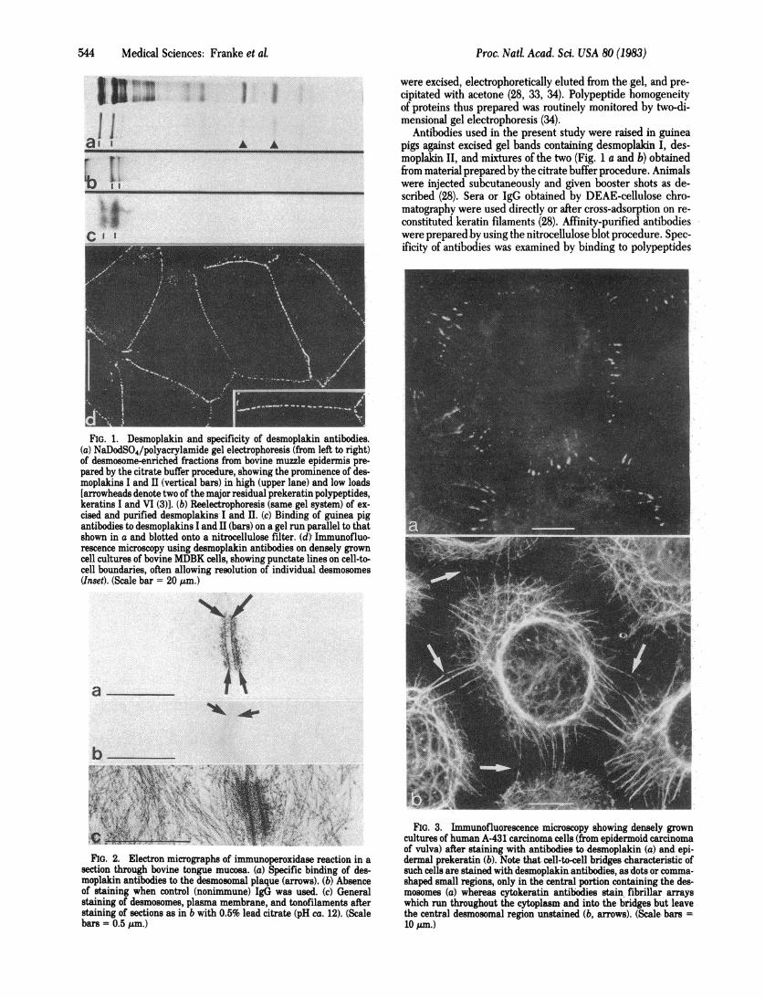
Antibodies used in the present study were raised in guineapigs against excised gel bands containing desmoplakin I, des-moplakin II, and mixtures of the two (Fig. 1 a and.b) obtainedfrom material prepared by the citrate buffer procedure. Animalswere injected subcutaneously and given booster shots as de-scribed (28). Sera or IgG obtained by DEAE-cellulose chro-matography were used directly or after cross-adsorption on re-constituted keratin filaments (28). Affinity-purified antibodieswere prepared by using the nitrocellulose blot procedure. Spec-ificity of antibodies was examined by binding to polypeptides
FIG. 1. Desmoplakin and specificity of desmoplakin antibodies.(a) NaDodSO4/polyacrylamide gel electrophoresis (from left to right)of desmosome-enriched fractions from bovine muzzle epidermis pre-pared by the citrate buffer procedure, showing the prominence of des-moplakins I and II (vertical bars) in high (upper lane) and low loads[arrowheads denote two of the major residual prekeratin polypeptides,keratins I and VI (3)]. (b) Reelectrophoresis (same gel system) of ex-cised and purified desmoplakins I and II. (c) Binding of guinea pigantibodies to desmoplakins I and II (bars) on a gel run parallel to thatshown in a and blotted onto a nitrocellulose filter. (d)- Immunofluo-rescence microscopy using desmoplakin antibodies on densely growncell cultures of bovine MDBK cells, showing punctate lines on cell-to-cell boundaries, often allowing resolution of individual desmosomes(Inset). (Scale bar = 20 Am.)
3'k-
FIG. 2. Electron micrographs of immunoperoxidase reaction in a
section through bovine tongue mucosa. (a) Specific binding of des-moplakin antibodies to the desmosomal plaque (arrows). (b) Absenceof staining when control (nonimmune) IgG was used. (c) Generalstaining of desmosomes, plasma membrane, and tonofilaments afterstaining of sections as in b with 0.5% lead citrate (pH ca. 12). (Scalebars = 0.5,um.)
FIG. 3. Immunofluorescence microscopy showing densely growncultures of human A-431 carcinoma cells (from epidermoid carcinomaof vulva) after staining with antibodies to desmoplakin (a) and. epi-dermal prekeratin (b). Note that. cell-to-cell bridges characteristic ofsuch cells are stained with desmoplakin antibodies, as dots or comma-shaped small regions, only in the-central portion containing the des-mosomes (a) whereas cytokeratin antibodies stain fibrillar arrayswhich run throughout the cytoplasm and into the bridges but leavethe central desmosomal region unstained (b, arrows). (Scale bars =
10 pam.)
a!r T'i !1
C II
a
b
Proc. Nad Acad. Sci. USA 80 (1983)
i!.on 4"&A

Proc. Natl. Acad. Sci. USA 80 (1983) 545
separated by gel electrophoresis and blotted onto nitrocellulosefilters (34, 35).
Immunolocalization. Immunofluorescence microscopy wasperformed on cells grown on cover slips or on frozen sections(3, 5, 6, 28, 33). For comparison, antibodies against cytokeratins(3, 5, 18, 19, 33) and vimentin (2, 29) were used. Electron mi-croscopic localization was done by using the immunoperoxidaseprocedure (28).
RESULTSSpecificity of Antibodies. Guinea pig antibodies raised
against bovine desmoplakin I or II or mixtures of I and II (Fig.lb) reacted, on nitrocellulose blots, with both desmoplakinbands (Fig. 1c), in agreement with the close similarity of des-moplakins I and II demonstrated by tryptic peptide maps (34).These antibodies specifically stained the desmosomal plaquesidentified by immunofluorescence (Fig. Id) and immunoelec-tron (Fig. 2) microscopy, in a fashion similar to a recently de-scribed antibody which reacted with some minor desmosomalplaque polypeptides (28). In immunofluorescence microscopy,individual desmosomes appeared as arrays offluorescent "dots"demarcating cell-to-cell boundaries (Figs. Id and 3a). Some-times positively stained dots were also seen away from the lat-eral cell membrane contacts, and such sites have been corre-lated with hemidesmosomes at the bottom surface or withendocytotically internalized desmosomal domains (28, 36).The antibodies to desmoplakins used in this study showed
crossreaction with desmosomal proteins of human (Fig. 3a),murine, and chicken cells. Desmoplakin antibodies were spe-cific for epithelial cells but also reacted with the desmosome-like junctions in intercalated disks of myocardiac cells and cul-tured cardiac myocytes (34). Positive cell lines were MDBK,BMGE, MH1C1, HEL, HeLa, A-431, and MCF-7. All othercell lines examined (lens-forming cells, 3T3, rat RVF smoothmuscle, and WI-38) were negative. In certain epithelial cell
cultures which formed intercellular bridges (A-431, BMGE,HEL) only the central portions of the bridges-i.e., the des-mosomes proper-were stained whereas antibodies to cytoker-atins stained the attached tonofilament bundles but not thedesmosomes (Fig. 3).When examined on frozen sections of human tissues, anti-
bodies to desmoplakins stained plasma membranes ofepithelialcells (Fig. 4), including those of thymic reticulum, kidney tu-bules, mesothelium, and myoepithelia of various glands. Theantibodies did not stain nonepithelial cells such as connectivetissue, striated and smooth muscle, blood vessel walls, spleen,adipose tissue, thymocytes, nerve tissue, and eye lens or tissuesin which certain junctions morphologically similar to desmo-somes ("desmosome-like junctions"; cf. refs. 26 and 37) havebeen described such as Sertoli cells, glia cells, endothelial cells,and retinal cells. The only exception ofnonepithelial cells show-ing a protein immunologically related to desmoplakins is rep-resented by the desmosome-like junctions of myocardiac inter-calated disks and of Purkinje fiber cells (ref. 34; cf. ref. 28).
Reaction of Desmoplaldn Antibodies on Tumor Sections.Epithelium-derived human tumors were invariably stained bydesmoplakin antibodies, usually revealing dotted arrays on cell-to-cell boundaries. This was found for differentiated nonmeta-static tumors such as basal cell epithelioma, myoepithelial-likecells of pleomorphic adenoma of the parotid gland, and ada-mantinoma and papillomas of the bladder (not shown) as wellas for squamous cell carcinomas of skin, tongue, epiglottis,esophagus, cervix, and rectum (Fig. 5). Metastatic lesions ofsquamous cell carcinoma in lymph nodes were also positive (notshown). Surrounding tumor-associated stromal tissue and bloodvessels were negative. Desmoplakin staining in dotted arrays,showing typical apical enrichment, was characteristic ofadeno-carcinomas of stomach and colon as well as metastatic lesionsderived from them (Fig. 6). Among liver tumors, those posi-tively identified as carcinomas by strong reaction with cytoker-
FIG. 4. Immunofluorescence microscopy showing desmosome-specific staining with desmoplakin antibodies on frozen sections through normalhuman tissues. (a) Esophagus, in survey, showing cell membrane specificity in all layers of mucosa and absence of reaction in connective tissueof lamina propria (LP). (Scale bar = 40 pm.) (b) Higher magnification of region as shown in a, presenting resolution of desmosomes in all layers,including the basal layer (bottom). (Scale bar = 20 gum.) (c) Liver, showing fluorescent dots in hepatocytes, especially rich in pericanalicular arrays,and bile duct cells (not shown) but not in mesenchymal cells such as endothelial and Kupffer cells. Arrows denote sinusoids (S). (Scale bar = 20Am.)
Medical Sciences: Franke et aL

546 Medical Sciences: Franke etaLP
FIG. 5. Immunofluorescence microscopy of frozen sections ofsquamous cell carcinoma of rectoanal origin (primary tumor) afterreaction with desmoplakin antibodies, showing specific punctate des-mosomal reaction in carcinoma cells but not in cells of stroma (S).(Scale bar = 20 ,Pm.)
atin antibodies were also stained by desmoplakin antibodiessuch as hepatocellular carcinomas, cholangiocarcinomas, andother carcinomas metastatic to the liver (not shown). Strongpunctate staining with anti-desmoplakin was also seen in ductaland lobular carcinomas of the breast (Fig. 7a), transitional cellcarcinoma ofthe bladder, various forms ofbronchial carcinomas(not shown), and carcinomas of upper respiratory tract epithe-
FIG. 6. Immunofluorescence microscopy of anti-desmoplakinstaining of frozen section of adenocarcinoma of the stomach. (a) Pri-mary tumor; (b) lymph node metastasis. L, lumina of glandular for-mations; S, stromal tissue. (Scale bars = 20 ,um.)
FIG. 7. Immunolocalization of desmoplakin on an infiltratingductal carcinoma of mammary gland (a) and a lymph node metastasisof solid carcinoma of maxillary sinus (b). Note absence of staining incarcinoma-associated stroma (S). (Scale bars = 20 pm.)
hum (Fig. 7b). Poorly differentiated or undifferentiated carci-nomas such as in lung, parotid gland, and bone were also pos-itive.
Negative reaction with desmoplakin antibodies was observedin all cases ofnonepithelial tumors examined, including uterineleiomyomas and leiomyosarcomas and endometrial stromal sar-comas, rhabdomyosarcomas, fibrosarcomas, hemangiomas ofthe liver, centroblastic and lymphoblastic lymphomas, malig-nant fibrous histiocytomas, melanomas, and neuroblastomas.All tissues negative for desmoplakin were also negative for stain-ing with antibodies to cytokeratins.
DISCUSSIONDesmoplakins I and II are large isoelectric polypeptides (Mr250,000 and 215,000) which are the major constituent proteinsof the desmosomal plaque (34). Both polypeptides have beenidentified in epithelia from various species (34), and the identityof desmoplakins I and II in various epithelia has been demon-strated by biochemical techniques (unpublished data). Anti-bodies to these proteins allow the specific detection of des-mosomes and other structures containing desmosomal plaquematerial [hemidesmosomes, internalized desmosomal domains(28, 36)] but do not react with any other type of junction.Expression of desmoplakin correlates with the appearance oftypical desmosomal ultrastructure and has been found only inepithelial cells and myocardium.
Because, in most epithelial cells, desmoplakins are amongthe major proteins (34) and show broad immunological cross-species reactivity, indicating their conservation during evolu-tion ofvertebrates, they are suitable markers for distinguishing
Proc. Natl. Acad. Sci. USA 80 (1983)

Proc. NatL Acad. Sci. USA 80 (1983) 547
epithelial (and myocardial) cells from other cell types and, con-sequently, carcinomas from other types ofmalignant neoplasms.Whether certain desmosome-like structures described in somekinds of tumors of unclear origin such as Ewing sarcoma andmeningiomas of unclear cell character are related to desmo-somes and contain desmoplakins remains to be seen. In addi-tion, the combined use of antibodies to both independent mo-lecular markers-cytokeratins and desmoplakins-makes itpossible now to define epithelial and carcinoma cells by theconcomitant expression oftwo characteristic major cytoskeletalproteins, irrespective of the morphological appearance of thespecific cells. We propose that immunocytochemical studieswith antibodies to desmoplakins, together with antibodies tocytokeratin already proven to be valuable in clinical diagnosis,be used for establishing the positive diagnosis or the exclusionof carcinoma in difficult or doubtful cases.
Antibodies to desmoplakins also can be used in pathologicalexaminations of exfoliative preparations as has been shown forsuspensions of cells from epithelia of urinary and genital tractas well as intestine (28). Immunolocalization ofdesmoplakin fur-ther facilitates quantitation ofdesmosomal densities at the lightmicroscope level and allows the detection of disorders andchanges of desmosomal arrays on surfaces of normal and neo-plastic cells.
1. Lazarides, E. (1980) Nature (London) 283, 249-256.2. Franke, W. W., Schmid, E., Osborn, M. & Weber, K. (1978)
Proc. Nati Acad. Sci. USA 75, 5034-5038.3. Franke, W. W., Weber, K., Osborn, M., Schmid, E. & Freu-
denstein, C. (1978) Exp. Cel Res. 116, 429-445.4. Sun, T.-T. & Green, H. (1978) Cell 14, 469-476.5. Franke, W. W., Appelhans, B., Schmid, E., Freudenstein, C.,
Osborn, M. & Weber, K. (1979) Differentiation 15, 7-25.6. Franke, W. W., Schmid, E., Weber, K. & Osborn, M. (1979)
Exp. Cell Res. 118, 95-109.7. Sun, T.-T., Shih, C. H. & Green, H. (1979) Proc. NatL Acad. Sci.
USA 76, 2813-2817.8. Battifora, H., Sun, T.-T., Bahu, R. M. & Rao, S. (1980) Hum.
Pathot 11, 635-641.9. Bannasch, P., Zerban, H., Schmid, E. & Franke, W. W. (1980)
Proc. Nati Acad. Sci. USA 77, 4948-4952.10. Schlegel, R., Banks-Schlegel, S., McLeod, J. A. & Pinkus, G. S.
(1980) Am. J. PathoL 110, 41-49.11. Altmannsberger, M., Osborn, M., H6lscher, A., Schauer, A. &
Weber, K. (1981) Virchows Arch. B 37, 277-284.12. Osborn, M., Geisler, N., Shaw, G., Sharp, G. & Weber, K.
(1982) Cold Spring Harbor Symp. Quant. BioL 46, 413-429.
13. Asch, B. B., Burstein, N. A., Vidrich, A. & Sun, T.-T. (1981)Proc. Natl Acad. Sci. USA 78, 5643-5647.
14. Gabbiani, G., Kapanci, Y., Barrazone, P. & Franke, W. W.(1981) Am. J. Pathot 104, 206-216.
15. Krepler, R., Denk, H., Weirich, E., Schmid, E. & Franke, W.W. (1981) Differentiation 20, 242-252.
16. Wu, Y.-J. & Rheinwald, J. G. (1981) Cell 25, 627-635.17. Fuchs, E. & Green, H. (1980) Cell 19, 1033-1042.18. Franke, W. W., Denk, H., Kalt, R. & Schmid, E. (1981) Exp.
Cell Res. 131, 299-318.19. Franke, W. W., Schiller, D. L., Moll, R., Winter, S., Schmid,
E., Engelbrecht, I., Stadler, J., Denk, H., Krepler, R. &Platzer, B. (1981)J. Mol Biol 153, 933-959.
20. Farquhar, M. G. & Palade, G. E. (1963)J. Cell Biol 17, 375-412.21. Weinstein, R., Zel, G. & Merk, F. B. (1974) in Membrane Trans-
formations in Neoplasia, eds. Schultz, J. & Block, R. E. (Aca-demic, New York), pp. 127-150.
22. Pauli, B. U., Cohen, S. M., Alroy, J. & Weinstein, J. R. (1978)Cancer Res. 38, 3276-3285.
23. Alroy, J., Pauli, B. U. & Weinstein, R. S. (1981) Cancer (Phila-delphia) 47, 104-112.
24. Ghadially, F. N. (1980) Diagnostic Electron Microscopy of Tu-mors (Butterworth, London), pp. 51-67.
25. Erlandson, R. A. (1981) Diagnostic Transmission Electron Mi-croscopy ofHuman Tumors (Masson USA, New York), pp. 107-116; 169-186.
26. Ghadially, F. N. (1982) Ultrastructural Pathology of the Cell andMatrix (Butterworth, London), pp. 797-820.
27. Denk, H., Krepler, R., Lackinger, E., Artlieb, U. & Franke, W.W. (1982) Lab. Invest. 46, 584-596.
28. Franke, W. W., Schmid, E., Grund, C., Mueller, H., Engel-brecht, I., Moll, R., Stadler, J. & Jarasch, E.-D. (1981) Differ-entiation 20, 217-241.
29. Franke, W. W., Schmid, E., Winter, S., Osborn, M. & Weber,K. (1979) Exp. Cell Res. 123, 25-46.
30. Ramaekers, F. C. S., Osborn, M., Schmid, E., Weber, K., Bloe-mendal, H. & Franke, W. W. (1980) Exp. Cell Res. 127, 309-327.
31. Skerrow, C. J. & Maltoltsy, A. G. (1974)J. Cell Biol 63, 515-523.32. Drochmans, P., Freudenstein, C., Wanson, J. C., Laurent, L.,
Keenan, T. W., Stadler, J., Leloup, R. & Franke, W. W. (1978)J. Cell Biol 79, 427-443.
33. Franke, W. W., Schmid, E., Freudenstein, C., Appelhans, B.,Osborn, M., Weber, K. & Keenan, T. W. (1980)J. Cell Biol 84,633-654.
34. Mueller, H. & Franke, W. W. (1982)J. Mol BioL, in press.35. Krohne, G., Stick, R., Kleinschmidt, J. A., Moll, R., Franke, W.
W. & Hausen, P. (1982)J. Cell BioL 94, 749-754.36. Kartenbeck, J., Schmid, E., Franke, W. W. & Geiger, B. (1982)
EMBOJ. 1, 725-732.37. Fawcett, D. W. (1981) The Cell (Saunders, Philadelphia), 2nd
Ed., pp. 156-167.
Medical Sciences: Franke et aL