immunofluorescence and studies of visna virus in cell...
TRANSCRIPT

JOURNAL OF VIROLOGY, Dec. 1967, p. 1265-1270Copyright © 1967 American Society for Microbiology
Vol. 1, No. 6Prinited inl U.S.A.
Immunofluorescence and Cytochemical Studies ofVisna Virus in Cell CultureD. H. HARTER, K. C. HSU, AND H. M. ROSE
Departments of Neurology and Microbiology, College of Physicianis anid Surgeonls,Columbia University, New York, New York 10032
Received for publication 3 July 1967
Sequential morphological changes occurring in sheep choroid plexus cells in-fected with visna virus were studied by direct immunofluorescence, acridine orange,
and hematoxylin and eosin staining methods. Specific immunofluorescence wasfirst detected in the perinuclear cytoplasm of solitary cells 24 hr after infection. Asthe infection progressed, viral antigen appeared in an increasing number of cells,and rounded globular cells with long slender processes harboring intense fluores-cence were seen. Nuclear fluorescence was not observed in infected monolayers.Polykaryocytes formed within 6 hr after inoculation due to the direct cell-fusingeffect of the virus inoculum did not show specific fluorescence. Viral antigen was
found, however, in the cytoplasm of multinucleated giant cells in cover slips har-vested after new infective virus had been released, and later in the course of infec-tion circular fluorescent inclusions were seen in the cytoplasm of polykaryocytes.Comparable eosinophilic inclusions were observed in hematoxylin and eosinpreparations, and acridine orange staining of infected monolayers demonstratedsimilar inclusions which fluoresced with the color characteristic of single-strandednucleic acid and were susceptible to digestion with ribonuclease. Visna virusappears to be a ribonucleic acid virus which replicates in the cytoplasm.
Visna virus is a yet unclassified virus whichcauses a slow, progressive, and fatal disease ofthe nervous system of sheep (31, 32). Propagationin cell cultures derived from the sheep choroidplexus (33) has resulted in characterization of anumber of the biological, physical, and chemicalproperties of the virus (16, 38-40).Although electron microscopic studies indicate
that virus particles are formed by budding on thecytoplasmic membrane of host cells, the finestructure of the nuclei and cytoplasm of infectedcells looks much like that of uninfected controlcells (36), and the cellular site of virus synthesishas not yet been established.The present communication describes experi-
ments performed to determine the site of visnavirus replication in sheep choroid plexus cellsby use of immunofluorescence and cytochemicaltechniques. The results suggest that visna virusis a ribonucleic acid (RNA) virus which issynthesized in the cytoplasm of infected cells.
MATERIALS AND METHODS
Cell cultures. Sheep choroid plexus (SCP) cells wereprepared by trypsin dispersion of choroid plexuses re-moved from the brains of exsanguinated domesticHampshire or Suffolk sheep as previously described
(16). Cells were grown in reinforced Eagle's medium(1) containing 10%o fetal bovine serum in 250-mlplastic flasks and incubated at 37 C. Cell lines pre-pared in this manner consist of elongated fibroblasticcells which survive 10 to 12 serial passages.
Virus. Visna virus K485 was obtained from H. Thor-mar and P. A. Palsson, Institute for ExperimentalPathology, University of Iceland, and was carriedthrough eight serial passages in SCP cells. Eighthpassage virus containing 3.4 X 10⁶.° TCID5o/ml wasused in the experiments. Concentrated visna viruscontaining 108.0 TcID50/ml was prepared by clarifica-tion at 1,500 X g for 10 min, followed by centrifuga-tion at 78,000 X g for 6 hr and suspension of thepelleted material in one-fiftieth of its original volumein reinforced Eagle's medium plus 0.5%c bovine plasmaalbumin (Fraction V, Armour Pharmaceutical Co.,Kankakee, Ill.); this concentrated preparation wasused in an experiment to produce rapid cell fusion. Allvirus stocks were stored at -70 C until they wereused.
Vislia viruts anitiserum. Serum 4992 from a sheep in-fected with visna virus was kindly supplied by H.Thormar. This serum has a specific neutralizationtiter of 1:1,024 and was used in the preparation offluorescein-labeled antiserum.
Ihifectiont of cell cultures. Experiments were per-formed by infecting confluent SCP monolayers grownin 60-mm plastic tissue culture dishes containing two18-mm square glass cover slips. Replicate dishes were
1265
on Novem
ber 12, 2018 by guesthttp://jvi.asm
.org/D
ownloaded from

HARTER, HSU, AND ROSE
washed twice with 5.0 ml of phosphate-buffered saline(PBS), pH 7.2 (9), and inoculated with 0.5 ml of un-
concentrated or concentrated virus. After adsorptionfor 3 hr at 37 C, the inoculum was removed and thecell sheet was washed with PBS. Maintenance medium(reinforced Eagle's medium and 2% inactivated lambserum) was added, and the cultures were incubatedat 37 C in a humidified atmosphere of 5% carbon di-oxide. At intervals after infection, the medium was
harvested, and bovine plasma albumin (BSA) was
added to a concentration of 0.5%/O. The harvestedmedium was then frozen and stored at -70 C until itwas assayed for infective virus. Cover slips were re-
moved and fixed for cytological studies. Uninfectedcultures inoculated with 0.5 ml of reinforced Eagle'smedium and 0.5% BSA were handled in the same
manner and served as controls.Assay of inifective virus. Confluent SCP monolayers
in 60-mm plastic petri dishes, four plates per dilution,were washed twice with PBS and inoculated with serial10-fold dilutions of virus in reinforced Eagle's mediumwith 0.5% BSA. After a 3-hr adsorption period at37 C, 5.0 ml of maintenance medium was added, andthe cultures were incubated at 37 C in a humidifiedatmosphere of 5% carbon dioxide. Cultures were ex-
amined after 14 days for cytopathic changes, and 50%linfectivity end points were calculated by the method ofReed and Muench (29).
Staininlg procedures. Immunofluorescence studieswere performed by the direct staining method ofCoons and co-workers (7, 8). The globulin fraction ofvisna virus antiserum 4992 was precipitated with so-
dium sulfate and conjugated with fluorescein isothio-cyanate (35). Fluorescein-labeled serum was ab-sorbed with rat and mouse liver powder before use
and retained a neutralizing titer of 1:200 against20,000 TCID5o of visna virus. Cover slips to be stainedby the immunofluorescence method were fixed inacetone, washed with PBS, and stained for 30 min at25 C with a 1:4 dilution of fluorescein-labeled visnavirus antiserum. Cover slips were then washed withPBS and mounted on microscope slides in bufferedglycerin.
Hematoxylin and eosin staining was done on cover
slips fixed in Zenker's fluid for 60 min by the methodof Enders and Peebles (11).
Acridine orange staining was performed on cover
slips fixed in Carnoy's fixative, stained with 0.05%
acridine orange (Chroma-Gesellschaft, Stuttgart,Germany) in acetate buffer (pH 5.4), and mounted inglycerin and acetate buffer (12).
Digestion with nucleases was performed by in-cubating Carnoy-fixed cover slips with either 0.05%five times crystallized ribonuclease in acetate buffer(pH 5.4) containing 0.003 M MgCl2 or 0.01% once
crystallized deoxyribonuclease in 0.02 M tris(hydroxy-methyl)aminomethane (Tris) buffer (pH 7.3) contain-ing 0.003 M MgCl2 at 37 C for 1 hr. Nucleases were
obtained from Sigma Chemical Co., St. Louis, Mo.Control cover slips were incubated with buffer alone.
Fluorescent-antibody- and acridine orange-stainedpreparations were examined by use of a ReichertFluorex microscope with a BG12 exciter filter and an
OG4 barrier filter. Photographs were taken on Ansco-chrome D-200 or Kodachrome X film.
RESULTS
Growth of visna virus in SCP cells. The amountof visna virus sequentially released in SCP culturesinfected at a multiplicity of 4 TCID5o per cell isshown in Table 1.Newly released virus was first detected in
medium harvested 24 hr after infection. An ex-
ponential increase then occurred during thenext 24 hr, and peak titers were found in mediumharvested at 96 and 120 hr after infection. Thispattern of viral multiplication is in general agree-ment with previously reported studies on thegrowth of visna virus in SCP cell cultures (16,37).
Morphological changes in visna virus-infectedSCP cells. Fluorescent-antibody staining ofvisna-infected cell monolayers was performed on
cover slips harvested at 6, 24, 31, 48, 72, 96, ane120 hr after infection. The results of these studie'are summarized in Table 1.
Specific fluorescence was first detected inisolated cells 24 hr after infection. Fluorescentantibody was found to be localized in the cyto-plasm of scattered fibroblastic cells constitutingless than 1% of the cell population of the mono-
layer. Such fluorescence was often noted to bemore intense about the nucleus of the cell, and
TABLE 1. Immunofluorescenice of visna virus-inifected sheep choroid plexus cells
Time after Infective virus Intensity of specific fluorescence Approximateinfection (hr)a (TC!D5o/m1) Inclusions percentage of
Fibroblasts Polykaryocytes Rounded cells cells stained
24 1.0 x 10³ + 0 0 0 <131 6.3 X 10³ + 0 0 0 1048 3.5 X 10⁵ ++ + + 0 1572 6.3X 10⁵ ++ + + 0 3096 2.2 X 10⁷ +++ ++ ++ + 70120 4.3 X 10⁷ +++ +++ +++ +++ 70
a Time after inoculation of sheep choroid plexus monolayers with visna virus at a multiplicity of 4TCID,0 per cell and incubation at 37 C.
J. VIROL.1 266
on Novem
ber 12, 2018 by guesthttp://jvi.asm
.org/D
ownloaded from

VISNA VIRUS 1267
lb
Id
3
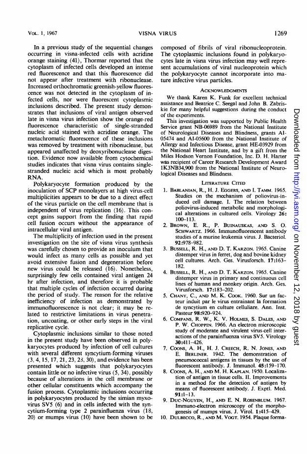
FIG. la to d. Immunofluorescent staining of shleep choroid plexus cells inlfected with visnia virus. Cells were
inioculated at a multiplicity of4 TCID50 per cell and at intervals were fixed antd stainted by thle direct immuniofluores-ceiice technique. (la) 72 hr after inifectiont, specific fluorescent stailning in the cytoplasm of a multilnucleated giantcell, X 125; (lb) 96 hr after infection, fluorescenit anitibody localized to the cytoplasm offibroblasts and in severalrounid globular cells with elonigated processes, X 50; (Ic) 120 hr after inifection, specific fluorescenice in the cyto-plasm offibroblast and multinucleated giant cell, X 125; (Id) 120 hr after infectioni, aggregates of fluorescentantibody appearinig as inclusions in thle cytoplasm of a multilnucleated giant cell, X 125.
FIG. 2. Circular eosinophilic i,icluisioni in polykaryocyte 120 hr after inoculationt with visna virus. Hematoxylinanld eosini stain. X 100.
FIG. 3. Intracytoplasmic inclusionis in polykaryocytes stained with acridine oranige 120 hr after inoculation withvisiia virus. Iniclusionis demonstrate the redfluorescence associated with sinigle-stranded RNA. X 100.
was fairly homogeneous in character. Polykaryo-cytes were observed at 6 and 24 hr after infection,and were probably the result of the direct fusingeffect of the visna virus inoculum (16). Suchmultinucleated cells did not show fluorescence.
The findings 31 hr after infection were similarto those at 24 hr, but at 31 hr viral antigen wasseen in the cytoplasm of a larger number offibroblastic cells.
After 48 hr, approximately 15%ao of the cells
VOL. 1, 1967
on Novem
ber 12, 2018 by guesthttp://jvi.asm
.org/D
ownloaded from
HARTER, HSU, AND ROSE
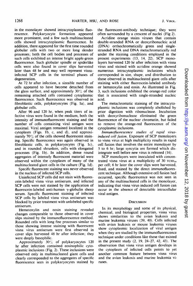
in the monolayer showed intracytoplasmic fluo-rescence. Polykaryocyte formation appearedmore prominent, and a few such multinucleatedcells showed intracytoplasmic fluorescence. Inaddition, there appeared for the first time roundedglobular cells with two or more long slenderprocesses; both the cell bodies and processes ofsuch cells exhibited an intense bright apple-greenfluorescence. Such globular spindle or spiderlikecells were also found in cover slips harvestedlater than 48 hr and may well represent visna-infected SCP cells in the terminal phases ofdegeneration.At 72 hr after infection, a sizeable number of
cells appeared to have become detached fromthe glass surface, and approximately 30%,7O of theremaining attached cells contained cytoplasmicfluorescence. Such fluorescence was observed infibroblastic cells, polykaryocytes (Fig. la), andglobular cells.
After 96 and 120 hr, when peak titers of in-fective virus were found in the medium, both theintensity of immunofluorescent staining and thenumber of cells containing viral antigen weremaximal. Viral antigen remained localized in thecytoplasm (Figs. lb, c, and d), and approxi-mately 70% of the cells showed fluorescent stain-ing. Specific fluorescence was again observed infibroblastic cells, in polykaryocytes (Fig. Ic),and in rounded shrunken, cells with elongatedprocesses (Fig. lb). In addition, dense roundaggregates of intensely fluorescent material wereobserved within the cytoplasm of many of themultinucleated giant cells in the culture (Fig. Id).
Specific fluorescent staining was never observedin the nucleus of infected SCP cells.
Uninfected SCP cells did not stain with fluores-cein-labeled visna virus antiserum, and infectedSCP cells were not stained by the application offluorescein-labeled anti-human y-globulin sheepserum. Specific fluorescent staining of infectedSCP cells by labeled visna virus antiserum wasblocked by prior treatment with unlabeled specificantiserum.
Hematoxylin and eosin staining revealedchanges comparable to those observed in coverslips stained by the immunofluorescence method.Rounded cells with long fine processes similar tothose showing intense staining with fluorescentvisna virus antiserum were first observed incover slips harvested 48 hr after infection; theywere deeply basophilic.
Approximately 30%/ of polykaryocytes 120hr after infection contained eosinophilic cyto-plasmic inclusions (Fig. 2). These inclusions wereobserved only in multinucleated giant cells andclearly corresponded to the aggregates of specificfluorescence seen in polykaryocytes stained by
the fluorescent-antibody technique; they wereoften surrounded by a crescent of nuclei (Fig 2).
Acridine orange stains viruses that containdouble-stranded RNA or deoxyribonucleic acid(DNA) orthochromatically green and single-stranded RNA and DNA metachromatically redunder the staining conditions employed in thepresent experiments (13, 14, 22). SCP mono-layers harvested 120 hr after infection with visnavirus and stained with acridine orange showednumerous intracytoplasmic inclusions whichcorresponded in size, shape, and distribution tothose observed in multinucleated giant cells afterstaining with either fluorescein-labeled antibodyor hematoxylin and eosin. As illustrated in Fig.3, such inclusions exhibited the orange-red colorthat is associated with single-stranded nucleicacid.The metachromatic staining of the intracyto-
plasmic inclusions was completely abolished byprior digestion with ribonuclease. Incubationwith deoxyribonuclease eliminated the greenfluorescence of the nuclear chromatin, but failedto remove the orange-red fluorescence of thecytoplasmic inclusions.
Immunofluorescence studies of rapid virus-induced cell fusion. Exposure of SCP monolayersto visna virus at high multiplicity results in rapidcell fusion that involves the entire monolayer by5 to 6 hr; large syncytia are formed which dis-integrate well before new virus is released (16).SCP monolayers were inoculated with concen-
trated visna virus at a multiplicity of 30 TCID1Oper cell; 6 hr later the cover slips were harvested,fixed, and stained by the direct immunofluores-cent technique. Although extensive cell fusion hadoccurred, specific fluorescence was not seen inany of the multinucleated cells in the monolayer,indicating that visna virus-induced cell fusion canoccur in the absence of detectable intracellularviral antigen.
DIscuSSION
In its morphology and some of its physical,chemical, and biological properties, visna virusshows similarities to the avian leukosis andmurine leukemia viruses (39, 40). Cells infectedwith avian leukosis or mouse leukemia virusesshow cytoplasmic localization of viral antigenwhen they are studied by the immunofluorescencetechnique under conditions like those that existedin the present study (2, 19, 24-27, 42, 43). Theobservation that visna virus antigen develops inthe cytoplasm of infected cells points to yetanother common feature between visna virusand the avian leukosis and murine leukemia vi-ruses.
1268 J. VIROL.
on Novem
ber 12, 2018 by guesthttp://jvi.asm
.org/D
ownloaded from
VISNA VIRUS
In a previous study of the sequential changesoccurring in visna-infected cells with acridineorange staining (41), Thormar reported that thecytoplasm of infected cells developed an intensered fluorescence and that this fluorescence didnot appear after treatment with ribonuclease.Increased orthochromatic greenish-yellow fluores-cence was not detected in the cytoplasm of in-fected cells, nor were fluorescent cytoplasmicinclusions described. The present study demon-strates that inclusions of viral antigen observedlate in visna virus infection show the orange-redfluorescence characteristic of single-strandednucleic acid stained with acridine orange. Themetachromatic fluorescence of these inclusionswas removed by treatment with ribonuclease, butappeared unaffected by deoxyribonuclease diges-tion. Evidence now available from cytochemicalstudies indicates that visna virus contains single-stranded nucleic acid which is most probablyRNA.
Polykaryocyte formation produced by theinoculation of SCP monolayers at high virus-cellmultiplicities appears to be due to a direct effectof the virus particle on the cell membrane that isindependent of virus replication (16). This con-cept gains support from the finding that rapidcell fusion occurs without the appearance ofintracellular viral antigen.The multiplicity of infection used in the present
investigation on the site of visna virus synthesiswas carefully chosen to provide an inoculum thatwould infect as many cells as possible and yetavoid extensive fusion and degeneration beforenew virus could be released (16). Nonetheless,surprisingly few cells contained viral antigen 24hr after infection, and therefore it is probablethat multiple cycles of infection occurred duringthe period of study. The reason for the relativeinefficiency of infection as demonstrated byimmunofluorescence is not clear; it may be re-lated to restrictive limitations in virus penetra-tion, uncoating, or other early steps in the viralreplicative cycle.
Cytoplasmic inclusions similar to those notedin the present study have been observed in poly-karyocytes produced by infection of cell cultureswith several different syncytium-forming viruses(3, 4, 15, 17, 21, 23, 28, 30), and evidence has beenpresented which suggests that polykaryocytescontain little or no infective virus (5, 34), possiblybecause of alterations in the cell membrane orother cellular constituents which accompany thefusion process. Cytoplasmic inclusions occurringin polykaryocytes produced by the simian myxo-virus SV5 (6) and in cells infected with the syn-cytium-forming type 2 parainfluenza virus (18,20) or mumps virus (10) have been shown to be
composed of fibrils of viral ribonucleoprotein.The cytoplasmic inclusions found in polykaryo-cytes late in visna virus infection may well repre-sent accumulations of viral nucleoprotein whichthe polykaryocyte cannot incorporate into ma-ture infective virus particles.
ACKNOWLEDGMENTSWe thank Karen K. Funk for excellent technical
assistance and Beatrice C. Seegal and John B. Zabris-kie for many helpful suggestions during the conductof the experiments.
This investigation was supported by Public HealthService grant NB-06989 from the National Instituteof Neurological Diseases and Blindness, grants Al-05474 and AI-05600 from the National Institute ofAllergy and Infectious Disease, grant HE-03929 fromthe National Heart Institute, and by a gift from theMiles Hodson Vernon Foundation, Inc. D. H. Harterwas recipient of Career Research Development AwardlK3NB34,900 from the National Institute of Neuro-logical Diseases and Blindness.
LITERATURE CITED1. BABLANIAN, R., H. J. EGGERS, AND I. TAMM. 1965.
Studies on the mechanism of poliovirus-in-duced cell damage. I. The relation betweenpoliovirus-induced metabolic and morphologi-cal alterations in cultured cells. Virology 26:100-113.
2. BROWN, E. R., P. BUINAUSKAS, AND S. 0.SCHWARTZ. 1966. Immunofluorescent antibodystudies of a murine leukemia virus. J. Bacteriol.92:978-982.
3. BUSSELL, R. H., AND D. T. KARZON. 1965. Caninedistemper virus in ferret, dog and bovine kidneycell cultures. Arch. Ges. Virusforsch. 17:163-182.
4. BUSSELL, R. H., AND D. T. KARZON. 1965. Caninedistemper virus in primary and continuous celllines of human and monkey origin. Arch. Ges.Virusforsch. 17:183-202.
5. CHANY, C., AND M. K. COOK. 1960. Sur un fac-teur induit par le virus entrainant la formationde syncytium en culture celluliare. Ann. Inst.Pasteur 98:920-924.
6. COMPANS, R. W., K. V. HOLMES, S. DALES, ANDP. W. CHOPPIN. 1966. An electron microscopicstudy of moderate and virulent virus-cell inter-actions of the parainfluenza virus SV5. Virology30:411-426.
7. COONS, A. H., H. J. CREECH, R. N. JONES, ANDE. BERLINER. 1942. The demonstration ofpneumococcal antigens in tissues by the use offluorescent antibody. J. Immunol. 45:159-170.
8. COONS, A. H., AND M. H. KAPLAN. 1950. Localiza-tion of antigen in tissue cells. II. Improvementsin a method for the detection of antigen bymeans of fluorescent antibody. J. Exptl. Med.91:1-13.
9. DUC-NGUYEN, H., AND E. N. ROSENBLUM. 1967.Immuno-electron microscopy of the morpho-genesis of mumps virus. J. Virol. 1:415-429.
10. DULBECCO, R., AND M. VOGT. 1954. Plaque forma-
1 269VOL. 1, 1967
on Novem
ber 12, 2018 by guesthttp://jvi.asm
.org/D
ownloaded from

HARTER, HSU, AND ROSE
tion and isolation of pure lines with poliomyeli-tis viruses. J. Exptl. Med. 99:167-182.
11. ENDERS, J. F., AND T. C. PEEBLES. 1954. Propaga-tion in tissue cultures of cytopathogenic agentsfrom patients with measles. Proc. Soc. Exptl.Med. 86:277-286.
12. FRANKLIN, R. M. 1962. A cytochemical descrip-tion of the multiplication of mengovirus in L-929 cells. J. Cell Biol. 12:1-16.
13. GOMATOS, P. J., AND I. TAMM. 1963. The sec-
ondary structure of reovirus RNA. Proc. Natl.Acad. Sci. U.S. 49:707-714.
14. GOMATOS, P. J., I. TAMM, S. DALES, AND R. M.FRANKLIN. 1962. Reovirus type 3: physicalcharacteristics and interaction with L cells.Virology 17:441-454.
15. GRESSER, I., AND J. F. ENDERS. 1961. Cytopatho-genicity of mumps virus in cultures of chickembryo and human amnion cells. Proc. Soc.Exptl. Biol. Med. 107:804-807.
16. HARTER, D. H., AND P. W. CHOPPIN. 1967. Cell-fusing activity of visna virus particles. Virology31:279-288.
17. HOLMES, K. V., AND P. W. CHOPPIN. 1966. On therole of the response of the cell membrane indetermining virus virulence. Contrasting effectsof the parainfluenza virus SV5 in two cell types.J. Exptl. Med. 124:501-520.
18. HOWE, C., C. MORGAN, C. DE VAUX ST. CYR,K. C. Hsu, AND H. M. ROSE. 1967. Morpho-genesis of type 2 parainfluenza virus examinedby light and electron microscopy. J. Virol. 1:215-237.
19. KELLOFF, G., AND P. K. VOGT. 1966. Localizationof avian tumor virus group-specific antigen incell and virus. Virology 29:377-384.
20. KUHN, N. O., AND C. G. HARFORD. 1963. Elec-tron microscopic examination of cytoplasmicinclusion bodies in cells infected with parain-fluenza virus, type 2. Virology 21:527-530.
21. LEPINE, P., C. CHANY, B. DROZ, AND F. ROBBE-FOSSAT. 1959. Cytopathogenic effect of twonewly recognized myxovirus strains: mecha-nism of syncytial formation. Ann. N.Y. Acad.Sci. 81:62-72.
22. MAYOR, H. D., AND N. 0. HILL. 1961. Acridineorange staining of single-stranded DNA bac-teriophage. Virology 14:264-266.
23. MILOVANOVIC, M. V., J. F. ENDERS, AND A.MITUS. 1957. Cultivation of measles virus inhuman amnion cells and in developing chickembryo. Proc. Soc. Exptl. Biol. Med. 95:120-127.
24. NOYES, W. F. 1960. Development of Rous sar-
coma virus antigens in cultured chick embryocells. Virology 12:488-492.
25. OSATO, T., E. A. MIRAND, AND J. T. GRACE, JR.1964. Propagation and immunofluorescent in-vestigations of Friend virus in tissue culture.Nature 201:52-54.
26. OSATO, T., E. A. MIRAND, AND J. T. GRACE, JR.1965. Hemadsorption and immunofluores-cence of Friend virus in cell culture. Proc. Soc.Exptl. Biol. Med. 119:1187-1191.
27. PAYNE, F. E., J. J. SOLOMON, AND H. G. PUR-
CHASE. 1966. Immunofluorescent studies ofgroup-specific antigen of the avian sarcoma-leukosis viruses. Proc. Natl. Acad. Sci. U.S.55:341-349.
28. PLOWRIGHT, W., AND R. D. FERRIS. 1959. Studieswith rinderpest virus in tissue culture. I.Growth and cytopathogenicity. J. Comp.Pathol. 69:152-172.
29. REED, L. J., AND H. MUENCH. 1938. A simplemethod of estimating fifty per cent endpoints.Am. J. Hyg. 27:493-497.
30. RUCKLE, G. 1957. Studies with measles virus. I.Propagation in different tissue culture systems.J. Immunol. 78:330-340.
31. SIGURDSSON, B., AND P. A. PALSSON. 1958. Visnaof sheep. A slow demyelinating infection. Brit.J. Exptl. Pathol. 39:519-528.
32. SIGURDSSON, B., P. A. PALSSON, AND H.GRIMSSON. 1957. Visna, a demyelinating trans-missible disease of sheep. J. Neuropathol.Exptl. Neurol. 16:389-403.
33. SIGURDSSON, B., H. THORMAR, AND P. A. PALSSON.1960. Cultivation of visna virus in tissue culture.Arch. Ges. Virusforsch. 10:368-381.
34. STOKER, M. G. P. 1958. Mode of intracellulartransfer of herpes virus. Nature 182:1525-1526.
35. STRAUSS, A. J. L., B. C. SEEGAL, K. C. Hsu, P. M.BURKHOLDER, W. L. NASTUK, AND K. E.OSSERMAN. 1960. Immunofluorescence demon-stration of a muscle binding, complement-fixing serum globulin fraction in myastheniagravis. Proc. Soc. Exptl. Biol. Med. 105:184-191.
36. THORMAR, H. 1961. An electron microscope studyof tissue cultures infected with visna virusVirology 14:463-475.
37. THORMAR, H. 1963. The growth cycle of visnavirus in monolayer cultures of sheep cells.Virology 19:273-278.
38. THORMAR, H. 1965. A comparison of visna andmaedi viruses. I. Physical chemical and biologi-cal properties. Res. Vet. Sci. 6:117-129.
39. THORMAR, H. 1965. Physical, chemical and bi-ological properties of visna virus and its rela-tionship to other animal viruses, p. 335-340.In Slow, Latent and Temperate Virus Infections.National Institute of Neurological Diseases andBlindness Monograph No. 2, Washington, D. C.
40. THORMAR, H. 1966. A study of visna and maediviruses and their relationship to other viruses ofanimals. Dansk Videnskabs Forlag, Copen-hagen.
41. THORMAR, H. 1966. Observations on visna virus-infected cell cultures stained with acridineorange. Acta Pathol. Microbiol. Scand. 68:54-58.
42. VOGT, P. K., AND H. RUBIN. 1961. Localization ofinfectious virus and viral antigen in chick fibro-blasts during successive stages of infection withRous sarcoma virus. Virology 13:528-544.
43. YOSKIDA, K., K. L. SMITH, AND D. PINKEL. 1966.Studies of murine leukemia viruses. I. Detectionof Moloney and Rauscher leukemia viruses byindirect immunofluorescence. Proc. Soc. Exptl.Biol. Med. 121:72-81.
1270 J. VIROL.
on Novem
ber 12, 2018 by guesthttp://jvi.asm
.org/D
ownloaded from