immunoglobulin and t-cell receptor gene rearrangement
TRANSCRIPT

FESTSCHRIFT SECTION
Immunoglobulin and T-cell receptor gene rearrangement
Implications for the diagnosis of lymphoproliferative disorders
ANDREW J. FISHLEDER, MD
• Recent studies have significantly broadened understanding of immunoglobulin production and T-cell receptor formation at the gene level. A limited number of genes can be rearranged during the course of B-cell or T-cell development to yield unique D N A sequences that will code for specific antibodies and T-cell receptor proteins. Southern blot hybridization analysis allows sensitive examination of lymphocyte D N A for the presence or absence of gene rearrangements. General mechanisms that underlie immunoglobulin and T-cell receptor rearrangement are reviewed, along with the diagnostic applications of detection of gene rearrangement by Southern blot hybridization technique. • INDEX TERMS: ANTIBODY FORMATION; ANTIBODY SPECIFICITY; LYMPHOPROLIFERATIVE DISORDERS; T LYMPHOCYTES • CLEVE CLIN J MED 1989; 56:716-721
ADVANCES in molecular biology over the past decade have resulted in a wealth of in-formation regarding the genes responsible for immunoglobulin production and T-cell re-
ceptor ( T Ç R ) formation. This discussion will first address the normal organization of the immunoglobulin gene system and the implications of the detection of im-munoglobulin gene rearrangement in the diagnosis of lymphoproliferative disorders. It will then examine the TCR beta chain gene system and the role it plays in the specificity of T cells for foreign antigens.
The immunoglobulin molecule is well known to con-tain light chains and heavy chains, both of which con-tain variable and constant regions. The variable regions
From the Department of Laboratory Hematology, The Cleveland Clinic Foundation. Submitted July 1987; accepted Feb 1988.
Address reprint requests to A.J.F., The Cleveland Clinic Founda-tion, 9500 Euclid Avenue, One Clinic Center, Cleveland, Ohio 44195.
are responsible for the specificity of an individual anti-body for a particular antigen while the constant regions code for sequences that allow recognition of heavy chain or light chain class, as well as functions common to antibodies of similar class. Scientists have been puzzled for many years about how the human genome, with its limited amount of DNA, can code for the tre-mendous antibody diversity required to provide an ade-quate immune system. It is now understood that the ex-planation is based upon the ability of a limited number of immunoglobulin genes to rearrange prior to the com-mitment of an individual B lymphocyte to immuno-globulin production.1,2 The rearranged DNA sequence is responsible for the production of a unique antibody by a specific B-cell clone.
The kappa light-chain genes, lambda light-chain genes, and heavy-chain genes are found on chromo-somes 2, 22, and 14, respectively.3 In general, different types of genes are responsible for the production of seg-ments of either the heavy-chain or light-chain portion
716 CLEVELAND CLINIC JOURNAL OF MEDICINE VOLUME 56 NUMBER 7
on December 2, 2021. For personal use only. All other uses require permission.www.ccjm.orgDownloaded from
GENE REARRANGEMENT • FISHLEDER
Germline Configuration 5
E O O RI E C O RI . 1 4 K b
L V K i L V ^ L V ^ L V ^ J , J 2 J 3 J 4 J 5
G e n e Rearrangement /V ! V !
\ / v " ^ \ / VI-
L V K I L V K 2 J 3 J 4 J 5 C .
8Kb E C O RI E C O RI
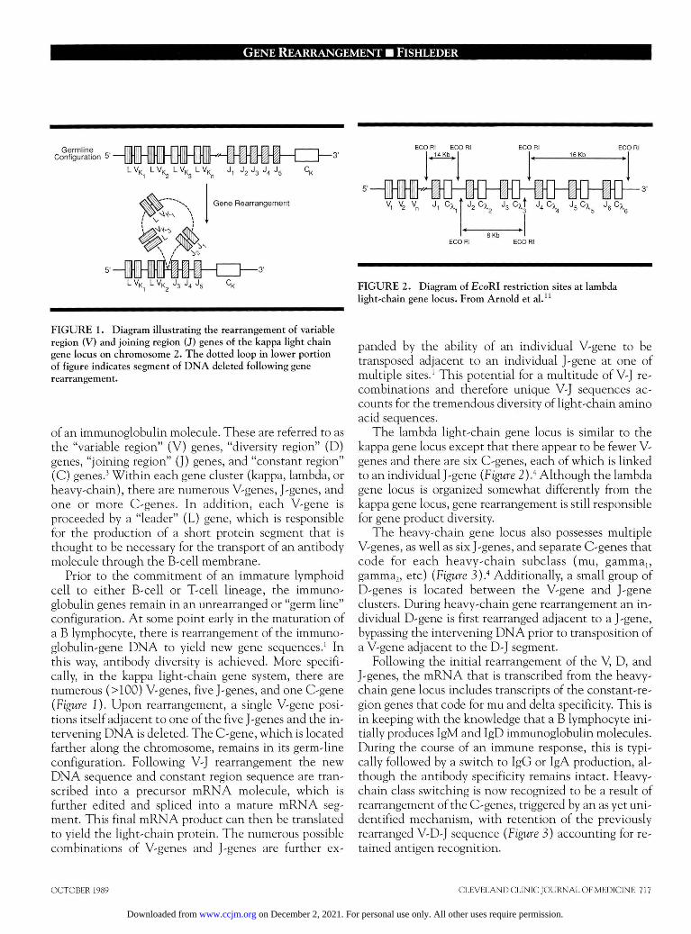
FIGURE 1. Diagram illustrating the rearrangement of variable region (V) and joining region (J) genes of the kappa light chain gene locus on chromosome 2. The dotted loop in lower portion of figure indicates segment of DNA deleted following gene rearrangement.
of an immunoglobulin molecule. These are referred to as the "variable region" (V) genes, "diversity region" (D) genes, "joining region" (J) genes, and "constant region" (C) genes.' Within each gene cluster (kappa, lambda, or heavy-chain), there are numerous V-genes, J-genes, and one or more C-genes. In addition, each V-gene is proceeded by a "leader" (L) gene, which is responsible for the production of a short protein segment that is thought to be necessary for the transport of an antibody molecule through the B-cell membrane.
Prior to the commitment of an immature lymphoid cell to either B-cell or T-cell lineage, the immuno-globulin genes remain in an unrearranged or "germ line" configuration. At some point early in the maturation of a B lymphocyte, there is rearrangement of the immuno-globulin-gene DNA to yield new gene sequences.1 In this way, antibody diversity is achieved. More specifi-cally, in the kappa light-chain gene system, there are numerous (>100) V-genes, five J-genes, and one C-gene (Figure I). Upon rearrangement, a single V-gene posi-tions itself adjacent to one of the five J-genes and the in-tervening DNA is deleted. The C-gene, which is located farther along the chromosome, remains in its germ-line configuration. Following V-J rearrangement the new DNA sequence and constant region sequence are tran-scribed into a precursor m R N A molecule, which is further edited and spliced into a mature mRNA seg-ment. This final m R N A product can then be translated to yield the light-chain protein. The numerous possible combinations of V-genes and J-genes are further ex-
FIGURE 2. Diagram of EcoRI restriction sites at lambda light-chain gene locus. From Arnold et al."
panded by the ability of an individual V-gene to be transposed adjacent to an individual J-gene at one of multiple sites.1 This potential for a multitude of V-J re-combinations and therefore unique V-J sequences ac-counts for the tremendous diversity of light-chain amino acid sequences.
The lambda light-chain gene locus is similar to the kappa gene locus except that there appear to be fewer V-genes and there are six C-genes, each of which is linked to an individual J-gene (Figure 2).4 Although the lambda gene locus is organized somewhat differently from the kappa gene locus, gene rearrangement is still responsible for gene product diversity.
The heavy-chain gene locus also possesses multiple V-genes, as well as six J-genes, and separate C-genes that code for each heavy-chain subclass (mu, gamma,, gamma2, etc) (Figure 3).4 Additionally, a small group of D-genes is located between the V-gene and J-gene clusters. During heavy-chain gene rearrangement an in-dividual D-gene is first rearranged adjacent to a J-gene, bypassing the intervening D N A prior to transposition of a V-gene adjacent to the D-J segment.
Following the initial rearrangement of the V, D, and J-genes, the mRNA that is transcribed from the heavy-chain gene locus includes transcripts of the constant-re-gion genes that code for mu and delta specificity. This is in keeping with the knowledge that a B lymphocyte ini-tially produces IgM and IgD immunoglobulin molecules. During the course of an immune response, this is typi-cally followed by a switch to IgG or IgA production, al-though the antibody specificity remains intact. Heavy-chain class switching is now recognized to be a result of rearrangement of the C-genes, triggered by an as yet uni-dentified mechanism, with retention of the previously rearranged V-D-J sequence (Figure 3) accounting for re-tained antigen recognition.
OCTOBER 1989 CLEVELAND CLINIC JOURNAL OF MEDICINE 717
on December 2, 2021. For personal use only. All other uses require permission.www.ccjm.orgDownloaded from

GENI: REARRANGEMENT • FISHLEDF.R
H H H H H -S C u V H , . „ D H , J H ,
1 St D N A R E A R R A N G E M E N T T O Y I E L D IgM A N D IgD P R O D U C T I O N
HMO-B-KH V H 3 D H 2 J H 4 J H 5 S C | j C 8 S C y 3 S C y , S C a ,
2 n d D N A R E A R R A N G E M E N T R E S U L T S IN H E A V Y C H A I N C L A S S S W I T C H (eg : I g G , ) W I T H I D I O T Y P E R E T E N T I O N
5'-
V H 3 D H 2 J H 4 J H 5 S P S y , C y , S C o
T Lymphocyte Macrophage
FIGURE 3. Diagram illustrating the rearrangement of variable region (V), diversity region (D), joining region (J), and constant region (C) genes of the heavy-chain gene locus on chromosome 14. Center portion of figure indicates initial rearrangements of variable and diversity region genes as part of primary immune response. Lower portion of figure indicates rearrangement of heavy-chain genes resulting in heavy-chain class switching. From Waldmann et al.18
Finally, it is now understood that the individual im-munoglobulin gene loci undergo an ordered sequence of rearrangement: the heavy-chain genes rearrange prior to the light-chain genes and the kappa light-chain genes rearrange prior to the lambda light-chain genes.5
Furthermore, if the kappa light-chain gene rearrange-ment yields a functional DNA sequence then the lambda light-chain genes do not undergo rearrange-ment.6 However, if the kappa light-chain gene rear-rangement results in a nonfunctional rearrangement or deletion of the genes then the lambda light-chain genes will rearrange. This presumably explains the 2 -3 :1 kappa to lambda light-chain ratio that is normally en-countered in polyclonal B-cell proliferations, as well as the excess of lymphomas producing kappa light chains relative to those producing lambda light chains. (It should also be recognized that when an individual gene locus, such as the heavy-chain genes or kappa light-chain genes, undergoes rearrangement, only one of the particular chromosome pair attempts rearrangement ini-tially. Should that rearrangement be successful then the genes located on the second chromosome of the pair will
FIGURE 4. T lymphocyte with dimeric T-cell receptor (TCR) and T 3 receptor on surface. T 3 receptor recognizes major histocompatibility complex (MHC) restricting element on macrophage. TCR recognizes bound antigen on macrophage.
remain in the germ-line configuration. Only if the rear-rangement of the genes on the first chromosome is un-successful will the second chromosome attempt rear-rangement.)
Unlike B cells, which produce immunoglobulins that recognize free antigens, T cells recognize antigens that are bound to the surface of antigen-presenting cells such as macrophages and monocytes. The ability of a T cell to recognize such foreign antigens is dependent upon a complex of two receptors on the T-cell surface (Figure 4).7 The T3 antigen recognizes major histocompatibility complex (MHC) restricting elements on the presenting cell surface and this allows the T C R to recognize the for-eign antigen. It is the T C R , therefore, that must have the ability to recognize specifically a variety of antigens in a manner similar to the immunoglobulin molecule.
The T C R is a dimer composed of an alpha chain and a beta chain. Each chain has both constant and variable regions similar to the component chains of the immuno-globulin molecule; the combination of the variable re-gions of the alpha and beta chains is responsible for an-tigen recognition. Although the genes coding for both the alpha and beta chains have been characterized, eval-uation of the beta-chain gene family is more widely used to recognize T-cell clonal expansion because it rear-ranges prior to the alpha-chain gene family. For this rea-son, the remainder of this section will deal with the T C R beta-chain genes.
The T C R beta-chain genes are located on chromo-some 7q and are organized somewhat differently from the immunoglobulin loci (Figure 5).7 The V-genes, of which there are approximately 50, are located in the 5' direction from all of the D, J, and C-genes. There are
7 1 8 C L E V E L A N D C L I N I C J O U R N A L O F M E D I C I N E V O L U M E 5 6 N U M B E R 7
B O U N D A N T I G E N
M H C R E S T R I C T I N G E L E M E N T
on December 2, 2021. For personal use only. All other uses require permission.www.ccjm.orgDownloaded from

GENE REARRANGEMENT • FISHLEDER
ECO RI ECO RI
0 J , J , J , J 4 J s J 6 D J , J, J 3 J 4 J 5 J 6 J,
HIND III HIND Mi HIND III HIND III
, Germline Configuration
G E N E R E A R R A N G E M E N T C H A N G E S R E L A T I V E P O S I T I O N O F B A M HI D I G E S T S I T E S
FIGURE 5. Diagram illustrating EcoRI, BamHl, and Hmdlll restriction sites of the T-cell receptor beta-chain gene locus.
B A M HI . . . . . .. B A M HI Al tered Length
" f t
Rearranged D N A
two C-genes, C(3, and CP2, which are located approxi-mately 10 kb apart. There are six and seven J-genes, re-spectively, in the 5' direction from each C-gene and ad-jacent to the 5 ' end of each of these J segments is a single D-gene. In contrast to the immunoglobulin genes, in which there is a sequential ordering of V, D, J, and C-genes, in the T C R beta chain gene system there is a single group of V-genes upstream from clusters of D, J, and C-genes. Regardless of this minor dissimilarity, rear-rangement of the V, D, and J-genes is again responsible for the creation of new DNA sequences that code for unique beta-chain proteins. Also in a manner analogous to the immunoglobulin genes, the T C R genes undergo sequential rearrangement; the T-cell beta-chain genes are rearranged and expressed prior to expression of the alpha-chain genes.
Detection of immunoglobulin and T C R gene rear-rangements is accomplished by restriction endonuclease digestion of DNA followed by Southern blotting and hy-bridization to probes specific for individual genes. Those most commonly used include probes for the C-genes of the kappa, lambda, or T C R beta-chain gene loci or the joining region gene of the heavy-chain locus. In the germ-line configuration, specific restriction endonu-cleases yield identifiable gene segments of constant lengths. For example, BamHl digestion of the kappa gene locus yields a 12-kb D N A segment when hy-bridized with the kappa C-gene probe (Figure 6). In con-trast, the same BamHl digestion would yield a 24-kb DNA segment when hybridized to the T C R beta-chain C-gene probe (Figure 5). The heavy-chain genes are more commonly examined following digestion with EcoRI or a combination of BamHl and Hindlll to yield 17-kb and 5.6-kb germ-line segments, respectively, when hybridized to the probe for the J-genes (Figure 7). (Caution must be exercised when using Hindlll diges-tion alone [11-kb germ-line], however, because of poly-
FIGURE 6. Diagram illustrating alteration of BamHI-restricted fragment size at kappa light-chain gene locus following gene rearrangement. This figure is a modification of Figure 1 from Arnold et al . "
FIGURE 7. Diagram of EcoRI, BamHl, and HmdIII restriction sites of heavy-chain gene locus.
morphisms observed in the 5' direction from the JH re-gion.)8 Finally, when examining the lambda light-chain gene locus with a probe for the C-genes an EcoRI diges-tion is most typically used and, in contrast to the pre-viously mentioned simple germ-line configurations, yields three DNA segments, most typically of 8 kb, 14 kb, and 16 kb (Figure 2). However, this is further compli-cated by polymorphisms within the population that can yield additional germ-line fragment sizes of 18 kb, 21 kb, and 23 kb in the lambda gene system.9
In the T C R beta chain gene system, EcoRI and Hin-dlll restriction endonucleases are commonly used to de-tect rearrangements involving Cp, and Cp,, respectively (Figure 5) . Germ-line bands following EcoRI digestion are 11.0 and 4-0 kb, however, an additional 9.5-kb band may be identified due to an EcoRI site that is resistant to
OCTOBER 1989 CLEVELAND CLINIC JOURNAL OF MEDICINE 719
on December 2, 2021. For personal use only. All other uses require permission.www.ccjm.orgDownloaded from

GENI: REARRANGEMENT • FISHLEDF.R
endonuclease digestion.4 Recognition of this problem is important to prevent misinterpretation of this "new fragment" as a true gene rearrangement.
When the V, D, and J-genes rearrange during the course of B-cell or T-cell maturation and an altered DNA sequence is formed, sites of restriction endonu-clease cleavage are shifted, resulting in DNA digestion products that contain DNA fragments of altered length (Figure 5). This unique gene sequence is then a marker of that specific B-cell or T-cell clone.4
This information is very useful in the diagnosis of lymphoproliferative disorders since the technique of re-striction endonuclease digestion, Southern blotting, and DNA hybridization is sensitive enough to detect a clonal expansion of only 1%—3% of the DNA in a sample. In typical reactive processes reported to date, a multitude of B-cell or T-cell clones proliferate, with no individual clone more than 1% of the total lymphocyte population. Only residual germ-line DNA is identifiable and no rearrangements can be detected.
On the other hand, B-cell or T-cell clonal expansions associated with lymphoproliferative disorders commonly account for more than l % - 3 % of a lymphoid population in a lymph node or in the peripheral blood. When ex-amining the DNA from a lymphoma, then, one would expect to identify on a Southern blot a DNA segment of altered size (non-germ-line) indicating a monoclonal proliferation.1011 This is particularly useful in the diagno-sis of lymphoproliferative disorders in cases that histo-logically appear to represent partial involvement of a lymph node by lymphoma, in SIg-negative B-cell lym-phomas, or in cases where immunophenotyping yields equivocal results. In addition, detectable immuno-globulin or TCR rearrangement can help to differentiate lymphomas from other poorly differentiated nonlym-phomatous neoplasms.
Unlike the B-lymphocyte system, in which uniform surface immunoglobulin can indicate monoclonality, there is no antigenic marker of clonality in the T-cell system. However, T C R beta-chain gene rearrangement does indicate a monoclonal T-cell proliferation1213 and therefore is helpful in identifying T-cell neoplasms. In particular, examination of the TCR beta-chain genes can be useful in the diagnosis of mycosis fungoides and in the subsequent detection of Sezary cells in enlarged
lymph nodes in those patients.14 Although the histologic distinction between dermatopathic lymphadenopathy and Sezary-cell involvement of a lymph node can be quite difficult, the detection of a rearranged TCR beta-chain gene segment would suggest Sezary involvement of the lymph node, especially if that new DNA fragment were of the same kilobase length as that detected in the primary mycosis involvement of the skin.
Although we have equated monoclonality with neo-plastic proliferations in the past, more sensitive detec-tion of gene rearrangements by Southern blotting tech-niques may identify some conditions that are not clinically malignant but that are associated with clonal lymphocyte proliferations. We cannot assume that mon-oclonality at the gene level has the same clinical impli-cation as immunologic monoclonality. This can be il-lustrated by immunoglobulin gene rearrangements that have been identified in the benign lymphoepithelial le-sion of the salivary gland typically found in association with Sjögrens syndrome. This has previously been con-sidered to be a benign polyclonal lymphocyte prolifera-tion but is now recognized as possessing a monoclonal B-cell component.15 Similarly, the disorder lymphomatoid papulosis has been considered a chronic inflammatory process involving the skin, but has now been demon-strated to possess TCR beta-chain gene rearrangements indicating a monoclonal T-cell component.16 Finally, we now recognize that immunocompromised individuals, such as transplant patients, may develop monoclonal or oligoclonal lymphocytic proliferations that resolve when immunosuppressive agents like cyclosporine are removed.17
CONCLUSION
In conclusion, molecular biologic advances have sig-nificantly broadened our understanding of the immuno-globulin and TCR gene systems and have provided the tools necessary to allow identification of monoclonal B-cell and T-cell proliferations at the gene level. The sen-sitivity of these techniques can provide a useful adjunct to the diagnosis of lymphoproliferative disorders; however, we must be cautious and recognize that the de-tection of gene rearrangement does not necessarily equate with malignant lymphocytic clonal expansion.
REFERENCES
1. Leder P. The genetics of antibody diversity. Scientific Am 1982; 246:(5):102—115.
2. Korsmeyer SJ, Hieter PA, Sharrow SO, Goldman CK, Leder P,
Waldmann TA. Normal human B cells display ordered light chain gene rearrangements and deletions. J Exper Med 1982; 156 :975-985.
3. Goldman JN, Goldman MB. The genetics of antibody production. JAMA 1984; 251:774-786.
4- Waldmann TA. The arrangement of immunoglobulin and T cell
720 CLEVELAND CLINIC JOURNAL OF MEDICINE VOLUME 56 NUMBER 7
on December 2, 2021. For personal use only. All other uses require permission.www.ccjm.orgDownloaded from

GENE REARRANGEMENT • FISHLEDER
receptor genes in human lymphoproliferative disorders. [In] Dixon FJ, et al, eds. Advances in Immunology. Vol. 40. San Diego, Academic Press, 1987, pp 247-321 .
5. Korsmeyer SJ, Hieter PA, Ravetch JV, Poplack DG, Waldmann TA, Leder P. Developmental hierarchy of immunoglobulin gene rearrange-ments in human leukemic pre-B cells. Proc Natl Acad Sci 1981; 7 8 : 7 0 9 6 - 7 1 0 0 .
6. Hieter PA, Korsmeyer S], Waldmann TA, Leder P. Human im-munoglobulin kappa light-chain genes are deleted or rearranged in lambda-producing B cells. Nature 1981; 2 9 0 : 3 6 8 - 3 7 2 .
7. Minden MD, Mak T W . The structure of the T cell antigen receptor genes in normal and malignant T cells. Blood 1986; 6 8 : 3 2 7 - 3 3 6 .
8. Fey MF, Wainscoat JS . DNA polymorphism 5 ' to the JH region of the human immunoglobulin heavy chain gene and immunoglobulin gene rearrangements in leukemia. Am J Clin Pathol 1988; 187-189.
9. Hieter PA, Hollis GF, Korsmeyer SJ, Waldmann TA. , Leder P. Clustered arrangement of immunoglobulin ^constant region genes in man. Nature 1981; 2 9 4 : 5 3 6 - 5 4 0 .
10. Cleary ML, Chao J, Warnke R, Sklar J. Immunoglobulin gene rearran-gement as a diagnostic criterion of B-cell lymphoma. Proc Natl Acad Sci 1 9 8 4 ; 8 1 : 5 9 3 - 5 9 7 .
11. Arnold A, Cossman J, Bakhshi A, Jaffe ES, Waldmann TA, Korsmeyer SJ. Immunoglobulin-gene rearrangements as unique clonal markers in
human lymphoid neoplasms. N Engl J Med 1983; 3 0 9 : 1 5 9 3 - 1 5 9 9 . 12. Flug F, Pier-Giuseppe P, Bonetti F, Knowles DM 11, Dalla-Favera R. T-
cell receptor gene rearrangements as markers of lineage and clonality in T-cell neoplasms. Proc Natl Acad Sci 1985; 8 2 : 3 4 6 0 - 3 4 6 4 .
13. Knowles DM 11, Pier-Giuseppe P, Dalla-Favera R. T-cell receptor beta chain gene rearrangements: genetic markers of T-cell lineage and clonality. Hum Pathol 1986; 17 :546-551 .
14. Weiss LM, Hu E, Wood G S , et al. Clonal rearrangements of T-cell receptor genes in mycosis fungoides and dermatopathic lym-phadenopathy. N Engl J Med 1985; 3 1 3 : 5 3 9 - 5 4 4 .
15. Fishleder A, Tubbs R, Hesse B, Levine H. Uniform detection of im-munoglobulin-gene rearrangement in benign lymphoepithelial lesions. N Engl J Med 1 9 8 7 ; 3 1 6 : 1 1 1 8 - 1 1 2 1 .
16. Weiss LM, Wood G S , Trela M, Wamke RA, Sklar J. Clonal T-cell populations in lymphomatoid papulosis: evidence of lymphoprolifera-tive origin for a clinically benign disease. N Engl J Med 1986; 3 1 5 : 4 7 5 -479.
17. Cleary ML, Warnke R, Sklar J. Monoclonality of lymphoproliferative lesions in cardiac-transplant recipients. N Engl J Med 1984; 3 1 0 : 4 7 7 -482.
18. Waldmann TA, et al. Molecular genetic analysis of human lymphoid neoplasms. Ann Intern Med 1985; 1 0 2 : 4 9 7 - 5 1 0 .
OCTOBER 1989 CLEVELAND CLINIC JOURNAL OF MEDICINE 721
on December 2, 2021. For personal use only. All other uses require permission.www.ccjm.orgDownloaded from