immunohematology - american red cross · immunohematology journal of blood ... hospital. each year...
TRANSCRIPT

ImmunohematologyJ O U R N A L O F B L O O D G R O U P S E R O L O G Y A N D E D U C A T I O N
V O L U M E 2 2 , N U M B E R 1 , 2 0 0 6

ImmunohematologyJ O U R N A L O F B L O O D G R O U P S E R O L O G Y A N D E D U C A T I O N
V O L U M E 2 2 , N U M B E R 1 , 2 0 0 6
C O N T E N T S
1External quality assessment scheme in red blood cell serology: a 5-year experience in Thailand
S. BEJRACHANDRA, J. SAIPIN, O. NATHALANG, U. SIRIBOONRIT, E. RUNGROUNG, AND S. UDEE
6Human platelet alloantigen systems in three Chinese ethnic populations
L.YAN, F. ZHU, J. HE, AND S.G. SANDLER
11Reduced red blood cell destruction by antibody fragments
A. MQADMI, S.ABRAMOWITZ, X. ZHENG, AND K.YAZDANBAKHSH
15H-deficient Bombay and para-Bombay red blood cells are most strongly agglutinated by the galactophilic
lectins of Aplysia and Pseudomonas aeruginosa that detect I and P1 antigensN. GILBOA-GARBER, D. SUDAKEVITZ, C. LEVENE, N. RAHIMI-LEVENE, AND V. YAHALOM
23A single base insertion of the 4-α-galactosyltransferase gene led to the deficiency of Gb3 biosynthesis
M.TANAKA, N.YAMASHITA, J.TAKAHASHI, F. HIRAYAMA,Y.TANI, AND H. SHIBATA
30ISBT 128 blood labeling: introduction and reference laboratory applications
S.H. BUTCH AND P.B. DISTLER
37C O M M U N I C A T I O N S
ERRATAVol. 21, No. 4, 2005, p. 146Vol. 21, No. 4, 2005, p. 171
38 41A N N O U N C E M E N T S A D V E R T I S E M E N T S
44I N S T R U C T I O N S F O R A U T H O R S

EDITORS-IN-CHIEF MANAGING EDITORSandra Nance, MS, MT(ASCP)SBB Cynthia Flickinger, MT(ASCP)SBB
Philadelphia, Pennsylvania Philadelphia, Pennsylvania
Connie M.Westhoff, SBB, PhDPhiladelphia, Pennsylvania
TECHNICAL EDITOR SENIOR MEDICAL EDITORChristine Lomas-Francis, MSc Geralyn Meny, MD
New York, New York Philadelphia, Pennsylvania
ASSOCIATE MEDICAL EDITORSDavid Moolton, MD Scott Murphy, MD Ralph Vassallo, MD
Philadelphia, Pennsylvania Philadelphia, Pennsylvania Philadelphia, Pennsylvania
EDITORIAL BOARD
EDITORIAL ASSISTANT PRODUCTION ASSISTANTJudith Abrams Marge Manigly
COPY EDITOR ELECTRONIC PUBLISHERLucy Oppenheim Paul Duquette
Immunohematology is published quarterly (March, June, September, and December) by the American Red Cross, National Headquarters,Washington, DC 20006.
Immunohematology is indexed and included in Index Medicus and MEDLINE on the MEDLARS system.The contents are also cited in the EBASE/Excerpta Medica and Elsevier BIOBASE/Current Awareness
in Biological Sciences (CABS) databases.
The subscription price is $30.00 (U.S.) and $35.00 (foreign) per year.
Subscriptions, Change of Address, and Extra Copies:Immunohematology, P.O. Box 40325
Philadelphia, PA 19106Or call (215) 451-4902
Web site: www.redcross.org/pubs/immuno
Copyright 2006 by The American National Red CrossISSN 0894-203X
Patricia Arndt, MT(ASCP)SBBPomona, California
James P.AuBuchon, MDLebanon, New Hampshire
Geoffrey Daniels, PhDBristol, United Kingdom
Richard Davey, MDWashington, District of Columbia
Sandra Ellisor, MS, MT(ASCP)SBBAnaheim, California
George Garratty, PhD, FRCPathPomona, California
Brenda J. Grossman, MDSt. Louis, Missouri
W. John Judd, FIBMS, MIBiolAnn Arbor, Michigan
Christine Lomas-Francis, MScNew York, New York
Gary Moroff, PhDRockville, Maryland
Ruth Mougey, MT(ASCP)SBBCarrollton, Kentucky
John J. Moulds, MT(ASCP)SBBShreveport, Louisiana
Marilyn K. Moulds, MT(ASCP)SBBHouston, Texas
Paul M. Ness, MDBaltimore, Maryland
Mark Popovsky, MDBraintree, Massachusetts
Marion E. Reid, PhD, FIBMSNew York, New York
Susan Rolih, MS, MT(ASCP)SBBCincinnati, Ohio
S. Gerald Sandler, MDWashington, District of Columbia
David F. Stroncek, MDBethesda, Maryland
Marilyn J.Telen, MDDurham, North Carolina

From 2000 to 2004, 36, 58, 72, 78, and 86 laboratories participatedin an external quality assessment scheme (EQAS) organized by theDepartment of Transfusion Medicine, Faculty of Medicine SirirajHospital. Each year the staff was requested to perform ABOgrouping, D typing, antibody screening, antibody identification, andDATs on eight blood samples. Each participant receivedinformation on the correct test results and a coded summary.Regarding ABO grouping, the error rate ranged from 0.3 to 1.3percent,mostly due to human errors. Error rates in D typing rangedfrom 0.7 to 5.7 percent, the most problematic being weak Dphenotype interpretation. Although every sample was negative bythe DAT, error rates due to false positive test results weredetermined to be 0.4 to 2.1 percent. Antibody screening errorswere also found; however, errors steadily decreased from 4.2percent in 2000 to 0.3 percent in 2004. Only 69.4 to 87.2 percentof laboratories performed antibody identification; however, correctresults increased from 78.4 to 91.0 percent. In conclusion,an EQASin RBC serology should be used to compare results from differentlaboratories and to identify those laboratories that needimprovement in testing procedures. Immunohematology2006;22:1–5.
Key Words: RBC serology,external quality assessment,Thailand
Quality assurance in transfusion medicine includesthe use of and participation in internal and externalquality programs. Quality management is essential toensure that laboratory performance is reliable andaccurate on a daily basis. However, an external qualityassessment scheme (EQAS) that compares results fromdifferent laboratories is essential to verify the accuracyand reliability of laboratory results.1–5 This study wasundertaken to evaluate RBC serology testing servicesamong hospital blood banks in Thailand.
Among Thai hospital laboratories, the qualityassurance program for blood transfusion services wasestablished in 1988. From 1994 to 1997, the Bureau ofLaboratory Quality Standards, Department of MedicalSciences,Ministry of Public Health, in cooperation withthe Subcommittee on Transfusion Medicine, set up thefirst proficiency test on RBC serology for hospital
blood banks throughout the country. The proficiencytesting samples obtained from the National BloodCentre, Thai Red Cross Society, were sent to everymember three times per year without charge. At first,all members were requested to perform only ABOgrouping and antibody screening tests. Eleven serumsamples were sent to 127 blood banks, revealing anoverall accuracy of 94.15 percent.6 Beginning in 2002,D typing and antibody identification tests were alsoperformed. The results of the tests performed wereevaluated using target values. Additionally, fromNovember 1996 to August 1997, 20 blood banks inBangkok were asked to participate in an externalquality control study in immunohematology that wasorganized by the Department of Transfusion Medicine,Faculty of Medicine Siriraj Hospital,Mahidol University.Each blood bank was requested to perform ABO and Dtyping, antibody screening, antibody identification, anddirect antiglobulin tests (DAT) on eight blood samples.Surprisingly, only three public hospital blood banksreported correct results for all tests on every bloodsample.
The second external quality assessment programfrom the Department of Transfusion Medicine, Facultyof Medicine Siriraj Hospital, Mahidol University, startedin 2000. In the beginning, 36 laboratories joined theprogram,with only 13.9 percent of them reporting 100percent correct results for tests on eight bloodsamples.7 Over time, more blood banks joined theprogram. The number of blood banks participating was58, 72, 78, and 86 in 2001, 2002, 2003, and 2004,respectively. At the end of each year, our laboratoryprepared a list of the errors made by each blood bankand the results were discussed at our annual meeting.
The purpose of the 2000 to 2004 EQAS was toevaluate the efficiency of the blood bank staff whoperform routine laboratory tests, such as compatibility
I M M U N O H E M A T O L O G Y, V O L U M E 2 2 , N U M B E R 1 , 2 0 0 6 1
External quality assessmentscheme in red blood cell serology:a 5-year experience in ThailandS. BEJRACHANDRA, J. SAIPIN, O. NATHALANG, U. SIRIBOONRIT, E. RUNGROUNG, AND S. UDEE

testing,antibody screening,and antibody identification.Analysis of the study results could help blood banksconsider more appropriate policies for increasing staffefficiency, performance, and accuracy.
Materials and Methods
Blood samplesFor each year in the study (2000 to 2004), eight
blood samples were distributed to participating bloodbanks. The Department of Transfusion Medicineprepared four blood samples and four were preparedby DiaMed AG, Switzerland. Samples were distributedfour times per year. Each time, two unknown samples,consisting of 5 ml serum and 5 ml RBC suspension inAlsever’s solution, were shipped to participants,packed in wet ice. The samples were distributed by thesupplier and each participant received them within 3days. The participants were instructed to handle thesesamples as part of their routine work, to have thesetests performed by a technician on duty within 3 daysafter receipt, and to return the results by surface mailor fax within 2 weeks. Late results were not includedin the analysis.
Tests performedEach hospital blood bank was requested to perform
five tests using their routine reagents and methods, suchas the conventional tube technique or gel test. Thesetests included ABO grouping, D typing, antibodyscreening and identification, and a DAT. A result formwas developed and used to record the individualhospital test results and the testing method used. Theresults from all blood banks were compiled and assessedand the correct test results, the individual laboratoryperformance, and a coded summary of the results of allparticipants were shared with all participants. Thisenabled them to have interlaboratory comparison.Additionally, at the end of the year, a summary of eachlaboratory’s performance was reported to the head ofthe blood bank laboratory and hospital director for eachfacility. This allowed them to be aware of capacitybuilding needed for laboratory personnel as well asequipment, reagents, and methods used.
Moreover, to educate and increase the awareness ofall members, a workshop was set up at the end of eachyear. Every participant was invited free of charge and alecture on relevant topics was given. Causes ofdiscrepant results, challenges, and any difficultiesoccurring during the program were discussed andresolved with an organizing team.
ResultsDetails of the samples with or without antibody
identification distributed in 2000 to 2004 aresummarized in Table 1. The number of blood banklaboratories participating in the EQAS on RBC serologyfrom 2000 to 2004 was 36, 58, 72, 78, and 86 for eachyear, respectively (Table 2). Causes of errors in ABOgrouping, D typing, DAT, and antibody screening testsare summarized in Table 3. For ABO grouping, allparticipants performed both RBC and serum groupingbut some errors in reporting still occurred. The errorrates for the 5 years were 0.7, 1.3, 0.9, 0.6, and 0.3percent, respectively (Table 2). Errors in ABO groupingtests were classified as human, misinterpretation, andtechnical. Human errors included those that occurredbecause of failure to interpret or record correct testresults, inadequate identification of blood specimenssuch as testing old EQAS samples instead of the currentones, and sample mixups. Errors classified asmisinterpretation included those that occurredbecause of the inability to interpret results because ofABO discrepancies caused by antibodies that wentundetected because antibody identification was notperformed. Technical errors included those thatoccurred because of the failure to detect antibodies ofthe ABO system; positive results were missed.Regarding D typing, error rates were 0.7, 5.7, 2.3, 4.8,and 3.2 percent, respectively (Table 2). The high errorrates in the year 2001 and 2003 were due to variationin the reporting of RBC samples with a weak Dphenotype as either D– or D+ (Table 3). These errorswere classified as those caused by misinterpretation.Again, human errors in this category were caused by amixup of specimens. Errors classified as reagent errorsoccurred because some commercial reagents gavestrong positive results in the immediate spin phasewhile others gave negative results. Technical errorsincluded those caused by D– RBCs that wereinterpreted as D+ or as a weak D phenotype.
Negative DAT results were correctly ascertained;however, error rates due to false positive test resultswere found to be 2.1, 1.1, 0.4, 1.6, and 1.2 percent,respectively (Table 2). The misinterpretation error thatoccurred in one laboratory was caused by a mixupbetween DAT and antibody screening tests results(Table 3). They reported positive DAT results instead ofantibody screening results. Errors caused by impropertesting procedures that resulted in the reporting ofDAT negative samples as DAT positive were classifiedas technical.
2 I M M U N O H E M A T O L O G Y, V O L U M E 2 2 , N U M B E R 1 , 2 0 0 6
S. BEJRACHANDRA ET AL.

I M M U N O H E M A T O L O G Y, V O L U M E 2 2 , N U M B E R 1 , 2 0 0 6 3
EQAS in red cell serology
Errors in antibody screening were also found.Errors rates were 4.2, 1.1, 1.4, 0.8, and 0.3 percent,respectively (Table 2). Errors caused by reagentsoccurred because inappropriate locally madescreening RBCs, which lacked some antigens such asMia, E, K, P1, or M, were used. Therefore, suchantibodies could not be detected. Technical errorsagain included those caused by improper testingtechniques that resulted in the reporting of negativeantibody screening test results as positive. On thecontrary, some positive antibody screening test resultswere reported as negative even though their screeningRBCs contained the specific antigens.
Antibody identification was not routinelyperformed in all blood bank laboratories participating
from 2000 to 2004. In the first year, only 25 out of 36laboratories (69.4%) performed this procedure.Routinely, the other blood bank laboratories, which didnot perform antibody identification, sent the bloodsamples with positive antibody screening results to theNational Blood Centre of the Thai Red Cross Society forinvestigation and crossmatching. Because of theexplanations and recommendations given at theworkshop, the number of participating laboratoriesperforming antibody identification increased from 79.3to 87.2 percent.
The results of antibody identification testing werethe most striking. In 2000, among 36 participants, only78.4 percent reported correct results for all eight bloodsamples. The correct results increased to 79.1 percentin 2001, 90.7 percent in 2002, 91.0 percent in 2003,and 91.0 percent in 2004, as shown in Table 2.Moreover, due to the workshop discussions on how todecrease errors, the number of participants reportingcorrect results for all tests on the eight blood sampleseach year gradually increased: 5 of 36 (13.9%) in 2000,8 of 58 (13.8%) in 2001,29 of 72 (40.3%) in 2002, 21 of78 (26.9%) in 2003, and 37 of 86 (43.0%) in 2004.
Every blood bank laboratory sent one or twomembers of its staff to attend the end-of-year workshopand each received a certificate of attendance. Inaddition, an award was given to the blood banklaboratories that reported all results correctly.
DiscussionExternal proficiency testing programs offer a
valuable management tool because they enablelaboratory personnel to compare their laboratoryresults with those obtained in other laboratories whenthe same material is examined. The proficiency testingsamples must be tested with the laboratory’s regularpatient workload, using routine testing methods. Inthis study, the organizing team also reported thelaboratory performance to the chief of laboratories aswell as hospital directors. Therefore, these EQAS helpevaluate the performance of procedures, equipment,materials, and personnel of the individual blood bankor transfusion service and suggest areas forimprovement.1–5
The results of this study indicated that only 13.9 to43.0 percent of the participating hospitals reported100 percent correct results for all tests on eight bloodsamples in each year, which is similar to previousstudies’ results.7 ABO grouping errors occurredbecause of human error in the interpretation of results.
Table 1. Distribution of proficiency testing samples from 2000 to 2004*
Number of samples
2000 2001 2002 2003 2004
ABO groupingA (12) 2 3 2 3 2B (9) 1 2 3 1 2AB (2) 1 0 0 0 1O (17) 4 3 3 4 3
D typingD+ (21) 5 5 4 3 4D– (12) 2 1 3 3 3Weak D phenotype (7) 1 2 1 2 1
DATPositive (0) 0 0 0 0 0Negative (40) 8 8 8 8 8
Antibody screeningPositive (33) 7 5 8 7 6Negative (7) 1 3 0 1 2
Antibody identification (33/40)Single antibody (20)
Inhouse (Siriraj)anti-E (2) 1 0 1 0 0anti-P1 (3) 0 1 1 1 0anti-Mia (2) 1 0 1 0 0anti-M (1) 0 0 0 0 1anti-D (1) 0 0 0 0 1
Import (DiaMed)anti-D (1) 0 0 1 0 0anti-c (3) 0 1 1 0 1anti-e (2) 1 0 0 1 0anti-K (3) 1 0 1 1 0anti-E (2) 0 0 0 1 1
Mixture of antibodies (13)Inhouse (Siriraj)
anti-D, -Mia (1) 0 0 0 1 0anti-D, -C (1) 1 0 0 0 0anti-D, -E (1) 0 0 1 0 0anti-E, -Mia (3) 0 1 0 1 1anti-P1, -Mia (1) 0 1 0 0 0
Import (DiaMed)anti-D, -Fya (1) 0 1 0 0 0anti-c, -K (2) 1 0 1 0 0anti-C, -D, -E (1) 1 0 0 0 0anti-D, -E (1) 0 0 0 1 0anti-D, -K (1) 0 0 0 0 1
*Number in parentheses is total number of samples from 2000 to 2004.

If a patient receives mistyped blood, especially an ABOmismatch, this can result in a life-threatening event.RBC samples that were D– as well as those of the weakD phenotype were also mistyped because of technicalerrors and result misinterpretation. Moreover, somecommercial monoclonal anti-D reagents gave strongagglutination with RBCs of the weak D phenotype.This should be noted because patients with anapparent weak D phenotype who may have a partial Dphenotype may be considered D+ and if D+ RBCs aregiven, anti-D could be made. On the other hand, donorRBCs of the weak D phenotype must be identified assuch. RBC products from these donors should belabeled as D+ and given to D+ patients in order toprevent the production of anti-D.8 Even though somereduction in the number of ABO and D typing testresult errors were found, the error rates were stillhigher than those found in the previous studies in theUnited Kingdom.9–10
The false positive results in the DAT should leadsome blood banks to reevaluate their reagents andtesting procedures. Because anti-E is one of thecommon Rh antibodies and anti-K is uncommonamong the Thai population, the use of locally madescreening RBCs, which lack these antigens, mightexplain the errors in antibody screening tests and whythese antibodies were not identified.11–14 When theappropriate screening RBCs, which included allclinically significant antigens, and methods, such assaline IAT,enzyme,and LISS IAT,were used, these bloodbank laboratories could identify antibodies in othersamples containing a mixture of antibodies such asanti-c, -K and anti-E, -Mia.
Moreover, for antibody identification of two orthree antibodies, such as anti-c, -K, anti-C, -D, and anti-C,-D, -E, only 40 to 80 percent of the hospitals couldreport the correct antibody specificity. Whenappropriate panel RBCs and second panel RBCstogether with different methods were used, the
hospitals could identify these antibody specificities. InThailand, in addition to inhouse screening and panelRBCs, laboratories can obtain these from the NationalBlood Centre at a reasonable price. The study showedthat errors in antibody identification could not beattributed to the reagents and the reason somelaboratories did not perform this test may be due to thepolicy of those laboratories. However, to differentiatewhether there is another antibody in the serum sampleor not, extra RBCs of known rare phenotypes areneeded. In addition, antigen typing of the patient’sRBCs should be performed before reaching anyconclusion. Moreover, staff who are experienced withappropriate testing procedures are needed to identifymore complex samples.3,4
Our results showed the same pattern as that of theNational External Quality Assessment Scheme in BloodGroup Serology, organized by the Bureau of LaboratoryQuality Standards, which sent unknown samples tovarious blood banks in the country (622 in 2002 and653 in 2003). They found that among 285 blood banks(51%) who continuously reported test results, 5
4 I M M U N O H E M A T O L O G Y, V O L U M E 2 2 , N U M B E R 1 , 2 0 0 6
S. BEJRACHANDRA ET AL.
Table 2. Summary of ABO, D, DAT, antibody screening, and antibody identification test results (2000–2004)
Antibody Antibody Year N ABO D DAT screening identification
✔ ✘ ✔ ✘ ✔ ✘ ✔ ✘ ✔ ✘
2000 36 93.3 0.7 99.3 0.7 97.9 2.1 95.8 4.2 78.4 21.6
2001 58 98.7 1.3 94.3 5.7 98.9 1.1 98.9 1.1 79.1 20.9
2002 72 99.1 0.9 97.7 2.3 99.6 0.4 98.6 1.4 90.7 9.2
2003 78 99.4 0.6 95.2 4.8 98.4 1.6 99.2 0.8 91.0 9.0
2004 86 99.7 0.3 96.8 3.2 98.8 1.2 99.7 0.3 91.0 9.0
N = number of blood bank laboratories ✔ = correct results (%) ✘ = wrong results (%)
Table 3. Summary of the nature of the errors in ABO grouping, D typing,DAT, and antibody screening tests (2000–2004)
Number of blood banks reportingincorrect results
2000 2001 2002 2003 2004
ABO groupingHuman errors 2 4 1 2 1Misrepresentation 0 1 2 1 0Technical errors 0 0 0 0 0
D typingHuman errors 0 2 0 1 0Misrepresentation 0 8 4 14 1Reagents 0 9 2 3 1Technical errors 2 3 6 7 20
DATMisrepresentation 1 0 0 0 0Technical errors 4 4 1 7 5
Antibody screeningReagents (screening RBCs) 4 0 5 3 2Technical errors 7 3 2 1 1
I M M U N O H E M A T O L O G Y, V O L U M E 2 2 , N U M B E R 1 , 2 0 0 6 5
EQAS in red cell serology
percent (15 of 285) and 0.3 percent (1 of 254) hadunacceptable results for ABO and D typing tests,respectively. In addition, of the 57.2 percent (163 of285) and 16.8 percent (48 of 285) of blood banks thatreported the results of antibody screening andantibody identification tests, 27 percent (44 of 163)and 73 percent (35 of 48) had excellent results.15
In conclusion, internal and external qualityassessment programs should be maintained in order toensure effective transfusion service and safety ofpatients. In addition to hospital accreditation, thehospital administration should support training andcontinuing education to improve the ability of theblood bank staff to perform all tests and evaluate theirresults.
AcknowledgmentsWe would like to thank all blood banks that
participated in our EQAS.
References1. Bert LM. Quality in Blood Banking. In: Harmening
DM, ed. Modern blood banking and transfusionpractices. 4th ed. Philadelphia: F.A. DavisCompany, 1999:326-42.
2. Petz LD, Swisher SW. Transfusion medicine in ahospital setting. In: Petz LD, Swisher SN, KleinmanS, Spence RK, Strauss RG, eds. Clinical practice oftransfusion medicine. 3rd ed. New York: ChurchillLivingstone, 1996:335-47.
3. Menitove JE. Process control. In: Standard forblood banks and transfusion services. 22nd ed.Bethesda: American Association of Blood Banks,2003:11-69.
4. Menitove JE. Assessments: internal and external.In: Standard for blood banks and transfusionservices. 22nd ed. Bethesda:American Associationof Blood Banks, 2003:93.
5. Brecher ME. Quality issues. In: Technical manual.14th ed. Bethesda: American Association of BloodBanks, 2002:1-88.
6. Petchoopong A and Thaworn C. The proficiencytest in antibody screening test of blood banklaboratories in different level hospitals 1994-1997.Thai J Hematol Transfus Med 1999; 9:185-93.
7. Bejrachandra S, Nathalang O, Saipin J, et al.External quality control program in red cellserology. In:Kanno T,Okabe H,Tatsumi N,Mori M,
Ichiyama S, eds. Global standardization andadvanced quality management ‘01. Quality controlin the clinical laboratory. Osaka: Eibun Press,2002:194-6.
8. Brecher ME.The Rh system. In:Technical manual.14th ed. Bethesda: American Association of BloodBanks, 2002:295-313.
9. Holburn AM, Prior DM. The UK national externalquality assessment scheme in blood groupserology. ABO and D grouping and antibodyscreening 1982-1983. Clin Lab Haematol1986;8:243-56.
10. Holburn AM, Prior DM, Whitton CM. The UKnational external quality assessment scheme inblood group serology. ABO and D grouping andantibody screening, direct antiglobulin test andantibody identification 1984-1985. Clin LabHaematol 1988;10:73-85.
11. Chandanayingyong D. Common problems inroutine pre-transfusion testing. Southeast Asian JTrop Med Public Health 1979;10:193-5.
12. Bejrachandra S,Chandanayingyong D.Unexpectedred cell antibodies in donors and patients at SirirajHospital. Southeast Asian J Trop Med PublicHealth 1979;10:204-6.
13. Nathalang O, Kuvanont S, Punyaprasidth P,Tasaneeyanond C, Sriphaisal T.A preliminary studyof the distribution of blood group systems in Thaiblood donors determined by the gel test.Southeast Asian J Trop Med Public Health2001;32:419-24.
14. Bejrachandra S, Nathalang O, Saipin J, et al.Distribution of blood group systems in Thai blooddonors determined by the gel test. Siriraj HospGaz 2002;54:403-9.
15. Soisangwan R. National external qualityassessment scheme in blood group serology inThailand, 2002-2003 experience. Thai J HematolTransf Med 2004;14:7-21.
Sasitorn Bejrachandra, MD, and Jariya Saipin, MSc,Department of Transfusion Medicine, Faculty ofMedicine Siriraj Hospital, Mahidol University,Bangkok 10700, Thailand; Oytip Nathalang, PhD,Phramongkutklao College of Medicine, Bangkok,Thailand; Usanee Siriboonrit, MSc, EkarajRungroung, MSc, and Sudjai Udee, BE, Faculty ofMedicine Siriraj Hospital, Mahidol University,Bangkok, Thailand.

6 I M M U N O H E M A T O L O G Y, V O L U M E 2 2 , N U M B E R 1 , 2 0 0 6
Knowledge of the prevalence of human platelet antigens (HPA) indifferent populations is important for effective diagnosis andmanagement of immune-mediated platelet disorders. The purposeof this study was to determine HPA gene frequencies in themajority Han ethnic population of China and in ethnic She and Tajikminority populations. Using PCR sequence specific primers, HPA-1, -2, -3, -4, -5, and -6, we determined genotypes for ethnic Han,She,and Tajik blood donors. HPA gene frequencies for Chinese Hanwere found to be similar to those of She, reflecting the historicaffinities of these two populations. HPA gene frequencies for Tajikwere closer to those for Caucasians than to Chinese Han, She, orother Asian populations, reflecting their disparate origin andhistoric geographic isolation. HPA gene frequencies in theseChinese populations reflect their historic origins. Knowledge ofthese findings may be used to better understand and treat immune-mediated platelet disorders in these populations.Immunohematology 2006;22:6–10.
Key Words: human platelet antigens,platelet serology,Chinese ethnic populations
The following report presents the results ofdetermining genotypes and gene frequencies for humanplatelet antigens (HPA) in indigenous Chinese Han, She,and Tajik populations, using PCR sequence specificprimers (PCR-SSP). We compared HPA gene frequenciesfor Han, the majority ethnic population in China, withthose for She and Tajik populations, two of the ethnicminority populations in China. Most (92%) Chinese areethnic Han. The remaining 8 percent of the populationare represented by 55 ethnic minorities, including She,who share certain historic, cultural, and linguisticaspects with the Han. Other minorities, such as Tajik,have historic origins, cultures, and languages that arequite different from those of the majority Han. Thepurpose of this study was to determine whether thehistoric affinities or disparities of these three ethnicChinese populations are reflected in their HPA genefrequencies. HPA gene frequencies in Chinese Han havebeen previously reported, but primarily in Chineselanguage publications that are not easily accessed bymost readers of Immunohematology. To facilitate
readers’ access to these data in Chinese-languagepublications, we include a summary of their results aswell as pertinent studies of HPA genes among otherAsian and Caucasian populations. We are unaware ofprevious studies of HPA gene frequencies among theindigenous She or Tajik minorities in China.
Materials and Methods
Populations studiedAfter obtaining informed consent, we collected 5
ml of peripheral venous blood from volunteer blooddonors who were self-identified to be Han, She, or Tajikby personal interviews. Samples were anticoagulatedwith EDTA and stored at 1°C to 6°C before testing.
HPA genotyping Genomic DNA was extracted from whole blood
samples using a standard salting-out method (Pel-Freez,Brown Deer,WI). HPA-1, -2, -3, -4, -5, and -6 genotypeswere determined by PCR-SSP method as previouslydescribed.1,2 In brief, the PCR reaction mixture (25 µL)contained 1X PCR buffer, 1.0 U of Taq DNApolymerase, 200 µmol/L of each dNTP, 1.8 mmol/LMgCl2, 0.5 µmol/L of each specific primer (Table 1), 0.2µmol/L of internal control primer (to amplify humangrowth hormone gene), and 50 to 100 ng genomicDNA. PCR amplification was performed with initialdenaturing at 95°C for 5 minutes followed by 30 cyclesof 30 seconds at 95°C, 1 minute at 58°C (67°C for HPA-2, 63°C for HPA-3, 57°C for HPA-5 and HPA-6), and 45seconds at 72°C plus a final extension at 72°C for 10minutes. Amplification products were verified in 2%agarose gel stained with ethidium bromide with DNAmarker and visualized by ultraviolet light (Genegenius,Syngene, UK).
Human platelet alloantigensystems in three Chinese ethnicpopulationsL.YAN, F. ZHU, J. HE,AND S.G. SANDLER

I M M U N O H E M A T O L O G Y, V O L U M E 2 2 , N U M B E R 1 , 2 0 0 6 7
Human platelet antigens
Gene frequencies and statistical analysisGene frequencies were calculated using the
formula gene frequency = allele numbers/samplenumber × 2. Chi-squared analysis was used to test forHardy-Weinberg equilibrium for HPA genes. Resultswith p < 0.05 were considered statistically significant.
Bibliographic searchWe reviewed the contents of medical publications
for original research on HPAs in Chinese populationsthat were published in the Chinese language from 1997to 2005. These journals included the Chinese Journalof Microbiology and Immunology and the ChineseJournal of Blood Transfusion.3–9 We also conducted asearch using Entrez PubMed for original researchpublications in English-language journals for articles onHPAs in Chinese10,11 and other Asian populations.12–16 Tocompare HPA gene frequencies in our Chinesepopulations with those in Caucasian populations, weestimated gene frequencies for Caucasians by selectingthe range (lower and upper limits) for HPA gene
frequencies reported for Danish, Finnish, Polish, Dutch,German,Austrian,French, Spanish,Welsh, and Americanwhite populations, as summarized by Halle et al.16
Results The results of genotyping blood samples from Han,
She, and Tajik blood donors are summarized in Table 2.All results were in Hardy-Weinberg equilibrium. Theresults of our calculations of HPA gene frequencies inthese populations as well as the results of otherinvestigations of HPA gene frequencies among HanChinese, Korean, Japanese, Northern Thai, Vietnamese,and Caucasian populations are summarized in Table 3.
The principal finding in this study is therecognition that HPA gene frequencies of Chinese Hanare similar to those of ethnic She, as well as thosepreviously reported for other indigenous Asian(Mongoloid) populations. In contrast, HPA genefrequencies for Tajik are closer to those for Caucasiansthan for Chinese Han, She, or other Asian populations.
Regarding HPA gene frequencies for She, our resultfor HPA-1a (1.000) is remarkably close to that for ourHan population (0.996), reports of other Hanpopulations (0.910–0.997), and reports of other Asianpopulations (0.972–0.991) (Table 3). In contrast, thefrequency for HPA-1a in She is well outside the rangefor Caucasians (0.820–0.890). Similarly, the frequencyfor HPA-5a in our She population (0.952) is close tothat for our Han population (0.967), reports of otherHan populations (0.910–0.996), and other Asianpopulations (0.963–0.980), but outside the range forCaucasian populations (0.890–0.903). Readers mayobserve additional examples of similarities of HPA genefrequencies for She and Han in Table 3.
Regarding HPA gene frequencies for Tajik, ourresult for HPA-1a (0.875) is within the range forCaucasians (0.820–0.890), but below that for our Hanpopulation (0.996), as well as outside the range for all
Table 1. Primers used for HPA gene amplification
SizePrimer Sequence (5´–3´) Strand (bp)
HPA-1a ACT TAC AGG CCC TGC CTC T Sense 196HPA-1b ACT TAC AGG CCC TGC CTC C Sense 196HPA-1c AGC CGG AGT GCA ATC CTC TG AntisenseHPA-2a CCC CCA GGG CTC CTG AC Sense 241HPA-2b CCC CCA GGG CTC CTG AT Sense 241HPA-2c GCA GCC AGC GAC GAA AAT A AntisenseHPA-3a GGA CTG GGG GCT GCC CAT Sense 138HPA-3b CAG GT GGA CTG GGG GCT GCC CAG Sense 143HPA-3c GAG AGC CTG CTC ACT ACG AG AntisenseHPA-4a CTG GCC ACC CAG ATG CG Sense 252HPA-4b CTG GCC ACC CAG ATG CA Sense 252HPA-4c GGT AGA AAG GAG CTA TAG TTT GGC AntisenseHPA-5a ATT AGT TTA TTT TTT TTT TTT TAC CTC Sense 178HPA-5b TAT TAG TTT ATT TTT TTT TTT TTA CCT T Sense 179HPA-5c ATT GGC TCC TAT TTT GGT AGT G AntisenseHPA-6a GAC GAG TGC AGC CCC CG Sense 237HPA-6b GGA CGA GTG CAG CCC CCA Sense 238HPA-6c CCT ATG TTT CCC AGT GGT TGC A AntisenseHGH-F TGC CTT CCC AAC CAT TCC CTT A Sense 432HGH-R CCA CTC ACG GAT TTC TGT TGT GTT TC Antisense
Table 2. Genotypes in ethnic Han, She, and Tajik populations
Han She Tajik
n Genotype n Genotype n Genotype
a+b– a+b+ a–b+ a+b– a+b+ a–b+ a+b– a+b+ a–b+
HPA-1 120 119 1 0 63 63 0 0 100 76 23 1
HPA-2 124 100 24 0 63 62 1 0 100 82 18 0
HPA-3 108 46 55 7 63 25 30 8 100 28 46 26
HPA-4 136 135 1 0 63 62 1 0 100 99 1 0
HPA-5 120 112 8 0 63 57 6 0 100 73 25 2
HPA-6 115 113 2 0 NT NT NT NT NT NT NT NT
NT = not tested

8 I M M U N O H E M A T O L O G Y, V O L U M E 2 2 , N U M B E R 1 , 2 0 0 6
L.YAN ET AL.
seven other studies in Han (0.910–0.997) (Table 3).The frequency for HPA-5a in our Tajik (0.855) is withinthe range for Caucasians (0.890–0.903),but well belowthat for our Han (0.967), as well as all seven otherstudies in Han (0.910–0.996). Readers may observeadditional examples of the similarities of HPA genefrequencies for Tajik and Caucasians in Table 3.
DiscussionWe interpret the HPA gene frequencies in the Han
and She populations to be consistent with theirhistoric and geographic affinities. In contrast, thehistoric origins of the Tajik in areas of present-day Iran,as well as their geographic isolation from centers ofHan populations, have resulted in significantdifferences in culture and language. These disparitiesare reflected in different HPA gene frequencies in thispopulation compared to Han and She.
The majority ethnic population in China (92%) isHan. The name Han is derived from the Han dynastythat ruled regions of China where Han Chineseoriginated. Genetically, as well as culturally, the Hanpeople are relatively homogeneous; they are believedto be so because of the relative ease of travel andinternal communications in their country.17
Accordingly, the Han are a suitable population forcomparing with other Asian populations, as well aswith ethnic minorities within China itself.17 AlthoughHan people speak many dialects, their spoken languageis Han, a member of the Sino-Tibetan language family.
She and Tajik are minority populations in China.Presently, the She ethnic population consists of morethan 630,000 individuals who live primarily in Fujian,Zhejiang, and Jiangxi provinces. During the Tangdynasty, ancestors of contemporary She migrated toregions that are within present-day Fujian, Guangdong,and Jiangxi provinces. She and Han peoples sharemany similarities of language and culture. The Shelanguage resembles the Hakka Han dialect. Most Shespeak Chinese (Han) rather than their own ethniclanguage. She men and children wear Han dress,although women wear distinctive, brightly colored,embroidered native costumes. She marital customs aresimilar to those of the Han. Like Han Chinese, Shecelebrate the Spring Festival, Lantern Festival, PureBrightness Festival (memory of the deceased), Dragon-Boat Racing Festival, Moon Festival, and Double-NinthFestival.
Most of China’s 33,200 Tajik live in XinjiangProvince in northwest China. In contrast to the She,the Tajik have been geographically isolated from the
Table 3. HPA gene frequencies in different populations
Population HPA-1a HPA-1b HPA-2a HPA-2b HPA-3a HPA-3b HPA-4a HPA-4b HPA-5a HPA-5b HPA-6a HPA-6b
Chinese Han 0.996 0.004 0.903 0.097 0.681 0.319 0.996 0.004 0.967 0.033 0.991 0.009
Chinese She 1.000 0.000 0.992 0.008 0.635 0.365 0.992 0.008 0.952 0.048 NA NA
Chinese Tajik 0.875 0.125 0.910 0.090 0.510 0.490 0.995 0.005 0.855 0.145 NA NA
Chinese Han3 0.995 0.005 0.900 0.100 0.775 0.225 1.000 0.000 0.970 0.030 NA NA
Chinese Han4 0.997 0.003 0.950 0.050 0.602 0.398 0.997 0.003 0.993 0.007 NA NA
Chinese Han5 0.970 0.030 0.980 0.020 0.820 0.180 0.990 0.010 0.910 0.090 NA NA
Chinese Han6 0.977 0.023 0.880 0.120 0.740 0.260 0.964 0.046 0.977 0.023 NA NA
Chinese Han7 0.910 0.090 0.815 0.185 0.630 0.370 1.000 0.000 0.925 0.075 NA NA
Chinese Han8 0.996 0.004 0.918 0.082 0.671 0.329 0.990 0.010 0.996 0.004 NA NA
Chinese Tibetan9 0.936 0.064 0.873 0.127 0.745 0.255 0.973 0.027 0.836 0.164 1.000 0.000
Chinese Han10 0.911 0.089 0.937 0.063 0.830 0.170 0.976 0.024 0.967 0.033 NA NA
Chinese Hong Kong11 0.995 0.005 0.975 0.025 0.525 0.475 1.000 0.000 0.965 0.035 NA NA
Korean12 0.990 0.010 0.920 0.080 0.550 0.450 0.990 0.010 0.980 0.020 0.980 0.020
Japanese13,14 0.991 0.009 0.900 0.100 0.718 0.282 0.982 0.018 0.973 0.027 0.973 0.027
Northeastern Thais15 0.972 0.028 0.937 0.063 0.533 0.467 1.000 0.000 0.963 0.037 0.985 0.015
Vietnamese16 0.986 0.014 0.953 0.047 0.486 0.514 1.000 0.000 0.972 0.028 0.986 0.014
German16 0.820 0.180 0.920 0.080 0.635 0.365 NA NA 0.900 0.100 NA NA
Austrian16 0.852 0.148 0.918 0.082 0.612 0.388 NA NA 0.892 0.108 NA NA
American White16 0.890 0.110 0.920 0.080 0.670 0.330 NA NA 0.890 0.110 NA NA
Welsh16 0.825 0.175 0.902 0.098 0.607 0.393 NA NA 0.903 0.097 1.000 0.000
NA = not available

I M M U N O H E M A T O L O G Y, V O L U M E 2 2 , N U M B E R 1 , 2 0 0 6 9
Human platelet antigens
Han and the two populations share minimal history,language, and culture. The Tajik people trace theirorigins to eastern Iranians who settled in the easternregions of the Pamir Mountains more than 20 centuriesago. Present-day Tajik live in the Taxkorgan TajikAutonomous County. They are Muslims and, in contrastto most Chinese, Tajik speak a language derived fromthe Indo-European language family. Most Tajik useUygur script for writing.
We conclude that study of HPA gene frequencies,like those of red cell blood group systems,17,18 HLA,19–23
and FCGR3B polymorphisms,24,25 is useful forcharacterizing the diversity among the majority andminority ethnic Chinese populations. In addition,specific human alloantigens are involved in neonatalalloimmune thrombocytopenia, platelet transfusionrefractoriness, and posttransfusion purpura26; studies ofthe HPA gene frequencies in isolated Chinese ethnicpopulations will be useful in determining the risk forthese diseases among the pertinent populations.
References1. Meyer O, Hildebrandt M, Schulz B, et al. Simul-
taneous genotyping of human platelet antigens(HPA) 1 through 6 using new sequence specificprimers for HPA 5.Transfusion 1999;39:256-8.
2. Tanaka S, Taniue A, Nagao N, et al. SimultaneousDNA typing of human platelet antigens 2, 3 and 4by an allele specific PCR method.Vox Sang 1995;68:223-5.
3. Shan XY,Zhang ZX,Li W,et al.Rapid determinationof platelet alloantigen genotypes of HPA -1, -2, -3, -4, -5 systems by AS-PCR method. Chin J MicrobiolImmunol 2000;20:377-9.
4. Lu ZL, Liu DZ, Bao YQ. Genotyping and genefrequency of HPA. Chin J Blood Transfusion2001;S1:91.
5. Luo CW, Hu LH,Wang L, et al. Genotyping of HPABlood Group by PCR-SSP in Hubei Han Populationof Chinese. J Huazhong Univ Sci Tech [Health Sci]2003;32:381-3.
6. Guo YM, Li JM, Wang XJ, et al. Polymorphism ofHPA, MN, NA in Qingdao Han population. Chin JBlood Transfusion 2001;14:112-3.
7. Chen M, Lian XH, Hu RH, et al. Polymorphism ofHPA in 100 Dalian Han population. Chin J BloodTransfusion 2001;14:111-2.
8. Zhang YH, Men Y, Zhu ML. Polymorphism of HPA1-5 system in 140 donors. Chin J BloodTransfusion 2002;15:351-2.
9. Liu ML, Jiang DL,Zhang KR,et al.Genotype of HPAand HNA-1 of Tibetan in Tibet. Chin J BloodTransfusion 2003;16:32-3.
10. Chen F, Jian Z,Xie Q,et al.Polymorphism of humanplatelet alloantigen in Chinese patients with acutemyocardial infarction and acute ischemic stroke.Chin Med J (Engl) 2000;113(8):702-5.
11. Chang YW, Mytilineos J, Opelz G, et al. Distributionof human platelet antigens in a Chinesepopulation.Tissue Antigens 1998;51:391-3.
12. Seo DH, Park SS, Kim DW, et al. Gene frequenciesof eight human platelet-specific antigens inKoreans.Transfus Med 1998;8:129-32.
13. Tanaka S, Ohnoki S, Shibata H, et al. Genefrequencies of human platelet antigens onglycoprotein IIIa in Japanese. Transfusion1996;36:813-7.
14. Legler TJ, Kohler M, Mayr WR, et al. Genotyping ofthe human platelet antigen systems 1 through 5by multiplex polymerase chain reaction andligation-based typing.Transfusion 1996;36:426-31.
15. Romphruk AV, Akahat J, Srivanichrak P, et al.Genotyping of human platelet antigens in ethnicNortheastern Thais by the polymerase chainreaction-sequence specific primer technique. JMed Assoc Thai 2000;83:333-9.
16. Halle L, Bach KH, Martageix C, et al. Eleven humanplatelet systems studied in the Vietnamese andMa’ohis Polynesian populations. Tissue Antigens2004;63:34-40.
17. Mourant AE. Blood relations/blood groups andanthropology. Oxford; New York: OxfordUniversity Press, 1985:60.
18. Yan L,Zhu F, Fu Q,et al.ABO,Rh,MNS,Duffy,Kidd,Yt, Scianna, and Colton blood group systems inindigenous Chinese. Immunohematol 2005;21:10-4.
19. Hu WH, Lu J, Lei YP, et al. HLA-DPB1 allelicfrequency of the Lisu ethnic group in theSouthwest China and evolutionary relationship ofLisu with other populations. Tissue Antigens2005;65:289-92.
20. Fu Y, Liu Z, Lin J, et al. HLA-DRB1, DQB1 and DPB1polymorphism in the Naxi ethnic group of South-western China.Tissue Antigens 2003;61:179-83.
21. Liu ZH, Lin J, Pan D, et al. HLA-DPB1 allelicfrequency of the Pumi ethnic group in south-western China and evolutionary relationship ofPumi with other populations. European JImmunogenet 2002;29:259.

10 I M M U N O H E M A T O L O G Y, V O L U M E 2 2 , N U M B E R 1 , 2 0 0 6
L.YAN ET AL.
22. Shaw CK, Chen LL, Lee A, et al. Distribution ofHLA gene and haplotype frequencies in Taiwan: acomparative study among Min-nan, Hakka,Aborigines and Mainland Chinese.Tissue Antigens1999;53:51-64.
23. Geng L, Imanishi T, Tokunaga K, et al.Determination of HLA class II alleles bygenotyping in a Manchu population in thenorthern part of China and its relationship withHan and Japanese populations. Tissue Antigens1995;46:111-6.
24. Tong Y, Jin J,Yan L,et al. FCGR3B gene frequenciesand FCGR3 variants in a Chinese population fromZhejiang Province.Ann Hematol 2003;82:574-8.
25. Yan L, Zhu F, Jin L, Lv Q, Fu Q. FCGR3Bpolymorphism in three ethnic Chinesepopulations. Immunohematol 2005;21:25-8.
26. Kroll H, Kiefel V, Santoso S. Clinical aspects andtyping of platelet alloantigens. Vox Sang1998;74(suppl):345-54.
Lixing Yan, MD, Faming Zhu, J He, MD, Blood Centerof Zhejiang Province, Hangzhou, Zhejiang Province,China, and S. Gerald Sandler, MD, Director,Transfusion Medicine, Department of LaboratoryMedicine, Georgetown University Hospital, 3800Reservoir Road, NW, Washington, DC 20007.

I M M U N O H E M A T O L O G Y, V O L U M E 2 2 , N U M B E R 1 , 2 0 0 6 11
Antibodies to blood group antigens can cause immune RBCdestruction directly (extravascular destruction) or indirectlythrough subsequent complement activation (intravascularhemolysis). The Fc portion of the IgG antibody is responsible forthe effector functions of immune RBC destruction. Wehypothesized that sensitization of RBCs with blood groupantigen–specific IgG antibodies lacking their Fc portion wouldescape from the recipient’s immune system, allowing for a longersurvival period of the RBCs in the circulation. Direct injection ofmouse RBC-specific Ter-119 monoclonal antibody into miceresulted in a more severe anemia compared with that in miceinjected with the Ter-119 F(ab´)2 fragment. We found that mouseRBCs coated in vitro with the Ter-119 F(ab´)2 fragment, whentransfused into mice, survived longer in circulation compared withRBCs coated with whole Ter-119 IgG molecule. The data supportthe conclusion that antibodies can be rendered less pathogenicthrough removal of their Fc portion. Immunohematology2006;22:11–14.
Key Words: antibody fragments, F(ab´)2, red bloodcells, RBC, survival, destruction, glycophorin A,Ter-119,mice, transfusion
Alloantibodies and autoantibodies to blood groupantigens can cause immune RBC destruction, resultingin transfusion reactions and autoimmune hemolyticdiseases.1 Antibodies to human glycophorin A(GPA)–associated antigens can cause hemolytictransfusion reactions and HDN.2 Ter-119, a rat IgG2bmonoclonal antibody, is a mouse erythroid-specificantibody that recognizes GPA3 and causes autoimmunehemolytic anemia after injection into mice.4 Thisindicates that the Ter-119 antibody pathogenicity isanalogous to that of human GPA antibodies. Becausethe Fc portion of the antibody molecule mediates itseffector functions, including binding to Fc receptorsand complement components,5 we hypothesized thatblood group–specific antibodies would be renderednonpathogenic by removal of their Fc domains. Wechose to use the F(ab´)2 fragment as compared withthe monovalent Fab fragment because of its higherbinding affinity (avidity) for the target antigens.Although the use of antibody fragments directedagainst a variety of antigens has been described, therehave been only two published studies that addressed
their in vivo application for antibodies directed againstRBC antigens. Specifically, in two previous reports, thein vivo effects of F(ab´)2 prepared from anti-D werecompared to whole IgG but conflicting results hadbeen obtained.6,7 In one report, the antibody fragmentpreparation resulted in complete clearance of D+RBCs,7 whereas in the other report, only about one-third of the RBCs were destroyed.6 The explanationgiven for these unexpected results was that theantibody fragment preparations used for the studieshad different degrees of contamination with intact IgGmolecules, which were responsible for RBCdestruction.
In this study, we compared the in vivo effects ofintact Ter-119 antibody with those of a highly purifiedpreparation of its F(ab´)2 fragment. We found thatdirect injection of Ter-119 antibody into mice resultedin a more severe anemia compared with that observedin mice injected with the F(ab´)2 fragment. In addition,mouse RBCs sensitized in vitro with the Ter-119F(ab´)2 fragment exhibited prolonged survival in vivoas compared with RBCs coated with the whole IgGmolecule.
Materials and Methods
Injection of antibodies into mice Ter-119 whole IgG and its F(ab´)2 fragment were
purchased (BD Pharmingen, San Diego, CA). Ter-119F(ab´)2 fragment had approximately 1 percent IgGcontamination as determined by SDS-PAGE and HPLCanalysis (data not shown). Eight to 10-week-oldC57BL/6 mice were injected intraperitoneally withnonagglutinating doses of Ter-119 whole IgG moleculeor its F(ab´)2 fragment. Blood samples (25 µl) wereobtained by retro-orbital sinus bleeding at the timepoints indicated after antibody injection and wholeblood count was performed on the samples using Advia120 Hematology System (Bayer,Tarrytown, NY).
Reduced red blood celldestruction by antibody fragments A. MQADMI, S.ABRAMOWITZ, X. ZHENG,AND K.YAZDANBAKHSH

12 I M M U N O H E M A T O L O G Y, V O L U M E 2 2 , N U M B E R 1 , 2 0 0 6
Transfusion of antibody-sensitized RBCs into mice Mouse RBCs were opsonized in vitro with Ter-119
or its F(ab´)2 fragment by incubating 1 × 10⁹/mL RBCswith various nonagglutinating concentrations ofantibody for 30 minutes at 37°C (data not shown). Thesensitized RBCs were checked by flow cytometry usinga FITC-conjugated goat anti-rat IgG (VectorLaboratories, Burlingame, CA) to ensure uniformcoating of all RBCs. In addition, the percentage ofantigen sites that were occupied by the antibodies wasmeasured by flow cytometry using a PE-conjugated Ter-119 antibody (BD Pharmingen). After they werelabeled with PKH-26 (Sigma, St. Louis, MO) accordingto manufacturer’s instructions, the antibody-coatedRBCs were injected by the tail vein into 8- to 10-week-old C57BL/6 female mice. Blood samples wereobtained by retro-orbital sinus bleeding at the timepoints indicated after transfusion and the clearance offluorescent RBCs was measured by flow cytometry aspreviously described.8 Ter-119 sensitization levels onthe transfused RBCs were determined by two-colorflow cytometry using FITC-conjugated rabbit anti-ratIgG heavy and light chains (Vector Laboratories)adsorbed to prevent any cross-reactivity with mouseimmunoglobulins. We also checked the transfusedRBCs for possible sensitization with mouse C3, IgG, orIgM components by flow cytometry but none weredetected (data not shown).
Statistical analysisThe significance of differences between groups of
mice was calculated using ANOVA; only p values lessthan 0.05 were considered significant.
ResultsIn a previous study by Jordan et al., it was shown
that nonagglutinating doses of Ter-119 antibody causedanemia when injected into mice and that RBCdestruction was mediated by erythrophagocytosis bysplenic macrophages.4 To compare the ability of RBC-specific Ter-119 antibody and its F(ab´)2 fragment tocause anemia, C57BL/6 mice were directly injectedwith either whole IgG molecule or its F(ab´)2
fragment. After a single injection of the antibodies at anonagglutinating concentration (80 µg) in both groupsof mice, all circulating RBCs of the mice weresensitized with the injected antibodies (Fig. 1A). Inmice injected with the Ter-119 F(ab´)2 fragment, therewas a slight decrease in the Hb levels of all treated mice(n = 4) over a 24-hour period (Fig.1B). In contrast,after
injection of an equivalent amount of whole IgG Ter-119antibody, one of four mice was dead by 8 hourspostinjection and the other three had developedanemia. By 24 hours postinjection, only one mousewas alive and it had severe anemia (Fig. 1B). When weinjected a lower concentration of the whole IgG Ter-119 (50 µg), all treated mice (n = 4) survived butdeveloped anemia (Hb levels of 8 ± 1.2 g/dL at 24 hpostinjection). These data indicate that the F(ab´)2
fragment of Ter-119 is less pathogenic than the wholeantibody molecule and that anemia caused by the RBC-specific Ter-119 antibody can be reduced by removal ofits Fc portion.
We next opsonized equivalent numbers of mouseRBCs in vitro with different nonagglutinatingconcentrations (7 µg/mL, 5 µg/mL, or 3 µg/mL) ofeither Ter-119 or its F(ab´)2 fragment and injected theminto C57BL/6 mice. RBCs opsonized in vitro with 7µg/mL or 5 µg/mL of the whole IgG Ter-119 antibody
A. MQADMI ET AL.
Fig. 1. Direct injection of Ter-119 whole IgG and its F(ab´)2 fragmentinto mice. (A) A representative flow cytometric analysis of RBCsbled from C57BL/6 mice that had been injected with 80 µg Ter-119 whole IgG or F(ab´)2 fragment at 8 hours postinjection usinga goat anti-rat secondary antibody, demonstrating that allcirculating RBCs were coated with the antibodies. RBCs frommice not injected with antibody (“Normal”) were used as acontrol. (B) Mice were bled 8 and 24 hours after injection of 80 µg of the Ter-119 antibody (whole IgG or F[ab´]2 fragment) inorder to determine blood Hb concentration. Control mice (n =4) were not injected with antibody (where n is the number ofanimals per group). One of four mice injected with Ter-119whole IgG died by 8 hours postinjection and another two weredead by 24 hours. None of the mice (n = 4) injected with Ter-119F(ab´)2 fragment died. Data are presented as means ± standarderrors.

I M M U N O H E M A T O L O G Y, V O L U M E 2 2 , N U M B E R 1 , 2 0 0 6 13
Prolonged RBC survival with F(ab´)2
were found to be rapidly cleared from the circulationso that by 5 hours posttransfusion only about 20percent of these RBCs remained in circulation (Fig. 2Aand 2B). In contrast, RBCs that were opsonized withsimilar concentrations of the F(ab´)2 fragment wereonly slightly reduced in numbers compared withcontrol, nonopsonized RBCs (Fig. 2A and 2B). Even atlower (3 µg/mL) antibody concentrations, the Ter-119coated RBCs were cleared more rapidly than those
opsonized with similar concentrations of the F(ab´)2
fragment (Fig. 2A). To ensure that the improved in vivosurvival of transfused F(ab´)2 -coated RBCs was not dueto differences in the levels of antibody sensitization,wemeasured relative antibody levels on the transfusedRBCs and found comparable levels of antibody coatingon the circulating RBCs of both groups of mice (Fig.2B). Thus, RBCs opsonized with the Ter-119 F(ab´)2
fragment have a higher survival in vivo than wholeIgG–sensitized RBCs.
DiscussionWe have shown that RBC destruction by the RBC-
specific Ter-119 antibody can be reduced by removal ofits Fc portion. In addition, we demonstrated that RBCsopsonized with the F(ab´)2 fragment of the Ter-119antibody have a higher survival in vivo than RBCsopsonized with similar concentrations of whole IgGmolecule. Although the protective effects of antibodyfragments in vivo for RBC survival have beenpreviously investigated, the results obtained wereconfounded by the contamination of their antibodyfragment preparation with intact IgG.6,7 Ourexperiments were performed with an F(ab´)2 fragmentpreparation that was about 99 percent pure.Altogether, the data are consistent with the conceptthat the Fc portion of the antibody fragment mediatesRBC destruction.
The data suggest that antibody fragments lackingthe Fc portion may be used to escape recognition fromthe recipient immune system, although future studiesare needed to assess the survival of F(ab´)2-coatedRBCs in mice that have preexisting antibodies. There isprecedent for masking target antigens using F(ab´)2
fragment. For example, direct injection of F(ab´)2
fragment with specificity against human plateletantigens to patients with certain thromboembolicdisorders induces dose-related inhibition of plateletaggregation by blocking the target antigen.9 Althoughit remains to be tested, the data suggest that antibodyfragments may have potential for masking blood groupantigens to prevent antibody-mediated RBCdestruction. In the future, using the transfusionprotocols described here, we hope to test thispossibility by directly injecting F(ab´)2 fragments orTer-119 F(ab´)2-coated RBCs into mice with preexistingTer-119 antibodies and measuring RBC survival.
Fig. 2. Survival of transfused RBCs, opsonized in vitro with Ter-119whole IgG or its F(ab´)2 fragment, in mice. RBCs were opsonizedin vitro with different concentrations of Ter-119 or its F(ab´)2
fragment. Although all the RBCs were uniformly coated with allantibody concentrations tested, these antibody concentrationswere nonsaturating and at 5 µg/mL antibody dose used to coatRBCs, about 50 percent of the target antigenic sites were stillreactive with a fluorescently labeled Ter-119 antibody (data notshown). Following labeling with fluorescent dye PKH-26, theRBCs were injected intravenously at a final Hct of 25% in 200 µLinto C57BL/6 mice. At times indicated, venous blood wassampled and analyzed by flow cytometry for the fraction offluorescent RBCs. To show the clearance kinetics, injected RBCsat 1 minute posttransfusion were taken as 100 percent and theremaining RBCs were calculated at different time points as theaverage for each group of mice. Error bars depict the standarderror of the mean (SEM). Survival of transfused RBCs opsonizedin vitro with (A) 7 µg/mL,5 µg/mL,and 3 µg/mL of Ter-119 wholeIgG or Ter-119 F(ab´)2 fragment in mice. Control (n = 7) micewere injected with an identical number of dye-labeled, but notopsonized, RBCs. Ter-119 whole IgG coated RBCs had lowersurvival rates at all concentrations tested over a 24-hour period(p < 0.05 as compared to control). The level of antibody coatingof the transfused RBCs opsonized in vitro with (B) 7 µg/mL, 5µg/mL, and 3 µg/mL of Ter-119 whole IgG or Ter-119 F(ab´)2
fragment was measured by flow cytometry using a FITC-conjugated anti-rat IgG at indicated times posttransfusion and ispresented in fluorescence intensity units on the y-axis as theaverage for each group of mice (error bars depict the SEM). Asexpected, the secondary antibody did not react with control,nonopsonized, transfused RBCs.

14 I M M U N O H E M A T O L O G Y, V O L U M E 2 2 , N U M B E R 1 , 2 0 0 6
A. MQADMI ET AL.
AcknowledgmentsWe are grateful to Mr. Thomas Hoffman and other
members of the Coller Lab (Rockefeller University,New York, NY) for help using the Advia 120hematology system. This study was supported in partby NIH grant R01 HL69102 and the American HeartAssociation Grant-in-Aid Heritage Affiliate.
References1. Nance, ST, ed. Immune destruction of red blood
cells.Arlington,VA:American Association of BloodBanks, 1989.
2. Issitt PD,Anstee DJ.Applied blood group serology.4th ed. Durham, NC: Montgomery ScientificPublications, 1998.
3. Auffray I, Marfatia S, De Jong K, et al. GlycophorinA dimerization and band 3 interaction duringerythroid membrane biogenesis: in vivo studies inhuman glycophorin A transgenic mice. Blood2001;97:2872-8.
4. Jordan MB, Van Rooijen N, Izui S, Kappler J,Marrack P. Liposomal clodronate as a novel agentfor treating autoimmune hemolytic anemia in amouse model. Blood 2003;101:594-601.
5. Duncan AR,Winter G.The binding site for C1q onIgG. Nature 1988;332:738-40.
6. von dem Borne AE, Beckers D, Engelfriet CP.Mechanisms of red cell destruction mediated bynon-complement binding IgG antibodies: theessential role in vivo of the Fc part of IgG. Br JHaematol 1977;36:485-93.
7. Steenken C,Borner P,Mariss P,et al. [Elimination ofrhesus (D)-incompatible erythrocytes by (Fab')2G-anti-D (author's transl)]. Z ImmunitatsforschExp Klin Immunol 1975;150:283-99.
8. Yazdanbakhsh K, Kang S, Tamasauskas D, Sung D,Scaradavou A. Complement receptor 1 inhibitorsfor prevention of immune-mediated red celldestruction: potential use in transfusion therapy.Blood 2003;101:5046-52.
9. Gold HK, Gimple LW, Yasuda T, et al.Pharmacodynamic study of F(ab')2 fragments ofmurine monoclonal antibody 7E3 directed againsthuman platelet glycoprotein IIb/IIIa in patientswith unstable angina pectoris. J Clin Invest1990;86:651-9.
Amina Mqadmi, MSc, Steven Abramowitz, BSc,Xiaoying Zheng, MS, and Karina Yazdanbakhsh,PhD, Department of Complement Biology, New YorkBlood Center, New York, NY.

I M M U N O H E M A T O L O G Y, V O L U M E 2 2 , N U M B E R 1 , 2 0 0 6 15
H-deficient Bombay and para-Bombay red blood cells are moststrongly agglutinated by thegalactophilic lectins of Aplysiaand Pseudomonas aeruginosathat detect I and P1 antigensN. GILBOA-GARBER, D. SUDAKEVITZ, C. LEVENE, N. RAHIMI-LEVENE, AND V. YAHALOM
The galactophilic lectins Aplysia gonad lectin (AGL) andPseudomonas aeruginosa lectin (PA-IL),which detect human I andP1 RBC antigens, were examined for hemagglutination of H+(group O and B) and H-deficient (Bombay and para-Bombayphenotype) RBCs. The results were compared with those obtainedusing two other galactophilic lectins, Maclura pomifera lectin(MPL) and Arachis hypogaea (peanut) agglutinin (PNA), whichshare T-antigen affinity, and two fucose-binding H-specific lectins,Ulex europaeus (UEA-I) and Pseudomonas aeruginosa lectin (PA-IIL), as well as with those achieved with anti-I serum. The resultsrevealed that, in contrast to UEA-I and PA-IIL, which preferentiallyagglutinated H+ RBCs, and to MPL and PNA, which similarlyagglutinated all examined RBCs, AGL, PA-IL, and the anti-I serumagglutinated the H-deficient RBCs more strongly than did the H+RBCs. These findings could be attributed to increased levels of Iand P1 antigens on those RBCs resulting from the use of the freecommon H-type 2 precursor for their synthesis. Since both PA-ILand PA-IIL are regarded as potential pathogen adhesins, it would beinteresting to statistically compare the sensitivities of individuals ofH+ and H-deficient RBC populations to P. aeruginosa infections.Immunohematology 2006;22:15–22.
Key Words: Bombay phenotype, para-Bombayphenotype, anti-I, Pseudomonas aeruginosa lectins,Aplysia lectin
Lectins, by virtue of their selective carbohydrate-binding capability, are powerful tools for use in theinvestigation of human RBC membrane antigenic lipid-and protein-associated glycans. In addition to their pastcrucial contribution to the disclosure of blood groupantigen structures,1–4 lectins displaying blood groupspecificities are important for blood groupidentification and for antigen typing.5–7 Several lectins
are widely used in blood banks: Ulex europaeus lectin(UEA-I) as an anti-H8 and as an aid in determining“secretor” status, replacing anti-H sera from individualsof the rare Bombay phenotype9; Arachis hypogaea(peanut) agglutinin (PNA) as anti-T10; Helix pomatiaand Dolichos biflorus lectins for determining Asubgroups11; and Salvia horminum and Salvia sclareaas anti-Tn lectins.12 It is important to note that multiplelectins with differing specificities can be obtained fromthe same organism. Examples of this include the seriesof U. europaeus (UEA-I and UEA-II) and Griffoniasimplicifolia lectins. An advantage of lectins, inaddition to their use in determining unique bloodgroup antigen specificities and providing additionalbiochemical information as to the composition ofantigen glycotopes, is their resistance to denaturation,enabling long-term use of their standardized stocks.
Lectin panels are generally used to obtain moredetailed information on human RBC antigens and toelucidate sugar variations associated with antigendepletion or modification. Such changes may occurdue to genetic mutations, diverse disease states (e.g.,infections, cancer13), and developmental processes.14
For example, the unmasking of RBC subterminalgalactose (Gal) by desialylation is demonstrated by Tantigen–detecting lectins, including PNA10 andMaclura pomifera lectin (MPL).15 Lectins can alsoassist in the study of biochemical pathways leading todiverse antigen formation (Fig. 1) subsequent to

16 I M M U N O H E M A T O L O G Y, V O L U M E 2 2 , N U M B E R 1 , 2 0 0 6
consecutive sequential degradation of theiroligosaccharide moieties.
In the present study, four galactophilic lectins andanti-I serum were used together with two L-fucosebinding lectins for the study of variations associatedwith H-antigen deficiency in RBCs from individuals ofBombay and para-Bombay phenotypes. The galacto-philic lectins used in this study were an animal lectin,Aplysia gonad lectin (AGL),16,17 isolated from the giantmarine snail; a bacterial lectin of the opportunisticpathogen P. aeruginosa (PA-IL)18–20; and the previouslydescribed plant seed lectins MPL15 and PNA.10 Thefucose-binding lectins included the plant seed lectinUEA-I8 and the second lectin of P. aeruginosa, PA-IIL.20–23 The hemagglutinating activities of these lectins(listed in Table 1) and the anti-I serum were examinedin parallel using H+ (group O and B) RBCs versus RBCsfrom individuals with H-deficient Bombay and para-Bombay phenotypes.
The logic behind that strategy was based on thebiochemical information showing that the RBCantigens H-2 (with or without A and B), Ii, and P1 areconstructed upon a common paragloboside precursor
(Fig. 1).33 According to that information,the deficiency in Bombay and para-Bombay phenotype RBCs of an α 1,2fucosyltransferase (FUT1) that catalyzesfucosylation of the type 2 precursor chainleads to the absence of the H-2 antigen(Fucα1,2Galβ1,4GlcNAcβ1,3Galβ1,4Glc-R), leaving unfucosylated paraglobosideprecursor in the crossroads of competingpathways. The latter include α or β 1,4galactosylations to produce P1 (Galα1,4Galβ1,4GlcNAcβ1,3Galβ1,4Glc-R) or I(1,6-LacNAc bi-antennary branched poly1,3LacNAc) antigens,respectively. In theirabsence, sialylation to sialylparaglobosidemay occur.33 The galactophilic lectinsAGL and PA-IL (Tables 1–3) were chosenbecause of the major anti-I specificity ofthe first17 and P1I (P1 combined with I)sensitivity of the second.22 Because theselectins may also interact with T antigen(Table 1),24 MPL and PNA were added tothe lectin array. Papain treatment of theRBCs removed the glycoprotein glycansencompassing the T antigen whilesialidase treatment unmasked thatantigen, enabling PNA and MPL
interactions with T antigen as well as with other RBCmembrane glycoprotein glycans.
Materials and Methods
Lectins and anti-I serumThe lectins used (Table 1) were galactose-binding
AGL,MPL,PNA,and PA-IL and fucose-binding PA-IIL andUEA-I.MPL,PNA,and UEA-I were purchased from Sigma(Aldrich, St. Louis, MO). AGL, PA-IL, and PA-IIL wereprepared in our laboratory as previously described.16,20
The anti-I serum (Stephenson) was obtained from Dr.Marie Cutbush-Crookston (Canada). All lectins andantiserum were used at a concentration that gave a titerof 16 units (four positive twofold dilutions) with thepapain- or sialidase-treated group O RBCs (the lectinconcentration being around 5 µg/mL).
Human RBCsHuman RBCs from adults were obtained from the
National Blood Group Reference Laboratory (NBGRL)at Magen David Adom (MDA) National Blood Services,Ramat-Gan, Israel. The RBCs were separated bycentrifugation and washed three times with
N. GILBOA-GARBER ET AL.
Fig. 1. Schematic presentation of the ABO, Ii, P1, Pk, and T antigen structures andinterrelationships (adapted from Moulds et al.33).
Fuc = L-fucose; Gal = D-galactose; GalNAc = N-acetyl-D-galactosamine; Glc = D-glucose;GlcNAc = N-acetyl-D-glucosamine; SA = sialic acid.

I M M U N O H E M A T O L O G Y, V O L U M E 2 2 , N U M B E R 1 , 2 0 0 6 17
H-deficient RBC agglutination by lectins
phosphate-buffered isotonic NaCl solution (PBS).Samples included RBCs from group O, group B,Bombay, and para-Bombay phenotype individuals (allpossessing I and P system antigens). RBC samples fromindividuals of the Bombay and para-Bombayphenotypes were obtained from MDA blood donors orthrough SCARF contributions.
Papain-treated RBCs were prepared as previouslydescribed20 and sialidase-treated RBCs were similarlyprepared by incubating the washed 5% RBCsuspension in PBS with 0.025 U/mL sialidase (of Vibriocholerae, Behring Diagnostics GmbH, Marburg,Germany).
Hemagglutination testsThe washed papain- or sialidase- treated RBCs were
suspended in PBS to 5% (v/v) concentration. A volumeof 0.05 mL of these suspensions was added to a seriesof tubes containing 0.05 mL of twofold dilutions of thelectin or antiserum preparations in PBS. Thehemagglutinating activities were examined after a 30-minute incubation at room temperature and a 30-second centrifugation.
The diverse hemagglutinins were calibrated to anagglutination titer of 16 toward the group O (H+)RBCs. For MPL, PNA, and anti-I serum, sialidase-treatedgroup O, group B, and Bombay phenotype RBCs wereused, while UEA-I, PA-IIL, PA-IL, and AGL were assayedwith papain-treated group O, group B, Bombay, andpara-Bombay phenotype RBCs.
ResultsAgglutination of the group O, group B, Bombay, and
para-Bombay phenotype RBCs by the chosen lectinsand anti-I yielded a very clear-cut segregation of theapplied hemagglutinins into three groups (Fig. 2). Asseen in Figure 2A, the first group included UEA-I andPA-IIL, which, because of fucose-binding, preferentiallyagglutinated the H+ RBCs. UEA-I differed from PA-IIL indisplaying lower activity toward group B RBCs. Thesecond group included the galactophilic lectins MPLand PNA, which agglutinated H+ and H– RBCs with nosignificant distinction between them (Fig. 2B). Thethird group, comprising AGL, PA-IL, and anti-I serum, asopposed to the previous two groups, most stronglyagglutinated the H-deficient RBCs (Fig. 2A). PA-ILagglutinated them more strongly than did AGL andboth lectins did so more strongly than did the anti-I.Figure 2A shows that PA-IL agglutinating activitytoward the group B RBCs was also slightly stronger
Table 1. Lectin characteristics
Carbohydrate Antigen Abbreviation Source binding detection References
AGL Aplysia GalUA>Gal I,T, (P1, Pk)* 16,17,24,25,26gonad
MPL Maclura Gal, GalNAc T plus† 15,27pomifera
PA-IL Pseudomonas Gal, GalNAc PI‡, BI, (T)* 18,20,22,26,29aeruginosa
PA-IIL Pseudomonas Fuc>Man Lea, Lex, H-1, H-2 20,22,23aeruginosa
PNA Arachis Galβ1,3GalNAc T 10,30hypogaea
UEA-I Ulex Fucα1,2 Gal H-2 8,31europaeus
* = relatively minor interactions.† = interactions with T antigen and additional glycotopes.‡ = P system antigens P1, Pk, P in association with I.7,28
Table 2. Relative sensitivities* of PA-IL and AGL to D-GalNAc andterminal nonreducing galactose-containing disaccharides, ascompared with D-Gal (=1)25,32
Relative sensitivity
Compound PA-IL AGL
Gal α1,4 Gal 1.8 55
Gal α1,3 Gal 0.8 0.7
D-GalNAc 0.8 0
Gal β1,4 Glc 0.5 0.6(lactose)
Gal β1,3 GlcNAc 0.4 9.2(type 1 precursor)
Gal β1,4 GlcNAc 0.2 0.5(type 2 precursor)
Gal β1,3 GalNAc 0.1 2
*The relative sensitivities were previously obtained using microtiter plate enzyme-linked lectino-sorbent assay (ELLSA) and inhibition of lectin-glycan binding.25,32
Table 3. Relative sensitivities* of PA-IL and AGL to galactosylated humanblood group B, Ii, P1, and T active glycoproteins† as compared toD-Gal (=1)25,32
Glycoprotein Blood group Relative sensitivity(gps) specificity PA-IL AGL
Human ovarian B 1.85 0.66cyst phenol
insolubleglycoprotein
Human ovarian Ii 1.99 2.4cyst Tighe P-1glycoprotein
Sheep hydatid P1 3 1.7cyst glycoprotein
Asialo fetuin I,T 0.11 0.97
*For details see Table 2 footnote.†The description of the preparation of the cyst fluid blood group activeglycoproteins (by pepsin digestion, precipitation with ethanol, and extraction withphenol) as well as their proposed carbohydrate side chain structures that display B,Ii, P1, and T blood group activities are presented in the previous studies.25,32

18 I M M U N O H E M A T O L O G Y, V O L U M E 2 2 , N U M B E R 1 , 2 0 0 6
N. GILBOA-GARBER ET AL.
than that of AGL. The stronger agglutination of groupB RBCs than of group O RBCs by PA-IL did notapproach that of the H-deficient RBCs. Reactions withAGL and PA-IL, both of which strongly agglutinated theH-deficient RBCs (PA-IL more than AGL), and with thefucose-binding lectins, which negligibly agglutinatedthose RBCs, did not differentiate between the Bombayand para-Bombay phenotype RBCs.
DiscussionThe diverse H antigen types are formed by the β-
galactoside α1,2 fucosyltransferases, FUT1 and FUT2,which are encoded by the H and Se structural genes,respectively.34,35 They have in common a nonreducingterminal Fucα1,2Gal β-linked to an oligosaccharidechain. The differences between them are in thesaccharide preceding the subterminal galactose, whichmay be GlcNAc or GalNAc, and the galactose linkage toit (α/β or 3/4).36 The very rare Bombay9 and para-Bombay35 phenotypes issue from mutations affectingeither FUT1 (H)37,38 and FUT2 (Se)39 or the genes thatgovern the metabolic pathways supplying FUT1 andFUT2 substrates or coenzymes (especially guanosine 5'-diphosphate [GDP]-fucose).40,41 The H-encoded FUT1mainly catalyzes the production of the H type 2 (H-2)antigen on human RBCs, vascular endothelium, andother cells of mesodermal origin, while the Se-encodedFUT2 is expressed in tissues of endodermal origin,
including the exocrine secretion system,42–44
contributing to presence of type 1 and other Hepitopes in secretions of “secretors” (80% of the Whitepopulation). Bombay phenotype is defined by totallack of H epitopes on the RBCs and in secretions (hh,nonsecretors) associated with the existence of anti-H,-A, and -B in the serum. In para-Bombay individuals,there might be detectable heterogeneous traces of Hepitopes on the RBC due to very low transferaseactivities. Their serum can also contain some anti-A, -B,or -H.
Lectins exhibiting α1,2 fucose affinity are useful forH antigen detection and for Bombay or para-Bombay-phenotype identification, characterized by no or veryweak agglutination. The most widely used lectin forthis goal is that of the plant U. europaeus,8 which ishighly specific for α1,2 fucose of H-2. This lectin ismost efficient for H epitope detection on human RBCsand in secretions. It strongly agglutinates human groupO RBCs, in which the α1,2 fucose is fully exposed,withsignificant decrease of its hemagglutinating activitieswith B, A, and AB RBCs. The reason for the weakeragglutination of the latter is the stearic hindranceexerted by the added α1,3Gal (of B), α1,3GalNAc (ofA), or both (in AB) in the vicinity of the H antigen α1,2fucose (Fig. 1). There are several additional fucose-binding anti-H lectins from plants, animals, and bacteriathat also agglutinate group O RBCs more strongly than
Fig. 2A. Hemagglutination of papain-treated human group O (horizontal lines); group B (diagonal lines), Bombay (blank bars), and para-Bombay (dottedbars) RBCs by UEA-I, by fucose-binding PA-IIL and galactophilic PA-IL lectins, and by AGL.
Fig. 2B. Hemagglutination of sialidase-treated human group O (horizontal lines), group B (diagonal lines), and Bombay (blank bars) RBCs by MPL, PNA, andhuman anti-I serum.
The data represent means + SEM of five experimental results obtained with seven group O, B, and Bombay, and two different para-Bombay RBCsamples.The asterisks denote significant differences (p < 0.05) in the hemagglutinating activities toward group B, Bombay, or para-Bombay versusgroup O RBCs.

I M M U N O H E M A T O L O G Y, V O L U M E 2 2 , N U M B E R 1 , 2 0 0 6 19
H-deficient RBC agglutination by lectins
Bombay and para-Bombay phenotype RBCs. Thoselectins differ from UEA-I in their affinities to thesaccharides and their discrimination range for subtlevariations as well as in their sensitivities to the Hantigen phenotypes and to interferences by the B andA epitopes.5–7 One of them is the bacterial fucose-binding lectin PA-IIL (Table 1) isolated from theopportunistic pathogen P. aeruginosa.20 This lectindisplays the highest fucose affinity (µmolar level)21,23
resembling that of antibodies rather than lectins,including UEA-I. Unlike that of UEA-I, its reaction withthe α1,2 fucosyl of the H antigen is neither restrictedto H-222,23 nor significantly interfered with by theneighboring galactose (of B) or GalNAc (of A). Itsreactions with H type 1 (H-1) and Le glycotopes arestronger than those with H-2 antigen,23 and those withgroup B (Fig. 2A) and group A RBCs are only slightlyweaker than with group O RBCs.22 The very weakagglutination of Bombay phenotype RBCs by PA-IILmight be attributed to the Lea molecules that areadsorbed onto them.4,7 The latter, which are notdetected by UEA-I, attract PA-IIL.23
In individuals of the Bombay and para-Bombayphenotypes with mutated FUT1, the type 2 precursorthat terminates in Galβ1,4GlcNAc (LacNAc) is notfucosylated. Eventually its location at the crossroads ofadditional antigen syntheses exposes it to competingglycosyltransferases that might add either α1,4galactose or poly LacNAc (including branching),leading to increased formation of P1 and I antigens,respectively (Fig.1). These considerations led us toexamine the interactions of H-deficient RBCs withgalactose-binding lectins that interact with Ii, P system,and T antigens, as well as with the nonfucosylatedtypes 1 and 2 precursors (Table 2), all of which containterminal galactose β or α (1,3 or 1,4)-linked to GlcNAc,Gal, or GalNAc. Among the lectins used, MPA and PNAbind to human RBCs galactosylated glycoprotein O-glycans;AGL binds both to the glycoproteins and to theglycolipids; and PA-IL,mainly to the latter. Desialylationof the RBCs (for unmasking the Galβ1,3GalNAc of theglycoprotein-bound T antigen) enabled MPA and PNAinteractions. Papain treatment facilitated AGL and PA-ILinteractions with the RBC glycolipid Ii and P systemantigens and type 2 precursor.
The data presented in Figure 2 show that thefucose-specific H-binding lectins UEA-I and PA-IIL (thefirst mainly H-2 specific and the second exhibiting awider reactivity spectrum) strongly agglutinated theH+ group O RBCs while negligibly agglutinating the H-
deficient Bombay and para-Bombay RBCs (Fig.2A). Thegalactophilic anti-T lectins MPL and PNA did notdifferentiate between the group O and Bombayphenotype RBCs (Fig.2B). In contrast, the galactophilicanti-I serum and P1 lectins AGL and PA-IL, whichweakly bind to the type 2 precursor (Table 2),25,26,29,32
resembled anti-I in agglutinating the H-deficient RBCsof the two types significantly more strongly than theydid the H+ group O RBCs (Fig. 2A).
Previous studies using B, Ii, and P1 blood groupactive glycoproteins isolated from human ovarian cystand sheep hydatid cyst showed AGL preference of Iover P1 (Table 3).25 In addition, our previous datademonstrated that pp (Tj[a–])RBCs45 were agglutinatedby AGL, as well as by anti-I , more strongly than wereP1+ RBCs.26 Therefore it may be suggested that thestronger agglutination of H-deficient RBCS by AGL isbecause of elevated I antigenicity on those RBCs.
In addition, our previous findings showed PA-ILhaving the highest affinity to P1I combination29 (theexistence of that complex had been described byseveral investigators46–48), but neither to I nor to P1alone. The later finding was based on the observationsthat PA-IL weakly agglutinated not only P1+ I-deficientRBCs (like AGL and anti-I), but (unlike them) also the I-rich pp RBCs.26,29 Hence, its stronger agglutination ofthe H-deficient RBCs indicates that there is aconcomitant increase in P1 and I antigen levels onthem.
It may be concluded that the parallel use of thegalactophilic lectins AGL and PA-IL efficientlydemonstrated that the nonfucosylated type-2precursor, located at the central crossroads of thesyntheses of human RBC ABO, P1, and Ii antigens(Fig.1), is used for concomitantly increased formationof both I and P1 antigens on the H-deficient Bombayand para-Bombay RBCs.
Because PA-IL and PA-IIL might serve the pathogenin adhesion to human RBCs via their galactosylated P1Iand BI antigen combinations or their H or Lewisantigens, respectively,22,23,49 it would be interesting tocompare the prevalence of P. aeruginosa infections inH+ and H-deficient populations. Results of such a studymight provide some indirect information as to therelative contribution of PA-IL and PA-IIL to thisbacterium infectivity.
AcknowledgmentsThe authors thank Ms. Sharon Victor and Ms. Ela
Gindy for their great help with the manuscript

20 I M M U N O H E M A T O L O G Y, V O L U M E 2 2 , N U M B E R 1 , 2 0 0 6
N. GILBOA-GARBER ET AL.
preparation and graphic presentations. The authorsacknowledge Dr. Phyllis P. Moores and Ms. ElizabethSmart, National Institute of Immunology, Durban, SouthAfrica; Ms. Cornelia Lonicer, München, Germany; Dr.Amy Chung, Canadian Red Cross Society, Ottawa,Canada; and Dr. Michael Bennet, HaEmek MedicalCenter, Israel, for contributing the rare RBCs used inthis study. The authors also thank Dr. Orna Asher andMs.Lea Mendelson (MDA—National Blood Services) forpreparing the blood samples.
This manuscript is dedicated to the memory ofthe late Dr.W.M.Watkins, whose contribution to thisfield is unforgettable. She passed away while thiswork was in progress.
References1. Morgan WTJ, Watkins WM. The inhibition of the
haemagglutinins in plant seeds by human bloodgroup substances and simple sugars. Br J ExpPathol 1953;34:94-103.
2. Kabat EA.The blood group substances. New York:Academic Press, 1956:1-330.
3. Race RR, Sanger R. Blood groups in man. Oxford:Blackwell Scientific Publications, 1975.
4. Daniel GL. Human blood groups. 2nd ed. Oxford,UK: Blackwell Science, 2002.
5. Judd WJ. The role of lectins in blood groupserology. Crit Rev Clin Lab Sci 1980;12:171-214.
6. Bird GWG. Lectins in immunohematology.Transfus Med Rev 1989;3:55-62.
7. Levene C, Gilboa-Garber N, Garber NC. Lectin-blood group interactions. In: Doyle RJ, Slifkin M,eds. Lectin-microorganism interactions. New York:Marcel Dekker, Inc., 1994:327-93.
8. Hindsgaul O, Khare DP, Bach M, Lemieux RU.Molecular recognition. III. The binding of the H-type 2 human blood group determinant by thelectin 1 of Ulex europaeus. Can J Chem1985;63:2653-8.
9. Bhende YM, Deshpande CK, Bhatia HM, et al. A"new" blood-group character related to the ABOsystem. Lancet 1952;1:903-4.
10. Bird GWG. Anti-T in peanuts. Vox Sang1964;25:748-9.
11. Bird GWG. Relationship of the blood sub-groupsA1,A2 and A1B,A2B to haemagglutinins present inthe seeds of Dolichos biflorus. Nature1952;170:674.
12. Bird GWG, Wingham J. Seed agglutinin for rapididentification of Tn-polyagglutination. Lancet1973;1:677.
13. Hakomori S.Aberrant glycosylation in tumors andtumor-associated carbohydrate antigens. AdvCancer Res 1989;52:257-331.
14. Hakomori S. Blood group ABH and Ii antigens ofhuman erythrocytes: chemistry, polymorphism,and their developmental change. Semin Hematol1981;18:39-62.
15. Sarkar M, Wu AM, Kabat EA. Immunochemicalstudies on the carbohydrate specificity ofMaclura pomifera lectin. Arch Biochem Biophys1981;209:204-18.
16. Gilboa-Garber N, Susswein AJ, Mizrahi L,AvichezerD. Purification and characterization of the gonadlectin of Aplysia depilans. FEBS Lett 1985;181:267-70.
17. Gilboa-Garber N, Wu AM. Binding properties andapplications of Aplysia gonad lectin.Adv Exp MedBiol 2001;491:109-26.
18. Gilboa-Garber N. Inhibition of broad spectrumhemagglutinin from Pseudomonas aeruginosaby D-galactose and its derivatives. FEBS Lett 1972;20:242-4.
19. Gilboa-Garber N, Katcoff DJ, Garber NC.Identification and characterization of Pseudo-monas aeruginosa PA-IIL lectin gene and proteincompared to PA-IL. FEMS Immunol Med Microbiol2000;29:53-7.
20. Gilboa-Garber N. Pseudomonas aeruginosalectins. Methods Enzymol 1982;83:378-85.
21. Garber N, Guempel U, Gilboa-Garber N, Doyle RJ.Specificity of the fucose-binding lectin ofPseudomonas aeruginosa. FEMS Microbiol Lett1987;48:331-4.
22. Gilboa-Garber N, Sudakevitz D, Sheffi M, Sela R,Levene C. PA-I and PA-II lectin interactions withthe ABO(H) and P blood group glycosphingolipidantigens may contribute to the broad spectrumadherence of Pseudomonas aeruginosa tohuman tissues in secondary infections. GlycoconjJ 1994;11:414-7.
23. Mitchell E, Houles C, Sudakevitz D, et al. Structuralbasis for oligosaccharide-mediated adhesion ofPseudomonas aeruginosa in the lung of cysticfibrosis patients.Nature Struct Biol 2002;9:918-21.
24. Gilboa-Garber N, Sudakevitz D. Usage of Aplysialectin interactions with T antigen and poly N-acetyllactosamine for screening of E. coli strains

I M M U N O H E M A T O L O G Y, V O L U M E 2 2 , N U M B E R 1 , 2 0 0 6 21
H-deficient RBC agglutination by lectins
which bear glycoforms cross-reacting withcancer-associated antigens. FEMS Immunol MedMicrobiol 2001;30:235-40.
25. Wu AM, Song S-C, Chen Y-Y, Gilboa-Garber N.Defining the carbohydrate specificities of Aplysiagonad lectin exhibiting a peculiar D-galacturonicacid affinity. J Biol Chem 2000;275:14017-24.
26. Sudakevitz D, Levene C, Gilboa-Garber N. Thegalactophilic lectins of Pseudomonas aeruginosaand Aplysia differentiate between I-positive and I-negative human erythrocytes. In:Van Driessche E,Rouge P, Beeckmans TC, Bog-Hansen TC, eds.Lectins: biology, biochemistry, clinical bio-chemistry. Hellerup (Denmark): Textop, 1996:207-11.
27. Bausch JN, Poretz RD. Purification and propertiesof the hemagglutinin from Maclura pomiferaseeds. Biochemistry 1977;16:5790-4.
28. Allen FHJ, Marsh WL, Jensen L, Fink J. Anti-IP: anantibody defining another product of interactionbetween the genes of the I and P blood groupsystems.Vox Sang 1974;27:422-6.
29. Sudakevitz D, Levene C, Sela R, Gilboa-Garber N.Differentiation between human erythrocytes of PKand p blood types using Pseudomonasaeruginosa PA-I lectin.Transfusion 1996;36:113-6.
30. Lotan R, Skutelsky E, Danon D, Sharon N. Thepurification, composition, and specificity of theanti-T lectin from peanut (Arachis hypogaea). JBiol Chem 1975;250:8518-23.
31. Debray H, Decout D, Strecker G, Spik G, MontreuilJ. Specificity of twelve lectins towardsoligosaccharides and glycopeptides related to N-glycosylproteins. Eur J Biochem 1981;117:41-55.
32. Chen C-P, Song SC, Gilboa-Garber N, Chang KSS,Wu AM. Studies on the binding site of thegalactose-specific agglutinin PA-IL fromPseudomonas aeruginosa. Glycobiology 1998;8:7-16.
33. Moulds JM, Nowicki S, Moulds JJ, Nowicki BJ.Human blood groups: incidental receptors forviruses and bacteria.Transfusion 1996;36:362-74.
34. Oriol R, Danilovs J, Hawkins BR. A new geneticmodel proposing that the Se gene is a structuralgene closely linked to the H gene. Am J HumGenet 1981;33:421-31.
35. Chen DP,Tseng CP,Wang WT, et al.Two prevalenth alleles in para-Bombay haplotypes among250,000 Taiwanese. Ann Clin Lab Sci 2004;34:314-8.
36. Daniels GL, Anstee DJ, Cartron JP, et al.International Society of Blood TransfusionWorking Party on Terminology for Red CellSurface Antigens.Vox Sang 2001;80:193-7.
37. Yip SP, Chee KY, Chan PY, Chow EY, Wong HF.Molecular genetic analysis of para-Bombayphenotypes in Chinese: a novel non-functionalFUT1 allele is identified.Vox Sang 2002;83:258-62.
38. Kaneko M,Nishihara S,Shinya N,et al.Wide varietyof point mutations in the H gene of Bombay andpara-Bombay individuals that inactivate Henzyme. Blood 1997;90:839-49.
39. Kelly RJ, Ernst LK, Larsen RD, et al. Molecular basisfor H blood group deficiency in Bombay (Oh) andpara-Bombay individuals. Proc Natl Acad Sci U S A1994;91:5843-7.
40. Herron R, Greenwell P,Westwood MC, et al. An H-deficient blood with normal H transferase levels.Vox Sang 1980;39:186-94.
41. Shechter Y, Etzioni A, Levene C, Greenwell P. ABombay individual lacking H and Le antigens butexpressing normal levels of alpha-2- and alpha-4-fucosyltransferases.Transfusion 1995;35:773-6.
42. Khare DP, Hindsgaul O, Lemieux RU.The synthesisof monodeoxy derivatives of lacto-N-biose I andN-acetyl-lactosamine to serve as substrates for thedifferentiation of α-L-fucosyl transferases.Carbohydr Res 1985;136:285-308.
43. Le Pendu J, Cartron JP, Lemieux RU, Oriol R. Thepresence of at least two different H-blood-group-related beta-D-gal alpha-2-L-fucosyltransferases inhuman serum and the genetics of blood group Hsubstances.Am J Hum Genet 1985;37:749-60.
44. Sarnesto A, Kohlin T,Thurin J, Blaszczyk-Thurin M.Purification of H gene-encoded beta-galactosidealpha 1-2 fucosyltransferase from human serum. JBiol Chem 1990;265:15067-75.
45. Naiki M,Marcus DM.An immunochemical study ofthe human blood group P1, P, and PKglycosphingolipid antigens. Biochemistry1975;14:4837-41.
46. Tippett P, Noades J, Sanger R, Race RR, Laima S.Further studies of the I antigen and antibody.VoxSang 1960;5:107-21.
47. Beck ML.The I blood group collection. In: MouldsJM, Woods LL, eds. Blood groups: P, I, Sda and Pr.Arlington, VA: American Association of BloodBanks, 1991:23-52.
48. Feizi T, Kabat EA,Vicari G, Anderson B, Marsh WL.Immunochemical studies on blood groups. XLIX.

22 I M M U N O H E M A T O L O G Y, V O L U M E 2 2 , N U M B E R 1 , 2 0 0 6
N. GILBOA-GARBER ET AL.
The I antigen complex: specificity differencesamong anti-I sera revealed by quantitativeprecipitin studies; partial structure of the Ideterminant specific for one anti-I serum. JImmunol 1971;106:1578-92.
49. Imberty A, Wimmerova M, Mitchell EP, Gilboa-Garber N. Structures of the lectins fromPseudomonas aeruginosa: insights into themolecular basis for host glycan recognition.Microbes Infect 2004;6:221-8.
Nechama Gilboa-Garber, PhD, Professor emeritus,Laboratory Head, and Dvora Sudakevitz, PhD,Faculty of Life Sciences, Bar-Ilan University, Ramat-Gan 52900, Israel; Cyril Levene, MD, Magen DavidAdom, National Blood Services, Ramat-Gan, Israel;Naomi Rahimi-Levene, MD, Assaf Harofeh MedicalCenter, Zerifin, Israel; and Vered Yahalom, MD,NBGRL, Magen David Adom, National BloodServices, Ramat-Gan, Israel.

I M M U N O H E M A T O L O G Y, V O L U M E 2 2 , N U M B E R 1 , 2 0 0 6 23
cDNAs for α 1,4 galactosyltransferase (A4GALT) have beenisolated. To explore the molecular basis of the p phenotype inJapanese donors, we analyzed the A4GALT gene sequences ofnormal and p phenotype samples. The coding region in theA4GALT gene for DNA sequencing was amplified by PCRamplification. A4GALT expression vectors for individual mutantswere constructed by PCR amplification of the coding region usingprimers and subsequent subcloning into an expression vector. Theexpression of Gb3/CD77 antigen on the cell surface was evaluatedby flow cytometry and by immunochemical techniques. Allindividuals with the p phenotype were found to have a single baseinsertion (A4GALT/insC) at the same nucleotide position. Neitherthe transfectant cells with a mutant gene (A4GALT/insC) of donororigin or those with a synthesized mutant gene (A4GALT/insC-Mu)expressed Gb3 antigen indicating that the presence ofA4GALT/insC diminished the A4GALT enzyme activity. In addition,an allele-specific PCR (ASP) system was developed in which donorsof the p phenotype with A4GALT/insC can be unambiguouslydiscriminated from normal donors. Based on the finding that asingle base insertion (A4GALT/insC) diminishes A4GALT activity,anASP assay was developed to detect individuals with this particularp phenotype. Immunohematology 2006;22:23–29.
Key Words: P blood group system,p phenotype,α 1,4-galactosyltransferase
The Globoside blood group system comprises the Pantigen and the Globoside blood group collectionincludes the Pk and LKE antigens.1 The P blood groupsystem contains the P1 antigen.1 Five phenotypes (P1,P2,P2
k, P1k, and p) are possible, depending on the presence
or absence of the three antigens (P1, P, and Pk).2 Thepresence of all three antigens results in the P1
phenotype, but absence of the P1 antigen causes the P2
phenotype. If both P1 and P antigens are absent, thephenotype P2
k arises. Absence of P antigen but presenceof P1 and Pk antigens results in the P1
k phenotype.Absence of all three antigens results in the p phenotype.Individuals with RBCs of the p phenotype are very rare.2
Lactosylceramide (CDH) is the common precursorfor glycosphingolipids (GSLs), which are componentsof the plasma membranes and intracellular membranesof all vertebrate cells. Different series of GSLs areformed depending on which sugars are added to CDH.3
Pk (Gb3/CD77) and P (Gb4) are high-incidenceantigens expressed on RBCs, platelets, lymphocytes,fibroblasts, and uroepithelial cells.4 Individuals withthe p phenotype lack Gb3, Gb4, and P1 antigens,presumably as a result of a defective enzyme, α 1,4-galactosyltransferase (A4GALT) that is responsible forthe synthesis of Gb3 from CDH5,6 (Fig. 1). Naturallyoccurring antibodies are formed against the missingcarbohydrate structures and can cause hemolytictransfusion reactions.4
cDNAs for A4GALT have been isolated.3,7,8 Fivemutations of the A4GALT gene,T>A at nucleotide 548(M183K), G>A at 560 (G187D), C>T at 752 (P251L),C>T at 290 (S97L), and G>A at 783 (W261Stop), werereported to destroy enzyme activity, leading to the pphenotype. Silent and missense mutations, G>A atnucleotide 903 (P301P), G>A at 987 (T329T), and A>Gat 109 (M37V), with no effect on A4GALT activity, werealso found.3,9 In addition,Koda et al. found a novel one-base insertion at positions 1026 to 1029 (A4GALT/insC, p[1026insC]) in two Japanese p individuals and aCTT deletion at nucleotides 237 to 239 in one Japanesep individual.10 In this study, we independentlyidentified the same A4GALT/insC genes that Koda et al.found in four Japanese p individuals andunambiguously showed that A4GALT/insC isresponsible for diminishing A4GALT activity.
A single base insertion of the 4-α-galactosyltransferase gene led tothe deficiency of Gb3biosynthesisM.TANAKA, N.YAMASHITA, J.TAKAHASHI, F. HIRAYAMA,Y.TANI, AND H. SHIBATA

24 I M M U N O H E M A T O L O G Y, V O L U M E 2 2 , N U M B E R 1 , 2 0 0 6
Materials and Methods
Blood samples and DNA preparationFour p phenotype and 50 normal blood samples
were obtained from Japanese volunteer donors. DNAswere prepared from the samples by QIAamp DNABlood Kit (Qiagen, Chatsworth, CA) and dissolved inH2O to a concentration of 100 ng/L.
Blood group serologyThe RBC phenotypes were determined by standard
serologic techniques with our inhouse collections ofantisera and test RBCs.
PCR amplification and DNA sequencingThe coding region in the A4GALT gene was
amplified for DNA sequencing using originaloligonucleotide primers (GALT01, GW-GALT11, andGW-GALT12), as shown in Figure 2. For directsequencing, PCR amplification was performed withinitial denaturing at 95°C for 9 minutes followed by 30cycles at 94°C for 1 minute, 58°C or 62°C (for primercombinations GALT01/ GW-GALT11 and GALT01/ GW-GALT12, respectively) for 30 seconds, and 72°C for 1minute. The PCR products were excised from 1.1%agarose gels (Cambrex, East Rutherford, NJ) stainedwith ethidium bromide, and purified using SpinColumns (Bio-Rad, Hercules, CA). A cycle sequencingkit (BigDye Terminator v3.1 Cycle Sequencing Kit,Applied Biosystems, Foster City, CA) and a geneticanalyzer (ABI PRISM 3100 Genetic Analyzer, AppliedBiosystems) were used for direct DNA sequencing withcapillary electrophoresis.
Construction of expression vectors A4GALT expression vectors for the A4GALT gene
derived from normal (A4GALT/WT) or p phenotype(A4GALT/insC) individuals were constructed by PCRamplification of the coding region using the primers(GW-GALT01 and GW-GALT11) for pcDNA Gatewaydirectional TOPO expression kits (Invitrogen, Carlsbad,CA), as shown in Figure 2, and subsequent insertion ofthe PCR products into the pcDNA3.2/V5/GW/D-TOPO
M.TANAKA ET AL.
Fig. 1. Pathways for the biosynthesis of Pk (Gb3) and P (Gb4) antigens.CDH is the common precursor consisting of lipid andcarbohydrate. Pk (Gb3) and P (Gb4) belong to the Globosideblood group collection and the Globoside blood group system,respectively. GSLs are components of the plasma membrane andintracellular membranes of all vertebrate cells. GSLs aresynthesized by sequential actions of glycosyltransferases. Gb3 issynthesized by A4GALT from LacCer. Gb3 has been characterizedon RBCs as the Pk antigen of the Globoside blood groupcollection. Gb4 is synthesized by 1,3-N-acetylglucosaminyl-transferase from Gb3 and has been characterized on RBCs as theP antigen. The p phenotype lacks A4GALT, which causes thedeficiency of Gb3 biosynthesis.
Fig. 2. Genomic structure of exon 3 of the A4GALT gene and four setsof primers used for PCR. Primers (GALT01/GW-GALT11 andGALT01/GW-GALT12) were used for PCR amplification forsequencing and primers (GW-GALT01/GW-GALT11 and GW-GALT01/GW-GALT12) were used for PCR amplification forexpression. Primers (Ins01, Ins02, and Ins11) were used for theamplification of ASP.

I M M U N O H E M A T O L O G Y, V O L U M E 2 2 , N U M B E R 1 , 2 0 0 6 25
A4GALT/insC led to deficiency of G3b biosynthesis
vector (Invitrogen). The A4GALT/insC-Mu vector wasconstructed as follows: First, the coding region in anormal A4GALT gene was amplified using the primers(GW-GALT01 and GW-GALT12), as shown in Figure 2.The products were then subcloned into the pKF18Kvector and a mutation with one nucleotide insertion Cat positions 1026 to 1029 in A4GALT/WT was inducedby Site-Direct Mutagenesis (TaKaRa, Shiga, Japan).
Transfection and expressionNamalwa cells (Pk– cells) were purchased from a
cell bank (The Japan Health Sciences Foundation,Tokyo, Japan). Cells were harvested after 7 days’culture with 10% FCS in RPMI-1640 medium. TheA4GALT expression vectors were transfected into theharvested Namalwa cells by electroporation usingGenePulser (Bio-Rad). After 48 hours in culture, thetransfectants were harvested and cultured with 10%FCS in RPMI-1640 medium containing neomycin (750µg/mL) for use in further analysis.
Flowcytometric analysisThe Gb3 antigen expression on the surface of the
transfected cells was evaluated by FACS Calibur(Becton Dickinson, Franklin Lakes, NJ) usingmonoclonal anti-CD77/Gb3 antibody (MCA579,Serotec, Oxford, UK) and FITC-labeled anti-rat IgM.
Thin-layer chromatography and immunostainingassay
Glycolipids extracted from the transfectant cellswere used for thin-layer chromatography (TLC) afterlysis and chloroform-methanol (2:1) treatment.6
Twenty µg of glycolipids was applied to a TLC plate(Merck,Whitehouse Station, NJ) which was developedfor 1 hour using a solvent system of chloroform,methanol, and water (65:35:8). The TLC plate wasstained with 0.5% orcin (SIGMA,St.Louis,MO) in H2SO4
and baked at 120°C in an oven. TLC immunostainingwas performed using anti-CD77/Gb3. The TLC platewas incubated with anti-CD77/Gb3 and HRP-labeledanti-rat IgM (American Research Products, Belmont,MA). After incubation, antibody binding was detectedwith a Konica Immunostain HRP-1000 (Konica-Minolta,Tokyo, Japan).
Allele-specific PCR Allele-specific PCR (ASP) was newly designed to
detect A4GALT genes with or without A4GALT/insCand was performed on blood samples from 50 normal
Japanese donors and from four Japanese p phenotypedonors determined using serologic methods. Primers(Ins01, Ins02, and Ins11) for A4GALT genes andprimers for β-actin (internal control) were used for ASPamplification. PCR amplification was performed withinitial denaturing at 95°C for 9 minutes followed by 30cycles at 94°C for 1 minute, 62°C for 30 seconds, and72°C for 1 minute. PCR products were excised from1.1% agarose gels (Cambrex, East Rutherford, NJ)stained with ethidium bromide.
Results
Mutation in the A4GALT gene of Japanese pphenotype donors
The results of direct sequencing revealed that allfour Japanese donors with p phenotype in our centershowed a single C insertion at positions 1026 to 1029in the A4GALT gene (A4GALT/insC, Fig. 3 right). Noother reported mutations, deletions, and insertions, ormissense mutations presented in Table 1 were found.The A4GALT/insC alleles were therefore speculated tohave caused a reading frameshift and loss of the stopcodon, resulting in a loss of enzyme activity.
Expression of the Gb3 antigen in Namalwa cells To confirm the possibility that A4GALT/insC was
responsible for defective A4GALT activity, wedeveloped cells transfected with A4GALT/WT,A4GALT/insC, or A4GALT/insC-Mu and we examinedthe Gb3 antigen expression on their cell surfaces. Inflowcytometric analyses, cells transfected with
Fig. 3. Results of partial sequencing of the corresponding regions of theA4GALT/WT and A4GALT/insC genes. A single C insertion atpositions 1026 to 1029 is underlined.

26 I M M U N O H E M A T O L O G Y, V O L U M E 2 2 , N U M B E R 1 , 2 0 0 6
M.TANAKA ET AL.
A4GALT/WT gene revealed definite Gb3 expression ontheir surfaces (Fig. 4B). In contrast, cells transfectedwith mutant genes (A4GALT/insC and A4GALT/insC-Mu) had diminished expression of Gb3 antigen (Figs.4C and D). Figure 4A shows the negative control, i.e.,the cells transfected with expression vector(pcDNA3.2/V5/GW/D-TOPO) without A4GALT genedid not react with the monoclonal anti-CD77/Gb3antibody.
TLC assay to detect Gb3 antigenTo further confirm the possibility that
A4GALT/insC was responsible for the loss of A4GALTactivity,we examined the Gb3 antigen expression usingTLC and immunostaining. In TLC-orcinol staining,which detects glycolipids, including CDH, Gb3, andglycolipids extracted from RBCs (Fig. 5A, lane 1), cellstransfected with the A4GALT/WT gene (Fig. 5A, lane 2)revealed a Gb3 band. In contrast, glycolipids extractedfrom A4GALT/insC transfectant cells (Fig. 5A, lane 3)
and A4GALT/insC-Mu transfectant cells (data notshown) showed no Gb3 band. The CDH extractedfrom A4GALT/WT, A4GALT/insC, and A4GALT/insC-Mu transfectant cells were equivalent by orcinol.
TLC immunostaining revealed that these Gb3bands were stained with anti-CD77/Gb3 (Fig. 5B, lanes1 and 2). These expression patterns correlated withthe cell surface expression of the Gb3 antigendescribed previously.
Allele-specific PCR in screening for individuals withthe p phenotype
ASP systems were designed to detect the normalnucleotide sequence and a single C insertion atpositions 1026 to 1029 in the A4GALT gene. ASP wasperformed on blood samples from 50 Japanese donorswith normal phenotype and four with the pphenotype. The ASP system used to detect the normalA4GALT gene revealed that all 50 normal samplesshowed a 328-bp band (Fig. 6A, lane 1), but that the
Table 1. Summary of defined mutations in the A4GALT gene.
Alleles of A4GALT cDNA change Translation change Reference
68-69insT 68-69insT L23Frameshift; 53X Hellberg et al.14
237delCTT 237-239delCTT F80del Koda et al.10
241delTTC 241-243delTTC F81del Hellberg et al.11
287A 287G>A C96Y Hellberg et al.14
290T 290C>T, 109A>G, 987G>A S97L; P36P;T329T Hellberg et al.14
delG 300-301delG 101Frameshift Yan et al.13
470ins CGTACCCGAC 470-497 ins CGTACCCGAC, 907G>C 157Frameshift Hellberg et al.11
473A 473G>A W158x Hellberg et al.14
502-504insC 502-504insC, 914C>T 167Frameshift; 281X; P305P Hellberg et al.14
548A 548T>A M183K Steffensen et al.3, Furukawa et al.9, Hellberg et al.11
548A;987A 548T>A, 987G>A M183K Steffensen et al.3, Furukawa et al.11
560A 560G>A G187D Furukawa et al.9, Hellberg et al.11
656T 656C>T A219V Hellberg et al.14
657delG 657delG A219Frameshift; 348X Hellberg et al.11,14
733insG 732-733insG, 903G>C 245Frameshift;P301P Hellberg et al.14
751T 751C>T P251S Hellberg et al.14
752T 752C>T P251L Hellberg et al.14
752T;903C 752C>T, 903G>C P251; P301P Furukawa et al.9
752T,109G;987G 109A>.G, 903C>G, 987G>A M37V, P301P,T329T Furukawa et al.9, Hellberg et al.11
992-997del 972-997del 331fs: extention by 83 Hellberg et al.14
residues
992-997del; 109G; 903C 972-997del, 109A>G 331Frameshift: extention by Hellberg et al.14
83 residues; P36P; P301P
769delG 769delG, 903C>G 256Frameshift Hellberg et al.11
783A 783G>A W261X Furukawa et al.9
902 delC 900-902delC 301Frameshift; 349X Hellberg et al.14
1026insC(A4GALT/insC) 1026-1029insC 342Frameshift Koda et al.10, Hellberg et al.14

I M M U N O H E M A T O L O G Y, V O L U M E 2 2 , N U M B E R 1 , 2 0 0 6 27
A4GALT/insC led to deficiency of G3b biosynthesis
four novel mutants showed no band (Fig. 6A, lanes2–5). In contrast, in the ASP system designed to detectA4GALT/insC, the four novel mutants showed the 329-bp band (Fig. 6B, lanes 2–5) but all 50 normal samplesshowed no band (Fig. 6B, lane 1). Thus, genotyping ofthe p phenotype caused by A4GALT/insC wasstraightforward using this ASP system.
DiscussionThe molecular bases of the blood group p
phenotype were unknown until recently. The A4GALTgene for A4GALT was cloned in 2000.3,7,8 The A4GALT
gene consists of at least three exons, with the entirecoding region in exon 3, encoding a type II membraneprotein with 353 amino acids.12 Recently, many groupsreported mutations in the A4GALT gene of the pphenotype from Sweden, Italy, England, Poland, India,Norway, Israel, and Japan.3,9–11,13,14 We independentlyidentified the same A4GALT/insC genes that Koda et al.found in four Japanese individuals of the pphenotype.15 A4GALT/insC allele causes a readingframeshift and loss of the stop codon, resulting in agene product with an additional 92 amino acids.10
In this study, A4GALT/insC genes unambiguouslyshowed evidence that A4GALT/insC led to a loss of
Fig. 4. Cell surface expression of the Gb3 antigen on Namalwa cells after transfection of the A4GALT genes. A: Control vector (pcDNA3.2/V5/GW/D-TOPO) without A4GALT, B: A4GALT/WT type, C: A4GALT/insC type, and D: A4GALT/insC-Mu type. All expression vectors were transfected intoNamalwa cells (Gb3 negative), and the Gb3 expression was tested using an anti-CD77/Gb3 monoclonal antibody and FITC-labeled anti-rat IgMantibody.

28 I M M U N O H E M A T O L O G Y, V O L U M E 2 2 , N U M B E R 1 , 2 0 0 6
M.TANAKA ET AL.
Gb3 antigen expression on the cell surface,presumablydue to diminished A4GALT enzyme activity.
According to our study and that of Koda et al.,A4GALT/insC is most frequently found (6 of 7) inindividuals with the p phenotype in Japan. Wedesigned the ASP system to detect the normalnucleotide sequence and a single C insertion atpositions 1026 to 1029 in the A4GALT gene. Whenfetal blood group p determination is required butserologic tests fail or cannot be performed, our ASPsystem should be useful, at least, in Japan.
References1. Daniels GL, Fletcher A, Garratty G, et al. Blood
group terminology 2004: from the InternationalSociety of Blood Transfusion committee onterminology for red cell surface antigens. VoxSang. 2004;87:304-16.
2. Daniels GL. Human blood groups. 2nd ed. Oxford:Blackwell Science, 2002.
3. Steffensen R, Carlier K,Wiels J, et al. Cloning andexpression of the histo-blood group Pk UDP-galactose: Gal beta-4 Glc beta 1-Cer alpha1, 4-galactosyltransferase. J Biol Chem 2000;275:16723-9.
4. Issitt P, Anstee DJ. Applied blood group serology.4th ed. Miami: Montgomery ScientificPublications, 1998.
5. Naiki M,Marcus DM.An immunochemical study ofthe human blood group P1, P, and PKglycosphingolipid antigens. Biochemistry 1975;14:4837-41.
6. Marcus D, Naiki M, and Kundu S.Abnormalities inthe glycosphingolipid content of human Pk and perythrocytes PNAS. 1976;73:3263-7.
7. Kojima Y,Fukumoto S,Furukawa K,et al.Molecularcloning of globotriaosylceramide/CD77 synthase,a glycosyltransferase that initiates the synthesis ofglobo series glycosphingolipids. J Biol Chem2000;275:15152-6.
8. Keusch JJ, Manzella SM, Nyame KA, et al. Cloningof Gb3 synthase, the key enzyme in globo-seriesglycosphingolipid synthesis, predicts a family ofalpha 1, 4-glycosyltransferases conserved inplants, insects, and mammals. J Biol Chem 2000;275:25315-21.
9. Furukawa K, Iwamura K, Uchikawa M, et al.Molecular basis for the p phenotype.Identification of distinct and multiple mutationsin the 1,4-galactosyltransferase gene in Swedishand Japanese individuals. J Biol Chem 2000;275:37752-6.
10. Koda Y, Soejima M, Sato H, et al. Three-basedeletion and one-base insertion of the alpha (1,4)galactosyltransferase gene responsible for the pphenotype.Transfusion 2002;42:48-51.
11. Hellberg A, Steffensen R, Yahalom V, et al.Additional molecular bases of the clinicallyimportant p blood group phenotype. Transfusion2003;43:899-907.
Fig. 5. Orcinol stain pattern of neutral glycolipids extracted from RBCsand transfectant cells. TLC immunostaining of the Gb3 antigenwas performed using an anti-CD77/Gb3 antibody. 1: Glycolipidsextracted from normal RBCs, 2: Glycolipids extracted fromNamalwa cells after transfection of the A4GALT/WT gene. 3:Glycolipids extracted from Namalwa cells after transfection ofthe A4GALT/insC gene, A: Orcinol staining, B: Immunostainingusing anti-CD77/Gb3 with HRP-labeled anti-rat IgM
Fig. 6. Representative gel electrophoretogram showing ASP screeningfor insertion of C in the A4GALT gene. A: primer set (Ins01 andIns11), B: primer set (Ins02 and Ins11), 1: M.T. (A4GALT/WT), 2:H.A. (A4GALT/insC), 3:Y.M. (A4GALT/insC), 4: J.T. (A4GALT/insC),5:A4GALT/insC-Mu. A human β-actin oligo primer set is includedas an internal control.

I M M U N O H E M A T O L O G Y, V O L U M E 2 2 , N U M B E R 1 , 2 0 0 6 29
A4GALT/insC led to deficiency of G3b biosynthesis
12. Iwamura K, Furukawa K, Uchikawa M, et al. Theblood group P1 synthase gene is identical to theGb3/CD77 synthase gene.A clue to the solution ofthe P1/P2/p puzzle. J Biol Chem 2003;278:44429-38.
13. Yan L, Zhu F, Xu X, et al. Molecular basis for pblood group phenotype in China. Transfusion2003;43:899-907.
14. Hellberg A, Ringressi A, Yahalom V, et al. Geneticheterogeneity at the glycosyltransferase lociunderlying the GLOB blood group system andcollection. Br J Haematol 2004;125:528-36.
15. Tanaka M,Yamashita N, Takahashi J, et al. A singlebase insertion diminished alpha 1,4-galactosyltransferase activity (abstract).Transfusion 2004;44 (Suppl):112A.
Mitsunobu Tanaka, PhD, Naoko Yamashita, JunkoTakahashi, Fumiya Hirayama, MD, PhD, YoshihikoTani, MD, PhD, and Hirotoshi Shibata, MD, PhD;Japanese Red Cross Society Osaka Blood Center, 2-4-43 Morinomiya, Joto-Ku, Osaka, 536-8505, Japan.
IMMUNOHEMATOLOGY IS ON THE WEB!
www.redcross.org/pubs/immuno
Password “2000”
For more information or to send an e-mail message “To the editor”
Notice to Readers: Immunohematology, Journalof Blood Group Serology and Education, is printed on acid-free paper.

30 I M M U N O H E M A T O L O G Y, V O L U M E 2 2 , N U M B E R 1 , 2 0 0 6
ISBT 128 will be implemented in the United States during the nexttwo years. In addition to improving unit traceability and lookbacktracking, this information technology standard has the power todetect and prevent errors in data entry by using data identifiers andcheck characters. Additionally, its ability to encode special testingresults such as CMV and RBC phenotype on a label provideslaboratories a computerized mechanism to verify the accuracy ofsuch labels. Immunohematology 2006; 22:30–36.
Key Words: bar codes, ISBT 128, blood labeling,information technology standard
While the adoption of ISBT 128 labeling hasprogressed slowly in North America, it is anticipatedthat most facilities will be using this standard by theend of 2008. Unlike Codabar, ISBT 128 is more than alabeling guideline; it is an information technologystandard that can be applied to blood, cellular therapyproducts, and tissues.
A sample ISBT 128 blood component label isshown in Figure 1 and a Codabar label in Figure 2. TheISBT 128 label differs from the current Codabar labelin several ways.
• The donor identification number (DIN) is longer,consisting of 13 characters that include thecollection facility identification and collectionyear. This results in a significant advantage ofusing ISBT 128 labeling: a DIN that is notrepeated anywhere in the world for 100 years.
• The ABO bar code is in line with the DIN bar code,allowing for verification using concatenation(defined as the reading of two bar codes as a singlemessage). Not only does this allow for greaterefficiency, it ensures that the two bar codes areindeed on the same unit of blood. This, in turn,allows for better process control. For example, ifthe DIN and the ABO/Rh are read together, theuser cannot scan the DIN from one unit, getinterrupted, and scan the ABO/Rh from another.
• The expiration date bar code has been movedfrom a position above the ABO group label to aposition in the lower right quadrant of the label,aligning it with the product code bar code. Thischange was made because the expiration date is afunction of the product code and is unrelated tothe ABO/Rh. While this arrangement may be lessconvenient for those who arrange blood on theshelf by expiration date, it does improve labelingaccuracy. For example, when a RBC product isirradiated, not only does the product codechange, but the expiration date may change aswell. This label design allows the bottom twoquadrants of the label to be replaced withoutover-labeling the original ABO. Further,by placingthe expiration date opposite the product code,the design allows concatenation of the productcode bar code and the expiration date bar code.
ISBT 128 blood labeling:introduction and referencelaboratory applicationsS.H. BUTCH AND P.B. DISTLER
Fig. 1. ISBT 128 label
Donor identification number
Productcode
Expirationdate
Specialtesting
ABO/Rh type

I M M U N O H E M A T O L O G Y, V O L U M E 2 2 , N U M B E R 1 , 2 0 0 6 31
ISBT 128
• The label is entirely black and white to facilitateon-demand printing of labels during initiallabeling and when modifying a component.
• While Codabar provides a method to indicateCMV seronegative units, ISBT 128 providescoding for multiple RBC,HLA,and platelet antigentyping results in addition to the CMV status.
As in Codabar, an eye-readable representation ofthe bar code, without data identifiers or start and stopcodes, is displayed below each bar code.
Table 1 lists some of the advantages of ISBT 128.Possibly the most powerful feature of ISBT 128 is itsbuiltin error detection and prevention capabilities.The mechanisms to detect and prevent errors includedata identifiers, flag characters, an internal checkcharacter, and a manual data entry check character.
• Data identifiers are the first characters within eachdata structure and convey the type of information
encoded within the data structure (see Table 2 fora partial list of available data identifiers, theirstructure, and their location on the blood unitlabel). Data identifiers allow the software toidentify the data stream being read and eitherreject it if it is not the intended information oraccept it if it is the expected information.
• Flag characters may be used in process control toidentify the different locations on which a givenbar code may be found (e.g., identifying if thescanned DIN was found on the donor registrationform,on one of the blood component containers,
Table 1. Advantages of ISBT 128
• Uses internationally agreed-upon standards for labeling bloodcomponents, cellular therapy products, and tissue as well as for datatransfer
• Provides improved data security features using the widely supportedCode 128 symbology
• Can be read by all modern scanners
• Uses more compact bar codes so that more information can besqueezed into the same size space
• Can be used with linear bar codes (Code 128), two-dimensional barcodes, and RFID, making it technology independent
• Prevents data entry errors utilizing data identifiers and check characters
• Provides donation identification numbers that are not duplicated for100 years
• Allows concatenation of bar codes for error detection and prevention
• Includes data structures for tracking blood containers, filters, andequipment used to process components
• Allows encoding of special testing information such as RBC antigen orHLA phenotypes
Table 2. Selected ISBT 128 data identifiers and data structures
LabelData Data location
identifier structure (quadrant)
Donor identification =α pppyynnnnnnff Upper leftnumber (DIN)
ABO/Rh =% ggre Upper right
Product code =< αooootds Lower left
Expiration date and time &> cyyjjjhhmm Lower right
Special testing: general &( zzzzz Lower right
Special testing: RBC =\ aaaaaaaaaaaaaaaaii Lower rightAntigens — general
Special testing: &{ AAAABBBBCCCCCCCCDD Lower rightserologically determined platelet HLA and platelet-specific antigens
Special testing: =[ EEEEFFFFGGGGHHHHLM Lower rightgenomically determined HLA-A and -B alleles
Special testing: =“ IIIIJJJJMMMMMMMMMM Lower rightgenomically determined HLA-DRB1 alleles
Fig. 2. Codabar label
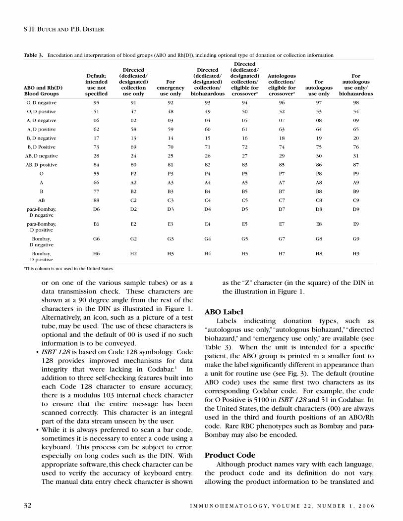
32 I M M U N O H E M A T O L O G Y, V O L U M E 2 2 , N U M B E R 1 , 2 0 0 6
S.H. BUTCH AND P.B. DISTLER
or on one of the various sample tubes) or as adata transmission check. These characters areshown at a 90 degree angle from the rest of thecharacters in the DIN as illustrated in Figure 1.Alternatively, an icon, such as a picture of a testtube, may be used. The use of these characters isoptional and the default of 00 is used if no suchinformation is to be conveyed.
• ISBT 128 is based on Code 128 symbology. Code128 provides improved mechanisms for dataintegrity that were lacking in Codabar.1 Inaddition to three self-checking features built intoeach Code 128 character to ensure accuracy,there is a modulus 103 internal check characterto ensure that the entire message has beenscanned correctly. This character is an integralpart of the data stream unseen by the user.
• While it is always preferred to scan a bar code,sometimes it is necessary to enter a code using akeyboard. This process can be subject to error,especially on long codes such as the DIN. Withappropriate software, this check character can beused to verify the accuracy of keyboard entry.The manual data entry check character is shown
as the “Z” character (in the square) of the DIN inthe illustration in Figure 1.
ABO LabelLabels indicating donation types, such as
“autologous use only,”“autologous biohazard,”“directedbiohazard,” and “emergency use only,” are available (seeTable 3). When the unit is intended for a specificpatient, the ABO group is printed in a smaller font tomake the label significantly different in appearance thana unit for routine use (see Fig. 3). The default (routineABO code) uses the same first two characters as itscorresponding Codabar code. For example, the codefor O Positive is 5100 in ISBT 128 and 51 in Codabar. Inthe United States, the default characters (00) are alwaysused in the third and fourth positions of an ABO/Rhcode. Rare RBC phenotypes such as Bombay and para-Bombay may also be encoded.
Product Code Although product names vary with each language,
the product code and its definition do not vary,allowing the product information to be translated and
Table 3. Encodation and interpretation of blood groups (ABO and Rh[D]), including optional type of donation or collection information
DirectedDirected Directed (dedicated/
Default: (dedicated/ (dedicated/ designated) Autologous Forintended designated) For designated) collection/ collection/ For autologous
ABO and Rh(D) use not collection emergency collection/ eligible for eligible for autologous use only/Blood Groups specified use only use only biohazardous crossover* crossover* use only biohazardous
O, D negative 95 91 92 93 94 96 97 98
O, D positive 51 47 48 49 50 52 53 54
A, D negative 06 02 03 04 05 07 08 09
A, D positive 62 58 59 60 61 63 64 65
B, D negative 17 13 14 15 16 18 19 20
B, D Positive 73 69 70 71 72 74 75 76
AB, D negative 28 24 25 26 27 29 30 31
AB, D positive 84 80 81 82 83 85 86 87
O 55 P2 P3 P4 P5 P7 P8 P9
A 66 A2 A3 A4 A5 A7 A8 A9
B 77 B2 B3 B4 B5 B7 B8 B9
AB 88 C2 C3 C4 C5 C7 C8 C9
para-Bombay, D6 D2 D3 D4 D5 D7 D8 D9D negative
para-Bombay, E6 E2 E3 E4 E5 E7 E8 E9D positive
Bombay, G6 G2 G3 G4 G5 G7 G8 G9D negative
Bombay, H6 H2 H3 H4 H5 H7 H8 H9D positive
*This column is not used in the United States.

I M M U N O H E M A T O L O G Y, V O L U M E 2 2 , N U M B E R 1 , 2 0 0 6 33
ISBT 128
understood internationally. Each product is definedusing component classes, modifiers, and attributes.
• Classes: general descriptions such as RBCs,platelets, or plasma
• Modifiers: terms that broadly differentiate theclasses, such as washed, frozen, thawed, orapheresis
• Attributes: further descriptions of thecomponents. Attributes are composed of:– core conditions that every blood component
must have (e.g., type of anticoagulant/additive,collection volume, and storage temperature)
– groups (such as system integrity, intended use,irradiation, and leukocyte reduction) andvariables within these groups (such as opensystem, for manufacturing use, irradiated, orresidual leukocyte count < 5 × 10⁶) that notevery component would have
On the blood unit label, the name of the product isgiven in the order of modifier,class, and attributes,withclass appearing in the larger font and modifiers andattributes in a smaller font (see Fig. 4).
Because ISBT 128 conveys more information aboutthe product in the bar code, there is not a one-to-onerelationship between ISBT 128 and Codabar productcodes. For example, CPD Whole Blood had a singlecode of 00150 in Codabar that did not encode thecollection volume or system integrity. In ISBT 128,there are four possible codes for this product:
Whole Blood |CPD/450mL/refg E0009Whole Blood |CPD/450mL/refg/Open E0013Whole Blood |CPD/500mL/refg E0023Whole Blood |CPD/500mL/refg/Open E0027.This additional encoded information allows for
computer verification that a label with the appropriatecollection volume and outdate was applied.
In addition to the product definition, the productcode data structure contains information about thetype of donation and a mechanism to identify divisionsof a given product.
Expiration DateThe bar code text display of the expiration date and
time is designed to avoid confusion over the monthand year and to comply with international usage. Theoutdate 6/23/05 2:30 pm is displayed as 23 JUN 200614:30. When a product expires at midnight, the time23:59 is encoded, but may not be printed in the barcode text.
Special Testing LabelsOf particular interest to laboratories that provide
antigen-typed blood is the ability to provide antigentyping information in a bar code format. In the lowerright quadrant of the label there is space for up to twobar codes for RBC antigen typing, other special testinginformation, or both. The data structures that can beused are:
• Special testing: general (e.g., CMV or IgA)• Special testing: RBC antigens• Special testing: serologically determined platelet
HLA and platelet-specific antigens (for plateletproducts)
• Special testing: genomically determined HLA-Aand -B alleles (for cellular therapy and tissueproducts)
• Special testing: genomically determined HLA-DRB1 alleles (for cellular therapy and tissueproducts)
Details about the data structures for thisinformation are displayed in Table 2. Tables 4, 5, and 6give the logic behind encoding RBC antigens. The
Fig. 3. ABO/Rh labels Fig. 4. Product label

34 I M M U N O H E M A T O L O G Y, V O L U M E 2 2 , N U M B E R 1 , 2 0 0 6
S.H. BUTCH AND P.B. DISTLER
Table 4. Encodation and interpretation of special testing: RBC antigens — General, positions 1 to 5
Position 1 2 3 4 5
Antibody
Antigen Rh K k Cw Mia† M N S sValue
0 C+c–E+e– nt nt nt nt nt nt nt nt
1 C+c+E+e– nt neg nt neg nt neg nt neg
2 C–c+E+e– nt pos nt pos nt pos nt pos
3 C+c–E+e+ neg nt neg nt neg nt neg nt
4 C+c+E+e+ neg neg neg neg neg neg neg neg
5 C–c+E+e+ neg pos neg pos neg pos neg pos
6 C+c–E–e+ pos nt pos nt pos nt pos nt
7 C+c+E–e+ pos neg pos neg pos neg pos neg
8 C–c+E–e+ pos pos pos pos pos pos pos pos
9 ni ni ni ni ni ni ni ni ni
Key: † most commonly associated with GP.Mur (Mi.III); nt — not tested; neg — negative; pos — positive; ni — no information (position not used)
Table 5. Encodation and interpretation of special testing: RBC antigens — General, positions 6 to 10
Position 6 7 8 9 10
Antibody
Antigen U P1 Lua Kpa Lea Leb Fya Fyb Jka Jkb
Value
0 nt nt nt nt nt nt nt nt nt nt
1 nt neg nt neg nt neg nt neg nt neg
2 nt pos nt pos nt pos nt pos nt pos
3 neg nt neg nt neg nt neg nt neg nt
4 neg neg neg neg neg neg neg neg neg neg
5 neg pos neg pos neg pos neg pos neg pos
6 pos nt pos nt pos nt pos nt pos nt
7 pos neg pos neg pos neg pos neg pos neg
8 pos pos pos pos pos pos pos pos pos pos
9 ni ni ni ni ni ni ni ni ni ni
Key: nt — not tested; neg — negative; pos — positive; ni — no information (position not used)
Table 6. Encodation and interpretation of special testing: RBC antigens — General, positions 11 to 16
Position 11 12 13 14 15 16
Antibody CMV
Antigen Doa Dob Ina Cob Dia VS/V Jsa res res res resValue
0 nt nt nt nt nt nt nt nt nt nt nt nt
1 nt neg nt neg nt neg nt neg nt neg nt neg
2 nt pos nt pos nt pos nt pos nt pos nt pos
3 neg nt neg nt neg nt neg nt neg nt neg nt
4 neg neg neg neg neg neg neg neg neg neg neg neg
5 neg pos neg pos neg pos neg pos neg pos neg pos
6 pos nt pos nt pos nt pos nt pos nt pos nt
7 pos neg pos neg pos neg pos neg pos neg pos neg
8 pos pos pos pos pos pos pos pos pos pos pos pos
9 ni ni ni ni ni ni ni ni ni ni ni ni
Key: res — reserved; nt — not tested; neg — negative; pos — positive; ni — no information (position not used)

I M M U N O H E M A T O L O G Y, V O L U M E 2 2 , N U M B E R 1 , 2 0 0 6 35
ISBT 128
position in the data structure is given in theinformation across the top of the charts. Down the farlefthand column, a value is given. To interpret thistable, if a 0 appears in position 1 of the RBC antigendata structure, the RBC product is C+,c–,E+,e–(phenotype given where the position 1 columnintersects the line with a value of 0 on Table 4). If this0 in the data structure is followed with the value 1 inposition 2, the RBCs are K not tested, k–. A morecomplete phenotype code (which makes use of Tables4, 5, 6, and 7) would be 880000008700000099. Thisdata string is decoded as follows: C–c+E-e+; K+k+; Cw,Mia, M, N, S, s, U, P1, Lua, Kpa, Lea, Leb not tested;Fy(a+b+); Jk(a+b–); Doa, Dob, Ina, Cob, Dia,VS/V, Jsa, CMVantibody not tested and default value for additionalRBC antigens.
Encoding the HLA type is based on tables similar toTables 4, 5, and 6, which can be found in the ISBT 128Standard,Technical Specification (not provided here).2
A CMV seronegative platelet unit with the serologicHLA class I typing HLA-A2, 3; B7, 44, untested forplatelet-specific antigens and IgA would be encodedthus:
020307440000000100
and the bar code text would appear as this:HLA-A 2, 3; B 7, 44Negative for antibodies to CMV
This has been a very brief overview of the ISBT128 information standard. The references listedbelow1–4 provide a much more indepth explanation ofthe features and logic behind the standard. Thesedocuments, and many others, may be found on theICCBBA Web site (www.isbt128.org). Additionally,information that may be used to plan implementationof ISBT 128 can be found in the ISBT Code 128Implementation Plan5 on the AABB Web site(www.aabb.org).
References1. Wray, B. Why code 128? The rationale behind
ISBT 128. Technical Bulletin 1, ICCBBA,November 1996.
2. ISBT 128 Standard Technical Specification Version2.1.0 August 2004.
3. United States industry consensus standard for theuniform labeling of blood and blood componentsusing ISBT 128, Version 1.2.0, November 1999.
Table 7. Encodation and interpretation of special testing: RBC antigens—General, positions 17 and 18*
Value Antigen
00 information elsewhere
01 Ena
02 ‘N’
03 Vw
04 Mur*
05 Hut
06 Hil
07 P
08 PP1Pk
09 hrS
10 hrB
11 f
12 Ce
13 G
14 Hro
15 CE
16 cE
17 Cx
18 Ew
19 Dw
20 hrH
21 Goa
22 Rh32
23 Rh33
24 Tar
25 Kpb
26 Kpc
27 Jsb
28 Ula
29 K11
30 K12
31 K13
32 K14
33 K17
34 K18
35 K19
36 K22
37 K23
38 K24
39 Lub
40 Lu3
41 Lu4
42 Lu5
43 Lu6
44 Lu7
45 Lu8
46 Lu11
47 Lu12
48 Lu13
49 Lu20
50 Aua
Value Antigen
51 Aub
52 Fy4
53 Fy5
54 Fy6
55 Dib
56 Sda
57 Wrb
58 Ytb
59 Xga
60 Sc1
61 Sc2
62 Sc3
63 Joa
64 removed
65 Hy
66 Gya
67 Co3
68 LWa
69 LWb
70 Kx
71 Ge2
72 Ge3
73 Wb
74 Lsa
75 Ana
76 Dha
77 Cra
78 IFC
79 Kna
80 Inb
81 Csa
82 I
83 Era
84 Vel
85 Lan
86 Ata
87 Jra
88 Oka
89 reserved for future use
90 reserved for future use
91 reserved for future use
92 reserved for future use
93 reserved for future use
94 reserved for future use
95 reserved for future use
96 reserved for future use
97 parvovirus B19
antibody present
98 IgA deficient
99 default
*The RBCs have been tested for the antigen indicated and found negative.

36 I M M U N O H E M A T O L O G Y, V O L U M E 2 2 , N U M B E R 1 , 2 0 0 6
S.H. BUTCH AND P.B. DISTLER
4. Draft United States industry consensus standardfor uniform labeling of blood and bloodcomponents using ISBT 128, Version 2.0.0,November 2005.
5. ISBT code 128 implementation planhttp://www.aabb.org/members_only/archives/other/isbt128plan.htm
Suzanne H. Butch, MA, Blood Bank and TransfusionService, UH 2F225/0054, 1500 E. Medical CenterDrive, Ann Arbor, MI 48109 and Patricia B. Distler,MS,ICCBBA, York, PA.
Attention SBB and BB Students: You are eligible for a free 1-year subscription to Immunohematology. Askyour education supervisor to submit the name and complete address for each student and the inclusive datesof the training period to Immunohematology, P.O. Box 40325, Philadelphia, PA 19106.

I M M U N O H E M A T O L O G Y, V O L U M E 2 2 , N U M B E R 1 , 2 0 0 6 37
E R R A T A
Vol. 21, No.4, 2005; page 146
Reactivity of FDA-approved anti-D reagents with partialD red blood cells
The author has informed the editors ofImmunohematology that there is an error on page146, second column, first sentence. The sentenceshould read “Individuals whose RBCS are characterizedas having a weak D phenotype usually do not makeanti-D if exposed to D+ RBCs…”
Vol. 21, No. 4, 2005; page 171
IN MEMORIAM Tibor J. Greenwalt, MD
A reader has informed the editors ofImmunohematology that there is an error on page171, second paragraph, sixth sentence. The sentenceshould read “He was Clinical Professor of Medicine atGeorge Washington University School of Medicine…”
Manuscripts: The editorial staff of Immunohematology welcomes manuscripts pertaining to blood groupserology and education for consideration for publication. We are especially interested in case reports, paperson platelet and white cell serology, scientific articles covering original investigations, and papers on newmethods for use in the blood bank. Deadlines for receipt of manuscripts for consideration for the March,June,September,and December issues are the first weeks in November,February,May,and August, respectively.For instructions for scientific articles, case reports, and review articles, see “Instructions for Authors” in everyissue of Immunohematology or on the Web. Include fax and phone numbers and e-mail address withyour manuscript.
Attention: State Blood Bank Meeting OrganizersIf you are planning a state meeting and would like copies of Immunohematology for distribution, pleasecontact Cindy Flickinger, Managing Editor, 4 months in advance, by fax or e-mail at (215) 451-2538 [email protected].

38 I M M U N O H E M A T O L O G Y, V O L U M E 2 2 , N U M B E R 1 , 2 0 0 6
Monoclonal antibodies available at no cost. The Laboratory of Immunochemistry at the New York BloodCenter has developed a wide range of monoclonal antibodies (both murine and humanized) that are useful forscreening for antigen–negative donors and for typing patients’ RBCs with a positive DAT. Monoclonal antibodiesavailable include anti-M, -Fya, -Fyb, -K, -k, -Kpa, -Jsb, -Dob, -Wrb, and –Rh17. For a complete list of available monoclonalantibodies, please see our Web site at http://www.nybloodcenter.org/framesets/FS-4C7.htm. Most of thoseantibodies are murine IgG and, thus, require the use of anti-mouse IgG for detection, i.e, anti-K, -k, and -Kpa. Someare directly agglutinating (anti-M, -Wrb, and -Rh17), and a few have been humanized into the IgM isoform and aredirectly agglutinating (anti-Jsb and -Fya). The monoclonal antibodies are available at no charge to anyone whorequests them. Contact: Marion Reid ([email protected]) or Gregory Halverson ([email protected]), New York Blood Center, 310 East 67th Street, New York, NY 10021.
A N N O U N C E M E N T S

I M M U N O H E M A T O L O G Y, V O L U M E 2 2 , N U M B E R 1 , 2 0 0 6 39
A N N O U N C E M E N T S C O N T ’ D
Masters (MSc) in Transfusion and Transplantation Sciences
At
The University of Bristol, England
Applications are invited from medical or science graduates for the Master of Science (MSc) degree in
Transfusion and Transplantation Sciences at the University of Bristol. The course starts in October 2006 and
will last for 1 year. A part-time option lasting 2 or 3 years is also available. There may also be opportunities
to continue studies for PhD or MD following MSc. The syllabus is organized jointly by The Bristol Institute
for Transfusion Sciences and the University of Bristol, Department of Cellular and Molecular Medicine.
It includes:
• Scientific principles of transfusion and transplantation
• Clinical applications of these principles
• Practical techniques in transfusion and transplantation
• Principles of study design and biostatistics
• An original research project
Applications can also be made for Diploma in Transfusion and Transplantation Science or a Certificate in
Transfusion and Transplantation Science.
The course is accredited by the Institute of Biomedical Sciences.
Further information can be obtained from the Web site:
http://www.blood.co.uk/ibgrl/MSc/MScHome.htm
For further details and application forms please contact:
Dr. Patricia Denning-Kendall
University of Bristol
Paul O’Gorman Lifeline Centre, Department of Pathology and Microbiology, Southmead Hospital
Westbury-on-Trym, Bristol
BS10 5NB, England
Fax +44 1179 595 342, Telephone +44 1779 595 455, e-mail: [email protected].

40 I M M U N O H E M A T O L O G Y, V O L U M E 2 2 , N U M B E R 1 , 2 0 0 6
S P E C I A L A N N O U N C E M E N T S
Meetings!
April 26–29 California Blood Bank Society (CBBS)The 53rd annual California Blood Bank Society (CBBS) conference will be held April 26 through 29, 2006, at theHyatt Regency Lake Tahoe, in Incline Village, Nevada. For more information, contact the CBBS central office at(520) 749-6908 or refer to the Web site at http://www.cbbsweb.org.
April 28–30 American Red Cross Immunohematology Reference Laboratory (IRL)Conference 2006
The American Red Cross Immunohematology Reference Laboratory (IRL) Conference 2006 will be held April 28through 30, 2006, at the Hawthorne Suites—Orlando Airport, in Orlando, Florida. For more information, contactCindy Flickinger at (215) 451-4909 or [email protected].
May 1–4 South Central Association of Blood Banks (SCABB)The 48th annual South Central Association of Blood Banks (SCABB) meeting and exhibit will be held May 1through 4, 2006, at the Wyndham DFW Hotel and Arlington Convention Center in Arlington, Texas. For moreinformation, refer to the Web site at http://www.scabb.org.
June 8–9 Heart of America Association of Blood Banks (HAABB)The spring meeting of the Heart of America Association of Blood Banks (HAABB) will be held June 8 and 9, 2006,at the Embassy Suites in Kansas City,Missouri.For more information,refer to the Web site at http://www.haabb.org.

I M M U N O H E M A T O L O G Y, V O L U M E 2 2 , N U M B E R 1 , 2 0 0 6 41
NATIONAL REFERENCE LABORATORY FORBLOOD GROUP SEROLOGY
Immunohematology ReferenceLaboratory
AABB,ARC, New York State, and CLIA licensed(215) 451-4901—24-hr. phone number
(215) 451-2538—Fax
American Rare Donor Program(215) 451-4900—24-hr. phone number
(215) 451-2538—[email protected]
Immunohematology(215) 451-4902—Phone, business hours
(215) 451-2538—[email protected]
Quality Control of Cryoprecipitated-AHF(215) 451-4903—Phone, business hours
(215) 451-2538—Fax
Granulocyte Antibody Detection and Typing
• Specializing in granulocyte antibody detectionand granulocyte antigen typing
• Patients with granulocytopenia can be classifiedthrough the following tests for proper therapyand monitoring:—Granulocyte agglutination (GA)—Granulocyte immunofluorescence (GIF)—Monoclonal Antibody Immobilization of
Granulocyte Antigens (MAIGA)
For information regarding services, call Gail Eiberat: (651) 291-6797, e-mail: [email protected],
or write to:Neutrophil Serology Reference Laboratory
American Red CrossSt. Paul Regional Blood Services
100 South Robert StreetSt. Paul, MN 55107
CLIA LICENSED
National Platelet Serology ReferenceLaboratory
Diagnostic testing for:• Neonatal alloimmune thrombocytopenia (NAIT)• Posttransfusion purpura (PTP)• Refractoriness to platelet transfusion• Heparin-induced thrombocytopenia (HIT)• Alloimmune idiopathic thrombocytopenia
purpura (AITP)Medical consultation available
Test methods:• GTI systems tests
—detection of glycoprotein-specific plateletantibodies
—detection of heparin-induced antibodies (PF4ELISA)
• Platelet suspension immunofluorescence test(PSIFT)
• Solid phase red cell adherence (SPRCA) assay• Monoclonal antibody immobilization of platelet
antigens (MAIPA) • Molecular analysis for HPA-1a/1b
For information, e-mail: [email protected] call:
Maryann Keashen-Schnell(215) 451-4041 office
(215) 451-4205 laboratory
Sandra Nance(215) 451-4362
Scott Murphy, MD(215) 451-4877
American Red Cross Blood ServicesMusser Blood Center
700 Spring Garden StreetPhiladelphia, PA 19123-3594
CLIA LICENSED
A D V E R T I S E M E N T S

42 I M M U N O H E M A T O L O G Y, V O L U M E 2 2 , N U M B E R 1 , 2 0 0 6
A D V E R T I S E M E N T S C O N T ’ D
IgA/Anti-IgA Testing
IgA and anti-IgA testing is available to do the following:• Monitor known IgA-deficient patients• Investigate anaphylactic reactions• Confirm IgA-deficient donors
Our ELISA assay for IgA detects antigen to 0.05 mg/dL.
For information on charges and sample requirements, call (215) 451-4909, e-mail:
[email protected],or write to:
American Red Cross Blood ServicesMusser Blood Center
700 Spring Garden StreetPhiladelphia, PA 19123-3594
ATTN: Cindy Flickinger
CLIA LICENSED
Reference and Consultation Services
Antibody identification and problem resolution
HLA-A, B, C, and DR typing
HLA-disease association typing
Paternity testing/DNA
For information regarding our services, contact
Mehdizadeh Kashi at (503) 280-0210, or write to:
Pacific Northwest Regional Blood Services
ATTENTION: Tissue Typing Laboratory
American Red Cross
3131 North Vancouver
Portland, OR 97227
CLIA LICENSED, ASHI ACCREDITED
National Neutrophil Serology ReferenceLaboratory
Our laboratory specializes in granulocyteantibody detection and granulocyte antigentyping.
Indications for granulocyte serology testinginclude:• Alloimmune neonatal neutropenia (ANN)• Autoimmune neutropenia (AIN)• Transfusion related acute lung injury (TRALI)
Methodologies employed:• Granulocyte agglutination (GA)• Granulocyte immunofluorescence by flow
cytometry (GIF)• Monoclonal antibody immobilization of
neutrophil antigens (MAINA)
TRALI investigations also include:• HLA (PRA) Class I and Class II antibody
detection
For further information contact:
Neutrophil Serology Laboratory(651) 291-6797
Randy Schuller(651) 291-6758
American Red Cross Blood ServicesNeutrophil Serology Laboratory
100 South Robert StreetSt. Paul, MN 55107
CLIA LICENSED
Notice to Readers: All articles published,including communications and book reviews,reflect the opinions of the authors and do notnecessarily reflect the official policy of theAmerican Red Cross.

I M M U N O H E M A T O L O G Y, V O L U M E 2 2 , N U M B E R 1 , 2 0 0 6 43
Donor IgA Screening
• Effective tool for screening large volumes ofdonors
• Gel diffusion test that has a 15-year proventrack record:– Approximately 90 percent of all donors
identified as IgA deficient by are confirmedby the more sensitive testing methods
For information regarding charging and samplerequirements, call Kathy Kaherl at:
(860) 678-2764, e-mail: [email protected] or write to:
Reference LaboratoryAmerican Red CrossConnecticut Region
209 Farmington Ave.Farmington, CT 06032
CLIA LICENSED
A D V E R T I S E M E N T S C O N T ’ D
REMEMBER: THE PASSWORD IS “2000” For www.redcross.org/pubs/immuno
Now, as a subscriber, you can enter the password, 2000, and access the back issues. That meanscover to cover! You will receive every article in total, every letter to the editor, every review, every ad, everynotice, and every literature review! All of the other services will continue to be available on the Web site,including sending a letter to the editor, subscribing with a credit card on the secure order site, performing aliterature search, reviewing instructions for authors, and linking to other important sites. Log on now to seethis great service!
Phone, Fax, and Internet Information: If you have any questions concerning Immunohematology,Journal of Blood Group Serology and Education, or the Immunohematology Methods and Proceduresmanual,contact us by e-mail at [email protected] information concerning the National ReferenceLaboratory for Blood Group Serology, including the American Rare Donor Program, please contact SandraNance, by phone at (215) 451-4362, by fax at (215) 451-2538, or by e-mail at [email protected]

44 I M M U N O H E M A T O L O G Y, V O L U M E 2 2 , N U M B E R 1 , 2 0 0 6
SCIENTIFIC ARTICLES, REVIEWS, AND CASE REPORTSBefore submitting a manuscript, consult current issues of
Immunohematology for style. Type the manuscript on white bondpaper (8.5" × 11") and double-space throughout. Number the pagesconsecutively in the upper right-hand corner, beginning with thetitle page. Each component of the manuscript must start on a newpage in the following order:1. Title page2. Abstract3. Text4. Acknowledgments5. References6. Author information7. Tables—see 7 under Preparation8. Figures—see 8 under Preparation
Preparation of manuscripts1. Title page
A.Full title of manuscript with only first letter of first word capitalized (bold title)
B. Initials and last name of each author (no degrees; all CAPS),e.g., M.T. JONES and J.H. BROWN
C.Running title of ≤ 40 characters, including spacesD.3 to 10 key words
2. AbstractA.One paragraph, no longer than 300 wordsB. Purpose, methods, findings, and conclusions of study
3. Key words—list under abstract4. Text (serial pages)
Most manuscripts can usually, but not necessarily, be divided intosections (as described below). Results of surveys and reviewpapers are examples that may need individualized sections.A. Introduction
Purpose and rationale for study, including pertinent back-ground references.
B. Case Report (if study calls for one)Clinical and/or hematologic data and background serology.
C.Materials and MethodsSelection and number of subjects, samples, items, etc. studiedand description of appropriate controls, procedures, methods,equipment, reagents, etc. Equipment and reagents should beidentified in parentheses by model or lot and manufacturer’sname, city, and state. Do not use patients’ names or hospitalnumbers.
D.ResultsPresentation of concise and sequential results, referring to perti-nent tables and/or figures, if applicable.
E. DiscussionImplications and limitations of the study, links to other studies;if appropriate, link conclusions to purpose of study as stated inintroduction.
5. AcknowledgmentsAcknowledge those who have made substantial contributions tothe study, including secretarial assistance; list any grants.
6. ReferencesA. In text, use superscript, arabic numbers.B. Number references consecutively in the order they occur in
the text.C.Use inclusive pages of cited references, e.g., 1431–7.D.Refer to current issues of Immunohematology for style.
7. TablesA.Head each with a brief title, capitalize first letter of first word
(e.g., Table 1. Results of ...), and use no punctuation at the endof the title.
B. Use short headings for each column needed and capitalize firstletter of first word. Omit vertical lines.
C.Place explanations in footnotes (sequence: *, †, ‡, §, ¶, **, ††).8. Figures
A.Figures can be submitted either by e-mail or as photographs (5″ × 7″ glossy).
B. Place caption for a figure on a separate page (e.g.,Fig.1.Resultsof ...), ending with a period. If figure is submitted as a glossy,place first author’s name and figure number on back of eachglossy submitted.
C.When plotting points on a figure, use the following symbols ifpossible: ●● ● ▲▲ ▲ ■■ ■.
9. Author informationA.List first name, middle initial, last name, highest academic
degree, position held, institution and department, and complete address (including zip code) for all authors. Listcountry when applicable.
SCIENTIFIC ARTICLES AND CASE REPORTS SUBMITTED AS LETTERS TO THE EDITOR
Preparation1. Heading—To the Editor:2. Under heading—title with first letter capitalized.3. Text—write in letter format (paragraphs).4. Author(s)—type flush right; for first author: name, degree,
institution, address (including city, state, ZIP code, and country);for other authors: name, degree, institution, city, and state.
5. References—limited to ten.6. One table and/or figure allowed.
ImmunohematologyJOURNAL OF BLOOD GROUP SEROLOGY AND EDUCATION
Instructions for Authors
Send all manuscripts by e-mail to:
Marge Manigly at [email protected]

I M M U N O H E M A T O L O G Y, V O L U M E 2 2 , N U M B E R 1 , 2 0 0 6 45
What is a certified Specialist in Blood Banking (SBB)?• Someone with educational and work experience qualifications who successfully passes the American Society for
Clinical Pathology (ASCP) board of registry (BOR) examination for the Specialist in Blood Banking.• This person will have advanced knowledge, skills, and abilities in the field of transfusion medicine and blood banking.
Individuals who have an SBB certification serve in many areas of transfusion medicine:• Serve as regulatory, technical, procedural, and research advisors• Perform and direct administrative functions • Develop, validate, implement, and perform laboratory procedures• Analyze quality issues, preparing and implementing corrective actions to prevent and document issues• Design and present educational programs• Provide technical and scientific training in blood transfusion medicine• Conduct research in transfusion medicine
Who are SBBs?Supervisors of Transfusion Services Managers of Blood Centers LIS Coordinators EducatorsSupervisors of Reference Laboratories Research Scientists Consumer Safety OfficersQuality Assurance Officers Technical Representatives Reference Lab Specialist
Why be an SBB?Professional growth Job placement Job satisfaction Career advancement
How does one become an SBB?• Attend a CAAHEP-accredited Specialist in Blood Bank Technology Program OR• Sit for the examination based on criteria established by ASCP for education and experience
Fact #1: In recent years, the average SBB exam pass rate is only 38%.Fact #2: In recent years, greater than 73% of people who graduate from CAAHEP-accredited programs pass the SBB
exam.
Conclusion:The BEST route for obtaining an SBB certification is to attend a CAAHEP-accredited Specialist in Blood BankTechnology Program
Becoming a Specialist in Blood Banking (SBB)
Contact the following programs for more information:PROGRAM CONTACT NAME CONTACT INFORMATION
Walter Reed Army Medical Center William Turcan 202-782-6210;[email protected]
Transfusion Medicine Center at Florida Blood Services Marjorie Doty 727-568-5433 x 1514; [email protected]
Univ. of Illinois at Chicago Veronica Lewis 312-996-6721; [email protected]
Medical Center of Louisiana Karen Kirkley 504-903-2466; [email protected]
NIH Clinical Center Department of Transfusion Medicine Karen Byrne 301-496-8335; [email protected]
Johns Hopkins Hospital Christine Beritela 410-955-6580; [email protected]
ARC-Central OH Region, OSU Medical Center Joanne Kosanke 614-253-2740 x 2270; [email protected]
Hoxworth Blood Center/Univ. of Cincinnati Medical Center Catherine Beiting 513-558-1275; [email protected]
Gulf Coast School of Blood Bank Technology Clare Wong 713-791-6201; [email protected]
Univ. of Texas SW Medical Center Barbara Laird-Fryer 214-648-1785; [email protected]
Univ. of Texas Medical Branch at Galveston Janet Vincent 409-772-4866; [email protected]
Univ. of Texas Health Science Center at San Antonio Bonnie Fodermaier SBB Program: 210-358-2807,Linda Smith [email protected]
MS Program: 210-567-8869; [email protected]
Blood Center of Southeastern Wisconsin Lynne LeMense 414-937-6403; [email protected]
Additional information can be found by visiting the following Web sites: www.ascp.org, www.caahep.org, and www.aabb.org


Musser Blood Center700 Spring Garden StreetPhiladelphia, PA 19123-3594
(Place Label Here)