immunohistochemical profile of mucins and their expression … · jurnal f bimedicine international...
TRANSCRIPT

INTERNATIONAL JOURNAL
OF BIOMEDICINE
International Journal of BioMedicine 3(2) (2013) 115-118
Immunohistochemical Profile of Mucins and their Expression in Precancerous Changes of the Stomach
Sergii V. Vernygorodskyi, PhD1*, Lyudmila V. Fomina, PhD, ScD1, Maksim V. Mnikhovich, PhD2, Nila A. Kaminska PhD1, Galina V. Datsenko, PhD1, Arthur M. Berezovskiy, PhD1, Olena
M. Shapoval, PhD1, Olena I. Bashynska, PhD1, Natalia V. Belik, PhD1
¹Vinnitsa National Pirogov Memorial Medical University, Vinnitsa, Ukraine,²Research Institute of Human Morphology of the Russian Academy of Medical Sciences, Moscow, Russia
AbstractThe aim of this study was to assess the profile of mucins (MUC1, MUC2, MUC5AC) in the intestinal metaplasia (IM) of
the gastric mucosa through the immunohistochemical method. Methods: To identify the metaplastic areas in the gastric mucosa, chromoendoscopy was employed using 0.5% solution of methylene blue. The expression of the profile of the mucins was determined using immunohistochemistry with MUC1, MUC5AC, and MUC2 antibodies (clone Ma695, clone CLH2, Ccp58 and CLH5, «Novocastra «Great Britain). Results: In the regions adjacent to the adenocarcinoma and neoplastic modified cells, a visible weak expression of MUC2 and MUC5AC was observed. In the case of complete IM, a visibly maximum MUC2 expression was observed in the goblet cells; thus, the MUC5AC, MUC1, and MUC6 marking were absent in the columnar epitheliocytes with the brush border. In the case of incomplete IM, along with the positive MUC2 markings of the goblet cells, the presence of gastric mucin (MUC5AC) has been observed in 25% of such patients with chronic atrophic gastritis (CAG) having incomplete IM; however, in the columnar epitheliocytes the characteristic occurrence of gastric mucin (MUC5AC) was observed in 100% of the patients while a small amount of MUC2 was recorded in 15% of patients. Conclusion: The MUC5AC expression of the gastric mucins in the columnar epithelial cells and the goblet exocrinocytes marks the formation of the gastrointestinal phenotype viz., incomplete intestinal metaplasia, along with the simultaneous production of the MUC2 by the goblet cells. The decrease with further loss of the protective MUC5AC production by the columnar epithelial cells and goblet exocrinocytes that were found in the regions of severe dysplasia and IM, adjacent to the neoplastic altered cells, may serve as additional criteria of early malignancy of the gastric mucosa.
Keywords: mucins, precancerous changes of the gastric mucosa.
IntroductionThe current understanding of the gastrointestinal barrier
links the ability of the surface and the glandular epithelial cells to synthesize mucin [1,2]. At the same time, the epithelial mucins produced by the epithelial cells are a large group of glycosylated proteins secreted and embedded in the plasmolemmal glycoproteins. In the epithelial tissues, 13 types of mucin are
found, forming two classes of compounds: the transmembrane and secretory mucins. MUC1 is one of the transmembrane mucins of the digestive tract cells. It is distributed on the cell surface and acts as a signaling molecule. The MUC3 and MUC4 present within the same cells are regarded as the original form of the secretory mucins. The gel-forming mucins of the digestive tract, however, include MUC2, MUC5AC, MUC4B, and MUC6. They are actively expressed by the epithelial cells and represent a variable number of peptides rich in serine, threonine and proline, coupled with a large number of oligosaccharide chains. According to the information available [3], the expression of the membrane glycoproteins increases nearly tenfold in the transformed cells, with shortening of the carbohydrate chains. Such changes triggered by the mucin-producing cells are
BASIC RESEARCH
*Corresponding author: Sergii V. Vernygorodskyi, PhD. Department of Pathological Anatomy, Forensic Medicine and Law, Vinnitsa National Pirogov Memorial Medical University. 34, Stakhursky st, apt 16, Vinnytsya, 21027, Ukraine.
Tel.: 0679565453. E-mail: [email protected]

116 S. V. Vernygorodskyi et al. / International Journal of BioMedicine 3(2) (2013) 115-118
considered to be a carcinogenic marker. Mechanisms that are responsible for the orientation of the protein molecules in the bilayer of the cytomembranes, stabilization of the spatial structure of the proteins, transmembrane and intracellular transport of substances (including the transfer of the hydrolytic enzymes from the Golgi complex to lysosomes), and the molecular mechanisms of intercellular recognition are associated with the specific structure of the carbohydrate determinants of the glycoproteins. As is evident, these mechanisms play an important role in the maturation and differentiation of cells, histogenesis and organ morphogenesis, with the contact inhibition of cell proliferation, providing immune surveillance. This forms the basis of the most important manifestations of malignant growth [4-7].
Today, it has been proved that CAG is one of the main elements responsible for the carcinogenesis. According to the recommendations of the International Atrophy Group (2002) two main types of atrophy are distinguishable, metaplastic and non-metaplasic [8]. Non-metaplasic atrophy is characterized by the loss of glands, accompanied by fibrosis or fibromuscular proliferation in the lamina propria of the gastric mucosa; however, metaplastic atrophy is characterized by the replacement of the gastric glands by metaplastic ones (including enteric), which can happen in the setting of other signs of atrophy. Any disturbance in the synthesis or secretion released by the glandular cells triggers a change in the chemical composition, under the influence of the exogenous and endogenous factors in the CAG with IM; however, this process has not yet been studied thoroughly enough. Such transformations are some of the most important components of the mucosal barrier and may bear great significance in relation to the morphogenetic and clinical differences of the CAG with IM in different patient groups.
Material and Methods Over a period of six years, 98 patients were examined.
They had been sent to the endoscopy departments of Vinnytsya hospitals to clarify the clinical diagnosis. Of these patients, 52 (53%) were male and 46 (47%) were female. From among these 98 patients, we selected 68 with the CAG with IM who constituted the main group for further investigation because IM was closely associated with this disease. The comparison group included 30 individuals with the CAG, but without IM. The average age of the patients examined in the dynamics was 52.96 ± 1.13, and the average duration of the disease at the time of IM diagnosis was 2.6 ± 0.63 years. Multiple biopsies were performed during the endoscopy and chromoendoscopy with 0.5% solution of methylene blue (two biopsies from the body and antrum, and one from an area in the angle of the stomach) taking into account the requirements of the modified Sydney system of chronic gastritis diagnosis. The areas of the gastric mucosa were stained for the following histological study of the biopsies. The biopsy material was fixed in 10% neutral formalin and after routine processing some paraffin blocks were produced. Sectioning was done to 5.7 micron thickness. To define the metaplastic changes of the gastric mucosa, routine histological (staining with H&E and Van Gieson) and histochemical (combined high iron diamine and alcian blue technique, orsein combined with alcian blue, Gomory aldehyde fuchsin, alcian blue with pH 1.0 and 2.5 together with the PAS reaction) methods were employed. To identify the presence of Helicobacter pylori (H. pylori), a rapid urease test was conducted,
the cytology was tested with a Pappenheim stain, and from the histological viewpoint staining was done using Romanovsky-Giemsa and toluidine blue stain.
Immunohistochemical studies were performed on paraffin sections using the streptavidin-biotin method (“DAKO”, Denmark, LSAB2 Systems, HRP). The expression of the mucin profile was determined using the antibodies of MUC1, MUC 5AC, MUC2, and MUC6 (clone Ma695, clone CLH2, Ccp58, and CLH5, “Novocastra “Great Britain). In preparations at 400-fold magnification the rate of intestinal differentiation was determined (nuclear label CDX2) in 5 randomly selected fields of view (≥ 500 cells) as a percentage of the positively stained nuclei of the epithelial cells of the gastric mucosa in three compartments (I - surface and foveolar epithelium; II - neck region, III - base of the glands, middle and lower third of the glands to the basal sections). To evaluate the expression of the mucins (MUC5AC, MUC2, and MUC6) in the gastric mucosa in similar areas, the semi-quantitative color intensity rating scale was used: 0 (none) - no positive reaction in the cells, 1 (weak) - up to 30% of the cells reacted positively, 2 ( moderate) - 31-60%, 3 (strong) - 60% or more stained cells [9].
Three independent investigators examined all the sections for histopathological study and a blind study for immunohistochemical evaluation was conducted by the third one. The expression of mucins was evaluated by cytoplasmic staining. The results were expressed semiquantitatively for each histological group as the number of positively labeled sections, the predominantly labeled cell type, and the average score of the positively labeled cells. Positive Control Sections included control tissues taken from the colon and stomach, with the previously identified MUC gene expression patterns included with each batch of sections for immunohistochemistry. Negative Control Sections involved eliminating the primary antibody as a negative control to test the specificity of the antibodies utilized for each section.
Statistical analysis: Results of the immunohistochemical alterations were compared with the clinical-pathologic features using the chi-square test for qualitative data, with a two- tailed p-value < 0.05 considered significant.
All the investigations were performed in accordance with the guidelines of the Ukraine National Bioethics Committee based on “Ethical and law problems of clinical trials and scientific experiments with humans and animals” (Decision of the Presidium of The National Academy of Sciences of Ukraine from 07.11.2007 № 288) corresponding to international standards of animal welfare.
ResultsThe study of mucins in the gastric mucosa showed that a
certain cell phenotype corresponds to a specific type of mucin. Therefore, the MUC5AC is found in the epithelium pits, while the MUC1 occurs in the mucous neck cells, the MUC6 occurs in the pyloric exocrinocytes, and the MUC2 occurs in goblet cells. The fact that the MUC 5AC is selectively synthesized by the epithelial cells of the pits and necks of the gland cells of normal gastric mucosa enables us to draw a conclusion that the epithelial cells of the necks of the gland cells may be identified as those producing the mucus. Due to the low specificity of the MUC 1 binding of the cytoplasmic mucus, we observed its weak
117S. V. Vernigorodskyi et al. / International Journal of BioMedicine 3(2) (2013) 115-118
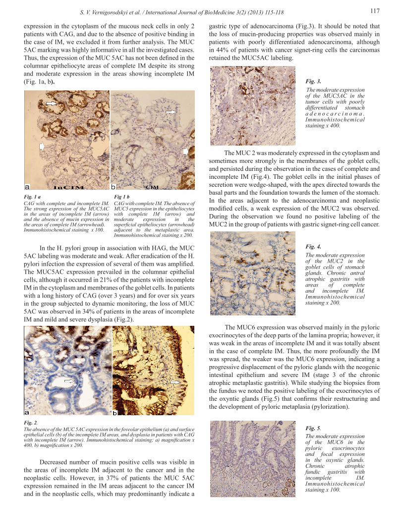
expression in the cytoplasm of the mucous neck cells in only 2 patients with CAG, and due to the absence of positive binding in the case of IM, we excluded it from further analysis. The MUC 5AC marking was highly informative in all the investigated cases. Thus, the expression of the MUC 5AC has not been defined in the columnar epitheliocyte areas of complete IM despite its strong and moderate expression in the areas showing incomplete IM (Fig. 1a, b).
In the H. pylori group in association with HAG, the MUC 5AC labeling was moderate and weak. After eradication of the H. pylori infection the expression of several of them was amplified. The MUC5AC expression prevailed in the columnar epithelial cells, although it occurred in 21% of the patients with incomplete IM in the cytoplasm and membranes of the goblet cells. In patients with a long history of CAG (over 3 years) and for over six years in the group subjected to dynamic monitoring, the loss of MUC 5AC was observed in 34% of patients in the areas of incomplete IM and mild and severe dysplasia (Fig.2).
Decreased number of mucin positive cells was visible in the areas of incomplete IM adjacent to the cancer and in the neoplastic cells. However, in 37% of patients the MUC 5AC expression remained in the IM areas adjacent to the cancer IM and in the neoplastic cells, which may predominantly indicate a
gastric type of adenocarcinoma (Fig.3). It should be noted that the loss of mucin-producing properties was observed mainly in patients with poorly differentiated adenocarcinoma, although in 44% of patients with cancer signet-ring cells the carcinomas retained the MUC5AC labeling.
The MUC 2 was moderately expressed in the cytoplasm and sometimes more strongly in the membranes of the goblet cells, and persisted during the observation in the cases of complete and incomplete IM (Fig.4). The goblet cells in the initial phases of secretion were wedge-shaped, with the apex directed towards the basal parts and the foundation towards the lumen of the stomach. In the areas adjacent to the adenocarcinoma and neoplastic modified cells, a weak expression of the MUC2 was observed. During the observation we found no positive labeling of the MUC2 in the group of patients with gastric signet-ring cell cancer.
The MUC6 expression was observed mainly in the pyloric exocrinocytes of the deep parts of the lamina propria; however, it was weak in the areas of incomplete IM and it was totally absent in the case of complete IM. Thus, the more profoundly the IM was spread, the weaker was the MUC6 expression, indicating a progressive displacement of the pyloric glands with the neogenic intestinal epithelium and severe IM (stage 3 of the chronic atrophic metaplastic gastritis). While studying the biopsies from the fundus we noted the positive labeling of the exocrinocytes of the oxyntic glands (Fig.5) that confirms their restructuring and the development of pyloric metaplasia (pylorization).
Fig. 1 a CAG with complete and incomplete IM. The strong expression of the MUC5AC in the areas of incomplete IM (arrow) and the absence of mucin expression in the areas of complete IM (arrowhead). Immunohistochemical staining x 100.
Fig 1 bCAG with complete IM. The absence of MUC5 expression in the epitheliocytes with complete IM (arrow) and moderate expression in the superficial epitheliocytes (arrowhead) adjacent to the metaplastic area. Immunohistochemical staining x 200.
Fig. 2. The absence of the MUC 5AC expression in the foveolar epithelium (a) and surface epithelial cells (b) of the incomplete IM areas, and dysplasia in patients with CAG with incomplete IM (arrow). Immunohistochemical staining; a) magnification x 400, b) magnification x 200.
Fig. 3. The moderate expression of the MUC5AC in the tumor cells with poorly differentiated stomach a d e n o c a r c i n o m a . Immunohistochemical staining x 400.
Fig. 4. The moderate expression of the MUC2 in the goblet cells of stomach glands. Chronic antral atrophic gastritis with areas of complete and incomplete IM. Immunohistochemical staining x 200.
Fig. 5.The moderate expression of the MUC6 in the pyloric exocrinocytes and focal expression in the oxyntic glands. Chronic atrophic fundic gastritis with incomplete IM. Immunohistochemical staining x 100.

118 S. V. Vernigorodskyi et al. / International Journal of BioMedicine 3(2) (2013) 115-118
Therefore, the findings of the immunohistochemical analysis of the profiles of the mucins indicate the presence of some mucin specificity in the different IM types (Tables 1-2).
As can be seen from Table 2, when the IM was complete, maximum expression of the MUC 2 was visible in the goblet cells, thus the MUC5AC, MUC1, and MUC6 marking were absent in the columnar epitheliocytes with a brush border. When the IM was incomplete and MUC2 marking of the goblet cells was positive, in 25% of patients with CAG with incomplete IM, the gastric mucin(MUC5AC) was observed as well; however, for the columnar epitheliocytes, it was characteristic that the gastric mucin was found in 100% of the patients observed, and as for the MUC2, it was observed in small amounts (15% of patients).
Discussion and ConclusionThe IM has now been recognized to be either a complete
or an incomplete type of IM, or a small- (Type I and Type II) or large- (Type III) intestinal-type IM [10,11]. Although these classifications have generally been accepted, they overemphasize the characteristics common to the cells in the small intestine, while neglecting the existing gastric phenotype. However, the results of our investigations coincide with other researches [12, 13] that suggested segregating the IM into two categories, i.e., a mixed gastric and intestinal (GI) type, and a solely intestinal (I) type, based on the residual gastric phenotype cells.
Thus, the separation of the gastrointestinal phenotype of IM, unlike the prior classification [10,11] which offered only intestinal types (mainly intestinal and colonic) enables us to consider the gastric and intestinal phenotypes. It helps us deepen our knowledge regarding the histogenesis of gastric cancer, as well as confirms the heterogeneity of IM.
1. The mucin profile can be used for the differential diagnosing of various types of metaplastic changes in the gastric epithelium and the histological variants of gastric cancer.
2. The MUC5AC expression of gastric mucins in the columnar epithelial cells and goblet exocrinocytes marks the formation of the gastrointestinal phenotype, incomplete intestinal metaplasia, along with the simultaneous production of MUC2 by goblet cells.
3. Most often the intestinal phenotype is characterized by the absence of MUC5AC in the columnar epitheliocytes and goblet exocrinocytes despite the MUC2 production by the goblet exocrinocytes.
4. MUC6 may serve as a marker for pyloric metaplasia, as its appearance in the cells of the glands shows pylorization and distribution of atrophic changes in the specialized glands of the stomach fundus.
5. The decrease with the further stop in the production of the protective MUC5AC by the columnar epithelial cells and goblet exocrinocytes that were in the areas of severe dysplasia and IM adjacent to the neoplastic altered cells may serve as additional criteria of the early malignancy of the gastric mucosa.
References1. Allen A, Flemstrom G. Gastroduodenal mucus
bicarbonate barrier: protection against acid and pepsin. Am J Pphysiol Cell Physiol 2005; 288(1):1-19.
2. Mogil’naia GM, Mogil’naia VL. Gastrointestinal protective barrier. Morphologiia 2007; 132(6): 9–16. [Article in Russian].
3. Gelesnaya LА. Structure and functions of mucus glycoproteins of mucus (mucins). Rus J Gastroenterol Hepatol Coloproctol1998; 1:30-37. [Article in Russian].
4. Tereshechenko VP, Koslova ТG, Pishykov VА. Pathology of mucous secretion in the stomach and duodenum in liquidators of Chernobyl Accident. Edited by VP Tereshechenko. К.: Medinform, 2004.
5. Karasawa F, Shiota A, Goso Y, Kobayashi M, Sato Y, Masumoto J, et al. Essential role of gastric gland mucin in preventing gastric cancer in mice. J Clin Invest 2012; 122(3):923-34.
6. Gomceli I, Demiriz B, Tez M. Gastric carcinogenesis. World J Gastroenterol 2012; 18(37): 5164-70.
7. Edaise Maria da Silva, José Humberto Tavares Guerreiro Fregnani. Molecular analyses of early-onset gastric cancer in Brazilian patients: TP53 mutations, cadherin-catenin and mucins proteins expression. JCT 2013; 4(1A): 33-42.
8. Rugge M, Correa P, Dixon MF, Fiocca R, Hattori T, Lechago J, et al. Gastric mucosal atrophy: interobserver consistency using new criteria for classification and grading. Aliment Pharmacol Ther 2002; 16(7):1249-59.
9. Cassaro M, Rugge M, Tieppo C, Giacomelli L, Velo D, Nitti D, et al. Indefinite for non-invasive neoplasia lesions in gastric intestinal metaplasia: the immunophenotype. J Clin Pathol 2007; 60(6):615-21.
10. Kawachi T, Kogure K, Tanaka N, Tokunaga A, Sugimura T. Studies of intestinal metaplasia in the gastric mucosa by detection of disaccharidases with ‘‘Tes-Tape’’. J Natl Cancer Inst 1974; 53(1):19–30.
11. Jass JR, Filipe MI. A variant of intestinal metaplasia associated with gastric carcinoma: a histochemical study. Histopathology 1979; 3(3):191–9.
12. Inada K, Nakanishi H, Fujimitsu Y, Shimizu N, Ichinose M, Miki K, et al. Gastric and intestinal mixed and solely intestinal types of intestinal metaplasia in the human stomach. Pathol Int 1997; 47(12):831-41.
13. Niwa T, Ikehara Y, Nakanishi H, Tanaka H, Inada K, Tsukamoto T, et al. Mixed gastric- and intestinal-type metaplasia is formed by cells with dual intestinal and gastric differentiation. J Histochem Cytochem 2005; 53(1):75-85.
Table 1. Chronic atrophic gastritis with complete intestinal metaplasia
Mucins
SpecificitySFE MNC OE PC PE GS СCBB
MUC1 - + - - - - -MUC2 +/- - - - - +++ -
MUC5AC - - ++ - - - -MUC6 - - - - +++ - -
Note: SFE - superficial and foveolar epithelium, MNC - mucous neck cells, OE – oxyntic exocrinocytes, PC - parietal cells, PE – pyloric exocrinocytes, GS - goblet cells, CCBB - columnar epithelial cells with brush border. MUC - mucin, - no expression, + / - weak or absent, + weak expression, + + moderate, + + + strong expression.
Table 2. Chronic atrophic gastritis with incomplete intestinal metaplasia
MucinsSpecificity
SFE MNC OE PC PE GS СCBBMUC1 + + - - - +++ -MUC2 - - - - - +++ +/-
MUC5AC +++ ++ ++ - - +/- +++MUC6 - + + - +++ - -
Note: see Table 1.