immunohistochemistry of human fetus...hiroshima journal of medical sciences vol. 33, no. 4, 625~632,...
TRANSCRIPT

Hiroshima Journal of Medical Sciences Vol. 33, No. 4, 625~632, December, 1984
HIJM 33-87
625
Immunohistochemistry of Human Fetus
-Ig G, A and M Cells in the Thymus and Spleen-*)
Yukio SATOWD, Naotaka AKIMOTOll, Juing-Yi LEED, Hiroshi SUMIDAD and Naomasa OKAMOT02)
1) Department of Geneticopathology, Research Institute for Nuclear Medicine and Biology, Hiroshima University, 1-2-3, Kasumi, Minami-ku, Hiroshima 734, Japan
2 ) Professor Emeritus (Received September 10, 1984)
Key words: lg G, A, M cells, ABC method, Human fetal thymus, Spleen
ABSTRACT
In order to study the development and differentiation of the immunological activity of human fetus, lg G: A and M cells are demor:strated in the thymus and the spleen by means of A vi din Biotin Peroxidase Complex method.
The results show (1) The Ig G cells appear in the thymus about 4 months of gastation and lg A and M cells about 6 months of gestation in the thymus and the spleen in our human fetal specimens. (2) The lg G cells are seen more often than Ig A and M cells both in the thymus and in the spleen. (3) lg G, A and M cells are observed mainly in the medulla of the thymus and in the red pulp of the spleen mostly around the capillary vessels.
INTRODUCTION For· a long time, we have been working on
autopsy of normal and abnormal human fetuses and newborns to make clear the mechanism of abnormal development. In this histological study, the Ig G, A and M cells in the thymus and spleen of normal human fetuses are examined by the immunoperoxidase method for the purpose of studying the development and differentiation of immunological activities of human fetuses, and to use them as control for the purpose of making a compari~ion with the group of abnormal development.
MATERIALS AND METHODS
About 5, 000 fetuses were autopsied by the Department of Pathology, Nagasaki University, School of Medicine (Director, Professor Takayoshi IKEDA, Professor Emeritus Ichiro HAYASHI) from 1947 to 1962, and about the same number of fetuses has been done by the Department of Geneticopathology, Research Institute for Nuclear Medicine and Biology, Hiro-
shima University (Director, Professqr Yukio SA TOW, Professor Emeritus Naomasa OKAMOTO) from 1962 to the present4l. Among the artificially aborted fetuses, we considered those with no abnormal :findings by autopsy to be normal control cases. In this study 9 cases of the normal fetuses in the gestation periods of 4, 6, 7, 8, and 10 months were examined by the light and electron microscopes and the immunoperoxidase staining method (Avidin Biotin Peroxidase Complex Method, Biomeda Corp. Foster City, CA., U.S. A., Maruzen Sekiyu Biochemical Co. Tokyo)
RESULT AND DISCUSSIONS
Histological findings in the thymus of human fetus
At the age of 4 months of gestation, the line demarcation between cortex and medulla is not clear yet. The connective tissue septa among lobules are relatively wide (Fig. 1).
About 6-7 months of gestation (Fig. 2), the line demarcation between cortex and medulla

626 Y. Satow et al.
is clear. The larger part of the lightly stained medulla is reticulum and the lymphocytes are few. In the medulla, the Hassall's corpuscles are beginning to appear.
Around 8-10 months of gestation, the darkstained cortex is a network of reticulum packed densely with free lymphocytes (Fig. 3).
Haematopoiesis is seen in the connective tissue septa among lobules.
Fig. 4 shows the electron microscopic finding of the border line between cortex and medulla. On the cortex side many lymphocytes with round and dense nuclei are seen, while around the capillary vessels in the medulla, the large cells with irregular shaped nuclei are observed.
Histological findings in the spleen of human fetus
About 4-6 months of gestation, two types of cells are seen in the spleen. One is a stellate cell with fibrils that attach themselves to one another and form a supporting reticulum network of the organ. The other is a free cell within the network. This free cell looks like a lymphocyte (Fig. 5).
About 7-10 months of gestation (Fig. 6 and 7), the initial figure of the white pulp appears around small branching arterial vessel. The red pulp is composed of reticulum cells with fibers supporting the free cells of blood and forming the so-called splenic cords. The white pulp scattered in the red pulp is packed with lymphocytes and grows to be typical lymphoid tissue. This white pulp is around the small branching arterial vessels.
Fig. 8 and Fig. 9 are the electron microscopic pictures of the white pulp and the red pulp of the organ at the 10 months of gestation respectively. In the red pulp, cells with irregular shaped nuclei and cytoplasm are observed among lymphoid cells, while in the white pulp many lymphoid cells are seen around the arterial vessels.
About the ABC (A vidin Biotin Peroxidase Complex) Method
The immunoperoxidase staining series are based on the avidin-biotin affinity cytochemistry technology5- 7, 9>. High affinity monospecific antibodies are labeled with a modified biotin structure which places the biotin residues away from the antiboby mass, resulting in a readily accessible and reactive biotin label. The avidinperoxidase complex is a stable and highly effi-
1) Add antibody reagent I (Biotin labeled antibody) i Avidin Biotin
2) Add 'd Peroxidase Complex perox1 ase reagent
(A vi din Peroxidase Complex)
i 3) Chromogen working reagent
i 4) Hematoxyl in
i 5) Bluing solution
Fig. 22. Schema of immunoperoxidase staining procedure
cient polymeric structure possessing very low non-specific binding characteristics. This peroxidase reagent recognizes the extended biotin residues associated with the specific antibody which is bound to the antigenic site under investigation. The peroxidase which becomes associated with those sites on the tissues is visualized by using a chromogenic substrate which forms insoluble deposits on the areas containing the enzyme. Finally, the unclear stain hematoxylin resolves the cells not stained by the immunohistochemical procedure.
In this study, such human immunoglobulin antigens as Ig G (r heavy chain), Ig A (a heavy chain) and Ig M (p. heavy chain) are demonstrated.
The simultaneous processing of pertinent ~ontrols is an absolute requirement in order to achieve meaningful interpretations of th~ immunoperoxidase staining.
Negative controls: (A) Delete antibody reagent and add phosphate buffered saline instead. (B) Delete peroxidase reagent and add phosphate buffered saline instead.
Any stain in control (A) is due either to nonspecific binding of the peroxidase reagent or to endogenous tissue peroxidase-like activity. Control (B) reveals the degree of endogenous peroxidase-like activity in the tissue.
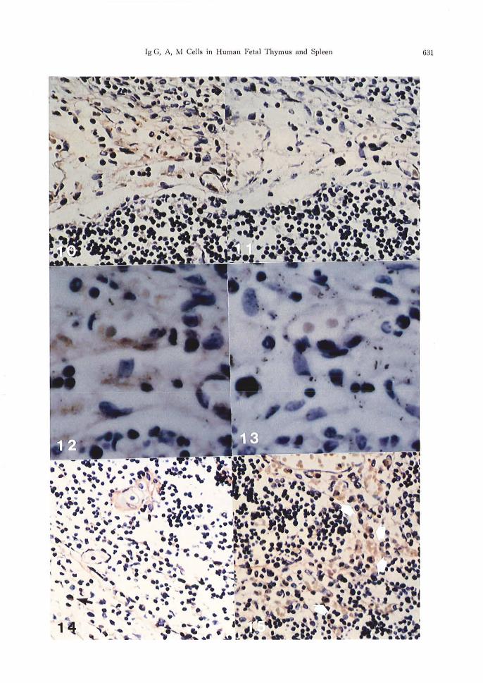
Fig. 10 and 12 (control A) shows a few cells with red stained cytoplasm and Fig. 11 and 13 (control B) shows no Ig positive cells.
Immunohistochemical findings in the thymus and spleen of the human fetus
a) lg G cells in the thymus About 4 months of gastation, the Ig G positive
cells are scarcely seen. About 6 months the number of the Ig G cells
is increasing slightly around the capillary vessels in the medulla (Fig. 14).

lg G, A, M Cells in Human Fetal Thymus and Spleen 627
About 7-10 months almost the same phenomenon is seen in the medulla (Fig. 15). The nuclei of these cells are round and the red stained cytoplasm are round and oval.
b) lg G cells in the spleen About 4 months of gestation, no Ig G positive
cells are seen in our specimens. About 6-7 months of gestation, small amount of the Ig G positive cells are seen mainly in the red pulp. About 8-10 months of gestation, the number of the Ig G cells is increasing slightly (Fig. 16 a~d 17). In the white pulp, dark-stained lymphocytes are seen, but lg G positive cells are scarcely seen.
c) lg A cells in the thymus The Ig A positive cells are hardly seen in
the period of about 4 months, and after 6-7 months of gestation, the cells are observed near
the capillary vessels in the medulla. Most of the lg A positive cells look like plasma cells (Fig. 18).
d) lg A cells in the spleen The lg A positive cells are scarcely seen about
4 months as in the thymus. The cells appear after 6 months, and the number of the cells increases gradually in the red pulp (Fig. 19).
e) lg M cells in the thymus The Ig M positive cells appear mainly in the
medulla around 6 months of gestation. The number of cells is increasing more rapidly than that of the lg A cells about 7-10 months (Fig. 20).
f) lg M cells in the spleen These lg M positive cells are also observed
mainly in the red pulp around 6 months (Fig. 21).
Table 1. Abstract of the amount and appearance of lg cells
Thymus fetal months
I I lg G cell lg A cell
10 * + 8 * +
7 + + 6 + + 4 +
The outline of the results are shown in Table 1.
In general, the lg G and A cells are believed to be produced from plasma cells and the lg M cells are from lymphoid cells, and its genetic regulation is thought not to be controlled by the simple Mendelism but by polygenic factors. In our study the number of Ig G cells is greater than that of lg A and M cells both in the thymus and in the spleen.
The same is observed to the amount of lg in the human fetus serum. Our observation shows that most of the lg G, A and M cells are seen near the capillary vessels in the medulla of the thymus and in the red pulp of the spleen. This suggests that the vicinity of the capillary vessels should play an important role to immunological activity.
The serum examination shows that the maternal lg G transfers to fetus after 37 weeks (about 8. 6 months) of gestation. Furth0 , using
Spleen
lgM cell lg G cell I
Ig A cell I
Ig M cell
+ + + + + + + +
+ + + +
+ + + +
the tissue culture or immuno:fluorescent method, demonstrates that the Ig G cells are produced from the lymphoid or plasma cells in the spleen after 20 weeks (about 4. 6 months) of gestation.
Tanaka8) reports that the average value of serum lg G is about 5. 4% of the adult value at 17-20 weeks (about 4 months), 21% at 21-27 weeks (about 5 months), 42% at 29-32 weeks (about 6. 7 months), 55% at 33-36 weeks (about 7. 7 months) and 86% at 37-40 weeks ·(about 8. 6-9. 3 months) of gestation.
About the Ig M, 0-2% of the adult value are measured at 17-20 weeks (about 4 months), 0-7. 1% at 21-24 weeks, 2. 6-12% at 29-32 weeks, 3. 5-4. 7 % at 33-36 weeks and 6-16 % at 37-40 weeks of gestation.
These fetal values of serum lg M are smaller than those of lg G. For this reason it is recognized that the maternal lg M does not tansfer to the fetus. Gitlin2> demonstrates that the ABO saline isoagglutinin, which does not move

628 Y. Satow et al.
from mother to fetus, belongs to the lg M. Gitlin et al. 2• 3 l observe that the maternal lg M labeled with 1311 does not transfer to the fetus. On the other hand it is suggested that the fetal lg M should come out around 17-20 weeks (about 3. 9 months) of gestation, because at this stage the lg M appear in the fetal serum. Furth1> also observes by the immunofiuorescent method that the fetal spleen produces lg M 19 weeks of gestation, and the thymus cells possess the lg M at 11 weeks of gestation8 l. We observe lg M cells both in the thymus and in the spleen about 6 months of gestation.
About of the lg A, small amount of lg A is measured in the fetal serum around 17-20 weeks (about 4 months) of gestation, and 1% of adult serum value at 32 weeks (about 6. 7 months) of gestation.
About 16% of the adult serum lg A value is calculated at 7-12 months old newborns, and it increases to 79 % at 6-8 years old. It is konwn that lg A does not move from mother to fetus. Although Furth et al.D by the immunofiuorescent method, point out that no cells producing lg A are observed around 13-31 weeks (4-6 months) of gestation, Tanaka8
> mentions that the presence of lg A cells both in the thymus at 11 weeks (2. 5 months) and in the spleen at 20 weeks (4. 6 months) of gestation. These observations and ours correspond each other.
REFERENCES l. Furth, R., Schuit, H. R. E. and Hijmans, W.
1965. The immunological development of the human fetus. J. Exp. Med. 122: 1173-1178.
2. Gitlin, D., Kumati, J., Urrusti, J. and Moales, C. 1964. The selectivity of the human placenta
in the transfer of plasma protein from mother to the fetus. J. Clin. Invest. 43 : 1938-1940.
3. Gitlin, D. and Biasucci, A. 1969. Development of rG, rA, rM, j3IC/ j3IA, C'l esterase inhibiter, ceruloplasmin, transferin, hemopexin, hepatoglobin, fibrinogen, plasmogen, a 1-antitrypsin, orosomucoid, j3-lipoprotein, a~-macroglobulin, and prealbumin in the human conceptus. J. Clin. Invest. 48 : 1433-1440.
4. Hayashi, I. and Okamoto, N. 1983. Outline on 10834 cases of autopsy. p. 96-104. In I. Hayashi and N. Okamoto (ed.), Atlas of human congental malformations-Embryos, fetuses and newborns-, Maruzen Hiroshima Publishing Service Center.
5. Hsu, S. M., Raine, L. and Fanger, H. 1981. Use of avidin-biotin-peroxidase complex (ABC) in immunoperoxidase techniques: a comparison between ABC and unlabeled antibody (PAP) procedures. J. Histochem. Cytochem. 29 : 577-580.
6. Hsu, S. M., Raine, L. and Fanger, H. 1981. A comparative study of the peroxidase anti-peroxidase method and avidin-biotin-complex method for studing polypeptide hormons with radioim -munoassay antibodies. Am. J. Clin. Pathol. 75 : 734-738.
7. Pinlms, G. S. and Said, J. W. 1977. Specific identification of intracellular immunoglobulin in parafin sections of multiple myeloma and macroglobulinemia using an immunoperoxidase thechnique. Am. J. Path. 87 : 47-58.
8. Tanaka, M., Tabata, K. and Okuda, R. 1969. Immunological changes during perinatal and childhood. Shanika Rinsho. 22 : 819-829. (in Japanese)
9. Taylor, C.R. and Mason, D. Y. 1974. Immunohistochemical detection of intracellular immunoglobulin in formalin paraffin sections from multiple myeloma using the immunoperoxidase technique. Clin. Exp. Immunol. 18: 417-429.

lg G, A, M Cells in Human Fetal Thymus and Spleen 629
EXPLANATIONS OF FIGURES Fig. 1. Human fetal thymus, 4 months of gestation. Autopsy number: HG 4169 ( Sj2 ), Body Weight (BW) 38. 3 g, Cranio-rump distance (CR) 7. 5 cm, thymus 0. 03 g. The borderline between cortex and medulla is not clear. Hematoxylin and Eosin (HE) staining. Fig. 2. Thymus, 6 months of gestation. HG 4402 ( o ), BW 350 g, CR 17 cm, thymus 0. 7 g. The line demarcation between cortex and medulla is clear. HE staining. Fig. 3. Thymus, 10 months of gestation. HG 3250 ( Sj2 ), BW 2950 g, GR 32. 6 cm, thymus 10.1 g. The darkstained cortex and lightly stained medulla are observed. Hassall's corpuscles are seen. HE staining. Fig. 4. Electron microscopic picture of the thymus, :LO months of gestation shows the borderline between the cortex (C) and medulla (M). Fig. 5. Human fetal spleen, 4 months of gestation. HG 4169, spleen 0. 06 g. Two types of cells are seen in the spleen. One is a stellate cell which forms a supporting reticulum network. The other is a free lymphocyte. HE staining. Fig. 6. Spleen, 6 months of gestation. HG 4402, spleen 0. 7 g. The lymphocyte are mainly observed around the arterial vessels (A). This shows initial figure of developing white pulp. HE staining. Fig. 7. Spleen, 7 months of gestation. HG 3574 ( o ), BW 250 g, CR 15. 4 cm, spleen 0. 2 g. The red pulp is composed of reticulum cells forming the so-called splenic cords. Fig. 8. Electron microscopic picture of red pulp of spleen, 10 months of gestation. Some irregular shaped reticulum and blood cells are observed. Fig. 9. Electron microscopic finding of white pulp of spleen, LO months of gestation. Many lymphoid cells are seen. Fig. 10. Negative control staining of delete antibody reagent of thymus, LO months of gestation. HG 3250. Few cells with red stained cytoplasm are seen. Fig. 11. Negative control staining of delete peroxidase reagent of thymus, LO months of gestation (HG 3250) shows no lg positive cells. Fig. 12. High magnification of Fig. 10 shows around blood vessels of the thymus. Few cells with red stained lg positive cells are observed. Fig. 13. High magnification of Fig. 11 shows next section of the preparat of Fig. l2. No lg positive cesll are observed. Fig. 14-21 show immunohistochemical findings. Fig. 14. lg G staining of the thymus, 6 months of gestation. HG 4402. Some lg G positive cells are observed around the capillary in the medulla ( I ). Fig. 15. lg G staining of the thymus, 8 months of gestation. HG 3080 ( o ), BW L8LO g, C~ 27. 5 cm, thymus 6. 0 g. lg G cells with red stained cytoplasm are observed (I ). Fig. 16. lg G staining of the spleen, 10 months of gestation. HG 3250 ( Si2) BW 2950g, CR 32. 6 cm spleen 8. 2 g. lg G positive cells look like plasma cells ( t ). Fig. 17. lg G staining of the spleen, 7 months of gestation. HG 3574. Some lg G positive cells are seen in the red pulp ( t ). Fig. 18. lg A staining of the thymus, 8 months of gestation. HG 3080. lg A positive cells are seen in the medulla ( t ). Fig. 19. lg A staining of the spleen, 8 months of gestation. HG 3080. Relatively small number of the cells are seen in the red pulp of the organ. Fig. 20. lg M staining of the thymus, 8 months of gestation. HG 3080. Most of the cells are observed in the medulla ( t ). Fig. 21. lgM staining of the spleen, 6 months of gestation. HG 4402. The cells are also mainly seen in
rad pulp ( t ).

630 Y. Satow et al.
Ig G, A, M Cells in Human Fetal Thymus and Spleen 631
i.t ,. ••

632 Y. Satow et al.