immunological properties of gold nanoparticles
TRANSCRIPT

ChemicalScience
PERSPECTIVE
Ope
n A
cces
s A
rtic
le. P
ublis
hed
on 1
6 N
ovem
ber
2016
. Dow
nloa
ded
on 3
/12/
2022
2:3
7:33
AM
. T
his
artic
le is
lice
nsed
und
er a
Cre
ativ
e C
omm
ons
Attr
ibut
ion
3.0
Unp
orte
d L
icen
ce.
View Article OnlineView Journal | View Issue
Immunological p
Dr Lev A. Dykman is a lead reseaLaboratory of the Russian AcadBiochemistry and Physiology of Phas published over 300 scientic won colloidal gold nanoparticles acurrent research interests are in imgold nanoparticles, and their amedical studies. In particular, hinteraction of nanoparticles and ctent cells and on the delivery oforgans, tissues and cells.
aInstitute of Biochemistry and Physiology
Academy of Sciences, 13 Prospekt Entuzi
[email protected]; [email protected] National Research State Univers
410012, Russia
Cite this: Chem. Sci., 2017, 8, 1719
Received 14th August 2016Accepted 14th November 2016
DOI: 10.1039/c6sc03631g
www.rsc.org/chemicalscience
This journal is © The Royal Society of C
roperties of gold nanoparticles
Lev A. Dykman*a and Nikolai G. Khlebtsov*ab
In the past decade, gold nanoparticles have attracted strong interest from the nanobiotechnological
community owing to the significant progress made in robust and easy-to-make synthesis technologies,
in surface functionalization, and in promising biomedical applications. These include bioimaging, gene
diagnostics, analytical sensing, photothermal treatment of tumors, and targeted delivery of various
biomolecular and chemical cargos. For the last-named application, gold nanoparticles should be
properly fabricated to deliver the cargo into the targeted cells through effective endocytosis. In this
review, we discuss recent progress in understanding the selective penetration of gold nanoparticles into
immune cells. The interaction of gold nanoparticles with immune cell receptors is discussed. As distinct
from other published reviews, we present a summary of the immunological properties of gold
nanoparticles. This review also summarizes what is known about the application of gold nanoparticles as
an antigen carrier and adjuvant in immunization for the preparation of antibodies in vivo. For each of the
above topics, the basic principles, recent advances, and current challenges are discussed. Thus, this
review presents a detailed analysis of data on interaction of gold nanoparticles with immune cells.
Emphasis is placed on the systematization of data over production of antibodies by using gold
nanoparticles and adjuvant properties of gold nanoparticles. Specifically, we start our discussion with
current data on interaction of various gold nanoparticles with immune cells. The next section describes
existing technologies to improve production of antibodies in vivo by using gold nanoparticles conjugated
with specific ligands. Finally, we describe what is known about adjuvant properties of bare gold or
functionalized nanoparticles. In the Conclusion section, we present a short summary of reported data
and some challenges and perspectives.
rcher at the Immunochemistryemy of Sciences' Institute oflants and Microorganisms. Heorks, including two monographsnd several book chapters. Hismunochemistry, fabrication ofpplications to biological andis research is focused on theonjugates with immunocompe-engineered particles to target
of Plants and Microorganisms, Russian
astov, Saratov 410049, Russia. E-mail:
ity, 83 Ulitsa Astrakhanskaya, Saratov
hemistry 2017
1. Introduction
Gold nanoparticles (GNPs) have attracted signicant interest asa novel platform in nanobiotechnology and biomedicinebecause of their convenient surface bioconjugation withmolecular probes1 and their remarkable optical2 and immuno-logical3 properties. Recently published examples include
Professor Nikolai G. Khlebtsov is head of the NanobiotechnologyLaboratory at the Russian Academy of Sciences' Institute ofBiochemistry and Physiology of Plants and Microorganisms. Healso is Professor at the Faculty of Nano- and Biomedical Tech-nologies at the Saratov State University. He has published over400 scientic works, including 12 monographs and book chapters.His current research interests are in biophotonics and nano-biotechnology of plasmon-resonant particles, biomedical applica-tions of metal nanoparticles, static and dynamic light scattering bysmall particles and clusters, and programming and computersimulation of light scattering and absorption by various metal anddielectric nanostructures. Professor Khlebtsov also serves asassociate editor for the Journal of Quantitative Spectroscopy andRadiative Transfer.
Chem. Sci., 2017, 8, 1719–1735 | 1719

Chemical Science Perspective
Ope
n A
cces
s A
rtic
le. P
ublis
hed
on 1
6 N
ovem
ber
2016
. Dow
nloa
ded
on 3
/12/
2022
2:3
7:33
AM
. T
his
artic
le is
lice
nsed
und
er a
Cre
ativ
e C
omm
ons
Attr
ibut
ion
3.0
Unp
orte
d L
icen
ce.
View Article Online
applications of GNPs to genomics, biosensorics, immunoas-says, clinical chemistry, detection and control of microorgan-isms, cancer cell photothermolysis, targeted delivery of drugs orother substances, and optical imaging and monitoring of bio-logical cells and tissues.4–6 Noteworthy is the fact that GNPs arebeing increasingly administered to animals and humansparenterally. In particular, they serve as carriers for the deliveryof drugs, genetic materials, and antigens. “Colloidal metallicgold is not bio-inert”—such is the name Brown et al.7 gave totheir article so as to stress the importance of nanometer size inbiological effects, even for such a seemingly inert material asgold.
It is natural to suppose that the rst cells that GNPsencounter on their way in the mammalian organism are thoseof the immune system, in particular its phagocytic link(neutrophils, monocytes, macrophages, dendritic cells andmast cells). Indeed, as early as in the rst attempts to investigatecolloidal gold biodistribution, which were performed in the1960s–80s on rabbits,8 mice,9 and rats10,11 it was found that aerparenteral administration, colloidal gold particles are capturedby liver cells, excreted through bile, and eliminated from theorganism with feces. Aer injection, gold was identied mostlyin Kupffer cells. Perhaps Scott et al.8 were the rst to note thatthe phagocytosis of GNPs is size dependent. Besides Hardonket al.,10 the important role of Kupffer cells in the elimination ofGNPs was established by Sadauskas et al.,12 who injected GNPsintravenously in mice. Electron microscopy showed that aerinjection, the GNPs accumulated in the macrophages of theliver (90%) and spleen (10%). The authors concluded that GNPspenetrate only phagocytes, primarily the Kupffer cells of theliver. In a subsequent study,13 Sadauskas et al. reported thatGNPs get localized in lysosomes (endosomes) of Kupffer cellsand can be retained there for up to six months. The inuence ofsize, solubility and surface modication on the biocompatibilityof GNPs and their use in biological applications is wellknown.14,15 However, the effects of nanoparticle properties onthe immune system are still being explored.
In this review, we discuss the selective penetration of GNPsinto immune cells and the interaction of GNPs with immunecell receptors. This review also summarizes what is knownabout the application of GNPs as an antigen carrier and adju-vant in immunization for the preparation of antibodies in vivo.
2. Interaction of gold nanoparticleswith immune cells
The immune system cells constitute the rst barrier to nano-particle penetration of animal tissues and cells. Therefore, thestudy of GNP interactions with phagocytes, the mechanismsof intracellular uptake, and the responses of immune cells toGNPs is undoubtedly of major interest. Perhaps the rstdetailed consideration of these issues can be found in Shuklaet al.,16 who, using three microscopic methods, examinedthe uptake of 3 nm GNPs into RAW264.7 macrophage cells.The conclusion from their study was that small GNPs entermacrophages through pinocytosis and get localized mostly in
1720 | Chem. Sci., 2017, 8, 1719–1735
lysosomes and in the perinuclear space. On the whole, Shuklaet al.'s data indicate that the GNPs are biocompatible, non-cytotoxic and nonimmunogenic and that they suppress theproduction of reactive oxygen species and do not cause elabo-ration of the proinammatory cytokines TNF-a and IL1-b(which contradicts the data of Yen et al.17). In contrast to databy Shukla et al.,16 Yen et al.17 noted that on the administrationof GNPs, the number of macrophages decreases and their sizeincreases, this being accompanied by elevated production ofIL-1, IL-6 and TNF-a. We emphasize that the data of Shuklaet al.16 were obtained for very small (3 nm) particles. However,Lim et al.,18 using much larger (60 nm) hollow NSphs cappedwith dextran, and Zhang et al.,19 using 60 nm GNPs, achievedresults similar to the ndings of Shukla et al.16 for the same cellculture. Sumbayev et al.20 showed that citrate-stabilized GNPsspecically downregulate, in a size dependent manner, thecellular responses induced by IL-1b both in vitro and in vivo. Ina recent study, Guevel et al.21 demonstrated that 12 nm goldnanoparticles induce cell mediated responses accompaniedby inammatory natural killer (NK) cell stimulation, whereas2 nm gold nanoparticles are more efficiently taken up withoutinducing dendritic cell maturation or lymphocyte proliferation.To summarize, the published data revealed strong effects ofthe GNP size and functionalization on production of proin-ammatory cytokines.
With some inspiration from data on GNP uptake by macro-phages, Choi et al.22 even proposed a new method for the pho-tothermal therapy of tumors that employs a “Trojan horse” inthe form of monocytes and macrophages laden with phagocy-tosed GNSs. For these purposes, Dreaden et al.23 suggested theuse of GNPs conjugated with macrolide antibiotics, which canaccumulate in tumor-specic macrophages and induce theircytotoxicity, causing tumor cells to die. Thus, particle size andstructure in these studies were not critical to macrophageuptake.
The inuence of colloidal gold on immunocompetent cellswas examined in vivo also by Tian et al.24 and by Lou et al.25 Inparticular, injection of nonconjugated GNPs into miceenhanced the proliferation of lymphocytes and normal killers,as well as increasing the IL-2 production.
Quite interesting data were acquired by Bastus et al.26,27 with10 nm nonconjugated GNPs. From their results, it follows thatindeed, on entry into murine bone marrow macrophages, GNPsdo not affect the production of proinammatory cytokines.However, if the GNP surface is modied with the peptideAGIP (amyloid growth inhibitory peptide, LPFFD) or SAP [sweetarrow peptide, (VRLPPP)3], GNPs, on entry into the macro-phages, involve the induction of NO synthase and proin-ammatory cytokines such as TNF-a, IL-1b and IL-6. Inaddition, they inhibit macrophage proliferation. The recogni-tion of GNP–peptide conjugates was made more effectivethrough toll-like receptors 4 (TLR-4) on the surface of themacrophages. Yet, Staroverov et al.28,29 demonstrated that both15 nm nonconjugated GNPs and their conjugates with high-and low-molecular-weight antigens, on entry into rat peritonealmacrophages, enhance their respiratory activity and the activityof macrophage mitochondrial enzymes (Fig. 1). GNPs also have
This journal is © The Royal Society of Chemistry 2017

Fig. 1 Changes in the concentration of reduced formazan dependingon the cultivation conditions of antigen (AG) with peritoneal ratmacrophages. Reproduced with permission from ref. 28, © 2009,Springer.
Perspective Chemical Science
Ope
n A
cces
s A
rtic
le. P
ublis
hed
on 1
6 N
ovem
ber
2016
. Dow
nloa
ded
on 3
/12/
2022
2:3
7:33
AM
. T
his
artic
le is
lice
nsed
und
er a
Cre
ativ
e C
omm
ons
Attr
ibut
ion
3.0
Unp
orte
d L
icen
ce.
View Article Online
greatly increased the production of IL-1, IL-6 and IFN-g (Fig. 2).Lee et al.30 reported that the penetration of gold nanorods(GNRs) and SiO2-coated GNRs into macrophages induces therelease of inammatory mediators (cytokines, prostaglandins,etc.) and the activation of immune response genes. Thus, inaddition to early observations by Shukla et al.16 for bare GNPs,the published data26–29 indicate a signicant role of surfacecoating in macrophage response aer GNP uptake.
The activation of macrophages by GNPs, found by severalauthors,26–28,30–34 can serve as a basis for new vaccine adjuvants.As in the usual cellular uptake, immunoactivity dependsstrongly on the particle size: 5 nm particles conjugated withdisaccharides performed far better than smaller, 2 nm ones.35
Yet another means of activating macrophages with GNPs wasproposed by Wei et al.36 For this purpose, they used 15 and30 nm GNPs conjugated to cytosine–phosphate–guanosine(CpG) oligodeoxynucleotides. As is known, these oligonucleo-tides are demethylated sites of microbial DNA that can activatemacrophage immune response by interacting with the TLR-9receptors and subsequently triggering a cascade of immune
Fig. 2 Changes in the serum IFN-g concentrations in rats immunizedwith different antigens. 1 – immunization with native antigen; 2 –immunization with antigen conjugated with GNPs; 3 – immunizationwith GNPs. Reproduced with permission from ref. 29, © 2011, Springer.
This journal is © The Royal Society of Chemistry 2017
response signals. The immunostimulating activity of syntheticoligonucleotides containing CpG motifs may be analogous tothat of oligonucleotides from bacterial DNA.37 According to Weiet al.,36 GNP–CpG conjugates were effective in enhancingnanoparticle internalization in RAW264.7 macrophages, andthey greatly increased the secretion of proinammatory cyto-kines such as TNF-a and IL-6 (15 nm conjugates did so toa greater degree than 30 nm ones did). The immunostimulatoryeffect of GNP–CpG was much greater than that of native CpG atthe same concentrations.
A recent study38 examined the inuence of the size ofPEGylated GNPs on the activation of the TLR-9 receptors ofRAW264.7 murine macrophages by CpG oligonucleotides. GNPswith diameters of 4, 11, 19, 35 and 45 nm inhibited CpG-induced elaboration of TNF-a and IL-6 and the activity of theTLR-9 receptors. This effect was markedly size dependent, witha peak for 4 nm GNPs, which penetrated the cells mostintensively.
Massich et al.39 reported on the immune response ofmacrophages aer the phagocytosis of GNPs functionalizedwith polyvalent oligonucleotides. The effectiveness of uptakeand the level of interferon production were found to depend onthe density of DNA molecules on the GNP surface. Kim et al.40
showed that the uptake effectiveness of oligonucleotide-func-tionalized GNPs differs for cells isolated from peripheral blood(mononuclear cells) and those introduced into a 293T culture.In addition, only in the rst type of cell did the uptake of GNPconjugates activate the expression of immune response genes.
A recent article by Walkey et al.41 described a thorough studyof the effect of coating GNPs with serum proteins and PEG onmacrophage uptake. The authors studied the adsorption of70 blood serum proteins to PEG-coated GNPs with differentdensities of PEG coating. Increasing the PEG coating densityreduced serum protein adsorption and changed the composi-tion of the adsorbed protein layer. Particle size also affectedserum protein adsorption through a change in the steric inter-actions between the PEG molecules. Both the density of PEGmolecules on the GNP surface and the size of GNPs determinedthe mechanism and effectiveness of macrophage uptake,possibly because the composition of the adsorbed blood serumproteins and their availability to cells were regulated. If thedensity of PEG coating was lower than�0.16 PEGmolecules pernm2, the macrophage uptake of GNPs depended on the pres-ence of adsorbed proteins (serum-dependent uptake). If thedensity was higher than �0.64 PEG molecules per nm2, serum-independent uptake was seen (Fig. 3).
Serum-dependent uptake was more effective than serum-independent uptake, apparently because of the difference in theenergy of the GNP–cell interaction. Interestingly, serum-inde-pendent uptake was more effective for large GNPs (90 nm)whereas serum-dependent uptake was maximal for 50 nmGNPs.
It should be noted that immediately on contact of GNPs withblood, lymph, gastric juice, or any other biological liquid in vivothe interaction between GNPs and solvable proteins and otherbiomolecules results in the formation of a protein “corona”.42,43
Similarly to the concept of functionalized GNPs, the concept of
Chem. Sci., 2017, 8, 1719–1735 | 1721

Fig. 3 Scheme for the influence of the PEG coating density on the adsorption of serum proteins to GNPs and their subsequent uptake bymacrophages. Reproduced with permission from ref. 41, © 2012, American Chemical Society.
Chemical Science Perspective
Ope
n A
cces
s A
rtic
le. P
ublis
hed
on 1
6 N
ovem
ber
2016
. Dow
nloa
ded
on 3
/12/
2022
2:3
7:33
AM
. T
his
artic
le is
lice
nsed
und
er a
Cre
ativ
e C
omm
ons
Attr
ibut
ion
3.0
Unp
orte
d L
icen
ce.
View Article Online
a GNP–protein corona is important in tuning the surfacephysicochemical properties of GNPs, such as charge, hydrody-namic size and colloidal stability. In fact, it is the GNP–proteincorona that forms the rst nano–bio interface and determinesthe rst interactions of GNPs with/or within living cells. This isbecause the GNP–protein corona is a dynamic biopolymer layerthat can strongly affect cellular uptake owing to modication ofthe particle properties (the overall size, charge, etc.). Althoughas much as 69 plasma proteins can bind to the GNP surface,44,45
only some of them, such as albumin, apolipoprotein, immu-noglobulin, complement and brinogen, are the most abun-dantly bound proteins forming the GNP–protein corona. Aerintravenous injection, the coating of GNPs by these proteinslargely determines the particles' fate in the body—bio-distribution over organs, tissues and cells, the efficiency ofcellular uptake and clearance, immunological properties, andso on.46,47
Ma et al.48 showed that GNPs attenuate LPS-induced NOproduction through the inhibition of nuclear factor-kB andIFN-b/STAT1 pathways in RAW264.7 cells. In contrast, Liuet al.49 demonstrated that PEGylated GNPs were internalizedmore quickly by lipopolysaccharide-activated RAW264.7 cellsthan by unstimulated cells, reaching saturation within 24 h. ThePEGylated GNPs enhanced LPS-induced production of NO andIL-6 and inducible nitric oxide synthase expression inRAW264.7 cells, partly by activating p38 mitogen-activatedprotein kinases and NF-kB pathways. Goldstein et al.50 showedthat GNPs and their plasmonic excitation could activate theNrf2-Keap1 pathway in macrophages.
Garcıa et al.51 studied the cellular uptake of GNPs with orwithout exposure of cells to latrunculin A, a phagocytosisinhibitor. The results indicate a size dependence of the inter-nalization mechanisms for macrophage (THP-1) cells. Theinternalization of larger GNPs (15 and 35 nm) was blocked inthe presence of latrunculin A, although they could attach to thecell membrane. Smaller GNPs (5 nm), though, were not blockedby actin-dependent processes.
Of considerable interest are studies on the uptake of GNPsnot only by macrophages but also by other cells of the immune
1722 | Chem. Sci., 2017, 8, 1719–1735
system, in particular dendritic cells. In the past decade,dendritic cells have attracted increased interest owing to theease of their isolation from peripheral blood monocytes and totheir ability to effectively present antigens to T cells. By now,a great deal of work has been done on the modulation ofimmune response in patients with chronic infections andoncological diseases by using antigen-primed dendritic cells.52
GNPs have been named, among other carriers, for applicationin antigen delivery to dendritic cells. For example, Cheunget al.53 described the use of 15 nmGNPs for presenting a peptideantigen associated with Epstein–Barr virus to dendritic cells.According to their TEM data, peptide-functionalized GNPspenetrated the dendritic cell cytoplasm but were not found inthe nuclei. The uptake of GNPs by dendritic cells resulted in anincreased content of g-interferon, the presentation by majorhistocompatibility complex I (MHC-I) of the antigen to CD4+ Tcells, and, correspondingly, activation of an epitope-specicimmune response by cytotoxic T cells.
Cruz et al.54 addressed dendritic cell uptake of and immuneresponse activation by 13 nm GNPs conjugated to prostatecancer peptide antigens. By TEM, LCM and ow cytometry,GNPs functionalized with the peptides and with Fc fragments ofIgG were shown to interact with the Fcg receptors of dendriticcells and were localized, upon uptake, in the cytoplasm ina diffuse way. Internalization of antigen-conjugated GNPs indendritic cells brought about an increase in the immuneresponse, as compared with the effect obtained from the use ofthe native antigen, which was manifested as enhancedlymphocyte proliferation. Such an approach, in the authors'opinion, opens up the way to the creation of an effective systemfor the development of antitumor and other vaccines.
Villiers et al.55 reported the effect of 10 nm non-antigen-functionalized GNPs on the immune functions of dendriticcells. From their ndings, the GNPs that had entered cellendosomes were not cytotoxic and had no effect on theproduction of the proinammatory cytokine IL-6. However, theydid promote the secretion of interleukin IL-12p70, whichis directly involved in the activation of T cells and, thus, inthe regulation of an antigen-specic immune response. The
This journal is © The Royal Society of Chemistry 2017

Perspective Chemical Science
Ope
n A
cces
s A
rtic
le. P
ublis
hed
on 1
6 N
ovem
ber
2016
. Dow
nloa
ded
on 3
/12/
2022
2:3
7:33
AM
. T
his
artic
le is
lice
nsed
und
er a
Cre
ativ
e C
omm
ons
Attr
ibut
ion
3.0
Unp
orte
d L
icen
ce.
View Article Online
authors also noted the development of long dendrites andan increase in the cell-surface amount of MHC-II molecules,which present antigens to T lymphocytes. Thus, even non-functionalized GNPs are immunostimulatory to both dendriticcells and macrophages.17
Ye et al.56 used TEM and ow uorocytometry to quantify theuptake of GNRs by dendritic cells and the particle effect on theirfunctions. Compared to spherical GNPs, GNRs entereddendritic cells more effectively and induced higher expressionof CD86 immunocostimulatory molecules, which are charac-teristic of dendritic cells.
Lin et al.57 reported that GNPs in complexes with peptidesderived from tumor-associated antigens are taken up effectivelyby dendritic cells. Moreover, dendritic cells take up GNPs withminimal toxicity and can process the vaccine peptides on theparticles to stimulate cytotoxic T lymphocytes. A high peptidedensity on the GNP surface can stimulate cytotoxic T lympho-cytes better than can free peptides. Thus, GNPs have greatpotential as carriers for various vaccine types.
GNP-mediated response of dendritic cells depends on thephysicochemical properties of the GNP surface. For example,Fytianos et al.58 clearly indicated that the chemical compositionand surface charge of GNPs modulate uptake by dendritic cellsand cytokine release. Further, in vivo GNP effects are dose-dependent. In particular, Małaczewska59,60 demonstrated thatmice, aer being orally administered with GNPs, showed anincreased activity of phagocytes and some changes in thelymphocyte phenotypes, i.e., an increased percentage of B andCD4+/CD8+ double positive T cells. The lowest dose had a pro-inammatory or immunostimulating effect, enhancing thesynthesis of proinammatory cytokines (IL-1b, IL-2, IL-6,TNF-a). The effect of the highest dose can be consideredproinammatory or immunotoxic, because the stimulatedcytokine synthesis was accompanied by a drastic decline in theproliferative activity of lymphocytes.
To estimate the functional impact of GNPs on B lympho-cytes, Sharma et al.61 treated a murine B lymphocyte cell line(CH12.LX) with 10 nm citrate-stabilized GNPs. This treatmentactivated an NF-kB-regulated luciferase reporter, and this acti-vation correlated with the altered B lymphocyte function (i.e.,with increased antibody expression). According to TEM images,GNPs could penetrate the cellular membrane and, therefore,
Fig. 4 TEM images of (a) spleen macrophages, (b) dendritic cells, (c) mpermission from ref. 64, © 2009, Elsevier; ref. 55, © 2010, Springer; ref. 6Society of Chemistry.
This journal is © The Royal Society of Chemistry 2017
could interact with the intracellular components of the NF-kBsignaling pathway.
In vitro, ex vivo and in vivo evidence suggests that GNPsactivate B cells and enhance IgG secretion.62 GNP treatmentupregulates blimp1, downregulates pax5, and enhances down-stream IgG secretion. This enhancement is size and timedependent. GNPs ranging from 2 to 12 nm had the maximumstimulatory activity for the production of antibody.
Moreover, GNPs augmented lymphocyte proliferation inresponse to phytohemagglutinin, and this effect was greater foras-synthesized than for capped gold nanoparticles. Release ofIL-10 and IFN-g from lymphocytes was increased and the effectwas again more marked for as-synthesized GNPs than it was forcapped GNPs.63
Bartneck et al.65,66 reported the interaction of variously sha-ped and sized particles GNPs with human neutrophil gran-ulocytes, monocytes and macrophages. On the basis of theirstudy, the mechanism of nanoparticle trapping can be classiedas macropinocytosis rather than phagocytosis. Particle shapewas found to affect strongly the particle trapping by cells of theimmune system; specically, CTAB-coated GNRs (50 � 15 nm)could be trapped faster than CTAB-coated 15 and 50 nm goldnanospheres. Replacing CTAB by poly(ethylene oxide) greatlyreduced uptake effectiveness for both types of GNPs. Nano-particle uptake by the immune cells was accompanied by anactivation of the genes of proinammatory cytokines and bya corresponding change in the cell phenotype. A characteristicfact is that the “professionally” phagocytic cells took up GNPstwo orders of magnitude more effectively than did, e.g., HeLacells. In addition, Bartneck et al. revealed an alternative elimi-nation mechanism whereby GNPs can be cleared from periph-eral blood via an extracellular network (“trap”) produced byneutrophil granulocytes.
The same group presented data67 on the uptake of GNPs intovarious cells of the reticuloendothelial system: monocytes,macrophages, immature and mature dendritic cells and endo-thelial cells. The greatest uptake ability was demonstrated bymacrophages, endothelial cells and immature dendritic cells.Positively charged GNPs penetrated into cells of the reticulo-endothelial systemmore effectively. Moreover, GNPs intensiedthe induction of several cytokines, including g-interferon, IL-8(both in dendritic cells and in macrophages), IL-1b and IL-6
onocytes and (d) lymphocytes treated with GNPs. Reproduced with5, © 2010, American Chemical Society; and ref. 61, © 2013, The Royal
Chem. Sci., 2017, 8, 1719–1735 | 1723
Chemical Science Perspective
Ope
n A
cces
s A
rtic
le. P
ublis
hed
on 1
6 N
ovem
ber
2016
. Dow
nloa
ded
on 3
/12/
2022
2:3
7:33
AM
. T
his
artic
le is
lice
nsed
und
er a
Cre
ativ
e C
omm
ons
Attr
ibut
ion
3.0
Unp
orte
d L
icen
ce.
View Article Online
(only in dendritic cells). Interestingly, in mature dendritic cells,GNPs accumulate in the MHC-II compartment and, conse-quently, may affect antigen processing.
Thus, GNPs can penetrate into various immune cells (Fig. 4)and activate the production of proinammatory cytokines(Table 1).
Phagocytic cells of the immune system have a multitude ofvarious receptors on their surface, through which they bindand take up foreign material.68,69 The interactions with varioustypes of receptors and, consequently, various types of GNPendocytosis depend in many ways on nanoparticle size andshape but especially on surface functionalization (includingopsonization by proteins from the culture medium or bloodplasma)70 and on the presence of mannose-containing poly-saccharides on the GNP surface.71 Some researchers areinclined to believe that the key role in macrophage uptake ofGNPs is played by scavenger receptors.72,73 These are mainlyinvolved in the endocytosis of apoptotic cells. A characteristicpeculiarity of their functioning, in contrast to the othermacrophage receptors, is the absence of release of proin-ammatory cytokines.
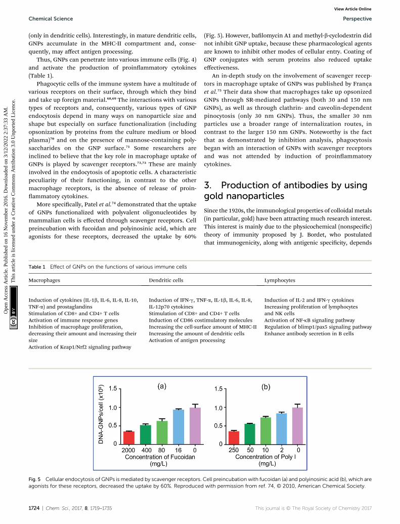
More specically, Patel et al.74 demonstrated that the uptakeof GNPs functionalized with polyvalent oligonucleotides bymammalian cells is effected through scavenger receptors. Cellpreincubation with fucoidan and polyinosinic acid, which areagonists for these receptors, decreased the uptake by 60%
Table 1 Effect of GNPs on the functions of various immune cells
Macrophages Dendritic cells
Induction of cytokines (IL-1b, IL-6, IL-8, IL-10,TNF-a) and prostaglandinsStimulation of CD8+ and CD4+ T cellsActivation of immune response genesInhibition of macrophage proliferation,decreasing their amount and increasing theirsizeActivation of Keap1/Nrf2 signaling pathway
Induction of IFN-g, TNIL-12p70 cytokinesStimulation of CD8+ aInduction of CD86 cosIncreasing the cell-surfIncreasing the amountActivation of antigen p
Fig. 5 Cellular endocytosis of GNPs is mediated by scavenger receptors.agonists for these receptors, decreased the uptake by 60%. Reproduced
1724 | Chem. Sci., 2017, 8, 1719–1735
(Fig. 5). However, balomycin A1 and methyl-b-cyclodextrin didnot inhibit GNP uptake, because these pharmacological agentsare known to inhibit other modes of cellular entry. Coating ofGNP conjugates with serum proteins also reduced uptakeeffectiveness.
An in-depth study on the involvement of scavenger recep-tors in macrophage uptake of GNPs was published by Françaet al.75 Their data show that macrophages take up opsonizedGNPs through SR-mediated pathways (both 30 and 150 nmGNPs), as well as through clathrin- and caveolin-dependentpinocytosis (only 30 nm GNPs). Thus, the smaller 30 nmparticles use a broader range of internalization routes, incontrast to the larger 150 nm GNPs. Noteworthy is the factthat as demonstrated by inhibition analysis, phagocytosisbegan with an interaction of GNPs with scavenger receptorsand was not attended by induction of proinammatorycytokines.
3. Production of antibodies by usinggold nanoparticles
Since the 1920s, the immunological properties of colloidal metals(in particular, gold) have been attracting much research interest.This interest is mainly due to the physicochemical (nonspecic)theory of immunity proposed by J. Bordet, who postulatedthat immunogenicity, along with antigenic specicity, depends
Lymphocytes
F-a, IL-1b, IL-6, IL-8,
nd CD4+ T cellstimulatory moleculesace amount of MHC-IIof dendritic cellsrocessing
Induction of IL-2 and IFN-g cytokinesIncreasing proliferation of lymphocytesand NK cellsActivation of NF-kB signaling pathwayRegulation of blimp1/pax5 signaling pathwayEnhance antibody secretion in B cells
Cell preincubation with fucoidan (a) and polyinosinic acid (b), which arewith permission from ref. 74, © 2010, American Chemical Society.
This journal is © The Royal Society of Chemistry 2017

Perspective Chemical Science
Ope
n A
cces
s A
rtic
le. P
ublis
hed
on 1
6 N
ovem
ber
2016
. Dow
nloa
ded
on 3
/12/
2022
2:3
7:33
AM
. T
his
artic
le is
lice
nsed
und
er a
Cre
ativ
e C
omm
ons
Attr
ibut
ion
3.0
Unp
orte
d L
icen
ce.
View Article Online
predominantly on the physicochemical properties of antigens,rst of all on their colloidal state. L. A. Zilber made successfulattempts to obtain agglutinating sera to colloidal gold76 (inter-estingly, a repeated attempt to prepare antisera to colloidal goldwas performed almost 80 years later, in 2006).77 Yet, severalauthors have shown that the introduction of a complete antigentogether with colloidal metals promotes the production of anti-bodies.78 Furthermore, some haptens may cause antibodyproduction when adsorbed to colloidal particles.79 Numerousdata on the inuence of colloidal gold on nonspecic immuneresponse are given in one of the best early reviews.80 In particular,it was noted that at 2 h aer an intravenous injection of 5 mL ofcolloidal gold into rabbits, there was a sizable increase in totalleucocytes in 1 mL of blood (from 10 000 to 19 800) againsta slight decline in mononuclear cells (from 5200 to 4900) anda considerable increase in polynuclear cells (from 4700 to14 900).81 On injection of other colloidal metals, no suchphenomena were observed. Unfortunately, with advances inimmunology and with denial of many postulates of Bordet'stheory, interest in the immunological properties of colloidsdecreased. There is no doubt, though, that the data obtained onthe enhancement of immune response to antigens adsorbed oncolloidal particles were utilized for the development of variousadjuvants.82,83
The size-dependent GNPs-induced changes (both increasingand decreasing) of the number of white blood cells have beenreported in several recent publications.84–86
It is known that antibody biosynthesis is induced bysubstances possessing sufficiently developed structures(immunogenicity). The substances include proteins, poly-saccharides, and some synthetic polymers. However, manybiologically active substances (vitamins, hormones, antibiotics,narcotics, etc.) have relatively small molecular masses and, as arule, do not elicit a pronounced immune response. In standardmethods of antibody preparation in vivo, this limitation isovercome by chemically attaching such substances (haptens) tohigh-molecular-weight carriers (most oen proteins), whichmakes it possible to obtain specic antisera. However, suchantisera usually contain attendant antibodies to the carrier'santigenic structures.87
Let us take a brief look at two interrelated problems incurrent immunology that have attracted much research atten-tion. These are the development of antibodies to non-immunogenic low-molecular-weight compounds (haptens) andthe creation of next-generation vaccines based on natural(microbial) or synthetic peptides.88–93 It is known that antibodybiosynthesis is induced by substances possessing sufficientlydeveloped structures (immunogenicity). These substancesinclude proteins, polysaccharides, and some synthetic poly-mers.94 However, many biologically active substances (neuro-transmitters, hormones, vitamins, antibiotics, etc.) haverelatively small molecular masses. Low-molecular-weight anti-gens are in the category “weak antigens,” i.e., they do not elicita pronounced immune response.
Because haptens are weakly immunogenic, the choice of anoptimal carrier (delivery system) providing a high immuneresponse, in parallel with the obtainment of pure enough
This journal is © The Royal Society of Chemistry 2017
antibody preparations, is an important task when producingantibodies to low-molecular-weight compounds. Traditionally,this problem is solved by chemical attachment of a hapten toa protein matrix called a schlepper (from the German schleppen“to drag”), and by the use of adjuvants and intensive schemes ofanimal immunization with the obtained conjugate.87,95 Bovineserum albumin (BSA), ovalbumin, thyreoglobulin, hemocyaninand diphtheria or tetanus toxoids (in the case of syntheticpeptides) are generally used as schleppers. However, thismethod yields antibodies to both the hapten and the immu-nodominant sites of the carrier. Note that when such a carrier isused, a pronounced immune response to weak antigens doesnot always develop. Besides, the subsequent purication andscreening of the obtained antibodies are laborious and expen-sive, and their titre and affinity are oen low. Most currentlyused adjuvants based on oil emulsions and on suspensions ofinorganic substances are, as a rule, liable to phase separation,are oen reactogenic, and their immunogenic properties varywith time. Many of these adjuvants cause local and systemictoxicity.82
In recent years, efforts have been made to develop “complexantigens”, i.e., articial molecular complexes formed from bothnecessary antigens and carriers or/and adjuvants. In particular,synthetic polyelectrolytes (poly-L-lysine, polyacrylic acid, poly-vinylpyridine, sulfonated polystyrene, coll, etc.) were proposedfor use as adjuvants.96 These polymer compounds are producedby chain-radical polymerization of the correspondingmonomers.The simplicity of polyelectrolyte composition and synthesis, thepossibility of obtainment of polymer chains with a wide range ofmolecular masses (i.e., of various lengths), their solubility inwater, and other properties (the capacity for conformationaltransitions, the formation of complexes with proteins, etc.)opened up possibilities for the use of polyelectrolytes in immu-nologic investigations. Such adjuvant carriers are capable ofantigen deposition at the sites of injection, enhancement ofantigen presentation to immunocompetent cells, and inductionof production of necessary cytokines. However, the low immu-nogenicity of such complexes, due to their small epitope density,prompts researchers to look for new nontoxic and effectivecarriers additionally possessing adjuvant properties.
In this respect, of special interest are nanoscale corpuscularcarriers: polymer nanoparticles [e.g., those made of poly-methylmethacrylate, polyalkylcyanoacrylate, polylactide-co-gly-colide, poly(g-glutamic acid), polystyrene, etc.]; liposomes,proteasomes and microcapsules; fullerenes; carbon nanotubes;graphene oxide; dendrimers; paramagnetic particles; silicananoparticles; titanium dioxide nanoparticles; aluminum andaluminum oxide nanoparticles; cobalt oxide nanoparticles;silver nanoparticles; selenium nanoparticles, and others. Whenthese are used, the forms of manifestation of immunogenicityof a given substance in the host's immune system vary. Anantigen, once adsorbed or encapsulated by nanoparticles, maybe used as an adjuvant for optimization of the immuneresponse aer vaccination.97–102
In 1986, Japanese researchers103 rst reported success ingenerating antibodies against glutamate by using colloidal goldparticles as a carrier. Subsequently, a number of papers were
Chem. Sci., 2017, 8, 1719–1735 | 1725

Fig. 7 (A) Specificity of antituberculin antibodies as determined by dotanalysis using primary labeling with rabbit antituberculin antibodiesand secondary labeling with conjugates of antirabbit antibodieswith 160/20 nm (SiO2 core/Au shell) nanoshells. Sampled antigens: 1 –rabbit anti-tuberculin antibodies; 2 – tuberculin; 3 – Mycobacteriabovis BCG; 4 – Escherichia coli XL-1 blue; 5 – Staphylococcus aureus209-R; 6 – Brucella abortus vaccine strain 82; 7 – brucellin. Forsamples 1, 2 and 7, the concentrations were 1 mg mL�1. (B–D) Dotimmunoassay of the mycobacteria M. bovis (B), M. smegmatis (C) andM. phlei (D) by using polyclonal antibodies to tuberculin (primaryantibodies) and conjugates of antirabbit antibodies with 15 nm GNPs(secondary antibodies). Note the weak nonspecific coloration ofM. smegmatis bacteria. (E) TEM image of an M. bovis cell treated withantituberculin antibodies and labeled with conjugates of antirabbitantibodies with 15 nm GNPs. The GNP accumulation on the bacterialsurface may reflect the localization of the tuberculin antigen. (F) Lightmicroscopy of M. bovis BCG treated with rabbit antituberculin anti-bodies and labeled with conjugates of antirabbit antibodies with 15 nmGNPs. The arrows point to mycobacteria. Reproduced with permissionfrom ref. 138, © 2013, Ivyspring International Publisher.
Chemical Science Perspective
Ope
n A
cces
s A
rtic
le. P
ublis
hed
on 1
6 N
ovem
ber
2016
. Dow
nloa
ded
on 3
/12/
2022
2:3
7:33
AM
. T
his
artic
le is
lice
nsed
und
er a
Cre
ativ
e C
omm
ons
Attr
ibut
ion
3.0
Unp
orte
d L
icen
ce.
View Article Online
published whose authors applied and further developed thistechnique to obtain antibodies to the following haptens andcomplete antigens: amino acids;104,105 platelet-activatingfactor;106,107 quinolinic acid;108 biotin;109 recombinantpeptides;110,111 lysophosphatide acid;112 endostatin;113 the capsidpeptide of hepatitis C,114 inuenza,115 foot-and-mouthdisease,116,117 and dengue118 viruses; a-amidated peptides;119
actin;120 antibiotics;121 ivermectin;122,123 azobenzene;124 Ab-pep-tide;125 clenbuterol;126 a-methylacyl-CoA racemase;127 Yersi-nia,128,129 Listeria monocytogenes,130 and Escherichia coli131 surfaceantigens; Neisseria meningitides,132 Streptococcus pneumoniae,133
and Burkholderia mallei134,135 carbohydrate antigens; Pseudo-monas aeruginosa agellin;136 the transmissible gastroenteritisvirus;29 tuberculin;137,138 the peptides of the malaria plasmo-dium surface proteins;139,140 opisthorchiasis excretory–secretoryantigen;141 tetanus toxoid.142 In all these studies, the haptens orcomplete antigens were directly conjugated to colloidal goldparticles, mixed with complete Freund's adjuvant or alum, andused for animal immunization. As a result, high-titer antiserawere obtained that needed no further purication fromcontaminant antibodies (Fig. 6).
Thus, to date almost 40 publications have demonstratedsuccessful application of functionalized GNPs to obtain anti-bodies against different antigens. In some cases the applicationof GNP conjugates produced higher titers and affinity. Oen thelevels of specic antibodies produced in the immunization ofanimals with gold nanoparticles conjugated antigens werehigher than that generated by classical adjuvants while theamount of antigen required to achieve this response was anorder of magnitude lower than for immunization with a stan-dard adjuvant.143 The reasons for this may be due to greateraccumulation of the antigen in cells such as dendritic cellsallowing greater presentation of the therapeutic antigen to theimmune system. The readers can nd below a similar consid-eration of a several studies for adjuvant properties of GNPs,although such unique examples is not sufficient to considera signicant massive of collected experimental data.
The use of antituberculin antibodies for immunoassay ofmycobacteria described for the rst time in ref. 137 and 138.Fig. 7 illustrates applications of the immunodot assay, and TEM
Fig. 6 Schematic representation of immunogen localization on thesurface of keyhole limpet hemocyanin (KLH) and GNPs, used asantigen carriers. (A) Antibodies toward the peptide–KLH conjugate areproduced to the epitopes of both peptide and KLH. (B) Antibodiestoward the peptide–GNP conjugate are produced only to the epitopesof the peptide. Reproduced with permission from ref. 116, © 2010, IOPPublishing.
1726 | Chem. Sci., 2017, 8, 1719–1735
and light microscopy imaging to mycobacteria, with the reac-tion products being visualized by using immunogold markers.In future work, the authors plan to use the GNP + tuberculinconjugates not only to obtain of diagnostic antibodies but alsoto develop of tuberculin-based anti-tuberculosis vaccines. Thiscan be considered as a new variant of theranostics, which can becalled “prophynostics” (prophylaxes + diagnostics).
In 1993, Pow and Crook144 suggested attaching a hapten(g-aminobutyric acid) to a carrier protein before conjugatingthis complex to colloidal gold. This suggestion was supportedin papers devoted to the raising of antibodies to somepeptides,145–149 amino acids,150–153 phenyl-b-D-thioglucoronide,154
diminazene.155 The antibodies obtained in this way possessedhigh specicities to the antigens under study and higher (asPow and Crook144 put it, “extremely high”) titers – from1 : 250 000 to 1 : 1 000 000, as compared with the antibodiesproduced routinely. At present, the Australian-based companyImmunoSolution offers antibodies, obtained according to ref.144, to some neurotransmitters and amino acids.
This journal is © The Royal Society of Chemistry 2017

Perspective Chemical Science
Ope
n A
cces
s A
rtic
le. P
ublis
hed
on 1
6 N
ovem
ber
2016
. Dow
nloa
ded
on 3
/12/
2022
2:3
7:33
AM
. T
his
artic
le is
lice
nsed
und
er a
Cre
ativ
e C
omm
ons
Attr
ibut
ion
3.0
Unp
orte
d L
icen
ce.
View Article Online
In 1996, Demenev et al.156 showed for the rst time thepossibility of using colloidal gold particles as part of an antiviralvaccine as carriers for the protein antigen of the tick-borneencephalitis virus capsid. According to the authors' data, theoffered experimental vaccine had higher protective propertiesthan its commercial analogs, despite the fact that the vaccinedid not contain adjuvants.
Subsequently, GNPs have been used to generate antibodiesand design experimental vaccines (both peptide and carbohy-drate) against inuenza A virus,157,158 West Nile virus,159 therespiratory syncytial virus,160 hepatitis E virus,161 coronavirus,162
as well as against tuberculosis137 and listeriosis.163 In addition,GNPs are being used in the development of experimentalvaccines against tumors164–170 and HIV/AIDS.171–173 In 2011,Wang et al.174 suggested a new therapeutic vaccine based on thecombination of myelin-associated inhibitors and GNPs for thetreatment of rat medullispinal traumas. Also, for GNP-assistedantigens, several groups reported new administration ways:oral, pulmonary, transcutaneous and transmucosal immuniza-tion.175–180 Table 2 summarizes the literature data on the anti-gens and haptens that have been conjugated with GNP carriersand then used for immunization of animals. The titers of theantibodies have been increased owing to GNPs.
A considerable number of papers devoted to the use of GNPsfor creating DNA vaccines have emerged as well. The principleof DNA immunization is as follows: gene constructions coding
Table 2 Conjugates of GNPs with antigens and haptens used for immu
Aminoacids
Neurotransmitters andhormones
Antibiotics andother drugs
Glutamate Acetylcholine ChloramphenicolAspartate Serotonin GentamicinGlycine Norepinephrine NeomycinSerine Histamine LincomycinCysteine Testosterone KanamycinTaurine g-Aminobutyric acid ClindamycinCitrulline Nortestosterone Ooxacinum
Estradiol TilmicosinIvermectinDiminazeneClenbuterolXylazine
This journal is © The Royal Society of Chemistry 2017
for the proteins to which one needs to obtain antibodies areintroduced into an organism. If the gene expression is effective,these proteins serve as antigens for the development of animmune response.181,182 In the early papers, immunization wasconducted by a subcutaneous or intramuscular injection ofa “naked” DNA. However, for this purpose, a “biolistic” trans-fection, using GNPs, began to be applied almost simulta-neously. It was found to be very effective, apparently because ofthe multiplicity of sites of transgene interaction with tissuesand because of transgene penetration directly into cells andnuclei.183,184 The method of gene immunization, oen calledDNA vaccination, which was well-developed in experiments withanimals, has shown high efficiency especially in respect of viralinfections: tick-borne encephalitis, HIV infection, hepatitis B,and some others.185
DNA immunization has some advantages over routinevaccination. A single recombinant vector can govern thesynthesis of several antigens simultaneously, reducing thenumber of separate immunizations. This results in erasingproblems connected with the difficulties of protein penetrationinto the organism and in reducing signicantly the risk of sideeffects, which depend on the toxicity of the contaminantproteins introduced during a routine immunization or on thevirulence of the bacteria and viruses used. One can expect thatDNA immunization will be among the most effective gene-therapy methods in the coming years.186–188
nization and vaccination of animals
Bacterial, protozoan andviral antigens Other substances
Yersinia pseudotuberculosis Platelet-activating factorYersinia pestis Quinolinic acidSalmonella typhimurium BiotinBrucella abortus Lysophosphatide acidMycobacterium tuberculosis ImmunophilinStreptococcus pneumoniae EndostatinNeisseria meningitides AzobenzeneBurkholderia mallei Phenyl-b-D-thioglucoronideEscherichia coli Indole-3-acetic acidListeria monocytogenes BacteriorhodopsinPseudomonas aeruginosa ActinPlasmodium malariae Bovine serum albuminPlasmodium falciparum FerritinOpisthorchis felineus TuberculinHepatitis C virus Tetanus toxoidHepatitis B virus a-Methylacyl-CoA racemaseHepatitis E virus Protein kinaseInuenza virus Carbonic anhydraseFoot-and-mouth disease virus Tumor antigensTransmissible gastroenteritisvirus
Recombinant and naturalpeptides
Tick-borne encephalitis virus OligosaccharidesWest Nile virusRespiratory syncytial virusRabies virusDengue virusDengue virusCoronavirusHIV-1
Chem. Sci., 2017, 8, 1719–1735 | 1727

Chemical Science Perspective
Ope
n A
cces
s A
rtic
le. P
ublis
hed
on 1
6 N
ovem
ber
2016
. Dow
nloa
ded
on 3
/12/
2022
2:3
7:33
AM
. T
his
artic
le is
lice
nsed
und
er a
Cre
ativ
e C
omm
ons
Attr
ibut
ion
3.0
Unp
orte
d L
icen
ce.
View Article Online
Recently, intramuscular injection of a “naked” DNA wasabandoned in DNA vaccination. Investigators have come to usenanoparticles as a carrier for genetic material and to introducethe injection substance subcutaneously, intracutaneously, epi-cutaneously and intranasally.189–191 Among the nanoparticlesused as DNA carriers, GNPs, both spherical and cylindrical(multivalent Au–Ni nanorods), are especially popular withresearchers.192–198 Besides DNA, polysaccharides, peptides andglycopeptides are used as vectors in such vaccines.53,199–205
Moreover, whereas gold was earlier used only as a carrier, Zhaoet al.206 noted: “Although the mechanism behind this is not wellunderstood, it appears that gold cartridges might enhanceimmune responses in vivo”.
4. Adjuvant properties of goldnanoparticles
Dykman et al.121,207–209 proposed a technology for the preparationof antibodies to various antigens, which uses colloidal gold asa carrier and adjuvant. In their method, antigens are adsorbeddirectly on the GNP surface, with no cross-linking reagents. Itwas found that animal immunization with colloidal gold–antigen conjugates (with or without the use of Freund'scomplete adjuvant) yielded specic, high-titer antibodies toa variety of antigens, with no concomitant antibodies. GNPs canstimulate antibody synthesis in rabbits, rats and mice, and theamount of antigen required is reduced, as compared with thatneeded with some conventional adjuvant (Table 3).
In summary, the experimental results give grounds to statethat:
(1) Using the method of “gold immunization,” one canobtain antibodies to those haptens to which it is very difficult toobtain antibodies conventionally (in particular, several antibi-otics, vitamins and nonimmunogenic peptides);
(2) The amount of antigen used for immunization in thiscase is much smaller than that used in conventional methods,even when the latter allow one to obtain an immune response;
(3) In the experiments with several antigens conjugated withGNPs, an immune response was obtained without the use ofother adjuvants;
(4) GNPs used as an antigen carrier stimulate the phagocyticactivity of lymphoid cells and induce the release of inamma-tory mediators.
Table 3 The antibody titers obtained during immunization of rabbitswith Yersinia antigen
Preparation1stimmunization
2ndimmunization Boosting
Colloidal gold + antigen(1 mg)
1 : 32 1 : 256 1 : 10 240
Complete Freund'sadjuvant + antigen(100 mg)
1 : 32 1 : 256 1 : 10 240
Physiologicalsaline + antigen (100 mg)
1 : 2 1 : 16 1 : 512
1728 | Chem. Sci., 2017, 8, 1719–1735
All the above facts show decisively that GNPs possessesadjuvant properties. With use of GNPs as an antigen carriersthey activated the phagocytic activity of macrophages andinuenced the functioning of lymphocytes (see above), whichapparently may be responsible for their immunomodulatingeffect. It also was found that GNPs and their conjugates withlow- and high-molecular weight antigens stimulate the respi-ratory activity of cells of the reticuloendothelial system and theactivity of macrophage mitochondrial enzymes,28 whichpossibly determines the adjuvant properties of colloidal gold.That GNPs act as both an adjuvant and a carrier (i.e., theypresent haptens to T cells) seems the most interesting aspect ofmanifestation of immunogenic properties by colloidal gold. Inparticular, GNPs conjugated to antigens were found to inuencethe activation of T cells: a tenfold increase in proliferation, ascompared with that observed on the addition of the nativeantigen, was found. This fact shows that there is a fundamentalpossibility of targeted activation of T cells followed by macro-phage activation and pathogen killing.
Several authors have reported a successful therapy of rheu-matoid arthritis with a colloidal gold solution.210–213 Accordingto the data of Graham,214 the effect of GNPs in this case is aninhibition of monocyte-induced lymphocyte proliferation. Thetransformation of Au(0) to Au(I) in the immune-system cellsunder the action of several amino acids was discussed byMerchant.215 It was noted by Eisler216 that injection of GNPs intolaboratory animals could result in an inammatory response,accumulation of gold in the reticular cells of lymphoid tissue,and activation of cellular and humoral immunity.
However, not a single paper available to us has reported dataon the mechanism of such properties of gold particles. In ouropinion, the reasoning given by Pow and Crook144 on the pref-erable macrophage response to corpuscular antigens, asopposed to soluble ones, is certainly valid. This fact has alsobeen conrmed by researchers studying the mechanism ofaction of DNA vaccines and using gold particles to delivergenetic material to cells.206 The role of Kupffer and Langerhanscells in the development of immune response was shownin those investigations. The inuence of dendritic cells onthe development of immune response upon injection ofa GNP-conjugated antigen was discussed by Vallhov et al.217 Inaddition, those authors noted that when using nanoparticles inmedical practice, one has to ensure that there are no lipopoly-saccharides on their surface. Similar results, for the interactionof GNPs with macrophages, were reported by Kingston et al.218
The interaction of cells of the immune system with GNPs wasvery actively examined by Dobrovolskaia's group.72,73,75,102,219,220
According to them, nanoimmunology is a new promising andrapidly developing eld. In spite of the many obstacles, signif-icant progress in our understanding of nanoparticle interactionwith the components of the immune system has been achieved.However, much is yet to be studied and understood.
Modern trends in the use of GNPs for vaccination is theapplication of multivalent glycopolymers202 and peptides;57
combined use of GNPs and other immunostimulants, inparticular CpG (including as conjugated with GNPs),36,38,221–226
polyvalent nucleic acid,39,227 and plant adjuvants, e.g., extracts
This journal is © The Royal Society of Chemistry 2017

Fig. 8 Mechanisms involved in NP-induced immunomodulation. Thestimulation/suppression of the immune system depends on the natureof the NPs and results in different outcomes. NPs, nanoparticles;NF-kB, nuclear factor kappa B; TLR pathway: toll-like receptorpathway; APC, antigen-presenting cell; DCs, dendritic cells; MCs, mastcells; GM-CSF, granulocyte-macrophage colony-stimulating factor;Th0, type 0 T-helper lymphocyte; Th1, type 1 T-helper lymphocyte;Th2, type 2 T-helper lymphocyte; solid line with arrow, activate/release/induce; solid line with vertical dashes at ends, inhibit; dottedline, possible influence; broken line, polarization/differentiation.Reproduced with permission from ref. 234, © 2014, HindawiPublishing Corporation.
Fig. 9 Schematic illustration of the proposed mode of action of the comantigen and soluble antigen mixed with blank nanoparticles. Reproduce
This journal is © The Royal Society of Chemistry 2017
Perspective Chemical Science
Ope
n A
cces
s A
rtic
le. P
ublis
hed
on 1
6 N
ovem
ber
2016
. Dow
nloa
ded
on 3
/12/
2022
2:3
7:33
AM
. T
his
artic
le is
lice
nsed
und
er a
Cre
ativ
e C
omm
ons
Attr
ibut
ion
3.0
Unp
orte
d L
icen
ce.
View Article Online
from Quillaja saponaria,228 Asparagus racemosus229 or Tamar-indus indica;230 and the application of GNPs of various sizesand shapes (including nanorods, nanocubic, nanocages,nanoclusters).159,231–233
However, those data do not answer the question about thefurther mechanisms of antigen presentation to T helpers.According to the current view,94 the presentation of an antigen to Tcells is preceded by its processing, i.e., cleavage into peptidefragments followed by the formation of bonds with molecules ofthemajor histocompatibility complex, which transport the antigenfragment to the surface of the antigen-presenting cell. It remainsunclear, then, how this process can proceed with a hapten. Thehypothesis of the multivalent antigen, i.e., the antigen formedbecause of the high local concentration of univalent antigens onthe surface of a gold particle, does not answer this question either.Hypothetical mechanisms of the immunomodulatory effects ofnanoparticles are shown in Fig. 8 and 9.234,235
Recently, many papers have been published in which theproblems associated with GNP use for targeted drug deliverywere discussed. In our opinion, one should deal with thisquestion very carefully, taking into account the possibility ofproduction in animals or humans of antibodies specic to theadministered drug adsorbed on gold particles. We believe thatthe discovery of adjuvant properties of GNPs creates favorableconditions for designing next-generation vaccines.
Alongside GNPs, other nonmetallic nanoparticles also canserve as antigen carriers. The published examples includeliposomes, proteosomes, microcapsules, fullerenes, carbonnanotubes, dendrimers and paramagnetic particles.208 In ourview, especially promising carriers are synthetic and natural
bined vaccine formulation composed of nanoparticles-encapsulatedd with permission from ref. 235, © 2014, Elsevier.
Chem. Sci., 2017, 8, 1719–1735 | 1729

Chemical Science Perspective
Ope
n A
cces
s A
rtic
le. P
ublis
hed
on 1
6 N
ovem
ber
2016
. Dow
nloa
ded
on 3
/12/
2022
2:3
7:33
AM
. T
his
artic
le is
lice
nsed
und
er a
Cre
ativ
e C
omm
ons
Attr
ibut
ion
3.0
Unp
orte
d L
icen
ce.
View Article Online
polymeric biodegradable nanomaterials [polymethyl methacry-late, poly(lactide-co-glycolid acid), chitosan, gelatin]. With theuse of such nanoparticles, the immunogenicity of a loadedsubstance and its representation in a host immune system willbe changed. A nanoparticle conjugate with an absorbed ora capsulated antigen can serve as an adjuvant for the optimi-zation of immune response aer vaccination.
The evident advantages of biodegradable nanoparticles istheir complete utilization in the vaccinated organism, highloading efficiency for the target substance, enhanced ability tocross various physiological barriers, and low systemic sideeffects. In all likelihood, the immune action of biodegradablenanoparticles and GNPs as corpuscular carriers are similar.Keeping inmind the recent data for the low toxicity of GNPs andtheir efficient excretion by the hepatobiliary system, we expectthat both nanoparticle classes – GNPs and biodegradablenanoparticles – will compete on equal footing for the develop-ment of next-generation vaccines.
5. Conclusions
Thus, GNP uptake into cells of the immune system activates theproduction of proinammatory cytokines, a nding whichindicates directly that GNPs are immunostimulatory. The acti-vation of immune cells by GNPs, shown by several authors, mayserve as a basis to develop new vaccine adjuvants. As in the caseof the usual cells, interactions with various types of receptors onthe surface of immune cells and, correspondingly, various typesof GNP endocytosis depend largely on the surface functionali-zation of GNPs. Many researchers believe that the key role inmacrophage uptake of GNPs is played by scavenger receptors.However, the interaction of functionalized GNPs with cells ofthe immune system is still far from being understood in moreor less detail and requires further study.236
In conclusion, it may be said that the time is probably rightto talk of not only the biochemistry but also the biophysics ofimmune response, because it is the unique biophysical prop-erties of metallic particles—in particular, the surface chargeand the electrostatic eld of the particle (inuencing, ina certain manner, the charge, orientation and polarization ofthe antigen molecules adsorbed on the particles)—that have tosignicantly affect the immune-response process.
Thus, the GNPs can serve as adjuvants to improve theeffectiveness of vaccines, stimulate antigen-presenting cells,and provide controlled release of antigens. In addition, theimmunogenicity of CNPs is determined by the physicochemicalproperties of particles such as size, shape, charge and surfacefunctionalization. Study of the immune response characteris-tics when using GNPs as a carrier and adjuvant for theproduction of antibodies will allow evaluating their potential forthe development of effective vaccines.
Acknowledgements
This work was supported by the Russian Science Foun-dation (project no. 14-13-01167 and 15-14-00002). We thankMr D. N. Tychinin for his help in preparation of the manuscript.
1730 | Chem. Sci., 2017, 8, 1719–1735
References
1 K. E. Sapsford, W. R. Algar, L. Berti, K. B. Gemmill,B. J. Casey, E. Oh, M. H. Stewart and I. L. Medintz, Chem.Rev., 2013, 113, 1904–2074.
2 N. G. Khlebtsov and L. A. Dykman, J. Quant. Spectrosc.Radiat. Transfer, 2010, 111, 1–35.
3 Handbook of Immunological Properties of EngineeredNanomaterials, ed. M. A. Dobrovolskaia and S. E. McNeil,World Scientic Publ., Singapore, 2013.
4 E. Boisselier and D. Astruc, Chem. Soc. Rev., 2009, 38, 1759–1782.
5 L. A. Dykman and N. G. Khlebtsov, Chem. Soc. Rev., 2012, 41,2256–2282.
6 E. C. Dreaden, A. M. Alkilany, X. Huang, C. J. Murphy andM. A. El-Sayed, Chem. Soc. Rev., 2012, 41, 2740–2779.
7 C. L. Brown, M. W. Whitehouse, E. R. T. Tiekink andG. R. Bushell, Inammopharmacology, 2008, 16, 133–137.
8 G. B. Scott, H. S. Williams and P. M. Marriott, Br. J. Exp.Pathol., 1967, 48, 411–416.
9 J. M. Singer, L. Adlersberg and M. Sadek, J. Reticuloendothel.Soc., 1972, 12, 658–671.
10 M. J. Hardonk, G. Harms and J. Koudstaal, Histochemistry,1985, 83, 473–477.
11 G. Renaud, R. L. Hamilton and R. Havel, Hepatology, 1989,9, 380–392.
12 E. Sadauskas, H. Wallin, M. Stoltenberg, U. Vogel,P. Doering, A. Larsen and G. Danscher, Part. FibreToxicol., 2007, 4, 10.
13 E. Sadauskas, G. Danscher, M. Stoltenberg, U. Vogel,A. Larsen and H. Wallin, Nanomed.: Nanotechnol., Biol.Med., 2009, 5, 162–169.
14 N. G. Khlebtsov and L. A. Dykman, Chem. Soc. Rev., 2011, 40,1647–1671.
15 L. A. Dykman and N. G. Khlebtsov, Chem. Rev., 2014, 114,1258–1288.
16 R. Shukla, V. Bansal, M. Chaudhary, A. Basu, R. R. Bhondeand M. Sastry, Langmuir, 2005, 21, 10644–10654.
17 H.-J. Yen, S.-h. Hsu and C.-L. Tsai, Small, 2009, 5, 1553–1561.
18 Y. T. Lim, M. Y. Cho, B. S. Choi, Y.-W. Noh and B. H. Chung,Nanotechnology, 2008, 19, 375105.
19 Q. Zhang, V. M. Hitchins, A. M. Schrand, S. M. Hussain andP. L. Goering, Nanotoxicology, 2011, 5, 284–295.
20 V. V. Sumbayev, I. M. Yasinska, C. P. Garcia, D. Gilliland,G. S. Lall, B. F. Gibbs, D. R. Bonsall, L. Varani, F. Rossiand L. Calzolai, Small, 2013, 9, 472–477.
21 X. le Guevel, F. Palomares, M. J. Torres, M. Blanca,T. D. Fernandez and C. Mayorga, RSC Adv., 2015, 5,85305–85309.
22 M.-R. Choi, K. J. Stanton-Maxey, J. K. Stanley, C. S. Levin,R. Bardhan, D. Akin, S. Badve, J. Sturgis, J. P. Robinson,R. Bashir, N. J. Halas and S. E. Clare, Nano Lett., 2007, 7,3759–3765.
23 E. C. Dreaden, S. C. Mwakwari, L. A. Austin, M. J. Kieffer,A. K. Oyelere and M. A. El-Sayed, Small, 2012, 8, 2819–2822.
This journal is © The Royal Society of Chemistry 2017

Perspective Chemical Science
Ope
n A
cces
s A
rtic
le. P
ublis
hed
on 1
6 N
ovem
ber
2016
. Dow
nloa
ded
on 3
/12/
2022
2:3
7:33
AM
. T
his
artic
le is
lice
nsed
und
er a
Cre
ativ
e C
omm
ons
Attr
ibut
ion
3.0
Unp
orte
d L
icen
ce.
View Article Online
24 Y. Tian, Y. Cui, H. Lou, J. Li and P. Yan, Chinese AgriculturalScience Bulletin, 2007, 23, 7–12.
25 H. Lou, Y. Tian, J.-Q. Gao, S.-Y. Deng and J.-L. Li, J. FoshanUniv., Nat. Sci. Ed., 2007, 25, 24–27.
26 N. G. Bastus, E. Sanchez-Tillo, S. Pujals, C. Farrera,M. J. Kogan, E. Giralt, A. Celada, J. Lloberas andV. Puntes, Mol. Immunol., 2009, 46, 743–748.
27 N. G. Bastus, E. Sanchez-Tillo, S. Pujals, C. Farrera,C. Lopez, M. J. Kogan, E. Giralt, A. Celada, J. Lloberas andV. Puntes, ACS Nano, 2009, 3, 1335–1344.
28 S. A. Staroverov, N. M. Aksinenko, K. P. Gabalov,O. A. Vasilenko, I. V. Vidyasheva, S. Y. Shchyogolev andL. A. Dykman, Gold Bull., 2009, 42, 153–156.
29 S. A. Staroverov, I. V. Vidyasheva, K. P. Gabalov,O. A. Vasilenko, V. N. Laskavyi and L. A. Dykman, Bull.Exp. Biol. Med., 2011, 151, 436–439.
30 J. Y. Lee, W. Park and D. K. Yi, Toxicol. Lett., 2012, 209, 51–57.
31 L. Xu, Y. Liu, Z. Chen, W. Li, Y. Liu, L. Wang, Y. Liu, X. Wu,Y. Ji, Y. Zhao, L. Ma, Y. Shao and C. Chen, Nano Lett., 2012,12, 2003–2012.
32 O. V. Zlobina, I. O. Bugaeva, G. N. Maslyakova, S. S. Firsova,A. B. Bucharskaya, N. G. Khlebtsov, B. N. Khlebtsov andL. A. Dykman, Russian Open Medical Journal, 2012, 1, 0302.
33 D. M. Brown, H. Johnston, E. Gubbins and V. Stone,J. Biomed. Nanotechnol., 2014, 10, 3416–3429.
34 S. Bancos, D. L. Stevens and K. M. Tyner, Int. J. Nanomed.,2015, 10, 183–206.
35 S. Fallarini, T. Paoletti, C. O. Battaglini, P. Ronchi, L. Lay,R. Bonomi, S. Jha, F. Mancin, P. Scrimin andG. Lombardi, Nanoscale, 2013, 5, 390–400.
36 M. Wei, N. Chen, J. Li, M. Yin, L. Liang, Y. He, H. Song,C. Fan and Q. Huang, Angew. Chem., Int. Ed., 2012, 51,1202–1206.
37 S. Rothenfusser, E. Tuma, M. Wagner, S. Endres andG. Hartmann, Curr. Opin. Mol. Ther., 2003, 5, 98–106.
38 C.-Y. Tsai, S.-L. Lu, C.-W. Hu, C.-S. Yeh, G.-B. Lee andH.-Y. Lei, J. Immunol., 2012, 188, 68–76.
39 M. D. Massich, D. A. Giljohann, D. S. Seferos, L. E. Ludlow,C. M. Horvath and C. A. Mirkin,Mol. Pharmaceutics, 2009, 6,1934–1940.
40 E.-Y. Kim, R. Schulz, P. Swantek, K. Kunstman, M. H. Malimand S. M. Wolinsky, Gene Ther., 2012, 19, 347–353.
41 C. D. Walkey, J. B. Olsen, H. Guo, A. Emili andW. C. W. Chan, J. Am. Chem. Soc., 2012, 134, 2139–2147.
42 I. Lynch and K. A. Dawson, Nano Today, 2008, 3, 40–47.43 A. E. Nel, L. Madler, D. Velegol, T. Xia, E. M. V. Hoek,
P. Somasundaran, F. Klaessig, V. Castranova andM. Thompson, Nat. Mater., 2009, 8, 543–547.
44 M. A. Dobrovolskaia, A. K. Patri, J. Zheng, J. D. Clogston,N. Ayub, P. Aggarwal, B. W. Neun, J. B. Hall andS. E. McNeil, Nanomed.: Nanotechnol., Biol. Med., 2009, 5,106–117.
45 S. H. D. P. Lacerda, J. J. Park, C. Meuse, D. Pristinski,M. L. Becker, A. Karim and J. F. Douglas, ACS Nano, 2010,4, 365–379.
This journal is © The Royal Society of Chemistry 2017
46 N. J. Braun, M. C. DeBrosse, S. M. Hussain andK. K. Comfort, Mater. Sci. Eng., C, 2016, 64, 34–42.
47 A. Sasidharan, P. Chandran and N. A. Monteiro-Riviere, ACSBiomater. Sci. Eng., 2016, 2, 1608–1618.
48 J. S. Ma, W. J. Kim, J. J. Kim, T. J. Kim, S. K. Ye, M. D. Song,H. Kang, D. W. Kim,W. K. Moon and K. H. Lee, Nitric Oxide,2010, 23, 214–219.
49 Z. Liu, W. Li, F. Wang, C. Sun, L. Wang, J. Wang and F. Sun,Nanoscale, 2012, 4, 7135–7142.
50 A. Goldstein, Y. Soroka, M. Frusic-Zlotkin, A. Lewis andR. Kohen, Nanoscale, 2016, 8, 11748–11759.
51 C. P. Garcıa, V. Sumbayev, D. Gilliland, I. M. Yasinska,B. F. Gibbs, D. Mehn, L. Calzolai and F. Rossi, Sci. Rep.,2013, 3, 1326.
52 H. Ueno, E. Klechevsky, R. Morita, C. Aspord, T. Cao,T. Matsui, T. Di Pucchio, J. Connolly, J. W. Fay,V. Pascual, A. K. Palucka and J. Banchereau, Immunol.Rev., 2007, 219, 118–142.
53 W.-H. Cheung, V. S.-F. Chan, H.-W. Pang, M.-K. Wong,Z.-H. Guo, P. K.-H. Tam, C.-M. Che, C.-L. Lin andW.-Y. Yu, Bioconjugate Chem., 2009, 20, 24–31.
54 L. J. Cruz, F. Rueda, B. Cordobilla, L. Simon, L. Hosta,F. Albericio and J. C. Domingo, Mol. Pharmaceutics, 2011,8, 104–116.
55 C. L. Villiers, H. Freitas, R. Couderc, M.-B. Villiers andP. N. Marche, J. Nanopart. Res., 2010, 12, 55–60.
56 F. Ye, H. Vallhov, J. Qin, E. Daskalaki, A. Sugunan,M. S. Toprak, A. Fornara, S. Gabrielsson, A. Scheynius andM. Muhammed, Int. J. Nanotechnol., 2011, 8, 631–652.
57 A. Y. Lin, J. Lunsford, A. S. Bear, J. K. Young, P. Eckels,L. Luo, A. E. Foster and R. A. Drezek, Nanoscale Res. Lett.,2013, 8, 72.
58 K. Fytianos, L. Rodriguez-Lorenzo, M. J. Cli, F. Blank,D. Vanhecke, C. von Garnier, A. Petri-Fink and B. Rothen-Rutishauser, Nanomed.: Nanotechnol., Biol. Med., 2015, 11,633–644.
59 J. Małaczewska, Pol. J. Vet. Sci., 2015, 18, 181–189.60 J. Małaczewska, Pol. J. Vet. Sci., 2015, 18, 273–282.61 M. Sharma, R. L. Salisbury, E. I. Maurer, S. M. Hussain and
C. E. W. Sulentic, Nanoscale, 2013, 5, 3747–3756.62 C.-H. Lee, S.-H. Syu, Y.-S. Chen, S. M. Hussain,
A. A. Onischuk, W. L. Chen and G. S. Huang,Nanotechnology, 2014, 25, 125103.
63 N. J. Liptrott, E. Kendall, D. J. Nieves, J. Farrell, S. Rannard,D. G. Fernig and A. Owen, Nanomedicine, 2014, 9, 2467–2479.
64 W.-S. Cho, M. Cho, J. Jeong, M. Choi, H.-Y. Cho, B. S. Han,S. H. Kim, H. O. Kim, Y. T. Lim, B. H. Chung and J. Jeong,Toxicol. Appl. Pharmacol., 2009, 236, 16–24.
65 M. Bartneck, H. A. Keul, S. Singh, K. Czaja, J. Bornemann,M. Bockstaller, M. Moller, G. Zwadlo-Klarwasser andJ. Groll, ACS Nano, 2010, 4, 3073–3086.
66 M. Bartneck, H. A. Keul, G. Zwadlo-Klarwasser and J. Groll,Nano Lett., 2010, 10, 59–63.
67 M. Bartneck, H. A. Keul, M. Wambach, J. Bornemann,U. Gbureck, N. Chatain, S. Neuss, F. Tacke, J. Groll and
Chem. Sci., 2017, 8, 1719–1735 | 1731

Chemical Science Perspective
Ope
n A
cces
s A
rtic
le. P
ublis
hed
on 1
6 N
ovem
ber
2016
. Dow
nloa
ded
on 3
/12/
2022
2:3
7:33
AM
. T
his
artic
le is
lice
nsed
und
er a
Cre
ativ
e C
omm
ons
Attr
ibut
ion
3.0
Unp
orte
d L
icen
ce.
View Article Online
G. Zwadlo-Klarwasser, Nanomed.: Nanotechnol., Biol. Med.,2012, 8, 1282–1292.
68 A. L. DeFranco, R. M. Locksley andM. Robertson, Immunity:The Immune Response to Infection, Oxford University Press,Oxford, 2007.
69 J. M. Blander and R. Medzhitov, Nat. Immunol., 2006, 7,1029–1035.
70 Z. J. Deng, M. Liang, M. Monteiro, I. Toth andR. F. Minchin, Nat. Nanotechnol., 2011, 6, 39–44.
71 B. Arnaiz, O. Martinez-Avila, J. M. Falcon-Perez andS. Penades, Bioconjugate Chem., 2012, 23, 814–825.
72 M. A. Dobrovolskaia and S. E. McNeil, Nat. Nanotechnol.,2007, 2, 469–478.
73 M. A. Dobrovolskaia, P. Aggarwal, J. B. Hall andS. E. McNeil, Mol. Pharmaceutics, 2008, 5, 487–495.
74 P. C. Patel, D. A. Giljohann, W. L. Daniel, D. Zheng,A. E. Prigodich and C. A. Mirkin, Bioconjugate Chem.,2010, 21, 2250–2256.
75 A. França, P. Aggarwal, E. V. Barsov, S. V. Kozlov,M. A. Dobrovolskaia and A. Gonzalez-Fernandez,Nanomedicine, 2011, 6, 1175–1188.
76 L. A. Zilber and W. W. Friese, Zh. Eksp. Biol. Med., 1929, 11,128–135.
77 G. S. Huang, Y.-S. Chen and H.-W. Yeh, Nano Lett., 2006, 6,2467–2471.
78 D. B. Steabben, Br. J. Exp. Pathol., 1925, 6, 1–13.79 J. Zozaya and J. Clark, J. Exp. Med., 1933, 57, 21–40.80 G. Pacheco, Mem. Inst. Oswaldo Cruz, 1925, 18, 119–149.81 O. Gros and J. M. O'Connor, Naunyn-Schmiedebergs Arch.
Pharmacol., 1911, 64, 456–467.82 H. F. Stills Jr, ILAR J., 2005, 46, 280–293.83 S. G. Reed, M. T. Orr and C. B. Fox, Nat. Med., 2013, 19,
1597–1608.84 X.-D. Zhang, D. Wu, X. Shen, P.-X. Liu, N. Yang, B. Zhao,
H. Zhang, Y.-M. Sun, L.-A. Zhang and F.-Y. Fan, Int. J.Nanomed., 2011, 2071–2081.
85 A. B. Bucharskaya, S. S. Pakhomy, O. V. Zlobina,G. N. Maslyakova, O. V. Matveeva, I. O. Bugaeva,N. A. Navolokin, B. N. Khlebtsov, V. A. Bogatyrev,N. G. Khlebtsov and V. V. Tuchin, J. Innovative Opt. HealthSci., 2016, 9, 1640004.
86 Q. Yu, J. Li, Y. Zhang, Y. Wang, L. Liu and M. Li, Sci. Rep.,2016, 6, 26667.
87 I. E. Kovalev and O. Y. Polevaya, Biochemical Foundations ofImmunity to Low-Molecular Chemical Compounds, Nauka,Moscow, 1985, in Russian.
88 R. Arnon and R. J. Horwitz, Curr. Opin. Immunol., 1992, 4,449–453.
89 T. Ben-Yedidia and R. Arnon, Curr. Opin. Biotechnol., 1997,8, 442–448.
90 B. R. Bloom and P.-H. Lambert, The Vaccine Book, AcademicPress, San Diego, CA, 2003.
91 A. A. Moisa and E. F. Kolesanova, Biochem. Suppl. Ser. B:Biomed. Chem., 2010, 4, 321–332.
92 W. Li, M. D. Joshi, S. Singhania, K. H. Ramsey andA. K. Murthy, Vaccines, 2014, 2, 515–536.
93 A. Vartak and S. J. Sucheck, Vaccines, 2016, 4, 12.
1732 | Chem. Sci., 2017, 8, 1719–1735
94 D. Male, J. Brostoff, D. Roth and I. Roitt, Immunology,Saunders, Philadelphia, 2012.
95 B. S. Kumar, V. Ashok, P. Kalyani and G. R. Nair, VeterinaryWorld, 2016, 9, 410–416.
96 R. V. Petrov and R. M. Khaitov, Immunogenes and vaccines ofnew generation, GEOTAR-Media, Moscow, 2011, in Russian.
97 M. Zaman, M. F. Good and I. Toth, Methods, 2013, 60, 226–231.
98 C. K. Prashant, M. Kumar and A. K. Dinda, J. Biomed.Nanotechnol., 2014, 10, 2317–2331.
99 M. Aklakur, M. A. Rather and N. Kumar, Crit. Rev. Food Sci.Nutr., 2016, 56, 2352–2361.
100 J. A. Salazar-Gonzalez, O. Gonzalez-Ortega and S. Rosales-Mendoza, Expert Rev. Vaccines, 2015, 14, 1197–1211.
101 A. Gupta, S. Das, B. Schanen and S. Seal, Wiley Interdiscip.Rev.: Nanomed. Nanobiotechnol., 2016, 8, 61–84.
102 A. N. Ilinskaya and M. A. Dobrovolskaia, Toxicol. Appl.Pharmacol., 2016, 299, 70–77.
103 S. Shiosaka, H. Kiyama, A. Wanaka and M. Tohyama, BrainRes., 1986, 382, 399–403.
104 A. Wanaka, Y. Shiotani, H. Kiyama, T. Matsuyama,T. Kamada, S. Shiosaka and M. Tohyama, Exp. Brain Res.,1987, 65, 691–694.
105 O. P. Ottersen and J. Storm-Mathisen, Trends Neurosci.,1987, 10, 250–255.
106 A. Tomii and F. Masugi, Jpn. J. Med. Sci. Biol., 1991, 44, 75–80.
107 N. Tatsumi, Y. Terano, K. Hashimoto, M. Hiyoshi andS. Matsuura, Osaka City Med. J., 1993, 39, 167–174.
108 J. R. Moffett, M. G. Espey and M. A. A. Namboodiri, CellTissue Res., 1994, 278, 461–469.
109 L. A. Dykman, L. Y. Matora and V. A. Bogatyrev, J. Microbiol.Methods, 1996, 24, 247–248.
110 L. D. Walensky, P. Gascard, M. E. Fields, S. Blackshaw,J. G. Conboy, N. Mohandas and S. H. Snyder, J. Cell Biol.,1998, 141, 143–153.
111 L. D. Walensky, T. M. Dawson, J. P. Steiner, D. M. Sabatini,J. D. Suarez, G. R. Klinefelter and S. H. Snyder, Mol. Med.,1998, 4, 502–514.
112 J. Chen, F. Zou, N. Wang, S. Xie and X. Zhang, Bioorg. Med.Chem. Lett., 2000, 10, 1691–1693.
113 A. L. Feldman, L. Tamarkin, G. F. Paciotti, B. W. Simpson,W. M. Linehan, J. C. Yang, W. E. Fogler, E. M. Turner,H. R. Alexander and S. K. Libutti, Clin. Cancer Res., 2000,6, 4628–4634.
114 L. V. Olenina, E. F. Kolesanova, Y. V. Gervaziev,I. S. Zaitseva, T. E. Kuraeva, B. N. Sobolev andA. I. Archakov, Med. Immunol., 2001, 3, 231.
115 Y.-S. Chen, Y.-C. Hung, I. Liau and G. S. Huang, NanoscaleRes. Lett., 2009, 4, 858–864.
116 Y.-S. Chen, Y.-C. Hung, I. Liau and G. S. Huang,Nanotechnology, 2010, 21, 195101.
117 L. A. Dykman, S. A. Staroverov, P. V. Mezhenny, A. S. Fomin,S. V. Kozlov, A. A. Volkov, V. N. Laskavy andS. Y. Shchyogolev, Gold Bulletin, 2015, 48, 93–101.
118 A. F. Versiani, L. M. Andrade, E. M. N. Martins, S. Scalzo,J. M. Geraldo, C. R. Chaves, D. C. Ferreira, M. Ladeira,
This journal is © The Royal Society of Chemistry 2017

Perspective Chemical Science
Ope
n A
cces
s A
rtic
le. P
ublis
hed
on 1
6 N
ovem
ber
2016
. Dow
nloa
ded
on 3
/12/
2022
2:3
7:33
AM
. T
his
artic
le is
lice
nsed
und
er a
Cre
ativ
e C
omm
ons
Attr
ibut
ion
3.0
Unp
orte
d L
icen
ce.
View Article Online
S. Guatimosim, L. O. Ladeira and F. G. da Fonseca, FutureVirol., 2016, 11, 293–309.
119 G. P. Mueller and W. J. Driscoll, in PosttranslationalModication of Proteins: Tools for Functional Proteomics,ed. C. Kannicht, Humana Press, Totowa, 2002, pp. 241–257.
120 L. A. Dykman, V. A. Bogatyrev, I. S. Zaitseva, M. K. Sokolova,V. V. Ivanov and O. I. Sokolov, Biophysics, 2002, 47, 587–594.
121 L. A. Dykman, M. V. Sumaroka, S. A. Staroverov,I. S. Zaitseva and V. A. Bogatyrev, Biology Bulletin, 2004,31, 75–79.
122 S. A. Staroverov, D. V. Pristensky, D. N. Yermilov,S. V. Semenov, N. M. Aksinenko, S. Y. Shchyogolev andL. A. Dykman, Biotechnology, 2007, 6, 100–109.
123 D. V. Pristensky, S. A. Staroverov, D. N. Ermilov,S. Y. Shchyogolev and L. A. Dykman, Biochem. Suppl. Ser.B: Biomed. Chem., 2007, 1, 249–253.
124 N. Ishii, F. Fitrilawati, A. Manna, H. Akiyama, Y. Tamadaand K. Tamada, Biosci., Biotechnol., Biochem., 2008, 72,124–131.
125 R. Kayed, E. Head, J. L. Thompson, T. M. McIntire,S. C. Milton, C. W. Cotman and C. G. Glabe, Science,2003, 300, 486–489.
126 O. A. Vasilenko, S. A. Staroverov, D. N. Yermilov,D. V. Pristensky, S. Y. Shchyogolev and L. A. Dykman,Immunopharmacol. Immunotoxicol., 2007, 29, 563–568.
127 L. A. Dykman, S. A. Staroverov, A. S. Fomin, E. V. Panlova,A. A. Shirokov, A. B. Bucharskaya, G. N. Maslyakova andN. G. Khlebtsov, Gold Bulletin, 2016, DOI: 10.1007/s13404-016-0186-4.
128 S. A. Staroverov, D. N. Ermilov, A. A. Shcherbakov,S. V. Semenov, S. Y. Shchyegolev and L. A. Dykman, Zh.Mikrobiol., Epidemiol. Immunobiol., 2003, 3, 54–57.
129 A. E. Gregory, E. D. Williamson, J. L. Prior, W. A. Butcher,I. J. Thompson, A. M. Shaw and R. W. Titball, Vaccine,2012, 30, 6777–6782.
130 E. Rodriguez-Del Rio, M. Marradi, R. Calderon-Gonzalez,E. Frande-Cabanes, S. Penades, N. Petrovsky andC. Alvarez-Dominguez, Vaccine, 2015, 33, 1465–1473.
131 W. Gao, R. H. Fang, S. Thamphiwatana, B. T. Luk, J. Li,P. Angsantikul, Q. Zhang, C.-M. J. Hu and L. Zhang, NanoLett., 2015, 15, 1403–1409.
132 F. Manea, C. Bindoli, S. Fallarini, G. Lombardi, L. Polito,L. Lay, R. Bonomi, F. Mancin and P. Scrimin, Adv. Mater.,2008, 20, 4348–4352.
133 D. Safari, M. Marradi, F. Chiodo, H. A. T. Dekker, Y. Shan,R. Adamo, S. Oscarson, G. T. Rijkers, M. Lahmann,J. P. Kamerling, S. Penades and H. Snippe, Nanomedicine,2012, 7, 651–662.
134 A. E. Gregory, B. M. Judy, O. Qazi, C. A. Blumentritt,K. A. Brown, A. M. Shaw, A. G. Torres and R. W. Titball,Nanomed.: Nanotechnol., Biol. Med., 2015, 11, 447–456.
135 A. G. Torres, A. E. Gregory, C. L. Hatcher, H. Vinet-Oliphant,L. A. Morici, R. W. Titball and C. J. Roy, Vaccine, 2015, 33,686–692.
136 F. Dakterzada, A. Mohabati Mobarez, M. Habibi Roudkenarand A. Mohsenifar, Vaccine, 2016, 34, 1472–1479.
This journal is © The Royal Society of Chemistry 2017
137 S. A. Staroverov and L. A. Dykman, Nanotechnology, 2013, 8,816–822.
138 N. G. Khlebtsov, V. A. Bogatyrev, L. A. Dykman,B. N. Khlebtsov, S. A. Staroverov, A. A. Shirokov,L. Y. Matora, V. A. Khanadeev, T. E. Pylaev,N. A. Tsyganova and G. S. Terentyuk, Theranostics, 2013,3, 167–180.
139 S. Parween, P. K. Gupta and V. S. Chauhan, Vaccine, 2011,29, 2451–2460.
140 R. Kumar, P. C. Ray, D. Datta, G. P. Bansal, E. Angov andN. Kumar, Vaccine, 2015, 33, 5064–5071.
141 A. K. Bulashev, S. S. Serikova and S. Z. Eskendirova,Biotechnology. Theory and practice., 2014, 36–42.
142 G. A. Barhate, S. M. Gaikwad, S. S. Jadhav andV. B. Pokharkar, Int. J. Pharm., 2014, 471, 439–448.
143 C. A. W. David, A. Owen and N. J. Liptrott, Nanomedicine,2016, 11, 1447–1464.
144 D. V. Pow and D. K. Crook, J. Neurosci. Methods, 1993, 48,51–63.
145 A. Baude, Z. Nusser, E. Molnar, R. A. J. McIlhinney andP. Somogyi, Neuroscience, 1995, 69, 1031–1055.
146 D. P. Harris, H.-M. Vordermeier, A. Arya, K. Bogdan,C. Moreno and J. Ivanyi, Immunology, 1996, 88, 348–354.
147 L. Pickard, J. Noel, J. M. Henley, G. L. Collingridge andE. Molnar, J. Neurosci., 2000, 20, 7922–7931.
148 M. K.-H. Schafer, H. Varoqui, N. Defamie, E. Weihe andJ. D. Erickson, J. Biol. Chem., 2002, 277, 50734–50748.
149 S. Holmseth, Y. Dehnes, L. P. Bjørnsen, J.-L. Boulland,D. N. Furness, D. Bergles and N. C. Danbolt, Neuroscience,2005, 136, 649–660.
150 M. J. Schell, M. E. Molliver and S. H. Snyder, Proc. Natl.Acad. Sci. U. S. A., 1995, 92, 3948–3952.
151 M. J. Schell, O. B. Cooper and S. H. Snyder, Proc. Natl. Acad.Sci. U. S. A., 1997, 94, 2013–2018.
152 M. J. L. Eliasson, S. Blackshaw, M. J. Schell andS. H. Snyder, Proc. Natl. Acad. Sci. U. S. A., 1997, 94, 3396–3401.
153 D. Huster, O. P. Hjelle, F.-M. Haug, E. A. Nagelhus,W. Reichelt and O. P. Ottersen, Anat. Embryol., 1998, 198,277–287.
154 N. Staimer, S. J. Gee and B. D. Hammock, Fresenius' J. Anal.Chem., 2001, 369, 273–279.
155 S. A. Staroverov, O. A. Vasilenko, K. P. Gabalov,D. V. Pristensky, D. N. Yermilov, N. M. Aksinenko,S. Y. Shchyogolev and L. A. Dykman, Int.Immunopharmacol., 2008, 8, 1418–1422.
156 V. A. Demenev, M. A. Shchinova, L. I. Ivanov,R. N. Vorobeva, N. I. Zdanovskaia and N. V. Nebaikina,Vopr. Virusol., 1996, 41, 107–110.
157 P. V. Mezhenny, S. A. Staroverov, A. A. Volkov, S. V. Kozlov,V. N. Laskavy, L. A. Dykman and A. Y. Isayeva, Bulletin,2013, 2, 29–32.
158 W. Tao, K. S. Ziemer and H. S. Gill, Nanomedicine, 2014, 9,237–251.
159 K. Niikura, T. Matsunaga, T. Suzuki, S. Kobayashi,H. Yamaguchi, Y. Orba, A. Kawaguchi, H. Hasegawa,
Chem. Sci., 2017, 8, 1719–1735 | 1733

Chemical Science Perspective
Ope
n A
cces
s A
rtic
le. P
ublis
hed
on 1
6 N
ovem
ber
2016
. Dow
nloa
ded
on 3
/12/
2022
2:3
7:33
AM
. T
his
artic
le is
lice
nsed
und
er a
Cre
ativ
e C
omm
ons
Attr
ibut
ion
3.0
Unp
orte
d L
icen
ce.
View Article Online
K. Kajino, T. Ninomiya, K. Ijiro and H. Sawa, ACS Nano,2013, 7, 3926–3938.
160 J. W. Stone, N. J. Thornburg, D. L. Blum, S. J. Kuhn,D. W. Wright and J. E. Crowe Jr, Nanotechnology, 2013, 24,295102.
161 H. Wang, Y. Ding, S. Su, D. Meng, A. Mujeeb, Y. Wu andG. Nie, Nanoscale Horiz., 2016, 1, 394–398.
162 H.-W. Chen, C.-Y. Huang, S.-Y. Lin, Z.-S. Fang, C.-H. Hsu,J.-C. Lin, Y. I. Chen, B.-Y. Yao and C.-M. J. Hu,Biomaterials, 2016, 106, 111–118.
163 R. Calderon-Gonzalez, H. Teran-Navarro, E. Frande-Cabanes, E. Ferrandez-Fernandez, J. Freire, S. Penades,M. Marradi, I. Garcıa, J. Gomez-Roman, S. Yanez-Dıazand C. Alvarez-Domınguez, Nanomaterials, 2016, 6, 151.
164 J. P. M. Almeida, E. R. Figueroa and R. A. Drezek, Nanomed.:Nanotechnol., Biol. Med., 2014, 10, 503–514.
165 R. Cao-Milan and L. M. Liz-Marzan, Expert Opin. DrugDelivery, 2014, 11, 741–752.
166 I.-H. Lee, H.-K. Kwon, S. An, D. Kim, S. Kim, M. K. Yu,J.-H. Lee, T.-S. Lee, S.-H. Im and S. Jon, Angew. Chem., Int.Ed., 2012, 51, 8800–8805.
167 Y.-M. Park, S. J. Lee, Y. S. Kim, M. H. Lee, G. S. Cha,I. D. Jung, T. H. Kang and H. D. Han, Immune Network,2013, 13, 177–183.
168 S. Ahn, I.-H. Lee, S. Kang, D. Kim, M. Choi, P. E. Saw,E.-C. Shin and S. Jon, Adv. Healthcare Mater., 2014, 3,1194–1199.
169 J. P. M. Almeida, A. Y. Lin, E. R. Figueroa, A. E. Foster andR. A. Drezek, Small, 2015, 11, 1453–1459.
170 S. Biswas, S. H. Medina and J. J. Barchi Jr, Carbohydr. Res.,2015, 405, 93–101.
171 F. Chiodo, P. M. Enrıquez-Navas, J. Angulo, M. Marradi andS. Penades, Carbohydr. Res., 2015, 405, 102–109.
172 P. D. Gianvincenzo, J. Calvo, S. Perez, A. Alvarez,L. M. Bedoya, J. Alcamı and S. Penades, BioconjugateChem., 2015, 26, 755–765.
173 Y. Liu and C. Chen, Adv. Drug Delivery Rev., 2016, 103, 76–89.
174 Y.-T. Wang, X.-M. Lu, F. Zhu, P. Huang, Y. Yu, L. Zeng,Z.-Y. Long and Y.-M. Wu, Biomaterials, 2011, 32, 7988–7998.
175 N. Marasini, M. Skwarczynski and I. Toth, Expert Rev.Vaccines, 2014, 13, 1361–1376.
176 M. Ballester, C. Nembrini, N. Dhar, A. de Titta, C. de Piano,M. Pasquier, E. Simeoni, A. J. van der Vlies, J. D. McKinney,J. A. Hubbell and M. A. Swartz, Vaccine, 2011, 29, 6959–6966.
177 P. N. Gupta and S. P. Vyas, Curr. Drug Targets, 2011, 12, 579–597.
178 S. Chadwick, C. Kriegel and M. Amiji, Adv. Drug DeliveryRev., 2010, 62, 394–407.
179 V. Pokharkar, D. Bhumkar, K. Suresh, Y. Shinde, S. Gairolaand S. S. Jadhav, J. Biomed. Nanotechnol., 2011, 7, 57–59.
180 D. Pissuwan, K. Nose, R. Kurihara, K. Kaneko, Y. Tahara,N. Kamiya, M. Goto, Y. Katayama and T. Niidome, Small,2011, 7, 215–220.
181 D. W. Kowalczyk and H. C. J. Ertl, Cell. Mol. Life Sci., 1999,55, 751–770.
1734 | Chem. Sci., 2017, 8, 1719–1735
182 U. A. Hasan, A. M. Abai, D. R. Harper, B. W. Wren andW. J. W. Morrow, J. Immunol. Methods, 1999, 229, 1–22.
183 N. S. Yang and P. Christou, Particle BombardmentTechnology for Gene Transfer, Oxford University Press,Oxford, 1994.
184 J. A. O'Brien and S. C. R. Lummis, BMC Biotechnol., 2011,11, 66.
185 J. J. Donnelly, B. Wahren and M. A. Liu, J. Immunol., 2005,175, 633–639.
186 DNA Vaccines: A New Era in Vaccinology, ed. M. A. Liu,M. R. Hillerman and R. Kurth, New York Academy ofSciences, New York, 1995.
187 S. Gurunathan, D. M. Klinman and R. A. Seder, Annu. Rev.Immunol., 2000, 18, 927–974.
188 J. Yang, Y. Li, S. Jin, J. Xu, P. C. Wang, X.-J. Liang andX. Zhang, Biomaterials Research, 2015, 19, 5.
189 P. Sundaram, W. Xiao and J. L. Brandsma, Nucleic AcidsRes., 1996, 24, 1375–1377.
190 Z. Cui and R. J. Mumper, Eur. J. Pharm. Biopharm., 2003, 55,11–18.
191 L. Zhang, G. Widera and D. Rabussay, Bioelectrochemistry,2004, 63, 369–373.
192 M. J. Roy, M. S. Wu, L. J. Barr, J. T. Fuller, L. G. Tussey,S. Speller, J. Culp, J. K. Burkholder, W. F. Swain,R. M. Dixon, G. Widera, R. Vessey, A. King, G. Ogg,A. Gallimore, J. R. Haynes and D. Heydenburg Fuller,Vaccine, 2000, 19, 764–778.
193 C. M. Leutenegger, F. Boretti, C. N. Mislin, J. N. Flynn,M. Schroff, A. Habel, C. Junghans, S. A. Koenig-Merediz,B. Sigrist, A. Aubert, N. C. Pedersen, B. Wittig andH. Lutz, J. Virol., 2000, 74, 10447–10457.
194 D. Chen and L. G. Payne, Cell Res., 2002, 12, 97–104.195 H. J. Dean, D. Fuller and J. E. Osorio, Comp. Immunol.,
Microbiol. Infect. Dis., 2003, 26, 373–388.196 M. Thomas and A. M. Klibanov, Proc. Natl. Acad. Sci. U. S. A.,
2003, 100, 9138–9143.197 A. K. Salem, C. F. Hung, T. W. Kim, T. C. Wu, P. C. Searson
and K. W. Leong, Nanotechnology, 2005, 16, 484–487.198 L. Xu, Y. Liu, Z. Chen, W. Li, Y. Liu, L. Wang, Y. Liu, X. Wu,
Y. Ji, Y. Zhao, L. Ma, Y. Shao and C. Chen, Nano Lett., 2012,12, 2003–2012.
199 R. Ojeda, J. L. de Paz, A. G. Barrientos, M. Martın-Lomasand S. Penades, Carbohydr. Res., 2007, 342, 448–459.
200 M. Marradi, P. Di Gianvincenzo, P. M. Enrıquez-Navas,O. M. Martınez-Avila, F. Chiodo, E. Yuste, J. Angulo andS. Penades, J. Mol. Biol., 2011, 410, 798–810.
201 R. P. Brinas, A. Sundgren, P. Sahoo, S. Morey,K. Rittenhouse-Olson, G. E. Wilding, W. Deng andJ. J. Barchi Jr, Bioconjugate Chem., 2012, 23, 1513–1523.
202 A. L. Parry, N. A. Clemson, J. Ellis, S. S. R. Bernhard,B. G. Davis and N. R. Cameron, J. Am. Chem. Soc., 2013,135, 9362–9365.
203 T. Mocan, C. Matea, F. Tabaran, C. Iancu, R. Orasan andL. Mocan, J. Cancer, 2015, 6, 583–592.
204 I. Tavernaro, S. Hartmann, L. Sommer, H. Hausmann,C. Rohner, M. Ruehl, A. Hoffmann-Roeder andS. Schlecht, Org. Biomol. Chem., 2015, 13, 81–97.
This journal is © The Royal Society of Chemistry 2017

Perspective Chemical Science
Ope
n A
cces
s A
rtic
le. P
ublis
hed
on 1
6 N
ovem
ber
2016
. Dow
nloa
ded
on 3
/12/
2022
2:3
7:33
AM
. T
his
artic
le is
lice
nsed
und
er a
Cre
ativ
e C
omm
ons
Attr
ibut
ion
3.0
Unp
orte
d L
icen
ce.
View Article Online
205 H. Cai, F. Degliangeli, B. Palitzsch, B. Gerlitzki, H. Kunz,E. Schmitt, R. Fiammengo and U. Westerlind, Bioorg.Med. Chem., 2016, 24, 1132–1135.
206 Z. Zhao, T. Wakita and K. Yasui, J. Virol., 2003, 77, 4248–4260.
207 L. A. Dykman, V. A. Bogatyrev, S. A. Staroverov,D. V. Pristensky, S. Y. Shchyogolev and N. G. Khlebtsov,Proc. SPIE–Int. Soc. Opt. Eng., 2006, 616404.
208 L. A. Dykman, S. A. Staroverov, V. A. Bogatyrev andS. Y. Shchyogolev, Gold nanoparticles as an antigen carrierand an adjuvant, Nova Science Publishers, New York, 2010.
209 L. A. Dykman, S. A. Staroverov, V. A. Bogatyrev andS. Y. Shchyogolev, Nanotechnology, 2010, 5, 748–761.
210 G. E. Abraham and P. B. Himmel, J. Nutr.Environ. Med.,1997, 7, 295–305.
211 G. E. Abraham, Original Internist, 2008, 15, 132–158.212 C. Y. Tsai, A. L. Shiau, S. Y. Chen, Y. H. Chen, P. C. Cheng,
M. Y. Chang, D. H. Chen, C. H. Chou, C. R. Wang andC. L. Wu, Arthritis Rheum., 2007, 56, 544–554.
213 C. L. Brown, G. Bushell, M. W. Whitehouse, D. S. Agrawal,S. G. Tupe, K. M. Paknikar and E. R. T. Tiekink, Gold Bull.,2007, 40, 245–250.
214 G. Graham, Agents Actions Suppl., 1993, 44, 209–217.215 B. Merchant, Biologicals, 1998, 26, 49–59.216 R. Eisler, Biol. Trace Elem. Res., 2004, 100, 1–18.217 H. Vallhov, J. Qin, S. M. Johansson, N. Ahlborg,
M. A. Muhammed, A. Scheynius and S. Gabrielsson, NanoLett., 2006, 6, 1682–1686.
218 M. Kingston, J. C. Pfau, J. Gilmer and R. Brey,J. Immunotoxicol., 2016, 13, 198–208.
219 M. A. Dobrovolskaia, D. R. Germolec and J. L. Weaver, Nat.Nanotechnol., 2009, 4, 411–414.
220 B. S. Zolnik, A. Gonzalez-Fernandez, N. Sadrieh andM. A. Dobrovolskaia, Endocrinology, 2010, 151, 458–465.
This journal is © The Royal Society of Chemistry 2017
221 A. Y. Lin, J. P. M. Almeida, A. Bear, N. Liu, L. Luo,A. E. Foster and R. A. Drezek, PLoS One, 2013, 8, e63550.
222 Y. Tao, Y. Zhang, E. Ju, H. Ren and J. Ren, Nanoscale, 2015,7, 12419–12426.
223 Q. Zhou, Y. Zhang, J. Du, Y. Li, Y. Zhou, Q. Fu, J. Zhang,X. Wang and L. Zhan, ACS Nano, 2016, 10, 2678–2692.
224 H. Zhang and X.-D. Gao, Mater. Sci. Eng., C, 2017, 70, 935–946.
225 Y. Wang, Y. Wang, N. Kang, Y. Liu, W. Shan, S. Bi, L. Renand G. Zhuang, Nanoscale Res. Lett., 2016, 11, 338.
226 D. M. Klinman, T. Sato and T. Shimosato, Wiley Interdiscip.Rev.: Nanomed. Nanobiotechnol., 2016, 8, 631–637.
227 P. Zhang, Y.-C. Chiu, L. H. Tostanoski and C. M. Jewell, ACSNano, 2015, 9, 6465–6477.
228 G. Barhate, M. Gautam, S. Gairola, S. Jadhav andV. Pokharkar, Int. J. Pharm., 2013, 441, 636–642.
229 G. Barhate, M. Gautam, S. Gairola, S. Jadhav andV. Pokharkar, J. Pharm. Sci., 2014, 103, 3448–3456.
230 M. M. Joseph, S. R. Aravind, S. Varghese, S. Mini andT. T. Sreelekha, Colloids Surf., B, 2013, 104, 32–39.
231 F. Ye, H. Vallhov, J. Qin, E. Daskalaki, A. Sugunan,M. S. Toprak, A. Fornara, S. Gabrielsson, A. Scheyniusand M. Muhammed, Int. J. Nanotechnol., 2011, 8, 631–652.
232 Y.-T. Wang, X.-M. Lu, F. Zhu and M. Zhao, Bio. Med. Mater.Eng., 2014, 24, 885–892.
233 E. Yavuz, H. Sakalak, H. Cavusoglu, P. Uyar, M. S. Yavuz andE. U. Bagriacik, Eur. J. Immunol., 2016, 46(1), 1223–1224.
234 Q. Jiao, L. Li, Q. Mu and Q. Zhang, BioMed Res. Int., 2014,2014, 426028.
235 W. Zhang, L. Wang, Y. Liu, X. Chen, Q. Liu, J. Jia, T. Yang,S. Qiu and G. Ma, Biomaterials, 2014, 35, 6086–6097.
236 J. D. Comber and A. Bamezai, Journal of Nanomedicine &Biotherapeutic Discovery, 2015, 5, 4.
Chem. Sci., 2017, 8, 1719–1735 | 1735