impact of aerobic exercice capacity and procedure …...2010/09/16 · perret-gentil, ch-1211...
TRANSCRIPT

1
Impact of Aerobic Exercice Capacity and
Procedure-Related Factors in Lung Cancer Surgery
Marc Licker1, Jean-Marie Schnyder2, Jean-Georges Frey2, John Diaper1, Vanessa Cartier1 , Cigdem Inan1, John Robert3 , Pierre-Olivier Bridevaux4, Jean-Marie Tschopp5
Running head: Peak VO2 and risk stratification in lung cancer surgery
Affiliation 1 Department of Anaesthesiology, Pharmacology and Intensive Care, Faculty of Medicine, University of Geneva, rue Gabrielle-Perret-Gentil, CH-1211 Geneva, Switzerland 2 Chest Medical Centre, CH-3960 Montana and Department of Internal Medicine, Hôpital of Sion, CH-1950 Sion, Switzerland 3 Department of Thoracic Surgery and Faculty of Medicine, rue Gabrielle-Perret-Gentil, University Hospital, CH-1211 Geneva, Switzerland 4 Department of Chest Medicine and Faculty of Medicine, University of Geneva, rue Gabrielle-Perret-Gentil, CH-1211 Geneva, Switzerland 5 Department of Internal Medicine, Chest Medical Centre, CH-3960 Montana and Faculty of Medicine, University of Geneva, CH-1211 Geneva, Switzerland
Key Words: cardiopulmonary exercise test; lung cancer; lung resection; peak oxygen
consumption; postoperative morbidity
Abstract Word Count: 186
Main Text Word Count: 3’243
Corresponding author:
Marc Licker, Department of Anaesthesiology, Pharmacology and Intensive Care, Faculty of Medicine, University Hospital of Geneva, rue Gabrielle-Perret-Gentil 4, CH-1211 Geneva 14, CH - 1211 Genève 14
Tel.: +41-22-3827439, Fax: 00-41-22-38 27 403
E-Mail: [email protected]
. Published on September 16, 2010 as doi: 10.1183/09031936.00069910ERJ Express
Copyright 2010 by the European Respiratory Society.
2
ABSTRACT
Over the past decades, major progress in patient selection, surgical techniques and anaesthetic
management have largely contributed to improve outcome in lung cancer surgery. The
purpose of this study was to identify predictors of postoperative cardiopulmonary morbidity
in patients with forced expired volume in one second less than 80% predicted value who
underwent cardiopulmonary exercise test (CPET).
In this observational study, 210 consecutive patients with lung cancer had CPET with
completed data over a 9-year period (2001-2009). Cardiopulmonary complications occurred
in 46 patients (22%), including 4 deaths (1.9%). On logistic regression analysis, peak oxygen
consumption (peakVO2) and anesthesia duration were independent risk factors of both
cardiovascular and pulmonary complications; age and the extent of lung resection were
additional predictors of cardiovascular complications whereas tidal volume (VT) during one-
lung ventilation was a predictor of pulmonary complications. Compared to patients with peak
VO2 > 17 ml/kg/min, those with a peak VO2 < 10 ml/kg/min had a four-fold higher incidence
of cardiac and pulmonary morbidity.
Our data support the use of preoperative CPET and the application of an intraoperative
protective ventilation strategy. Further studies should evaluate whether preoperative physical
training can improve postoperative outcome.

3
Introduction
To date, lung resection is the only curative option for the early stages of lung cancer. Overall,
perioperative mortality rates range between 2 to 5% and the incidence of postoperative
cardiopulmonary complications varies between 20 to 40% resulting in prolonged hospital stay
and increased health care costs [1-3]. Although up to 25% of patients may benefit from
surgical resection, in the remaining patients, either the cancer stage is considered too
advanced or comorbidies are judged too severe, precluding any benefit in terms of
postoperative survival and quality of life [4].
Basically, selection criteria for lung resection entail a stepwise approach with distinct targets,
first determining the resectability of the tumour, then assessing the severity of comorbidities
and finally questioning the expected functional capacity following lung resection [5-7].
Besides imaging techniques and lung functional investigations, cardiopulmonary exercise
testing (CPET) has recently emerged as a non-invasive tool providing valuable diagnostic and
prognostic information [8-10]. During incremental physical workload, the physiological
reserve of the heart, lungs and skeletal muscles can be quantified by monitoring ECG
changes, heart rate and blood pressure responses, respiratory volumes, oxygen consumption
and carbon dioxide production as well as grading dyspnea and subjective feelings.
Accordingly, interpretation of abnormal results during CPET is helpful for detecting
significant coronary artery disease, ventricular or valvular dysfunction, gas exchange or
ventilation abnormalities as well as skeletal muscle deconditioning [8, 11].
Several prospective studies have demonstrated that limitation of exercise capacity as
expressed by peak or maximal oxygen consumption (peakVO2 or VO2max) was strongly
predictive of perioperative mortality and cardio-pulmonary morbidity [12-15]. Based on these
data, CPET has been endorsed by the European Respiratory Society and European Cardio-
Thoracic Society (ERS/ESTS) and incorporated in a functional algorithm for the evaluation of
candidates for lung resection [16, 17]. These updated guidelines recommend that a formal
CPET should be performed in all surgical candidates with forced expiratory volume in one
sec (FEV1) or with diffusing capacity for carbon monoxide (DLCO) below 80% of predicted
values. Interestingly, this algorithm dates back to the mid-nineties, being tested and
subsequently validated in two cohorts of 80 and 135 patients [18, 19]. Since this pioneering
work, advances in surgical techniques and anaesthetic management have led to improved
perioperative outcomes despite increasing proportions of elderly and higher-risk patients [2,
20, 21].

4
In this study, we retrospectively analyzed all lung resection candidates who underwent formal
CPET according to the ERS/ECTS guidelines in our institution over the last decade. The main
objective was to re-examine predictors of in-hospital cardiac and pulmonary morbidity based
on patient’comorbidity indices, various parameters of CPET and intraoperative procedure-
related factors.
Material and Method
Study design and settings
This was an observational cohort analysis performed on data prospectively collected in
patients who underwent lung resection for lung cancer from January 2’001 to December 2009
in a tertiary referral hospital (N=243 patients). The study population included surgical
candidates with FEV1 less than 80% of predicted values who underwent CPET according to
an algorithm initially proposed by Bolliger et al [19] and recently updated by a Task Force of
the ERS/ESTS [16, 17]. The study and the database were approved by the local institutional
review board and informed consent was waived given the retrospective analysis of existing
documents and the anonymized database. All patients were operated on by a board-certified
thoracic surgeon and were managed by the same team of cardiothoracic anaesthetists and
chest physicians.
Patients and perioperative management
Besides clinical evaluation, ECG and laboratory screening, routine preoperative work up
included pulmonary function tests (Sensor Medics; Yonba Linda, CA), lung biopsy, CT-scan
and/or positron emission tomography of the chest and abdomen. Patients with abnormal
spirometric results (forced expiratory volume in 1 sec lower than 80% of predicted value),
impaired exercise tolerance or cardiac risk factors underwent complementary investigations
including CPET and diffusing capacity for carbon monoxide (DLCO).
A symptom-limited CPETs was performed on an upright electronically braked cycle
ergometer (Sensormedics 2200 SP; Sensormedics). After a 2-min warm-up period at 20 W, a
an incremental protocol with 20-W.min-1 workload increases was started. The exercise test
was stopped when the patients were exhausted, or at any ECG signs or clinical symptoms of
myocardial ischaemia, including a fall in systolic blood pressure. PeakVO2 was expressed in
ml.min-1.kg-1 body weight and in ml.min-1.kg -1 predicted body weight (PBW) [22].
Before surgical incision, antimicrobial prophylaxis was administered (cefuroxime 1.5g as a
single dose). Lung resection with systematic lymph node sampling was performed through an

5
anterolateral muscle-sparing thoracotomy while the contralateral lung was selectively
ventilated with tidal volume (VT) ranging from 4 to 10 ml/kg and with variable levels of
positive end-expiratory pressure (PEEP 0 – 10 cmH2O). Fast-track anaesthetic modalities
entailed the administration of short-acting anaesthetic agents and low doses of intravenous
opiates, provision of continuous thoracic epidural analgesia, monitoring neuromuscular
blockade and goal-directed intravenous fluid loading. All patients were extubated in the
operating theatre and were managed over the next 12 to 24 hours in the post-anaesthesia
recovery room before being transferred in the thoracic surgical unit. Postoperative
management was standardized with particular emphasis on early feeding with close control of
fluid balance, active mobilization with lung recruitment manoeuvres and provision of
multimodal analgesia including nonsteroidal anti-inflammatory drugs, paracetamol and
thoracic epidural blockade. Parenteral morphine was only administered to patients with
contraindication for epidural puncture or non-functioning/displaced epidural catheter (10 out
of 243 patients, 4.1%).
Data collection
The primary composite morbidity rate included operative mortality (death within 30 days
after surgery or later if the patient was still hospitalized) as well as cardiovascular
complications (myocardial infarct, arrhythmia’s, congestive heart failure, stroke, thrombo-
embolism, renal dysfunction) and pulmonary complications (atelectasis, pneumonia, acute
lung injury [ALI]) as detailed in annex 1 and consistent with previous studies [2, 13].
Demographic, clinical, surgical and anaesthetic data as well as pathologic stage and
perioperative complications were recorded on a case report form for the index hospitalization.
Respiratory functional data included ergometric parameters (peak VO2, workload, maximal
ventilatory capacity, arterial oxygen pressure), baseline spirometric values and the predicted
postoperative FEV1 (ppoFEV1) calculated by taking into account the number of open and
functional segments removed [5]. Intra-operatively, the following items were also recorded:
VT during one-lung ventilation, amount of vasopressor drugs, urine output and fluid intake
(colloids, crystalloids and blood products). All these data (94 items) were entered in a surgical
database and cross-checked for accuracy.
Statistical analysis
Perioperative clinical, surgical and ergometric characteristics of patients with and without
cardiovascular or respiratory complications (or both) were compared with the χ2 test for

6
categorical variables (expressed in percentage) and the Student t test (normal distribution) or
Wilcoxon rank test (non-Gaussian distribution) for continuous variables (all expressed as
mean ± SD).
Variables that had a univariate probability value < 0.20 or those judged to be clinically
important were selected for inclusion in a logistic regression model. To avoid multi-
colinearity, only one variable was retained in a set of variables with the highest correlation
coefficient (> 0.5). Independent predictors of cardiovascular and/or respiratory morbidity and
factor-adjusted odds ratios (ORs) with 95% confidence interval (CI) were calculated. To
further assess the reliability of the predictors, regression was repeated in 1’000 random
bootstrap samples from the original dataset. Given the small number of perioperative deaths
(only four cases), mortality was not separately analyzed and these cases were included in
morbidity analysis according to the type of complications. Model discrimination was
evaluated by the area under the receiver-operator-characteristic (ROC) curve, and calibration
was assessed with the Hosmer-Lemeshow goodness-of-fit statistic. All analysis were
performed using SPSS software (version 14.0 for Microsoft Windows; SPSS, Chicago, IL)
and Stata software (version 11.0, Stata Corp; College Station, TX) ). Statistical significance
was specified to a two-tailed type I error (p value) set below the 0.05 level.
Results
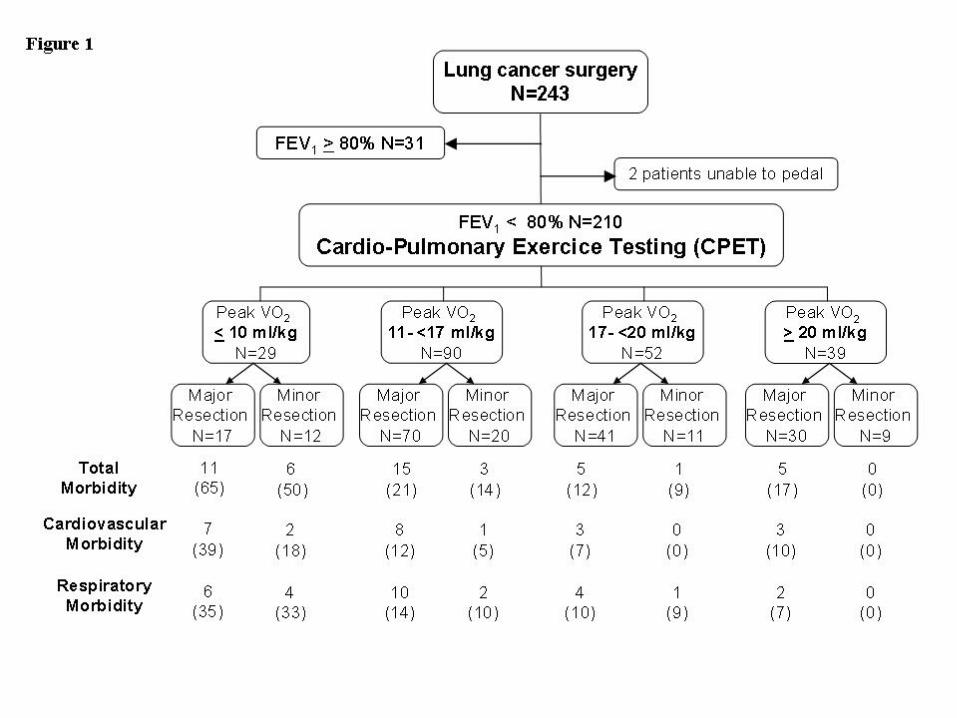
A total of 210 patients with FEV1 < 80% predicted yielded completed data sets and were
included in the study (Figure 1). Table 1 shows the baseline characteristics of patients
stratified by peak VO2. Patients with low peak VO2 (<17 ml/kg/min) were more likely to be
older, female and hypertensive; they also presented with a higher body mass index, higher
ASA risk classes, lower FEV1 (preoperative and predicted postoperative values) and with
earlier pathologic stages. Rates of coronary artery disease, hypercholesterolemia, diabetes and
mellitus as well as smoking and alcohol status did not differ significantly among the stratified
peak VO2 groups.
Peak VO2 expressed in ml/min/kg PBW was higher than peak VO2 expressed in ml/min/kg
absolute body weight (18.50±5.7 vs 16.0±5.3, P=0.0003).
As described in table 2, 30-day mortality was 1.9% and all four deaths occurred in patients
with peak VO2 < 17 ml/kg and were primarily related to cardiovascular complications (three
cases) and ALI (one case). Forty six patients experienced at least one cardiopulmonary
complication (incidence of 22%). Thirty-six pulmonary complications occurred in 28 patients

7
(pneumonia, 18 cases; atelectasis, 15 cases; ALI, 3 cases). Thirty cardiovascular
complications occurred in 24 patients (arrhythmias, 17 cases; stroke, 3 cases; acute heart
failure, 6 cases; myocardial infarct, 2 cases; pulmonary embolism, 2 cases). Sixteen patients
presented with both cardiovascular and pulmonary complications. Overall, the incidence of
complications did not differ in patients undergoing minor or major lung resection (19.2% vs.
20.5%, respectively; P=0.701).
In univariate analysis, patients experiencing cardio-pulmonary complications had lower
preoperative VO2, they underwent longer and more extensive procedures and required larger
doses of vasopressors at the time of surgery; in addition, a higher body mass index was
associated with a higher incidence of respiratory complications (Table 3). In contrast, gender,
smoking habits, alcohol consumption, the presence of coronary artery disease, hyperlipidemia
or diabetes mellitus did not differ between groups of patients with or without complications.
After adjusting for other perioperative factors by logistic regression, peak VO2 and
anaesthesia duration remained independent predictors of cardiovascular and/or pulmonary
complications; age and the extent of lung resection (pneumonectomy or bi-lobectomy) were
additional predictors of cardiovascular complications whereas tidal volume (VT) during one-
lung ventilation was an independent risk factor of respiratory complications (Table 4).
As detailed in Table 5, ROC curve analysis showed that the best cut-off values for peak VO2
to predict cardiac complications were 13.6 ml/min/kg and 16.2 ml/min/PBW (c-indices, 0.71
vs 0.74, respectively; P=0.445) whereas for pulmonary complications, the best cut-off values
were 12.8 ml/min/kg and 15.8 ml/min/PBW (c-indices, 0.72 vs 0.65, respectively; P=0.304).
The incidence of cardiovascular and pulmonary complications increased in parallel with the
reduction in peak VO2 during exercise testing (Figure 2). Compared to patients with peak
VO2 > 17 ml/kg/min, those with a peak VO2 < 10 ml/kg/min had a four-fold higher incidence
of cardiac and pulmonary morbidity.
Discussion
In this retrospective analysis of surgical candidates with FEV1 < 80%, we demonstrated that
markers of preoperative cardiopulmonary fitness and the duration of anesthesia were the best
predictors of cardio-pulmonary complications following lung cancer surgery. In addition, age
and the extent of lung resection were also considered independent risk factors of
cardiovascular complications whereas tidal volume during one-lung ventilation was a
predictor of pulmonary complications.

8
Among CPET parameters, peak VO2 values had the best discriminative power to identify the
high-risk group of patients. In agreement with previous reports, we confirmed that a peak VO2
> 20 ml.kg-1.min-1 is a safe cutoff value for lung resection since no mortality and less than 8%
respiratory and cardiac complications occurred above this threshold [13, 24-25]. In contrast,
below 10ml.kg-1.min-1peakVO2, patients experienced a fourfold higher incidence of
cardiovascular and pulmonary complications, compared with surgical candidates presenting
peak VO2 values > 20ml.kg-1.min-1. Nevertheless, the use of peak VO2 expressed in ml per kg
may lead to exclusion of obese candidates who are actually fit enough to undergo surgery. As
peak VO2 max better correlates with lean body mass than total body weight, several
physiologists have recommended the use of height and age in the calculation of predicted
peak VO2, particularly under condition of overweight [22, 26].
Interestingly, in the multivariate analysis, ppoFEV1 based on the “segment method” failed to
provide additional predictive value for cardiopulmonary complications. Actually, by adopting
a functional algorithm for selecting surgical candidates, we excluded most patients with
ppoFEV1 < 30%. Moreover, in patients with COPD, a “lung volume reduction” effect may
take place after resection, providing minimal loss or even improvement in lung function [27,
28]. Given the prognostic importance of ppoFEV1 regarding long-term quality of life, more
accurate estimation of functional segments might have been achieved by using perfusion
scintigraphy, quantitative CT or magnetic resonance imaging [29].
Although there is no firmly identified causal mechanism linking a low aerobic capacity with a
high risk profile, one explanation is that “unfit” patients are unable to face the increased
surgical stress-induced metabolic demand by mounting an adequate increase in oxygen
delivery [11]. Pre-existing disturbances in cardiac pumping performances, respiratory
function and oxygen utilization within the skeletal muscles may worsen during the early
postoperative period as a result of inadequate pain control, residual effects of anesthesia, fluid
overload, ventilator-induced lung injuries, direct surgical nerve damage, reflex inhibition of
diaphragmatic function and muscular fatigue associated with systemic inflammation and
enhanced protein catabolism. Indeed, patients with the lowest peak VO2 values (< 17
ml/kg/min) presented a higher index of comorbidity as reflected by ASA risk classes, a higher
prevalence of arterial hypertensive disease and greater impairments in pulmonary function.
Finally, oxidative stress has been shown to accelerate protein breakdown and to trigger
nuclear apoptosis in unloaded skeletal muscle, thereby rendering these patients unable to
sustain an adequate ventilatory response [30].
9
Previous investigators have limited their analysis mainly to preoperative clinical and
functional parameters that could affect the early postoperative outcome [13-15, 19, 25, 31]. In
the present study, we assessed more than 90 potential risk factors focusing on patient pre-
operative condition but also on intraoperative markers of processes of care. In this regard,
prolonged anaesthetic time likely reflected the dual aspects of a complex surgical approach as
well as difficulties with anesthesia emergence and weaning from the ventilator. Extended
tissue dissection involving ischemia-reperfusion injuries would obviously imposes larger
postoperative metabolic needs and greater loads on the cardiovascular and respiratory
systems.
Not surprisingly, we found that age and performing a pneumonectomy or bi-lobectomy were
additional risk factors for the occurrence of cardiac complications. Indeed, excess collagen
deposition in the extracellular matrix coupled with a progressive loss of cardiac myocytes by
apoptotic cell death are hallmarks of the senescent heart that have been incriminated in the
pathogenesis of ventricular dysfunction and atrial fibrillation [ 32]. Moreover, the pulmonary
circulatory system becomes less compliant after extended parenchymal lung resection
resulting in increased right ventricular afterload [33]. On the other hand, autonomic nerve
injuries occur inevitably when surgical dissection involves the hilar mediastinal structures
[34]. Accordingly, the majority of cardiovascular complications consisting in arrhythmias
(8%) and acute heart failure (3%) were likely related to age-associated myocardial
remodelling, cardiac autonomic nerve imbalance and greater vulnerability to fluid loading.
Besides markers of cardiopulmonary fitness and the duration of anesthesia, we found that the
intraoperative ventilatory strategy significantly influenced the development of pneumonia,
atelectasis and ALI, larger tidal volume being associated with higher operative risk.
Consistent with this finding, tidal volumes exceeding 7 to 8 ml/kg or elevated inspiratory
pressure during one-lung ventilation have been associated with greater risks to develop ALI
and with larger release of cytokines within the lungs and the systemic circulation [35,36].
Conversely, the application of “physiological” tidal volume (4-7 ml/kg) and external PEEP in
patients with healthy lungs undergoing oesophagectomy has been associated with an
attenuated systemic proinflammatory response, lower interstitial pulmonary edema and a
better oxygenation index [37]. Performing vital capacity manoeuvre has also been shown to
re-expand the collapsed dependent lung areas that develop in almost all anaesthetized patients
whereas moderate levels of PEEP are known to prevent a subsequent fall in functional
residual capacity by preventing cyclic opening-closing within pulmonary alveoli and

10
peripheral airways [21, 38]. Given the strong body of scientific knowledge related to
perioperative ventilator-associated injuries, we have adopted a lung-protective protocol
involving small tidal volume, external PEEP and periodical recruitment manoeuvres since
March 2003. Over the following 5-year period, implementation of this “open-lung” strategy
was associated with a reduced incidence of ALI and atelectasis along with fewer admissions
in intensive care [39].
There are some limitations and strengths of this observational study that should be mentioned.
First, although more than 90 items were prospectively collected, we assume some variability
in recorded data, unmeasured risk factors and diseases with low prevalence rate (e.g., diabetes
mellitus, pulmonary hypertension) that could partly confound the interpretation of the
multivariate analysis. Secondly, we obtained peakVO2 data using bicycle exercise that might
not be feasible in patients with lower extremities disabilities caused by vascular, neurological
or orthopedic conditions. In such cases, arm exercise testing should be considered as a
suitable alternative.[40] Third, our findings obtained in a referral thoracic centre, can not be
generalized to nonspecialized units in the absence of standardized clinical pathways.
Implementation of bundles of scientifically-based interventions has been shown to improve
perioperative outcome. If we compare the current data with those published previously [26],
we have witnessed a 25% reduction in cardiopulmonary morbidity over the last 15 years, that
was likely attributed to the beneficial effects of thoracic epidural analgesia, fluid titration
guided by Doppler flow monitoring and protective lung ventilation.[2, 39, 41]
Our selection criteria were less restrictive than in the initial algorithm since 29 patients with
peak VO2 < 10 ml/kg/min were considered suitable candidates for surgery, with 25 survivors
and 14 of whom without any serious complications. The majority of these very high-risk
patients presented with early cancer stages IA and IB (55%) and moderate-to-severe COPD.
Although, no firm conclusions could be drawn from these encouraging data, some patients
deemed inoperable (or at very high-risk) according to their very low peak VO2 values might
become eligible for curative surgery, particularly those with acceptable ppoFEV1/ppoDLCO
values (>30%). Moreover, data support that sublobar resection is a suitable alternative therapy
for patients bearing a small sized tumour (< 2-3 cm) whereas minimal deterioration of
postoperative FEV1 has been observed in COPD patients after anatomical lobar resection as a
result of a “volume reduction” effect [27,28].
Finally, future trials should question whether improvement in aerobic performances induced
by preoperative physical training may reduce perioperative mortality and morbidity in these

11
high risk patients. To date, experimental data indicates that short-term intense physical
training induces a cardioprotective phenotype similar to ischemic preconditioning and
enhances oxygen extraction from the skeletal muscle by increasing mitochondrial mass,
capillary density and oxidative capacity.[42, 43] Preliminary studies in elderly patients with
cancer, arterial disease or heart failure confirm the beneficial physiological effects of short-
term supervised training program, namely an increase in peak VO2 (+20%) and in anaerobic
threshold (10%), a reduction in plasma inflammatory markers and improvement in cardiac
function [44-47].
In conclusion, our data strongly support the use of CPT for risk stratification in patients with
preoperative FEV1 < 80%. Given substantial progress in surgical techniques and anaesthetic
management over the last decades, even high-risk patients (peakVO2 < 10 ml/kg/min) may be
considered for life-saving surgery. According to our results, several risk-reducing strategies
can be implemented: 1) patient referral to a qualified and experienced thoracic team in order
to shorten the time of intervention, 2) application of a protective ventilatory protocol to
minimize pulmonary injuries triggered by mechanical ventilation, 3) considering short-term
preoperative physical training in the “unfit” patients. Further studies are needed to replicate
our observations and to question whether improved aerobic capacity achieved by exercise
training is associated with better postoperative outcome in the high-risk group.
Acknowledgements
The Lancardis Fundation in Sion in Switzerland granted financial support for this study.

12
References 1. Boffa DJ, Allen MS, Grab JD, Gaissert HA, Harpole DH, Wright CD: Data from
The Society of Thoracic Surgeons General Thoracic Surgery database: the
surgical management of primary lung tumors. J Thorac Cardiovasc Surg 2008,
135(2):247-254.
2. Licker MJ, Widikker I, Robert J, Frey JG, Spiliopoulos A, Ellenberger C,
Schweizer A, Tschopp JM: Operative mortality and respiratory complications
after lung resection for cancer: impact of chronic obstructive pulmonary disease
and time trends. Ann Thorac Surg 2006, 81(5):1830-1837.
3. Kates M, Perez X, Gribetz J, Swanson SJ, McGinn T, Wisnivesky JP: Validation
of a model to predict perioperative mortality from lung cancer resection in the
elderly. Am J Respir Crit Care Med 2009, 179(5):390-395.
4. Spiro SG, Tanner NT, Silvestri GA, Janes SM, Lim E, Vansteenkiste JF, Pirker
R: Lung cancer: progress in diagnosis, staging and therapy. Respirology,
15(1):44-50.
5. Colice GL, Shafazand S, Griffin JP, Keenan R, Bolliger CT: Physiologic
evaluation of the patient with lung cancer being considered for resectional
surgery: ACCP evidenced-based clinical practice guidelines (2nd edition). Chest
2007, 132(3 Suppl):161S-177S.
6. Bolliger CT, Koegelenberg CF, Kendal R: Preoperative assessment for lung
cancer surgery. Curr Opin Pulm Med 2005, 11(4):301-306.
7. van Tilburg PM, Stam H, Hoogsteden HC, van Klaveren RJ: Pre-operative
pulmonary evaluation of lung cancer patients: a review of the literature. Eur
Respir J 2009, 33(5):1206-1215.
8. Palange P, Ward SA, Carlsen KH, Casaburi R, Gallagher CG, Gosselink R,
O'Donnell DE, Puente-Maestu L, Schols AM, Singh S et al: Recommendations on
the use of exercise testing in clinical practice. Eur Respir J 2007, 29(1):185-209.
9. Jones LW, Eves ND, Haykowsky M, Joy AA, Douglas PS: Cardiorespiratory
exercise testing in clinical oncology research: systematic review and practice
recommendations. Lancet Oncol 2008, 9(8):757-765.

13
10. ATS/ACCP statement on cardiopulmonary exercise testing. Am J Respir Crit
Care Med 2003, 167(2):211-77.
11. Albouaini K, Egred M, Alahmar A, Wright DJ: Cardiopulmonary exercise
testing and its application. Heart 2007, 93(10):1285-1292.
12. Brunelli A: Algorithm for functional evaluation of lung resection candidates:
time for reappraisal? Respiration 2009, 78(1):117-118.
13. Brunelli A, Belardinelli R, Refai M, Salati M, Socci L, Pompili C, Sabbatini A:
Peak oxygen consumption during cardiopulmonary exercise test improves risk
stratification in candidates to major lung resection. Chest 2009, 135(5):1260-1267.
14. Benzo R, Kelley GA, Recchi L, Hofman A, Sciurba F: Complications of lung
resection and exercise capacity: a meta-analysis. Respir Med 2007, 101(8):1790-
1797.
15. Win T, Jackson A, Sharples L, Groves AM, Wells FC, Ritchie AJ, Laroche CM:
Cardiopulmonary exercise tests and lung cancer surgical outcome. Chest 2005,
127(4):1159-1165.
16. Brunelli A, Charloux A, Bolliger CT, Rocco G, Sculier JP, Varela G, Licker M,
Ferguson MK, Faivre-Finn C, Huber RM et al: ERS/ESTS clinical guidelines on
fitness for radical therapy in lung cancer patients (surgery and chemo-
radiotherapy). Eur Respir J 2009, 34(1):17-41.
17. Brunelli A, Charloux A, Bolliger CT, Rocco G, Sculier JP, Varela G, Licker M,
Ferguson MK, Faivre-Finn C, Huber RM et al: The European Respiratory
Society and European Society of Thoracic Surgeons clinical guidelines for
evaluating fitness for radical treatment (surgery and chemoradiotherapy) in
patients with lung cancer. Eur J Cardiothorac Surg 2009, 36(1):181-184.
18. Wyser C, Stulz P, Soler M, Tamm M, Muller-Brand J, Habicht J, Perruchoud
AP, Bolliger CT: Prospective evaluation of an algorithm for the functional
assessment of lung resection candidates. Am J Respir Crit Care Med 1999, 159(5
Pt 1):1450-1456.
19. Bolliger CT, Jordan P, Soler M, Stulz P, Gradel E, Skarvan K, Elsasser S, Gonon
M, Wyser C, Tamm M et al: Exercise capacity as a predictor of postoperative

14
complications in lung resection candidates. Am J Respir Crit Care Med 1995,
151(5):1472-1480.
20. Tang SS, Redmond K, Griffiths M, Ladas G, Goldstraw P, Dusmet M: The
mortality from acute respiratory distress syndrome after pulmonary resection is
reducing: a 10-year single institutional experience. Eur J Cardiothorac Surg 2008,
34(4):898-902.
21. Licker M, Fauconnet P, Villiger Y, Tschopp JM: Acute lung injury and outcomes
after thoracic surgery. Curr Opin Anaesthesiol 2009, 22(1):61-67.
22. Hansen JE, Sue DY, Wasserman K: Predicted values for clinical exercise testing.
Am Rev Respir Dis 1984, 129(2 Pt 2):S49-55.
23. BTS guidelines: guidelines on the selection of patients with lung cancer for
surgery. Thorax 2001, 56(2):89-108.
24. Burke JR, Duarte IG, Thourani VH, Miller JI, Jr.: Preoperative risk assessment
for marginal patients requiring pulmonary resection. Ann Thorac Surg 2003,
76(5):1767-1773.
25. Brutsche MH, Spiliopoulos A, Bolliger CT, Licker M, Frey JG, Tschopp JM:
Exercise capacity and extent of resection as predictors of surgical risk in lung
cancer. Eur Respir J 2000, 15(5):828-832.
26. Buskirk E, Taylor HL: Maximal oxygen intake and its relation to body
composition, with special reference to chronic physical activity and obesity. J
Appl Physiol 1957, 11(1):72-78.
27. Brunelli A, Al Refai M, Monteverde M, Sabbatini A, Xiume F, Fianchini A:
Predictors of early morbidity after major lung resection in patients with and
without airflow limitation. Ann Thorac Surg 2002, 74(4):999-1003.
28. Edwards JG, Duthie DJ, Waller DA: Lobar volume reduction surgery: a method
of increasing the lung cancer resection rate in patients with emphysema. Thorax
2001, 56(10):791-795.
29. Ohno Y, Koyama H, Nogami M, Takenaka D, Matsumoto S, Yoshimura M,
Kotani Y, Sugimura K: Postoperative lung function in lung cancer patients:
comparative analysis of predictive capability of MRI, CT, and SPECT. AJR Am J
Roentgenol 2007, 189(2):400-408..

15
30. Chopard A, Hillock S, Jasmin BJ: Molecular events and signalling pathways
involved in skeletal muscle disuse-induced atrophy and the impact of
countermeasures. J Cell Mol Med 2009, 13(9B):3032-3050.
31. Bobbio A, Chetta A, Internullo E, Ampollini L, Carbognani P, Bettati S, Rusca
M, Olivieri D: Exercise capacity assessment in patients undergoing lung
resection. Eur J Cardiothorac Surg 2009, 35(3):419-422.
32. Chen W, Frangogiannis NG: The role of inflammatory and fibrogenic pathways
in heart failure associated with aging. Heart Fail Rev.
33. Amar D, Burt ME, Roistacher N, Reinsel RA, Ginsberg RJ, Wilson RS: Value of
perioperative Doppler echocardiography in patients undergoing major lung
resection. Ann Thorac Surg 1996, 61(2):516-520.
34. Roselli EE, Murthy SC, Rice TW, Houghtaling PL, Pierce CD, Karchmer DP,
Blackstone EH: Atrial fibrillation complicating lung cancer resection. J Thorac
Cardiovasc Surg 2005, 130(2):438-444.
35. Licker M, de Perrot M, Spiliopoulos A, Robert J, Diaper J, Chevalley C, Tschopp
JM: Risk factors for acute lung injury after thoracic surgery for lung cancer.
Anesth Analg 2003, 97(6):1558-1565.
36. Schilling T, Kozian A, Huth C, Buhling F, Kretzschmar M, Welte T, Hachenberg
T: The pulmonary immune effects of mechanical ventilation in patients
undergoing thoracic surgery. Anesth Analg 2005, 101(4):957-965, table of
contents.
37. Michelet P, D'Journo XB, Roch A, Doddoli C, Marin V, Papazian L, Decamps I,
Bregeon F, Thomas P, Auffray JP: Protective ventilation influences systemic
inflammation after esophagectomy: a randomized controlled study.
Anesthesiology 2006, 105(5):911-919.
38. Wolthuis EK, Choi G, Dessing MC, Bresser P, Lutter R, Dzoljic M, van der Poll
T, Vroom MB, Hollmann M, Schultz MJ: Mechanical ventilation with lower tidal
volumes and positive end-expiratory pressure prevents pulmonary inflammation
in patients without preexisting lung injury. Anesthesiology 2008, 108(1):46-54.

16
39. Licker M, Diaper J, Villiger Y, Spiliopoulos A, Licker V, Robert J, Tschopp JM:
Impact of intraoperative lung-protective interventions in patients undergoing
lung cancer surgery. Crit Care 2009, 13(2):R41.
40. Myers J, Arena R, Franklin B, Pina I, Kraus WE, McInnis K, Balady GJ;
American Heart Association Committee on Exercise, Cardiac Rehabilitation, and
Prevention of the Council on Clinical Cardiology, the Council on Nutrition,
Physical Activity, and Metabolism, and the Council on Cardiovascular Nursing.
Recommendations for clinical exercise laboratories: a scientific statement from
the american heart association. Circulation. 2009;119(24):3144-61.
41. Diaper J, Ellenberger C, Villiger Y, Robert J, Tschopp JM, Licker M:
Transoesophageal Doppler monitoring for fluid and hemodynamic treatment
during lung surgery. J Clin Monit Comput 2008, 22(5):367-374.
42. Powers SK, Quindry JC, Kavazis AN: Exercise-induced cardioprotection against
myocardial ischemia-reperfusion injury. Free Radic Biol Med 2008, 44(2):193-
201.
43. Befroy DE, Petersen KF, Dufour S, Mason GF, Rothman DL, Shulman GI:
Increased substrate oxidation and mitochondrial uncoupling in skeletal muscle of
endurance-trained individuals. Proc Natl Acad Sci U S A 2008, 105(43):16701-
16706.
44. Bobbio A, Chetta A, Ampollini L, Primomo GL, Internullo E, Carbognani P,
Rusca M, Olivieri D: Preoperative pulmonary rehabilitation in patients
undergoing lung resection for non-small cell lung cancer. Eur J Cardiothorac
Surg 2008, 33(1):95-98.
45. Kothmann E, Batterham AM, Owen SJ, Turley AJ, Cheesman M, Parry A,
Danjoux G: Effect of short-term exercise training on aerobic fitness in patients
with abdominal aortic aneurysms: a pilot study. Br J Anaesth 2009, 103(4):505-
510.
46. Wisloff U, Stoylen A, Loennechen JP, Bruvold M, Rognmo O, Haram PM,
Tjonna AE, Helgerud J, Slordahl SA, Lee SJ et al: Superior cardiovascular effect
of aerobic interval training versus moderate continuous training in heart failure
patients: a randomized study. Circulation 2007, 115(24):3086-3094.

17
47. Jones LW, Eves ND, Mackey JR, Peddle CJ, Haykowsky M, Joy AA, Tankel K,
Courneya KS, Reiman T: Systemic inflammation, cardiorespiratory fitness, and
quality of life in patients with advanced non-small cell lung cancer. J Thorac
Oncol 2008, 3(2):194-195.

18
Annex 1 Major outcomes
Mortality
Death within 30 days after surgery or for a longer period if the patient was still
hospitalized
Cardiovascular
1) Myocardial infarct : typical rise and fall of CPK (> 120 U/L) and CK-MB/CPK > 6%
or troponin-I > 1.5 ng/ml with at least one of the following criteria: ischemic
symptoms, development of pathological Q waves on the ECG, ST segment elevation
or depression (> 1 mm) or coronary artery intervention
2) Arrhythmia’s : supraventricular and ventricular tachyarrhythmia's on ECG causing
unstable hemodynamic condition and requiring anti-arrhythmic medications and/ or
electrical cardioversion
3) Congestive heart failure : need for sympathomimetic support, diuretics or vasodilators
consistent with clinical, hemodynamic (pulmonary artery pressure > 15 mmHg) and
radiological evidence of pulmonary congestion
4) Thrombo-embolism: acute occlusion of pulmonary arteries diagnosed by scintigraphy
or angiogram
5) Stroke : focal neurological deficit (transient or permanent)
6) Renal dysfunction: elevation of serum creatinine > 50% compared with preoperative
value
Respiratory
1. Atelectasis :Lobar collapse (Chest-X rays), need for CPAP and / or bronchoscopy
2. Bronchopneumonia: temperature > 38°C, hyperleucocytosis (neutrophils), new lung
infiltration (chest-X rays), positive culture (bronchial secretions or alveolar fluid)
3. Acute Lung Injury: 1) sudden onset of respiratory distress, 2) infiltrates on the chest
radiograph consistent with pulmonary edema, 3) impaired oxygenation with an arterial
oxygen pressure - to- inspired oxygen fraction ratio (PaO2/FIO2 or P/F ratio) < 300
mm of Hg, 4) absence of cardiac insufficiency or fluid overload, based on pulmonary
arterial catheterization, echocardiogram and/or clinical evaluation.

19
Table 1 Baseline Characteristics stratified by peak VO2 expressed in ml.min-1.kg-1 Characteristics VO2
< 10 VO2
11 - < 17 VO2
17- <20 VO2 > 20
P Value for Trend
N=29 N=90 N=52 N=39 Age (yr) 68 (10) 65 (9) 64 (9) 53 (11) 0.022 BMI (kg/[m2]2) 28.4 (5.1) 25.5 (4.6) 24.8 (5.8) 23.9 (4.7) 0.009 Male gender (n, %) 18 (62) 61 (68) 36 (69) 30 (76) 0.023 ASA risk classes 3 & 4 (n, %)
14 (48) 39 (43) 21 (40) 6 (15) 0.011
Smoking status Unit Pack-Years Current smoker (n, %) Ex-Smoker* (n, %)
46 (24) 18 (62) 4 (14)
42 (21) 59 (66) 10 (11)
43 (20) 32 (62) 6 (12)
44 (22) 24 (62) 4 (10)
0.652 0.712 0.694
Alcohol status (n, %) 4 (14) 17 (19) 9 (17) 1 (13) 0.751 Coronary artery disease (n, %)
3 (10) 7 (8) 4 (8) 2 (5) 0.854
Hypertension (n, %) 13 (45) 35 (39) 16 (31) 7 (18) 0.004 Hypercholesterolemia (n, %)
5 (17) 21 (23) 12 (23) 4 (10) 0.324
Diabetes mellitus (n, %)
1 (3.4) 9 (10) 4 (8) 3 (8) 0.852
Hematocrit (%) 45 (20) 42 (6) 42 (4) 45 (20) 0.325 Creatinine clearance, (ml.min-1)
86 (23) 79 (29) 79 (30) 82 (27) 0.309
FEV1 (% predicted) 65 (19) 79 (22) 82 (25) 88 (21) < 0.001 ppoFEV1 (% predicted) 50 (19) 55 (20) 58 (17) 64 (19) < 0.001 DLCO (% predicted) 64 (16) 74 (17) n.a. n.a. - ppoDLCO (% predicted)
51 (10) 59 (18) n.a. n.a. -
VO2max (ml-1.kg-1) 9.0 (1.6) 13.7 (1.2) 17.6 (1.1) 24.4 (4.6) < 0.001 VO2max (ml-1.kg-1 PBW)
11.6 (2.9) 15.8 (3.1) 20 (3.6) 25.4 (5.4) < 0.001
VO2 max (% predicted) 39 (11) 52 (13) 65 (14) 76 (16) < 0.001 Load (Watt) 83 (32) 93 (23) 113 (27) 137 (36) < 0.001 VEmax (L.min-1) 37 (10) 48 (11) 57 (16) 68 (15) < 0.001 VEmax (% pred) 75 (15) 78 (17) 89 (17) 99 (17) < 0.001 PO2 end exercise (mmHg)
63 (18) 66 (22) 73 (12) 74 (16) < 0.001
Neoadjuvant chemotherapy (n, %)
4 (14) 12 (13) 6 (12) 6 (15) 0.534
Pathologic stages Ia and Ib (n, %)
16 (55) 37 (41) 20 (38) 12 (31) 0.021
Pneumonectomy or bi-lobectomy (n, %)
5 (17) 16 (18) 13 (25) 5 (13) 0.458
Data are presented as n (%) or mean (SD) unless otherwise stated. *, > 6 months smoking cessation; n.a., not available; ASA, American Society Association; FEV1, Forced Expiratory Volume during the first second; ppoFEV1, predicted postoperative Forced Expiratory Volume during the first second.

20
Table 2 Clinical and surgical characteristics of patients with and without complications
Total Morbidity
Cardiovascular Complications
Respiratory
Complications Yes
(n=46) No
(n=164)Yes
(n=24) No
(n=186) Yes
(n=28) No
(n=182) Preoperative period
Age (yr) 66 (9) 62 (11) 69 (9) 62 (8)* 65 (9) 63 (11)
Body Mass Index (kg/[m2]2)
26.4 (6.0)
25.1 (4.8)
25.7 (4.5)
25.4 (5.2)
27.0 (6.8)*
25.2 (4.8)
Male gender (n, %) 34 (74) 110(67) 19 (79) 125 (67) 21 (72) 144 (80) ASA classes 3 & 4 (n, %)
22 (48) 58 (35) 13 (54) 67 (36) 15 (53) 80 (44)
Smoking status Unit Pack-Years Current smoker (n, %) Ex-Smoker* (n, %)
44 (21)
32 (69) 6 (13)
42 (23)
101(62) 18 (11)
45 (23)
17 (71) 3 (12)
41 (24)
116 (62) 21 (11)
46 (23)
20 (71) 3 (11)
43 (21)
113 (62) 21 (12)
Alcohol status (n, %) 8 (17) 23 (14) 4 (17) 27 (14) 6 (21) 25 (14) Coronary artery disease (n, %)
6 (13.0) 11 (6.7) 3 (12.5) 14 (7.5) 3 (10.7) 17 (9.4)
Hypertension (n, %) 21 (46) 51 (31) 13 (54) 59 (32) 13 (46) 72 (40) Hyperlipidemia (n, %) 11 (24) 32 (20) 8 (33) 35 (19) 4 (14) 43 (24) Diabetes mellitus (n, %)
2 (4.3) 15 (9.1) 1 (4.2) 16 (8.6) 2 (7.1) 17 (9.4)
FEV1 (% predicted) 77 (23) 80 (23) 78 (22) 80 (23) 74 (22) 80 (23) ppoFEV1 (% predicted)
56 (18) 61 (21) 55 (16) 61 (19) 54 (15)* 62 (17)
Hematocrit (%) 40.3 (6.7)
41.9 (4.4)
38.5 (6.4)*
42.1 (4.6)
41.4 (7.5)
41.6 (4.6)
Creatinine clearance (ml.min-1)
80 (33) 84 (25) 73 (18)* 85 (27) 78 (37) 82 (25)
VO2max (ml.min-1.kg-
1) 13.0
(4.4)* 16.9 (5.2)
13.0 (4.3)
16.5 (5.3)
15.2 (6.3)*
18.5 (5.6)
VO2max (ml.min-1.kg-1
PBW) 15.0
(5.5)* 18.9 (5.6)
14.3 (4.0)*
18.6 (5.8)
12.7 (4.6)*
16.6 (5.2)
VO2 max (% predicted)
50 (17)* 60 (17) 50 (14)* 59 (18) 49 (20)* 59 (17)
Load (Watt) 90 (30)*
109 (34)
89 (35)*
107 (33)
87 (26)*
108 (34)
VEmax (L.min-1) 47 (17)* 54 (16) 48 (18) 53 (16) 46 (15)* 53 (16) VEmax (%pred) 60 (21) 65 (17) 65 (27) 64 (16) 56 (12)* 65 (18) PO2 end exercice (mmHg)
64 (21) 70 (18) 58 (24)* 70 (17) 67 (20) 69 (18)
Intraoperative Period Surgical time (min) 111
(42)* 97
(29) 123
(42)* 97
(30) 109 (41)
99 (31)
Anesthesia time (min) 152 (64)*
123 (35)
160 (52)*
125 (42)
154 (74)*
125 (37)

21
Pneumonectomy or Bi-lobectomy (n, %)
12 (26) 27 (16) 10 (42)* 29 (15) 6 (21) 33 (18)
VT during TLV (ml.kg-
1 PBW) 8.1
(2.3) 7.4
(2.1) 7.9
(2.3) 7.6
(2.2) 8.8
(1.9)* 6.9
(2.0) VT during OLV (ml.kg-
1 PBW) 6.9
(2.2) 6.5
(2.1) 7.0
(2.9) 6.4
(2.7) 7.0
(1.9)* 5.6
(1.8) Phenylephrine (mg) 580
(442)* 384
(348) 597
(524)* 405
(348) 635
(431)* 397
(358) Fluid (ml.kg-1.h-1) 9.6
(5.9) 8.8
(3.5) 9.4
(7.1) 8.9
(3.6) 9.1
(4.1) 8.9
(4.2) Data are presented as n (%) or mean (SD) unless otherwise stated. ASA, American Society Association; FEV1, Forced Expiratory Volume during the first second; ppoFEV1, predicted postoperative Forced Expiratory Volume during the first second; VT, Tidal Volume; TLV, two-Lung Ventilation; OLV, One-Lung Ventilation; PBW, Predicted Body Weight.

22
Table 3 Characteristics of the Four Patients who Died within the Perioperative Period
Patient Age Gender BMI Operation Comorbidities peak VO2 ppoFEV1 Cause of Death
N° yr M/F kg/(m2)2 ml/kg/min§ % predicted % predicted
1 82 M 28.7 RUL Alcohol* 8.2 41 63% Stroke (day 6) 2 61 M 26.3 LP CAD 8.4 31 45% Myocardial Infarct, Stent Thrombosis, Hyperlipemia Heart Failure (day 10)
3 57 F 27.9 RP Hypertension 9.1 32 39% ALI, MOF (day 9) Alcohol*
4 68 M 27.2 LUL Hypertension 13.6 49 62% Pulmonary Thrombo-Embolism (day 4) Hyperlipemia Alcohol* BMI: Body Mass Index; M/F, Male/ Female; CAD, peakVO2
§ expressed in ml/min/kg absolute body weight; Alcohol*: > 21 drinks/week for men or > 14 drinks/week for women; RUL, Right Upper Lobectomy; LP, Left Pneumonectomy; RP, Right Pneumonectomy; LUL, Left Upper Lobectomy; ALI, Acute Lung Injury; MOF, Multiple Organ Failure

Table 4 Results of Logistic Regression Analysis for Different Dependent Variables Independent
predictor Coefficient SEM OR 95% CI P Value Boostrap
frequency,% Total Morbidity Constant 0.2980 79 Duration of
Anesthesia 0.0145 0.0041 1.015 1.006 - 1.023 0.0005
Peak VO2 in ml.min-1.kg-1
-0.2353 0.0546 0.79 0.71 - 0.88 <0.0001 81
Cardiovascular Complications
Constant -6.5727 Age 0.0804 0.0318 1.084 1.018 - 1.153 0.0115 62 Duration of
Anesthesia 0.0143 0.0045 1.014 1.005 - 1.024 0.0016 72
Pneumonectomyor Bilobectomy
1.7518 0.5674 5.76 1.90 - 17.53 0.0020 71
Peak VO2 in ml.min-1.kg-1
-0.2286 0.0768 0.80 0.68 - 0.92 0.0029 69
Pulmonary Complications
Constant -21.0904 Duration of Anesthesia
0.0133 0.0042 1.013 1.005 - 1.022 0.032 66
Peak VO2 in ml.min-1.kg-1
-0.1752 0.0590 0.840 0.749 - 0.942 0.021 70
VT ml.kg-1 PBW 2.8249 0.5384 1.65 1.27 – 4.49 0.003 77 VT , tidal volume

24
Table 5 Areas Under the Receiver Operating Characteristic Curves and Cutoff Values of Various Parameters for the Prediction of Cardiovascular and Pulmonary Complications
Area Under the
Curve
Standard Error
Lower – Upper Bound
P Value Cutoff Sensitivity (%)
Specificity (%)
Total Morbidity VO2 kg-1 0.717 0.045 0.651 to 0.777 0.0001 12.8 51 85 VO2 kg-1 PBW 0.710 0.045 0.643 to 0.771 0.0001 15.8 64 69 VO2 % pred. 0.657 0.045 0.589 to 0.722 0.0010 58 75 48 ASA score 0.593 0.039 0.523 to 0.661 0.0156 3 71 48 ppoFEV1 0.565 0.045 0.495 to 0.633 0.7880 64 65 49
Cardiovascular Complications
VO2 kg-1 0.708 0.065 0.640 to 0.771 0.0011 13.6 63 72 VO2 kg-1 PBW 0.738 0.054 0.671 to 0.798 0.0001 16.2 75 61 VO2 % pred. 0.633 0.061 0.562 to 0.700 0.0029 53 64 61 ASA score 0.630 0.046 0.560 to 0.695 0.0080 3 79 47 ppoFEV1 0.492 0.066 0.422 to 0.562 0.9030 80 29 79
Pulmonary Complications
VO2 kg-1 0.723 0.057 0.654 to 0.784 0.0001 12.3 56 86 VO2 kg-1 PBW 0.691 0.065 0.580 to 0.718 0.0020 12.1 45 94 VO2 % pred. 0.616 0.066 0.544 to 0.684 0.0169 37 30 95 ASA score 0.597 0.044 0.527 to 0.664 0.0269 3 73 47 ppoFEV1 0.545 0.051 0.480 to 0.614 0.3716 64 73 50