impact of increased helminth diagnostic sensitivity on
TRANSCRIPT

Impact of Increased Helminth Diagnostic Sensitivity on Estimation of Risk Factors and
Outcomes
Michael B. Arndt
A thesis
submitted in partial fulfillment of the
requirements for the degree of
Master of Public Health
University of Washington
2012
Committee:
Judd Walson
Grace John-Stewart
Program Authorized to Offer Degree:
Public Health- Epidemiology

ii
© Copyright 2012
Michael B. Arndt

iii
University of Washington
Abstract
Impact of Increased Helminth Diagnostic Sensitivity on Estimation of Risk Factors and
Outcomes
Michael B. Arndt
Chair of the Supervisory Committee:
Assistant Professor Judd Walson
Global Health, Medicine, Pediatrics, Epidemiology
Background: Traditional methods utilizing microscopy for the detection of helminth infections
have limited sensitivity to detect infection, particularly in populations with lower helminth
prevalence and burden. Newer polymerase chain reaction (PCR) assays may enhance detection
of helminth infections and improve identification of risk factors for infection. However, these
methods may detect low level helminth infections with limited impact on clinical outcomes.
Methods: This cross-sectional study was nested within a randomized clinical trial (RCT)
conducted at 3 HIV Care sites in Kenya. We performed three microscopy methods and real-time
multiplex PCR for the detection of Ascaris lumbricoides, hookworm spp., Strongyloides
stercoralis, and Schistosoma spp. in stool. Sensitivity for each diagnostic modality to detect
helminths was calculated using positive results from microscopy and PCR as the gold standard
positive. PCR cycle threshold is the amplification cycle in which the fluorescent probe signal
level exceeds background fluorescence, and correlates with parasite DNA load. We utilized
relative risk regression and linear regression models to evaluate the association between helminth
infection as detected by either microscopy or PCR and potential risk factors or outcomes.
iv
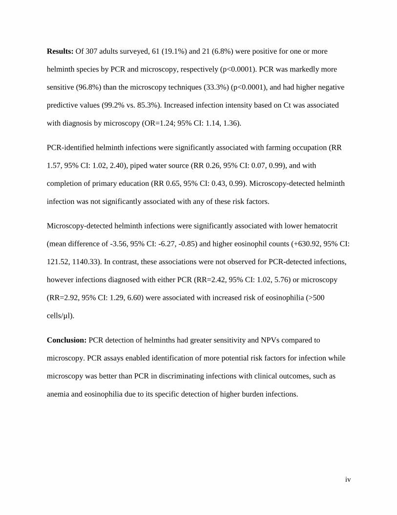
Results: Of 307 adults surveyed, 61 (19.1%) and 21 (6.8%) were positive for one or more
helminth species by PCR and microscopy, respectively (p<0.0001). PCR was markedly more
sensitive (96.8%) than the microscopy techniques (33.3%) (p<0.0001), and had higher negative
predictive values (99.2% vs. 85.3%). Increased infection intensity based on Ct was associated
with diagnosis by microscopy (OR=1.24; 95% CI: 1.14, 1.36).
PCR-identified helminth infections were significantly associated with farming occupation (RR
1.57, 95% CI: 1.02, 2.40), piped water source (RR 0.26, 95% CI: 0.07, 0.99), and with
completion of primary education (RR 0.65, 95% CI: 0.43, 0.99). Microscopy-detected helminth
infection was not significantly associated with any of these risk factors.
Microscopy-detected helminth infections were significantly associated with lower hematocrit
(mean difference of -3.56, 95% CI: -6.27, -0.85) and higher eosinophil counts (+630.92, 95% CI:
121.52, 1140.33). In contrast, these associations were not observed for PCR-detected infections,
however infections diagnosed with either PCR (RR=2.42, 95% CI: 1.02, 5.76) or microscopy
(RR=2.92, 95% CI: 1.29, 6.60) were associated with increased risk of eosinophilia (>500
cells/µl).
Conclusion: PCR detection of helminths had greater sensitivity and NPVs compared to
microscopy. PCR assays enabled identification of more potential risk factors for infection while
microscopy was better than PCR in discriminating infections with clinical outcomes, such as
anemia and eosinophilia due to its specific detection of higher burden infections.
1
INTRODUCTION
The burden of soil-transmitted helminth infections and schistosomiasis is considerable; there are
over a billion infections globally, with more than half of these infections (533 million) occurring
in sub Saharan Africa[1]. Helminth infections are a significant source of morbidity; contributing
to iron deficiency/anemia, as well as growth and cognitive deficiencies in school-age children[2].
In adults, helminth infections have been associated with reduced Vitamin A absorption[3],
economic productivity, and higher rates of anemia [4].
Traditionally, helminth and schistosoma infections have been detected using a variety of stool
microscopy techniques (Kato-Katz, Formol-ether, and Wet-prep technique), which have high
specificity in ruling out infection in those uninfected, but have limited sensitivity[5-8] especially
in populations where infection intensity (based on egg excretion) is low. New polymerase chain
reaction (PCR) assays enhance detection of helminth infections, having higher sensitivity and
high specificity[9-11].
There is significant geographic overlap in areas in which HIV and helminth infections are
prevalent [12]. Infection with HIV may alter excretion of parasite eggs in stool due to HIV-
associated immunodysregulation[13], potentially influencing the diagnostic accuracy of
microscopy. Because PCR-based diagnostic methods do not depend on egg excretion alone for
the detection of infection, they may be useful in detecting infections in HIV-infected individuals.
Previous studies have used microscopy diagnostic methods to identify several social and
environmental factors associated with helminth infection in adults. Age, education status, gender
and poor hygiene practices have all been associated with higher risk of helminth infection. The
prevalence and intensity of helminth infection is generally higher among younger adults and

2
correlates with age[14-20]. Lower education has been associated with increased risk of helminth
and Schistosoma infection [15,19,21,22]. Male gender has been associated with Schistosoma
infection[16,17]; but the association has not been observed consistently[23]. Environmental
factors, such as rural habitation or visitation have been associated with higher risk of helminth
infection[22,24,25]. Factors related to specific helminths include distance to large bodies of
water[19,20] or water contact[26] for Schistosoma spp. and contact with soil for hookworms, as
both farming occupation and lack of shoes are associated with higher risk[22,27]. Good hygiene
practices, such as hand-washing after defecation and latrine use, are also associated with lower
risk of helminth infections[16,21,22,24,27,28], which may account for the associations between
lower socioeconomic status and helminth infection [16,29].
Clinical consequences of microscopy-detected helminth infection in adults include reduced
Vitamin A absorption[3] and reductions in physical fitness and worker productivity[30,31].
Hookworm infections of moderate to high intensity have been associated with lower hemoglobin
levels and anemia in school-age children and adults[4,32,33], and with iron deficiency anemia in
pregnant women[34]. A 2010 systematic review found that hemoglobin in non-pregnant adults
decreased progressively with hookworm infection intensity[33], and a 2012 study using real-time
PCR demonstrated a load-dependent relationship between A. duodenale infection and iron
deficiency and anemia in school children[35]. A recent review described mixed findings
regarding the relationship between schistosomiasis and anemia[36].
In the present study we compared the helminth diagnostic performance of microscopy to that of
PCR in a cohort of HIV-positive Kenyan adults. We hypothesized that identification of risk
factors and clinical outcomes associated with helminth infection would be substantially
influenced by helminth diagnostic method. We explored whether the use of PCR for diagnoses

3
altered our ability to detect risk factors and outcomes of helminth infection by conducting
parallel correlates analyses: one using PCR and the other using microscopy for helminth
diagnosis.
METHODS
Study design: The study utilized a cross-sectional design nested in a 2-year randomized clinical
trial (RCT)-the Helminth Eradication to delay ART Trial (HEAT) study (ClinicalTrials.gov,
number NCT0050722), which compared an antihelminthic regimen consisting of single dose
albendazole (400mg) every three months and praziquantel (25 mg/kg) given annually to standard
care among ART naïve, HIV infected adults in Kenya. The methods and results of this clinical
trial have been previously reported in detail[37].
Study subjects: HIV-positive individuals were recruited from clinics at three sites in Kenya (Kisii
Provincial Hospital, Kisumu District Hospital, and Kilifi District Hospital) between February
2008, and June 2010. Individuals were included if they were aged 18 years or older, were HIV
seropositive, and did not meet WHO criteria for ART initiation (on the basis of disease stage and
CD4 cell count). Exclusion criteria included pregnancy at enrolment, anthelminthic use in the
previous 6 months, or prior ART use (except for the prevention of mother-to-child transmission).
Participants gave written consent in their preferred language (Kiswahili, Kisii, Luo, Giriama, or
English) or if illiterate, gave oral consent in the presence of a witness and confirmed by
thumbprint.
The University of Washington Human Subjects Review Committee and the Kenya Medical
Research Institute Ethical Review Committee approved the study protocol.
4
Data collection: For this study, among 877 individuals completing the RCT we randomly
selected (using a computer-generated algorithm) a subset of 309 individuals, 154 and 155
participants from the treatment and the standard of care arm (figure 1), respectively, to receive
additional PCR analysis for helminth infection. Stool was evaluated for infection with the
following helminths: A. lumbricoides, Hookworm, S. stercoralis, and Schistosoma spp.
Microscopy was performed by technicians with training and certification in the differentiation
and quantification of stool helminth species. Stool was prepared for examination by wet
preparation, Kato-Katz technique and formol-ether concentration within 20 minutes of
collection. Both qualitative and quantitative diagnoses were made for all helminths detected in
stool. Stool samples underwent real-time multiplex PCR assessment at the University of Leiden
(Leiden, Netherlands) to detect A. duodenale, N. americanus, A. lumbricoides, Strongyloides
stercoralis, Schistosoma haematobium, and Schistosoma mansoni[9,11,38].
Multiplex real-time Stool PCR: One gram of unpreserved stool was gathered from select samples
within 24 hours of production, sieved to remove debris, and stored in either Greiner 146361 tube
or cryo-tube (Corning430659). If stool was very dry, one small drop of distilled water was added
and mixed thoroughly. Tubes were clearly marked and stored at -20◦C or -80◦C. Tubes were
transported by a staff member to Leiden University Medical Center, in Amsterdam, Netherlands
where PCR assessment was performed by trained scientists using species-specific target
sequences listed in appendix 1. Phocin Herpes Virus 1 (PhHV-1) -specific primers and probe[39]
were used as positive controls. Procedures used have been detailed previously[9,38].
PCR output was expressed as the cycle threshold (Ct), meaning the amplification cycle in which
the level of fluorescent signal exceeds background fluorescence. The Ct reflects parasite species-
specific DNA load in the stool samples. We ran 50 PCR amplification cycles for each stool

5
sample. Infection intensity categories were defined by Verweij and colleagues[40]: low (>35
cycles), medium (30-35 cycles), high (≤30 cycles). Individuals with more than one infection
were assigned the highest intensity category of the two infections.
Laboratory measures included hemoglobin levels, CD4, counts, and eosinophilia, and HIV-1
RNA levels. CD4 measurements were assessed by FACSCalibur at the KEMRI/University of
Washington Flow Laboratory at the Centre for Clinical Research in Nairobi, Kenya. Full Blood
Counts with Differential were assessed at each individual study site. HIV-1 RNA levels (8 mL)
were quantified at the Kenya Medical Research Institute/Centers for Disease Control.
Data analysis: Data analysis was performed using STATA version 12.1 IC (College Station,
Texas, USA).
In analyses comparing sensitivity and negative predictive values (NPV), we compared PCR and
microscopy in all individuals with stool helminth testing (307 of the 309 with valid stool tests).
Socio-demographic and laboratory data from the parent RCT were used in comparison analyses.
As there is no true gold standard for helminth diagnosis, individuals identified as positive by
PCR or any microscopy method were considered as “true” positives. Sensitivity was calculated
for PCR, pooled microscopy, and each microscopy method. Inter-method agreement between
microscopy methods and PCR was measured using Cohen’s Kappa statistics. A continuous
measure of PCR infection intensity was created for the purpose of exploring an association
between intensity and detection by microscopy methods. As a total of 50 cycles were run per
each sample, this variable was created by subtracting the Ct from 50, and therefore a 1 unit
increase in intensity was the same as a 1 unit decrease in Ct.
6
Risk factors and clinical outcomes were compared in those with helminth infection versus those
without in two series of analyses – first using microscopy detection as the basis for classifying
individuals as helminth-infected and second using PCR detection as the basis for classifying
individuals as helminth infected. Risk factor and outcome analyses were conducted only in the
153 individuals from the standard of care group, due to near complete eradication of helminths in
the serially treated group. The species-specific microscopy results were combined into a pooled-
microscopy variable indicating positivity by any microscopy method to enhance helminth
detection in the absence of multiple samples[5,8].Analyses were conducted in parallel: one using
PCR for helminth diagnosis and the other using microscopy. Our choice of potential risk factors
was informed by a review of studies published within the past 5 years (appendix 2). Relative risk
regression was used to identify univariate risk factors for helminth infection, and focused on
variables gathered at baseline: CD4 count, HIV plasma viral load, age, number of children in
household, education, housing materials, water source, and rural setting. Linear regression was
used to examine associations between infection and clinical/laboratory outcomes measured at
month 24; including CD4 count, HIV plasma viral load, hemoglobin, hematocrit, eosinophil
count, and BMI. Patients’ anemia status was determined based on standard clinical cutoffs [41]:
males were considered anemic if hematocrit <41.0 percent and/or hemoglobin <13.5g/dl, and
females if hematocrit <36.0 percent and/or hemoglobin <12g/dl[41]. Patients were categorized as
having eosinophilia if their eosinophil count was >500 cells/µl[42].
RESULTS
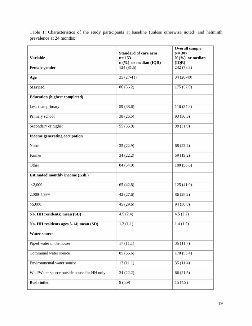
Patient characteristics: Overall summaries for 307 individuals from both RCT arms with both
PCR and microscopy at exit are provided in table 1, and were used for sensitivity and NPV
estimates. Participants had a median age of 34 years (IQR: 28-40) and were predominantly
7
women (78.8%). Just over half were married (57.0%), and nearly 40% of participants had less
than a primary education (37.8%). Less than a quarter (22.2%) was unemployed and 19.2% listed
farming as their primary occupation. Income levels among the cohort were generally low; 41.0%
of subjects reported monthly household incomes of less than 2,000 Kenyan shillings ($23.50),
and only 30.8% reported monthly household incomes over 5,000 shillings ($58.75). On average,
households contained 4.5 members, with 1.4 members between the age of 5 and 14. The majority
of households (55.4%) utilized a communal water source, and 4.9% reported using bush toilets
(the environment) rather than latrines (89.2%) or flush toilets (5.9%). These variables were
obtained at enrollment in the RCT two years prior to the time of the stool evaluation and were
assumed to remain fairly stable. At the time of the stool helminth study (last visit of the RCT),
median CD4 counts were 356.2 cells/mm3.
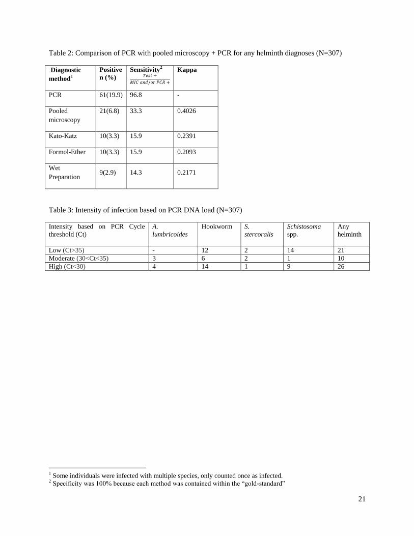
Prevalence and infection intensity: The prevalence of helminth infection in the overall sample of
307 participants was 6.8% as detected using microscopy and 19.9% by PCR. Among the
untreated standard of care group, infection prevalence was considerably higher (13.1% by
microscopy and 36.0% by PCR). PCR was more sensitive (96.8%) than microscopy (33.3%)
(p<0.0001). The NPV of PCR was significantly higher (99.2%) than microscopy (85.3%)
(p<0.0001). Comparing PCR and pooled microscopy, Cohen’s Kappa statistic was 0.40 (Table
2).
Table 3 summarizes the helminth infection intensity classified by PCR. In the overall sample
there were 28 low-intensity infections, 12 moderate intensity infections and 28 high intensity
infections. 7 hookworm infected individuals were also infected with Schistosoma (6) or S.
stercoralis (1). Infection intensity was positively associated with diagnosis by microscopy; a 1 Ct
difference in PCR was associated with 24% higher odds of microscopy detection (1.16, 1.32).
8
This association remained significant, in analyses restricted to the standard of care arm. Over
half (57%) of high intensity infections were detected by microscopy, compared to only 9.8% of
low intensity infections. The likelihood of being microscopy-positive for an individual of low,
medium, or high intensity infection was 12, 22.65, and 60.95 times that of an individual who was
considered uninfected by PCR, with a significant trend test (p<0.001).
Risk factor analysis: Because those in the treatment arm received comprehensive deworming
over a 2-year period, analyses of risk factors and outcomes was restricted to patients in the
standard of care arm who had not antihelminthic agents during the past year (n=153). Baseline
characteristics of individuals in the standard of care arm are summarized in and did not differ
considerably from the overall sample unless otherwise noted (table 1). Table 4 summarizes risk
factors for helminth infection identified by diagnosis with PCR compared to risk factors
identified by microscopy. Using microscopy, no significant associations between helminth
infection and the examined risk factors were detected in the cohort. However, using PCR yielded
significant associations between helminth infection and farming occupation (RR 1.57, 95% CI:
1.02, 2.40), piped water source (RR 0.26, 95% CI: 0.07, 0.99), and with completion of primary
education (RR 0.65, 95% CI: 0.43, 0.99).
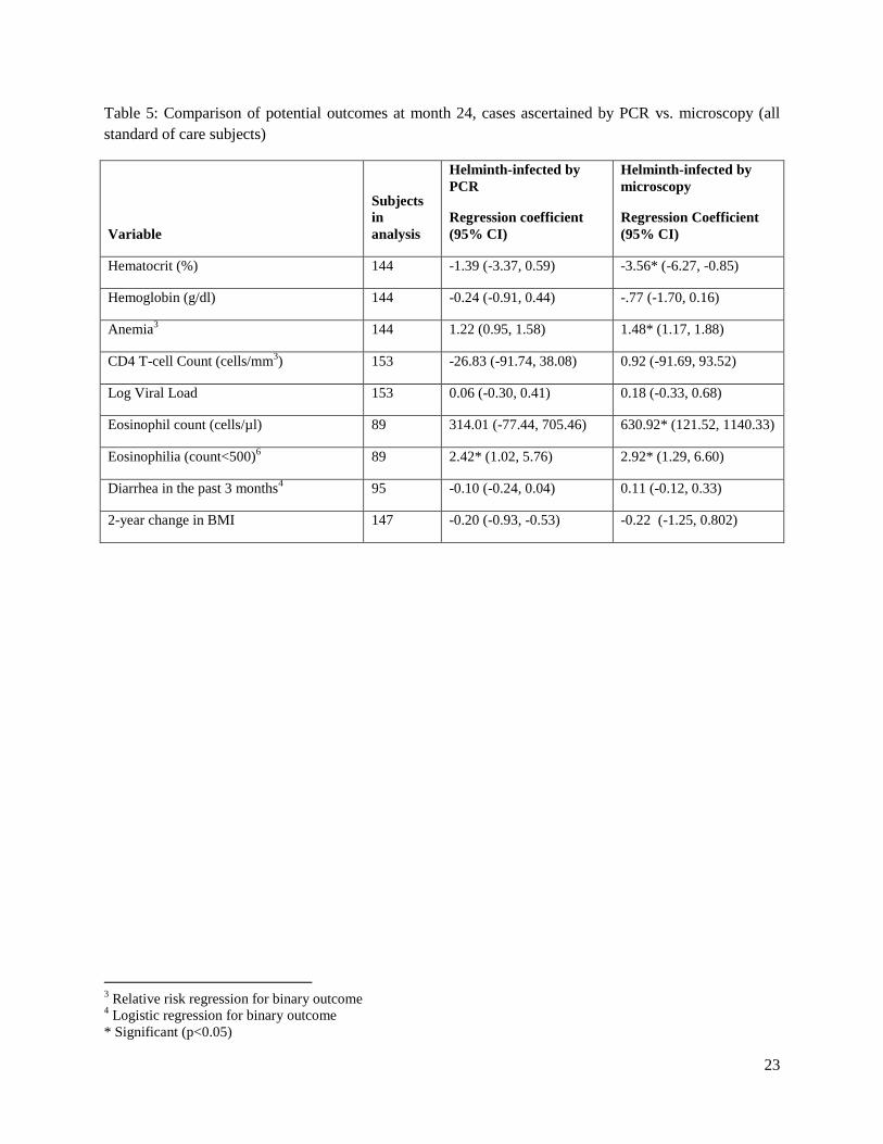
Outcome analysis: Table 5 summarizes the analysis of potential clinical and laboratory outcomes
from helminth infection, comparing associations in analyses using helminth diagnosis based on
PCR to analyses using microscopy. In analyses comparing individuals with microscopy-
diagnosed infections versus without microscopy-detected infection, those with infection had
significantly lower hematocrit levels (Mean difference of -3.56, 95% CI: -6.27, -0.85), higher
eosinophil counts (mean difference of 630.9 cells/µl, 95% CI: 121.5, 1140.3), and 48% higher
risk of anemia (RR=1.48, 95% CI: 1.17, 1.88). In contrast, these associations were not observed

9
in analyses comparing individuals with PCR-detected infections versus without. However,
significant associations between helminth infection and eosinophilia (>500 cells/μl) were
detected in analyses using PCR-detection (RR=2.50, 95% CI: 1.06, 5.92) and in those using
microscopy-detection (RR=2.88, 95% CI: 1.28, 6.51). Inclusion of ART use as a covariate did
not alter the findings.
Helminth intensity and outcomes: While the association between infection intensity and
eosinophil count was not significant (18.49 cells/µl per 1 unit increase, 95% CI: -1.01, 37.99),
the risk of eosinophilia was associated with infection intensity using both continuous (RR for 1
unit increase= 1.061, 95% CI: 1.017, 1.106) and categorical methods. The risk of eosinophilia in
the group with high-intensity infections was 3.4 times that in the uninfected group (RR=3.41,
95% CI: 1.39, 8.37).
Infection intensity and hematocrit were not significantly associated (-.08 per 1 unit increase, 95%
CI: -0.18, 0.03, p=0.141), there were trends for significance in the association between infection
intensity and risk of anemia using both continuous (RR for 1 unit increase= 1.01, 95% CI: 1.00,
1.02; p=0.083) and categorical methods. The risk of anemia was 29% higher among individuals
with high-intensity infections compared to uninfected individuals (RR=1.29, 95% CI: 0.96,
1.74).
DISCUSSION
In this study, we found that PCR helminth detection methods markedly increased detection of
helminth infections when compared with traditional microscopy in a cohort of HIV-infected
adults. The increased sensitivity of the PCR assay resulted in greater power to detect several risk
factors significantly associated with helminth infection in the cohort which were not detected in
10
analyses of the same cohort using stool microscopy. In contrast, less sensitive stool microscopy,
which preferentially detected higher intensity infections, detected infections associated with
clinically relevant outcomes. PCR-detected infections were not associated with as many
significant clinical outcomes, likely because the PCR assay detected many low-burden infections
with minimal clinical impact. Overall, our study illustrates several potential benefits and
limitations of each of these helminth detection methods.
Our findings regarding relative benefits of the diagnostic methods, though derived from an HIV-
positive cohort, likely extend to general adult populations in other settings. Past studies have
shown that correlates of helminth infection in HIV-positive populations are consistent with those
observed in the general population[22,25]. As patients with low baseline CD4 counts were
excluded from the study, it is unlikely that immune responses to helminth infection differed
markedly in this study from that of HIV-negative populations.
The observed differences in the sensitivity of PCR assays and microscopy are consistent with
those described previously in other cohorts[9,11,43,44]. The observed sensitivities for individual
microscopy methods were similar to the sensitivity of 17.7% observed for a single Kato-Katz test
in comparison to repeated collection and detection for detection of S. mansoni plus
hookworms[6]. As the NPV of microscopy was 85%, a negative microscopy results does not rule
out helminth infection as well as PCR(99.2%) in this setting. Past studies have found that Ct
value and egg counts derived from microscopy methods are correlated[9,11], which supports our
observation of an association between PCR Ct value and detection by microscopy.
Infections detected by PCR were associated known risk factors for helminth infection;
agricultural occupation[22,27], lack of piped water[22], and decreased education[15,19,21,22].
11
Given that detection by microscopy was not associated with these risk factors, microscopy likely
missed many infections, resulting in inadequate power to observe these associations. In previous
larger cohorts, microscopy did detect risk factors similar to the ones we detected with PCR
assays. However, the use of PCR in this study enabled the detection of risk factors using a much
smaller sample size, suggesting that these more sensitive diagnostics improve the ability to
identify risk factors.
While PCR and microscopy-identified infections were both associated with elevated risk of
eosinophilia, the greater risk increase of anemia and eosinophilia observed in cases diagnosed by
microscopy suggests that these higher intensity cases may be associated with higher levels of
hematologic and immune dysregulation. This was supported by the observed relationship
between the risk of eosinophilia and intensity of infection, as the risk of eosinophilia in
individuals with high-intensity infection was 3.4 times that of uninfected individuals(RR=3.41,
95% CI: 1.39, 8.37). The significantly lower hematocrit levels observed in subjects with
microscopy-diagnosed infections is consistent with previous studies documenting associations
between hookworm infection and anemia[4,33]. However, we did not detect an association
between helminth infection as determined by PCR and hematocrit. This is consistent with data
from another recent study using PCR, which did not find associations between low-intensity
hookworm infection and hematocrit or iron status[35]. While we did detect a trend towards a
significant association between infection intensity and risk of anemia using both continuous and
categorical methods, our sample may have been underpowered for this analysis.
Strengths of this study include the multi-site sample and the rigorous lab and specimen handling
practices. It is the first report to compare the risk factors and potential outcomes of helminth
infection using the two different diagnostic methods. It is also the first study to assess
12
performance of PCR for helminth infection among HIV-positive adults. Our PCR methods
included an internal control to determine efficiency of the PCR and detect inhibition of nucleic
acid replication in the sample which has been reported previously[45]. While the study was
conducted at two sites in Kenya and included populations with a range of socio-demographic and
other characteristics, there are several features of this population that affect the potential external
generalizability. The study only included HIV infected adults and was conducted in an area of
moderate to low baseline helminth prevalence. In addition, the two study sites were
geographically similar. The helminth infection burdens described by PCR were bi-modally
distributed, which was unusual in that helminth infections are generally overdispersed, with most
individuals harboring a small number of parasites and a small proportion carry the bulk of the
parasites[46,47]. The pattern observed may suggest that while there is high correlation between
PCR and egg count from microscopy, it is not a perfect measure of infection intensity. Infection
status was not gathered at enrollment, limiting the use of baseline laboratory measurements to
control for intra-individual variation. There is high variability in day-to-day shedding of eggs and
a multi-day stool sampling scheme could have considerably increased the number of
microscopy-identified cases, as has been shown in low-intensity settings[6]. Triplicate Kato-Katz
is the standard test for helminth diagnosis[48]; therefore our use of Cohen’s Kappa to compare
each microscopy method to PCR may be of limited value. As few infections were identified in
the treatment arm, the correlates analysis was restricted to the standard of care arm; which
decreased sample size and decreased our power to detect associations. Though species-specific
correlates analyses would have provided interesting additional data, the relatively small sample
size limited the power of this study for these analyses. Finally, the hematocrit and hemoglobin

13
cutoffs utilized for anemia are based on a population distribution[41] which may differ from that
of Kenya; therefore the cutoffs may not be optimal in our study population.
In conclusion, our findings suggest that while helminth infections (particularly hookworm and
Schistosoma spp.) may be underdiagnosed by microscopy methods, the use of the more sensitive
PCR diagnostic test may lead to the detection of more low-intensity infections; which may be
less clinically relevant. PCR can quickly and accurately diagnose helminth infections and may be
useful to discern risk factors and transmission epidemiology. However, in resource-limited
settings, the increased diagnostic sensitivity provided by PCR may capture many infections
which pose little health risk and which may not contribute substantially to transmission. Our
study suggests that interpretation of studies on risk factors and outcomes of helminth infection
should note the marked impact of the method used to detect helminths.
14
REFERENCES
1. de Silva NR, Brooker S, Hotez PJ, Montresor A, Engels D, et al. (2003) Soil-transmitted
helminth infections: updating the global picture. Trends Parasitol 19: 547-551.
2. Bethony J, Brooker S, Albonico M, Geiger SM, Loukas A, et al. (2006) Soil-transmitted
helminth infections: ascariasis, trichuriasis, and hookworm. Lancet 367: 1521-1532.
3. Mahalanabis D, Jalan KN, Maitra TK, Agarwal SK (1976) Vitamin A absorption in ascariasis.
Am J Clin Nutr 29: 1372-1375.
4. Stoltzfus RJ, Dreyfuss ML, Chwaya HM, Albonico M (1997) Hookworm control as a strategy
to prevent iron deficiency. Nutr Rev 55: 223-232.
5. Brown M, Bukusuba J, Hughes P, Nakiyingi J, Watera C, et al. (2003) Screening for intestinal
helminth infestation in a semi-urban cohort of HIV-infected people in Uganda: a
combination of techniques may enhance diagnostic yield in the absence of multiple stool
samples. Trop Doct 33: 72-76.
6. Booth M, Vounatsou P, N'goran EK, Tanner M, Utzinger J (2003) The influence of sampling
effort and the performance of the Kato-Katz technique in diagnosing Schistosoma
mansoni and hookworm co-infections in rural Côte d'Ivoire. Parasitology 127: 525-531.
7. Ebrahim A, El-Morshedy H, Omer E, El-Daly S, Barakat R (1997) Evaluation of the Kato-
Katz thick smear and formol ether sedimentation techniques for quantitative diagnosis of
Schistosoma mansoni infection. Am J Trop Med Hyg 57: 706-708.
8. Knopp S, Mgeni AF, Khamis IS, Steinmann P, Stothard JR, et al. (2008) Diagnosis of soil-
transmitted helminths in the era of preventive chemotherapy: effect of multiple stool
sampling and use of different diagnostic techniques. PLoS Negl Trop Dis 2: e331.
9. ten Hove RJ, Verweij JJ, Vereecken K, Polman K, Dieye L, et al. (2008) Multiplex real-time
PCR for the detection and quantification of Schistosoma mansoni and S. haematobium
infection in stool samples collected in northern Senegal. Trans R Soc Trop Med Hyg 102:
179-185.
10. Verweij JJ, Blange RA, Templeton K, Schinkel J, Brienen EAT, et al. (2004) Simultaneous
Detection of Entamoeba histolytica, Giardia lamblia, and Cryptosporidium parvum in
Fecal Samples by Using Multiplex Real-Time PCR. Journal of Clinical Microbiology 42:
1220-1223.
11. Verweij JJ, Brienen EA, Ziem J, Yelifari L, Polderman AM, et al. (2007) Simultaneous
detection and quantification of Ancylostoma duodenale, Necator americanus, and
Oesophagostomum bifurcum in fecal samples using multiplex real-time PCR. Am J Trop
Med Hyg 77: 685-690.
12. Fincham JE, Markus MB, Adams VJ (2003) Could control of soil-transmitted helminthic
infection influence the HIV/AIDS pandemic. Acta Trop 86: 315-333.
13. Karanja DM, Colley DG, Nahlen BL, Ouma JH, Secor WE (1997) Studies on
schistosomiasis in western Kenya: I. Evidence for immune-facilitated excretion of
schistosome eggs from patients with Schistosoma mansoni and human immunodeficiency
virus coinfections. Am J Trop Med Hyg 56: 515-521.
14. Downs JA, Mguta C, Kaatano GM, Mitchell KB, Bang H, et al. (2011) Urogenital
schistosomiasis in women of reproductive age in Tanzania's Lake Victoria region. Am J
Trop Med Hyg 84: 364-369.
15. Khalid A, Abdelgadir MA, Ashmaig A, Ibrahim AM, Ahmed AA, et al. (2012) Schistosoma
mansoni infection among prenatal attendees at a secondary-care hospital in central Sudan.
Int J Gynaecol Obstet 116: 10-12.
15
16. Knopp S, Mohammed KA, Stothard JR, Khamis IS, Rollinson D, et al. (2010) Patterns and
risk factors of helminthiasis and anemia in a rural and a peri-urban community in
Zanzibar, in the context of helminth control programs. PLoS Negl Trop Dis 4: e681.
17. Sarkinfada F, Oyebanji AA, Sadiq IA, Ilyasu Z (2009) Urinary schistosomiasis in the
Danjarima community in Kano, Nigeria. J Infect Dev Ctries 3: 452-457.
18. van Eijk AM, Hill J, Alegana VA, Kirui V, Gething PW, et al. (2011) Coverage of malaria
protection in pregnant women in sub-Saharan Africa: a synthesis and analysis of national
survey data. Lancet Infect Dis 11: 190-207.
19. Woodburn PW, Muhangi L, Hillier S, Ndibazza J, Namujju PB, et al. (2009) Risk factors for
helminth, malaria, and HIV infection in pregnancy in Entebbe, Uganda. PLoS Negl Trop
Dis 3: e473.
20. Ndassa A, Mimpfoundi R, Gake B, Paul Martin MV, Poste B (2007) Risk factors for human
schistosomiasis in the Upper Benue valley, in northern Cameroon. Ann Trop Med
Parasitol 101: 469-477.
21. Olsen A, Samuelsen H, Onyango-Ouma W (2001) A study of risk factors for intestinal
helminth infections using epidemiological and anthropological approaches. J Biosoc Sci
33: 569-584.
22. Walson JL, Stewart BT, Sangaré L, Mbogo LW, Otieno PA, et al. (2010) Prevalence and
correlates of helminth co-infection in Kenyan HIV-1 infected adults. PLoS Negl Trop Dis
4: e644.
23. Chu TB, Liao CW, D'Lamini P, Chang PW, Chiu WT, et al. (2010) Prevalence of
Schistosoma haematobium infection among inhabitants of Lowveld, Swaziland, an
endemic area for the disease. Trop Biomed 27: 337-342.
24. Belyhun Y, Medhin G, Amberbir A, Erko B, Hanlon C, et al. (2010) Prevalence and risk
factors for soil-transmitted helminth infection in mothers and their infants in Butajira,
Ethiopia: a population based study. BMC Public Health 10: 21.
25. Modjarrad K, Zulu I, Redden DT, Njobvu L, Freedman DO, et al. (2005) Prevalence and
predictors of intestinal helminth infections among human immunodeficiency virus type 1-
infected adults in an urban African setting. Am J Trop Med Hyg 73: 777-782.
26. Pinot de Moira A, Fulford AJ, Kabatereine NB, Ouma JH, Booth M, et al. (2010) Analysis of
complex patterns of human exposure and immunity to Schistosomiasis mansoni: the
influence of age, sex, ethnicity and IgE. PLoS Negl Trop Dis 4.
27. Humphries D, Mosites E, Otchere J, Twum WA, Woo L, et al. (2011) Epidemiology of
hookworm infection in Kintampo North Municipality, Ghana: patterns of malaria
coinfection, anemia, and albendazole treatment failure. Am J Trop Med Hyg 84: 792-800.
28. Stothard JR, Imison E, French MD, Sousa-Figueiredo JC, Khamis IS, et al. (2008) Soil-
transmitted helminthiasis among mothers and their pre-school children on Unguja Island,
Zanzibar with emphasis upon ascariasis. Parasitology 135: 1447-1455.
29. Muhumuza S, Kitimbo G, Oryema-Lalobo M, Nuwaha F (2009) Association between socio
economic status and schistosomiasis infection in Jinja District, Uganda. Trop Med Int
Health 14: 612-619.
30. Brooks RM, Latham MC, Crompton DW (1979) The relationship of nutrition and health to
worker productivity in Kenya. East Afr Med J 56: 413-421.
31. Ndamba J, Makaza N, Munjoma M, Gomo E, Kaondera KC (1993) The physical fitness and
work performance of agricultural workers infected with Schistosoma mansoni in
Zimbabwe. Ann Trop Med Parasitol 87: 553-561.

16
32. Stoltzfus RJ (2001) Iron-deficiency anemia: reexamining the nature and magnitude of the
public health problem. Summary: implications for research and programs. J Nutr 131:
697S-700S; discussion 700S-701S.
33. Smith JL, Brooker S (2010) Impact of hookworm infection and deworming on anaemia in
non-pregnant populations: a systematic review. Trop Med Int Health 15: 776-795.
34. Brooker S, Hotez PJ, Bundy DA (2008) Hookworm-related anaemia among pregnant
women: a systematic review. PLoS Negl Trop Dis 2: e291.
35. Jonker FA, Calis JC, Phiri K, Brienen EA, Khoffi H, et al. (2012) Real-time PCR
demonstrates Ancylostoma duodenale is a key factor in the etiology of severe anemia and
iron deficiency in Malawian pre-school children. PLoS Negl Trop Dis 6: e1555.
36. Friedman JF, Kanzaria HK, McGarvey ST (2005) Human schistosomiasis and anemia: the
relationship and potential mechanisms. Trends Parasitol 21: 386-392.
37. Walson J, Singa B, Sangaré L, Naulikha J, Piper B, et al. (2012) Empiric deworming to delay
HIV disease progression in adults with HIV who are ineligible for initiation of
antiretroviral treatment (the HEAT study): a multi-site, randomised trial. Lancet Infect
Dis.
38. Basuni M, Muhi J, Othman N, Verweij JJ, Ahmad M, et al. (2011) A pentaplex real-time
polymerase chain reaction assay for detection of four species of soil-transmitted
helminths. Am J Trop Med Hyg 84: 338-343.
39. Niesters HG (2002) Clinical virology in real time. J Clin Virol 25 Suppl 3: S3-12.
40. Verweij JJ (2012) PCR questions: Internal control, infection intensity, sequence choice. In:
Arndt MA, editor.
41. Schrier S (2012) Approach to the adult patient with anemia. pp. UpToDate.
42. Tefferi A (2005) Blood eosinophilia: a new paradigm in disease classification, diagnosis, and
treatment. Mayo Clin Proc 80: 75-83.
43. Pontes LA, Dias-Neto E, Rabello A (2002) Detection by polymerase chain reaction of
Schistosoma mansoni DNA in human serum and feces. Am J Trop Med Hyg 66: 157-
162.
44. Pontes LA, Oliveira MC, Katz N, Dias-Neto E, Rabello A (2003) Comparison of a
polymerase chain reaction and the Kato-Katz technique for diagnosing infection with
Schistosoma mansoni. Am J Trop Med Hyg 68: 652-656.
45. Monteiro L, Bonnemaison D, Vekris A, Petry KG, Bonnet J, et al. (1997) Complex
polysaccharides as PCR inhibitors in feces: Helicobacter pylori model. J Clin Microbiol
35: 995-998.
46. Guyatt HL, Bundy DA, Medley GF, Grenfell BT (1990) The relationship between the
frequency distribution of Ascaris lumbricoides and the prevalence and intensity of
infection in human communities. Parasitology 101 Pt 1: 139-143.
47. Croll NA, Ghadirian E (1981) Wormy persons: contributions to the nature and patterns of
overdispersion with Ascaris lumbricoides, Ancylosotma duodenale, Necator americanus
and Trichuris trichiura. Trop Geogr Med 33: 241-248.
48. Sturrock RF (1998) Guidelines for the evaluation of soil-transmitted helminthiasis and
schistosomiasis at community level: A guide for managers of control programmes: A.
Montresor, D. W. T. Crompton, D. A. P. Bundy, A. Hall & L. Savioli. Geneva: World
Health Organization, 1998. iv+46pp. Transactions of the Royal Society of Tropical
Medicine and Hygiene 92: 470-471.
17
49. Dada-Adegbola HO, Oluwatoba OA, Bakare RA (2010) Strongyloidiasis: prevalence, risk
factors, clinical and laboratory features among diarrhea patients in Ibadan Nigeria. Afr J
Med Med Sci 39: 285-292.
50. Ugbomoiko US, Ofoezie IE, Okoye IC, Heukelbach J (2010) Factors associated with urinary
schistosomiasis in two peri-urban communities in south-western Nigeria. Ann Trop Med
Parasitol 104: 409-419.
51. Garba A, Pion S, Cournil A, Milet J, Schneider D, et al. (2010) Risk factors for Schistosoma
haematobium infection and morbidity in two villages with different transmission patterns
in Niger. Acta Trop 115: 84-89.
52. van Eijk AM, Lindblade KA, Odhiambo F, Peterson E, Rosen DH, et al. (2009) Geohelminth
Infections among pregnant women in rural western Kenya; a cross-sectional study. PLoS
Negl Trop Dis 3: e370.
53. Kawai K, Saathoff E, Antelman G, Msamanga G, Fawzi WW (2009) Geophagy (Soil-eating)
in relation to Anemia and Helminth infection among HIV-infected pregnant women in
Tanzania. Am J Trop Med Hyg 80: 36-43.

18
Figure 1. Study Flow Chart
19
Table 1: Characteristics of the study participants at baseline (unless otherwise noted) and helminth
prevalence at 24 months:
Variable
Standard of care arm
n= 153
n (%) or median (IQR)
Overall sample
N= 307
N (%) or median
(IQR)
Female gender 124 (81.5) 242 (78.8)
Age 35 (27-41) 34 (28-40)
Married 86 (56.2) 175 (57.0)
Education (highest completed)
Less than primary 59 (38.6) 116 (37.8)
Primary school 38 (25.5) 93 (30.3)
Secondary or higher 55 (35.9) 98 (31.9)
Income generating occupation
None 35 (22.9) 68 (22.2)
Farmer 34 (22.2) 59 (19.2)
Other 84 (54.9) 180 (58.6)
Estimated monthly income (Ksh.)
<2,000 65 (42.8) 125 (41.0)
2,000-4,999 42 (27.6) 86 (28.2)
>5,000 45 (29.6) 94 (30.8)
No. HH residents; mean (SD) 4.5 (2.4) 4.5 (2.2)
No. HH residents ages 5-14; mean (SD) 1.3 (1.1) 1.4 (1.2)
Water source
Piped water in the house 17 (11.1) 36 (11.7)
Communal water source 85 (55.6) 170 (55.4)
Environmental water source 17 (11.1) 35 (11.4)
Well/Water source outside house for HH only 34 (22.2) 66 (21.5)
Bush toilet 9 (5.9) 15 (4.9)

20
M0 CD4 T-cell Count (cells/mm3) 409.9 (321.5-538.7) 421.9 (308.5-558.6)
M24 CD4 T-cell Count (cells/mm3) 365.2 (267.8-489.5) 373.5 (270.6-511.9)
Prevalence of helminth infection
By microscopy 20 (13.1) 21 (6.8)
By PCR 55 (36.0) 61 (19.9)
21
Table 2: Comparison of PCR with pooled microscopy + PCR for any helminth diagnoses (N=307)
Diagnostic
method1
Positive
n (%)
Sensitivity2
Kappa
PCR 61(19.9) 96.8 -
Pooled
microscopy
21(6.8) 33.3 0.4026
Kato-Katz 10(3.3) 15.9 0.2391
Formol-Ether 10(3.3) 15.9 0.2093
Wet
Preparation 9(2.9) 14.3 0.2171
Table 3: Intensity of infection based on PCR DNA load (N=307)
Intensity based on PCR Cycle
threshold (Ct)
A.
lumbricoides
Hookworm
S.
stercoralis
Schistosoma
spp.
Any
helminth
Low (Ct>35) - 12 2 14 21
Moderate (30<Ct<35) 3 6 2 1 10
High (Ct<30) 4 14 1 9 26
1 Some individuals were infected with multiple species, only counted once as infected.
2 Specificity was 100% because each method was contained within the “gold-standard”

22
Table 4: Comparison of risk factors, cases ascertained by PCR vs. microscopy
Category Variable
Subjects in
analysis
Risk of helminth
infection by PCR
RR (95% CI)
Risk of helminth
infection by
microscopy
RR (95% CI)
Age (continuous, per 10
year increase) 153 0.83 (0.66, 1.05) 0.63 (0.38, 1.04)
Completed primary
education 153 0.65* (0.43, 0.99) 0.41 (0.23, 1.16)
Farming occupation 153 1.57* (1.02, 2.40) 1.88 (0.82, 4.35)
Water
source
Communal water source 85 1.00 1.00
Environmental water
source 17 0.53 (0.22, 1.28) 1.15 (0.37, 3.62)
Piped water in the house 17 0.26* (0.07, 0.99) ----
Well/Water source outside
house for HH only 34 0.72 (0.42, 1.24) 0.77 (0.27, 2.19)
Bush toilet use 153 0.92 (0.36, 2.38) -----
Baseline CD4
count<median 153 1.27 (0.83, 1.96) 0.99 (0.44, 2.23)
HH with at least 1 child
between ages 5-14 153 1.11 (0.70, 1.78) 1.07 (0.44, 2.61)
HH with at least 1 child
under age 5 153 1.19 (0.78, 1.82) 1.30 (0.57, 2.97)
23
Table 5: Comparison of potential outcomes at month 24, cases ascertained by PCR vs. microscopy (all
standard of care subjects)
3 Relative risk regression for binary outcome
4 Logistic regression for binary outcome
* Significant (p<0.05)
Variable
Subjects
in
analysis
Helminth-infected by
PCR
Regression coefficient
(95% CI)
Helminth-infected by
microscopy
Regression Coefficient
(95% CI)
Hematocrit (%) 144 -1.39 (-3.37, 0.59) -3.56* (-6.27, -0.85)
Hemoglobin (g/dl) 144 -0.24 (-0.91, 0.44) -.77 (-1.70, 0.16)
Anemia3 144 1.22 (0.95, 1.58) 1.48* (1.17, 1.88)
CD4 T-cell Count (cells/mm3) 153 -26.83 (-91.74, 38.08) 0.92 (-91.69, 93.52)
Log Viral Load 153 0.06 (-0.30, 0.41) 0.18 (-0.33, 0.68)
Eosinophil count (cells/µl) 89 314.01 (-77.44, 705.46) 630.92* (121.52, 1140.33)
Eosinophilia (count<500)6
89 2.42* (1.02, 5.76) 2.92* (1.29, 6.60)
Diarrhea in the past 3 months4 95 -0.10 (-0.24, 0.04) 0.11 (-0.12, 0.33)
2-year change in BMI 147 -0.20 (-0.93, -0.53) -0.22 (-1.25, 0.802)

24
Appendix 1: Species-specific oligonucleotide PCR targets and GenBank accession numbers[9,38]
Organism
Gene
target Oligonucleotide name
GenBank
accession number
S. mansoni COX1
Smcyt748F
NC 002545 Smcyt847R
Smcyt785T
S. haematobium COX1
Sh307F
AY157209 Sh447R
Shaem377MGB
A. lumbricoides ITS1
Alum96F
AJ000895 Alum183R
Alum124T
S. stercoralis 18S
Stro18S-1530F
AF279916 Stro18S-1630R
Stro18S-1586T
A. duodenale ITS2
Ad125F
AJ001594 Ad195R
Ad155MGB
N. americanus ITS2
Na58F
AJ001599 Na158R
Na81MGB

25
Appendix 2: Risk factors for helminth infection among adults (or mixed ages)
Study Population Association
OR or RR (95% CI)
Method of
diagnosis
HIV
status
[15] 292 pregnant women at a
hospital in Geizera state, Sudan S. mansoni:
Maternal age less than 20 years OR= 9.8 (1.5, 16.3)
No education OR= 6.2 (2.8, 12.9)
Formol-ether and
duplicate Kato-Katz
slide examination
Not
reported
[27] 132 adults (age>15) from 62
households (HH) in Kintampo
North, Ghana
Hookworm:
Poor nutritional status (BMI≤23) OR= 0.7 (0.60, 0.85)
Not using a latrine OR= 6.10 (2.09, 17.54)
Not wearing shoes OR= 4.06 (1.57, 10.53)
Occupation (farming) OR= 4.89 (1.90, 12.61)
Single Kato-Katz
slide examination
Not
reported
[14] 457 women living in eight rural
villages in northwest Tanzania S. haematobium:
HIV OR= 4.0 (1.2–13.5)
Younger age
OR= 5.5 (1.2, 26.3 for ages < 25 yrs compared with 35+)
OR =8.2 (1.7, 38.4) for ages 25–29 yrs compared with age 35+)
Urine microscopy Mixed-
5.9%
positive
[23] Cross-sectional study of 295
villagers in Lowveld,
Swaziland.
S. haematobium:
Female gender: OR = 8.2 (1.8, 36.2)
Younger age
OR= 0.1 (0.02, 1.09) for ages 13-18 compared with age <5
OR= 0.2 (0.04, 0.57) for ages 19+ compared with age <5
Urine microscopy Not
reported
[49] Hospital-based cross-sectional
study of 1,090 diarrhea patients
from 5 hospitals in Ibadan,
Nigeria. 65.1% were adults
S. stercoralis:
Open field or bush for toilet OR= 2.83 (1.40, 5.76) (calculated from 2x2
in table 2)
Formol-ether
concentration
method, Wet-
preparation
concentration
method
Mixed-
5.2%
positive
[26] 290 villagers (age 5-66) from
Booma, Uganda S. mansoni:
Male gender OR= 2.17 (1.14, 4.13)
Water contact exposure OR= 4.19 (2.50, 7.04) (per SD increase in
exposure)
Cercarial exposure OR= 4.80 (2.89, 7.95) (per SD increase in exposure)
Duplicate Kato-Katz
slide examination
Not
reported

26
[50] Population-based cross-
sectional study of 1023
villagers in Osun State, Nigeria.
Ages 2-78, with median age of
12.
S. haematobium:
Family income of <$500/month OR= 3.73 (2.66, 5.21)
Number of children aged 10-15 in household OR= 1.6 (1.24, 2.06)
Literate family head OR= 0.28 (0.19, 0.42)
HH close to river OR= 1.38 (1.00, 1.89)
Being single OR= 4.27 (2.43, 7.50)
Not living with biological parents OR= 1.93 (1.29, 2.88)
Urine microscopy Not
reported
[16] 454 villagers from a
community-based cross-
sectional study in Zanzibar, TZ
A. lumbricoides:
Male OR= 1.94 (1.03, 3.65)
Age OR= 0.98 (0.96, 0.99)
Eating raw vegetables or salad OR= 2.54 (1.27, 5.10)
Hookworm:
Recent travel history OR= 5.06 (1.21, 21.06)
Very poor OR= 0.11 (0.02, 0.58)
Least poor OR= 0.12 (0.04, 0.42)
Eating unpeeled fruits OR= 0.28 (0.11, 0.73)
Male OR= 2.25 (1.23, 4.12)
S. stercoralis:
Male OR= 4.11 (1.21, 13.90)
Washing hands after defection OR= 0.29 (0.09, 0.96)
Recent travel history OR= 5.43 (1.08, 27.27)
Age OR= 0.97 (0.94, 1.00)
S. haematobium:
Age OR= 0.97 (0.95, 1.00)
Kato-Katz slide
examination,
Baermann method,
and Koga agar plate
method.
Not
reported
[51] 500 villagers from two different
villages in Niger (Lossa and
Tara)
S. haematobium:
Age OR= 3.4 (2.1, 5.7) for moderate or heavy infection (versus no
infection for ages 7-15 compared with 2-6)
Urine microscopy Not
reported
[24] 908 mothers from Butajira,
Ethiopia, 1 year after giving
birth
Pooled:
Infrequent use of soap by the mother OR= 1.40 (1.04-1.88) OR= 1.66
(0.92-2.99) for use at least once a week and less frequent than once a
week respectively relative to daily use; p for trend =0.018)
Urban place of residence OR= 0.45 (0.28-0.73)
Formol-ether
concentration
method.
Not
reported
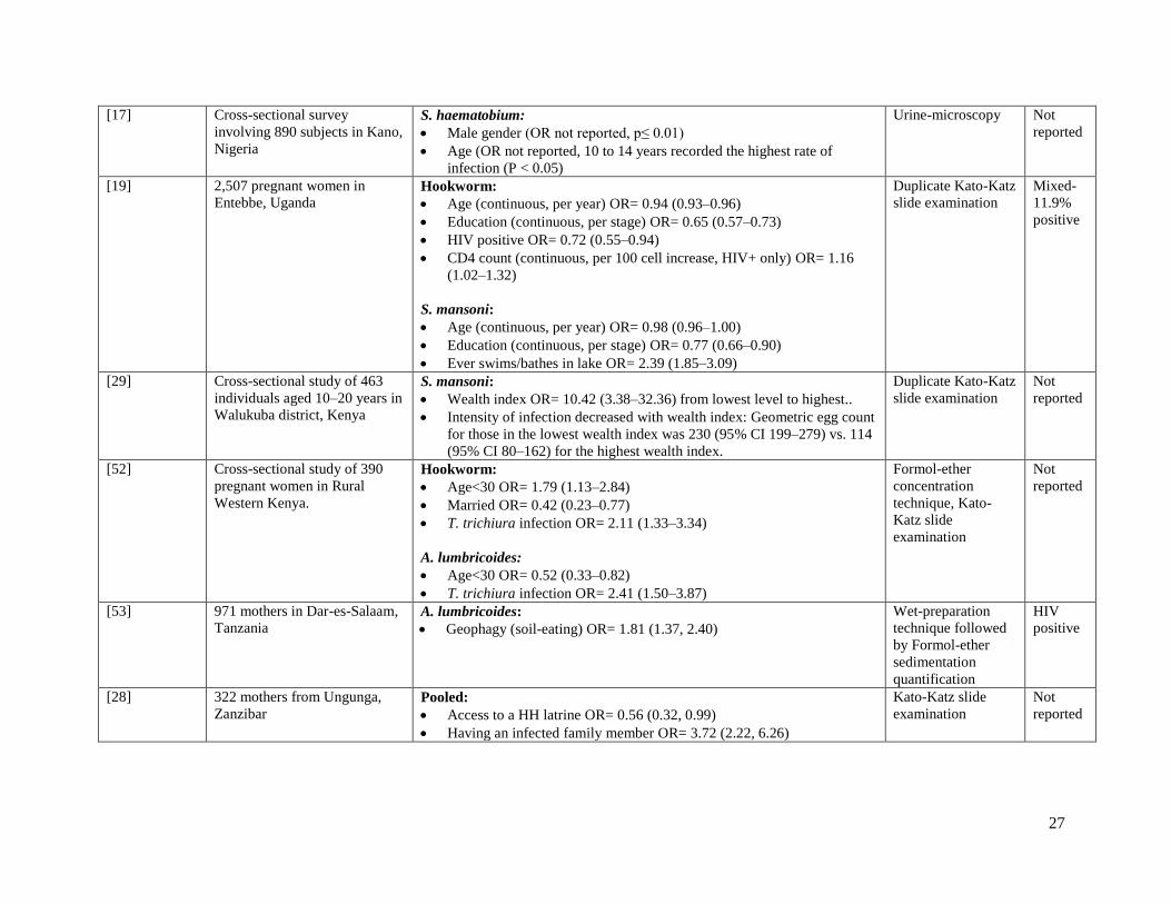
27
[17] Cross-sectional survey
involving 890 subjects in Kano,
Nigeria
S. haematobium:
Male gender (OR not reported, p≤ 0.01)
Age (OR not reported, 10 to 14 years recorded the highest rate of
infection (P < 0.05)
Urine-microscopy Not
reported
[19] 2,507 pregnant women in
Entebbe, Uganda Hookworm:
Age (continuous, per year) OR= 0.94 (0.93–0.96)
Education (continuous, per stage) OR= 0.65 (0.57–0.73)
HIV positive OR= 0.72 (0.55–0.94)
CD4 count (continuous, per 100 cell increase, HIV+ only) OR= 1.16
(1.02–1.32)
S. mansoni:
Age (continuous, per year) OR= 0.98 (0.96–1.00)
Education (continuous, per stage) OR= 0.77 (0.66–0.90)
Ever swims/bathes in lake OR= 2.39 (1.85–3.09)
Duplicate Kato-Katz
slide examination
Mixed-
11.9%
positive
[29] Cross-sectional study of 463
individuals aged 10–20 years in
Walukuba district, Kenya
S. mansoni:
Wealth index OR= 10.42 (3.38–32.36) from lowest level to highest..
Intensity of infection decreased with wealth index: Geometric egg count
for those in the lowest wealth index was 230 (95% CI 199–279) vs. 114
(95% CI 80–162) for the highest wealth index.
Duplicate Kato-Katz
slide examination
Not
reported
[52] Cross-sectional study of 390
pregnant women in Rural
Western Kenya.
Hookworm:
Age<30 OR= 1.79 (1.13–2.84)
Married OR= 0.42 (0.23–0.77)
T. trichiura infection OR= 2.11 (1.33–3.34)
A. lumbricoides:
Age<30 OR= 0.52 (0.33–0.82)
T. trichiura infection OR= 2.41 (1.50–3.87)
Formol-ether
concentration
technique, Kato-
Katz slide
examination
Not
reported
[53] 971 mothers in Dar-es-Salaam,
Tanzania A. lumbricoides:
Geophagy (soil-eating) OR= 1.81 (1.37, 2.40)
Wet-preparation
technique followed
by Formol-ether
sedimentation
quantification
HIV
positive
[28] 322 mothers from Ungunga,
Zanzibar Pooled:
Access to a HH latrine OR= 0.56 (0.32, 0.99)
Having an infected family member OR= 3.72 (2.22, 6.26)
Kato-Katz slide
examination
Not
reported
28
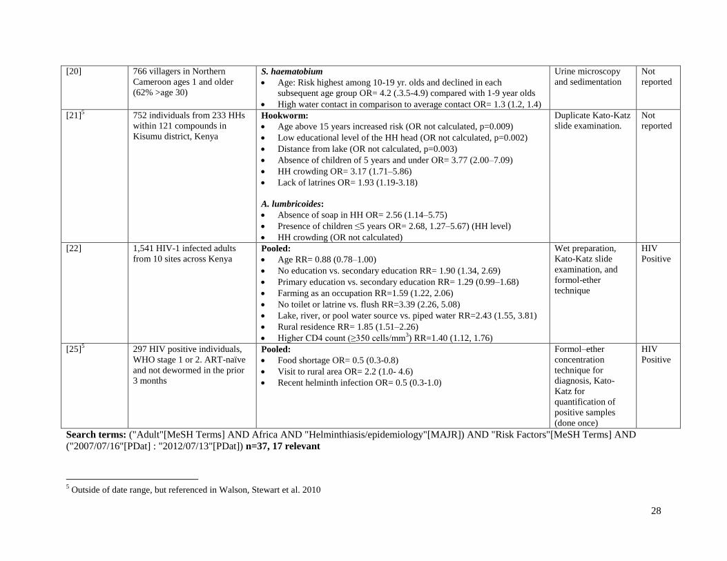
[20] 766 villagers in Northern
Cameroon ages 1 and older
(62% >age 30)
S. haematobium
Age: Risk highest among 10-19 yr. olds and declined in each
subsequent age group OR= 4.2 (.3.5-4.9) compared with 1-9 year olds
High water contact in comparison to average contact OR= 1.3 (1.2, 1.4)
Urine microscopy
and sedimentation
Not
reported
[21]5 752 individuals from 233 HHs
within 121 compounds in
Kisumu district, Kenya
Hookworm:
Age above 15 years increased risk (OR not calculated, p=0.009)
Low educational level of the HH head (OR not calculated, p=0.002)
Distance from lake (OR not calculated, p=0.003)
Absence of children of 5 years and under OR= 3.77 (2.00–7.09)
HH crowding OR= 3.17 (1.71–5.86)
Lack of latrines OR= 1.93 (1.19-3.18)
A. lumbricoides:
Absence of soap in HH OR= 2.56 (1.14–5.75)
Presence of children ≤5 years OR= 2.68, 1.27–5.67) (HH level)
HH crowding (OR not calculated)
Duplicate Kato-Katz
slide examination.
Not
reported
[22] 1,541 HIV-1 infected adults
from 10 sites across Kenya Pooled:
Age RR= 0.88 (0.78–1.00)
No education vs. secondary education RR= 1.90 (1.34, 2.69)
Primary education vs. secondary education RR= 1.29 (0.99–1.68)
Farming as an occupation RR=1.59 (1.22, 2.06)
No toilet or latrine vs. flush RR=3.39 (2.26, 5.08)
Lake, river, or pool water source vs. piped water RR=2.43 (1.55, 3.81)
Rural residence RR= 1.85 (1.51–2.26)
Higher CD4 count (≥350 cells/mm3) RR=1.40 (1.12, 1.76)
Wet preparation,
Kato-Katz slide
examination, and
formol-ether
technique
HIV
Positive
[25]5
297 HIV positive individuals,
WHO stage 1 or 2. ART-naïve
and not dewormed in the prior
3 months
Pooled:
Food shortage OR= 0.5 (0.3-0.8)
Visit to rural area OR= 2.2 (1.0- 4.6)
Recent helminth infection OR= 0.5 (0.3-1.0)
Formol–ether
concentration
technique for
diagnosis, Kato-
Katz for
quantification of
positive samples
(done once)
HIV
Positive
Search terms: ("Adult"[MeSH Terms] AND Africa AND "Helminthiasis/epidemiology"[MAJR]) AND "Risk Factors"[MeSH Terms] AND
("2007/07/16"[PDat] : "2012/07/13"[PDat]) n=37, 17 relevant
5 Outside of date range, but referenced in Walson, Stewart et al. 2010