impact of ultrasound in diagnosis of pediatric superficial
TRANSCRIPT

Original article
36
Impact of Ultrasound in Diagnosis of Pediatric Superficial Masses
in Head and Neck
Ahmed Farid, Ahmed Shalaan, Shirin Khedr
Abstract:
Background: Neck masses are frequently encountered in pediatric
medicine, and can present a diagnostic dilemma for the clinicians
involved. Aim: to determine the impact of ultrasound in diagnosis
of superficial masses in head and neck in pediatric. Methods: This
is a review article. The search was performed using MEDLINE,
Embase, Pubmed and CINAHL Plus in the same date range with
the following medical terms: ―Ultrasound; Pediatric; mass; head;
neck‖, including articles from 2000 to 2019. Results: In this
review, the US findings are described for a variety of common and
uncommon pediatric head and neck masses diagnosed in our
practice. Specifically, the entities covered include neonatal scalp
hematoma, craniosynostosis, dermoid and epidermoid cysts,
Langerhans cell histiocytosis, lymph nodes and their
complications, fibromatosis colli, thyroglossal duct cyst, branchial
cleft cyst, cervical thymus, congenital goiter, thyroid papillary
carcinoma, parathyroid adenoma, hemangioma, lymphangioma,
jugular vein phlebectasia, acute parotitis, leukemia and/or
lymphoma, neurogenic tumor, and rhabdomyosarcoma. Conclusion: Duplex US has emerged
as the first-line imaging modality for the evaluation of superficial pediatric masses. Without the
use of ionizing radiation, iodinated contrast material, or sedation and/or anesthesia, this safe,
quick, and cost-effective imaging modality allows rapid evaluation and characterization of
masses.
Keywords: Ultrasound; Pediatric; mass; head; neck
Department of Radiology,
Benha faculty of medicine,
Benha University, Egypt
Correspondence to: Shirin
Khedr, department of
Radiology, Benha faculty of
medicine, Benha University,
Egypt.
Email:
Received: 19 July 2020
Accepted: 15 August 2020

Benha Medical Journal, Vol. 37,Special issue (Radiology), 2020
36
Introduction
Superficial palpable masses of the head and
neck are common in the pediatric
population, with the vast majority of the
lesions ultimately proven to be benign (1).
Neck masses are frequently encountered in
pediatric medicine, and can present a
diagnostic dilemma for the clinicians
involved. There are several means by which
neck mass in children can be subdivided, for
example by age at presentation, anatomical
location including compartments and fascia
of the neck, their classical appearance when
imaged, or by etiology. When imaging
children the clinicians must be mindful of
radiation exposure and as such ultrasound
(US) is often attempted first (2).
A wide variety of scalp lesions are
identified as palpable masses or as
incidental findings on radiologic studies.
They represent a challenge, for that may
lead to diagnostic mistakes. Correct
interpretation of a scalp mass may lead to
reduced mortality and morbidity and may
guide physicians toward the most
appropriate management (3).
The US findings are described for a variety
of common and uncommon pediatric head
and neck masses diagnosed in our practice.
Specifically, neonatal scalp hematoma,
craniosynostosis, dermoid and epidermoid
cysts, Langerhans cell histiocytosis, lymph
nodes and their complications, fibromatosis
colli, thyroglossal duct cyst, branchial cleft
cyst, cervical thymus, congenital goiter,
thyroid papillary carcinoma, parathyroid
adenoma, hemangioma, lymphangioma,
jugular vein phlebectasia, Lemierre
syndrome, acute parotitis and parotid
abscess, leukemia and/or lymphoma,
neurogenic tumor, and rhabdomyosarcoma
(2).
Ultrasound is the second most common
method of imaging carried out in hospitals
worldwide after plain-film radiography.
Modern high-resolution ultrasound has
excellent spatial and contrast resolution for
the near field, ultrasound plays an
increasingly important role in head and neck
imaging (4).
Improved developments in digital
ultrasound technology and the use of high-
frequency broadband transducers make
ultrasound (US) imaging the first screening
tool in investigating superficial tissue
lesions. US is a safe (no ionizing radiation),
portable, easily repeatable, and cheap form
of imaging compared to other imaging
modalities. US is an excellent imaging
modality to determine the nature of a mass
lesion (cystic or solid) and its anatomic
relation to adjoining structures. Masses can

Impact of US in diagnosis of pediatric superficial masses in head and neck,2020
36
be characterized in terms of their size,
number, component, and vascularity with
US (5).
This study aimed to determine the impact of
ultrasound in diagnosis of superficial
masses in head and neck in pediatric.
Methods
This is a review article. The search was
performed using MEDLINE, Embase,
Pubmed and CINAHL Plus in the same date
range with the following medical terms:
―Ultrasound; Pediatric; mass; head; neck‖,
including articles from 2000 to 2019.
Excluded articles from review are those of
language other than English. Key words:
Ultrasound; Pediatric; mass; head; neck.
Results
Head Masses
Neonatal Scalp Hematoma
US is used to evaluate for superficial
hematomas when there is clinical concern
regarding the differential diagnosis, as well
as to determine if higher-tech imaging is
required. Precise knowledge of the relevant
scalp anatomy is essential in the evaluation
of neonatal scalp collections. The scalp is
divided into skin, connective tissue, the
aponeurosis, also known as the galea
aponeurotica, and the pericranium
(periosteum), which covers the skull.
Although many types of neonatal scalp
hematomas exist, this review focuses on
utilization of US for assessment of
suspected cephalohematomas and subgaleal
hematomas (6).
Craniosynostosis
Although the diagnosis of craniosynostosis
can often be made on the basis of the
findings at physical examination because of
the cosmetic and/or calvarial deformity, US
can be helpful in more subtle cases. At US,
there is loss of the normal hypoechoic
appearance of a segment of normal skull
suture secondary to fusion of the suture. It
may be quite challenging, in the presence of
patient motion and/or excess scalp hair, to
follow the entire course of the sutures to
confirm their patency. Both a lack of suture
patency and ridging of the sutures are
characteristic of craniosynostosis. Although
US is a good screening modality for
craniosynostosis, definitive diagnosis and
surgical planning require advanced imaging
(CT) with three-dimensional reconstruction
(7).
Dermoid and Epidermoid Cysts
At US, dermoid and epidermoid cysts are
generally well circumscribed, avascular, and
hypoechoic, compared with subcutaneous
fat, and occasionally contain hyperechoic
foci because of the presence of calcification,
fat, mucoid, and/or purulent material or soft

Benha Medical Journal, Vol. 37,Special issue (Radiology), 2020
33
tissue. To confirm the origin of the cyst, it is
imperative to demonstrate whether the cyst
is covered by periosteum or is superficial to
the periosteum, figure 1. (8)
Figure 1: Epidermoid cyst in a 9-month-old female
infant presenting with a small hard immobile right
frontal mass: Transverse gray-scale US image shows a
hypoechoic avascular well-defined complex cystic
mass containing debris, which arises from the right
frontal bone. The periosteum (arrows) is draped over
the mass. (Morrow Ms and Oliveira AM., 2014)
Langerhans Cell Histiocytosis
On radiographs, the bone lesions of
Langerhans cell histiocytosis are typically
described as ―punched-out‖ lytic lesions
without periosteal reaction or reactive
sclerosis. In patients with Langerhans cell
histiocytosis, asymmetric destruction of the
inner and outer tables of the skull results in
a beveled appearance. At US, Langerhans
cell histiocytosis manifests as a solid mass
with minimal vascularity, extending from
the diploic surface. Because the differential
diagnosis includes leukemia and/or
lymphoma as well as other neoplastic
processes that cannot be excluded with US
alone, cross-sectional imaging is required
for more precise tissue characterization and
to assess the extent of the lesion, especially
with regard to invasion of the bone, dura,
meninges, and/or cerebral cortex (9).
Neck Masses
Normal Cervical Lymph Nodes
At gray-scale US, normal lymph nodes will
be well defined and appear hypoechoic
when compared with the adjacent
musculature. A flattened or oval
configuration is typically depicted, with
distinct borders and a short axis–to–long
axis ratio of less than 0.5. By the end of the
1st decade of life, normal cervical lymph
nodes will measure up to 10 mm in size
when measured in the short axis, with nodes
larger than 10 mm considered enlarged.
During the 1st decade of life, the short axis
of normal lymph nodes generally measures
5 mm or less. A hyperechoic linear fatty
hilum should be able to be identified in a
normal lymph node. The vasculature
courses through the hilum before radially
branching from the central lymph node.
This pattern of central vascular flow may be
demonstrated at color Doppler US. The
perinodal soft tissues will be distinct, with
well-defined planes separating the dermis,
subcutaneous fat, and muscle (10).
Malignant lymphadenopathy is also a
consideration in a child presenting with
cervical lymphadenopathy. Malignant
lymphadenopathy in the setting of

Impact of US in diagnosis of pediatric superficial masses in head and neck,2020
36
metastatic disease or primary lymphoma
and/or leukemia will often manifest
differently, with nodal enlargement being
painless, hard, and non-mobile .If
lymphadenopathy persists after a trial of
antibiotic therapy, tissue sampling is often
the next step in the workup, to look for
malignancy. Please refer to the ―Leukemia
and/or Lymphoma‖ section for further
discussion of the US appearance of
lymphomatous and leukemic nodes (11).
Fibromatosis Colli
US imaging is the first step in the workup to
evaluate for fibromatosis colli and will
demonstrate either focal (most commonly in
the lower two-thirds of the muscle) or
diffuse enlargement of the
sternocleidomastoid muscle. The affected
fibrotic regions may have a variable
appearance, often demonstrating an
ellipsoid region of thickening that is
uniformly hyperechoic or hypoechoic
compared with the adjacent unaffected
muscle. The swollen belly of the
sternocleidomastoid muscle will smoothly
blend with the unaffected muscle fibers
without evidence of a focal well-defined
primary mass (12).
At real-time US, the mass will move
synchronously with the remaining
sternocleidomastoid muscle, confirming the
location of the mass and its relationship to
the muscle. Uncommonly, hyperechoic foci
with acoustic shadowing may be depicted,
owing to calcification, a finding that is
likely the sequela of prior hemorrhage.
Pertinent associated findings, such as
adjacent lymphadenopathy, extension of the
mass beyond the muscle planes, or irregular
mass margins, should be reported to prompt
further workup, because these suspicious
characteristics would not be expected in
fibromatosis colli figure 2. (12).
Figure 2: Fibromatosis colli in a 3-week-old female
newborn presenting with a right neck mass. Sagittal
gray-scale US images of the right (27) and left (28)
sternocleidomastoid muscles show an enlarged right
sternocleidomastoid muscle (arrows on 27),
compared with the uninvolved corresponding left
muscle (arrows on 28).(Lyshchik A et al.,2005)
Thyroglossal Duct Cyst
At US evaluation, TDCs will be a well-
circumscribed hypo- or anechoic cystic
structure with posterior acoustic
enhancement.
Imaging features of internal complexity,
such as septa and debris or internal echoes
from proteinaceous material, may be
depicted in the absence of infection. In the

Benha Medical Journal, Vol. 37,Special issue (Radiology), 2020
36
setting of an infected TDC, additional US
findings such as a thickened and irregular
cyst wall with increased peripheral
vascularity may be present. A soft-tissue
mass associated with a TDC may represent
ectopic thyroid rests or, rarely, a
carcinoma—with papillary thyroid
carcinoma diagnosed in most cases (>80%)
at histopathologic examination.
Given this small potential for malignant
transformation, as well as the potential for
infection and the challenge of differentiating
infection from tumor in the presence of
hypervascular wall nodules and
calcifications, elective resection is
recommended. Before surgical removal, the
presence of a properly positioned thyroid
gland must be confirmed, because the
ectopic thyroid rests may potentially be the
only functional thyroid tissue in the body
(13). (figure3).
Figure 3: Transverse gray-scale US image of the
midline anterior part of the neck at the level of the
hyoid bone in a 12-year-old girl shows a 3.9-cm
avascular cystic lesion with internal debris, findings
compatible with a minimally complex TDC.(Kollars
J etal.,2005)
Branchial Cleft Cyst
At US evaluation, BCCs are well-
circumscribed round or oval hypo- or
anechoic cystic structures with posterior
acoustic enhancement. A common location
for BCCs is the anterior triangle of the neck,
located anterior to the sternocleidomastoid
muscles. These cysts lie anterolateral to the
great vessels of the neck and may be
adherent to the internal jugular vein or
possibly protrude between the internal and
external carotid arteries. A variable complex
cystic appearance and peripheral
hypervascularity may be seen in the setting
of superimposed infection. When imaging a
BCC, it is important to look for a concurrent
branchial cleft sinus or branchial fistulas,
because resection of these tracts is necessary
to prevent recurrence of the cyst. Those
patients with a branchial cleft sinus or
branchial fistulas will often present earlier
and have more symptoms than those with a
BCC, owing to external and/or internal
mucosal drainage and an increased risk of
superinfection. Often, the entire tract will be
difficult to delineate but will be proven to be
present at surgical resection. (1)
Cervical Thymus
At US, a cervical thymus will demonstrate
the same characteristics as normally
positioned thymic tissue, thereby helping to
confirm the diagnosis. Multiple linear
hyperechoic septa and discrete

Impact of US in diagnosis of pediatric superficial masses in head and neck,2020
36
homogeneously distributed hyperechoic foci
give the thymus a speckled appearance.
Continuity with the dominant thymic mass
may be seen by way of the
thymopharyngeal duct, a finding that may
also aid in diagnosis, (14), figure 4.
Figure 4: Cervical thymus in a 19-year-old man
presenting with a left anterior neck mass that had
been unchanged for the previous 3 years. Sagittal
gray-scale US image of the neck left of midline
shows a painless well-defined hypoechoic
heterogeneous compressible avascular structure
containing a myriad of tiny bright hyperechoic foci
with comet tail artifacts. The superior margin of the
structure is adjacent to the undersurface of the body
of the hyoid bone, and the inferior margin overlies
the upper third of the thyroid anteriorly. (Ridgway
JM etal.,2010)
Congenital Goiter
Antenatal US evaluation is difficult, but
investigators have attempted to use US
findings to help correlate whether a fetal
goiter is associated with thyroid
dysfunction.
It is determined by identifying a diffusely
and homogeneously enlarged thyroid gland
with a circumference or diameter that is
greater than the 95th percentile for
gestational age on the basis of normative
values.
Once a goiter is identified, color Doppler
US may be used in conjunction with other
findings, such as bone maturation and fetal
heart rate, to help determine the cause of the
fetal goiter. Peripheral vascularity is thought
to be suggestive of an enlarged but inactive
thyroid gland, whereas central vascularity is
suggestive of an overactive thyroid gland,
although these findings are not exclusive to
their diagnoses. Postnatal US evaluation
will also demonstrate a diffusely and
homogeneously enlarged thyroid gland that
may show mass effect and narrowing of the
adjacent airway. The findings at physical
examination may include neck
hyperextension, and the birth history may
include a difficult vaginal delivery owing to
cervical dystocia (15)
Thyroid Papillary Carcinoma
At US evaluation, malignant thyroid
nodules are predominantly solid and
hypoechoic, compared with the adjacent
normal thyroid parenchyma, with prominent
perinodular and/or intranodular vascular
flow. The borders of malignant thyroid
nodules may be irregular or microlobulated,
but the presence of smooth well-defined
borders does not preclude the diagnosis.
In addition to suspicious-appearing thyroid
nodules, a diffusely enlarged thyroid gland
with multiple microcalcifications should be
managed similarly, and cases should be
followed up with fine needle aspiration (16).
Benha Medical Journal, Vol. 37,Special issue (Radiology), 2020
67
Parathyroid Adenoma
In the presence of a parathyroid adenoma,
one or more parathyroid glands will enlarge
and appear as round solid masses that are
hypoechoic compared with the adjacent
thyroid parenchyma.
This decreased echogenicity is thought to
correlate with the dense cellularity of the
adenoma seen at histopathologic
examination. As the adenoma enlarges, it
will adopt an oval and flattened shape as it
grows between the tissue planes. At color
Doppler US, there will be prominent
peripheral hypervascularity with a feeding
artery, which may or may not be identified
(17). (Figure 5).
Figure 5: Parathyroid adenoma in a 14-year-old female
adolescent presenting with hyperparathyroidism.
Sagittal gray-scale (38) and color Doppler (39) US
images of the inferior aspect of the right thyroid lobe
show a well-circumscribed hypoechoic mass (arrows
on 38) with minimal vascularity.(Heul C etal.,2009)
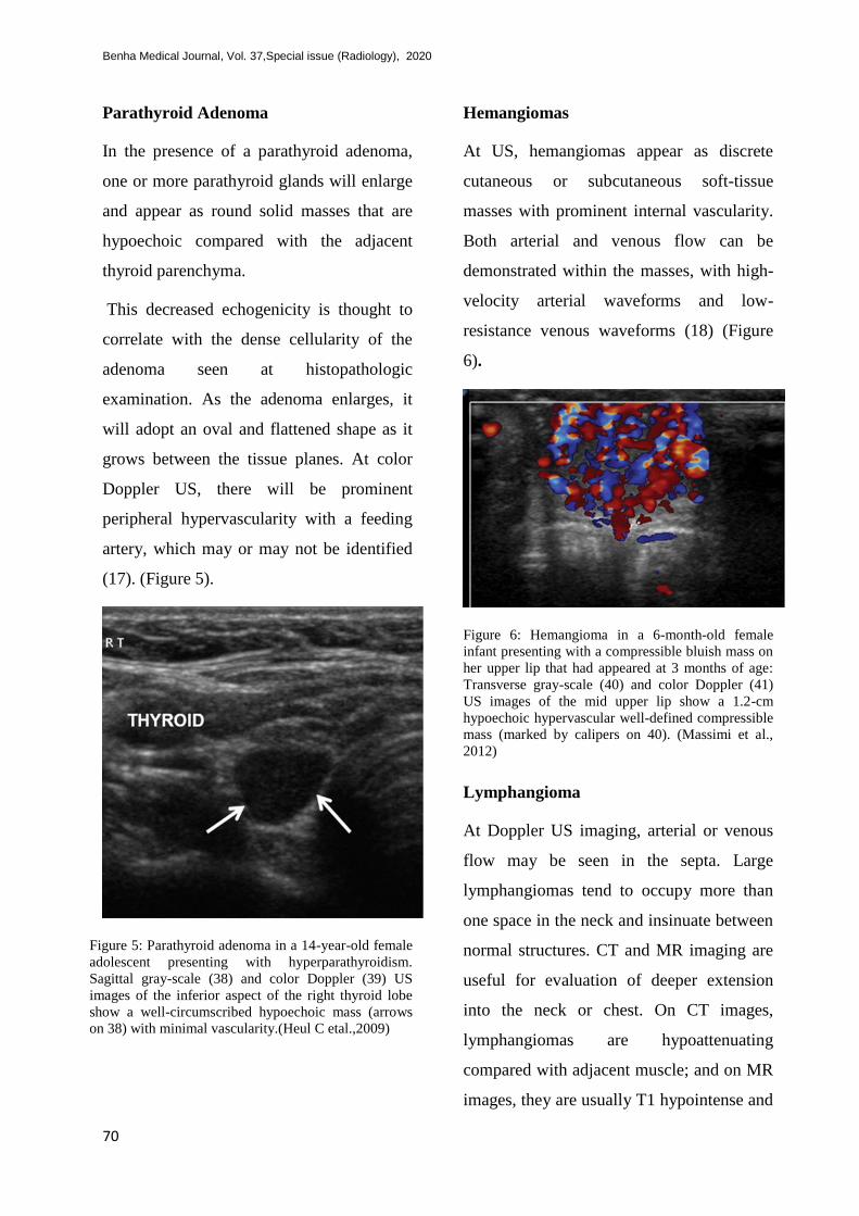
Hemangiomas
At US, hemangiomas appear as discrete
cutaneous or subcutaneous soft-tissue
masses with prominent internal vascularity.
Both arterial and venous flow can be
demonstrated within the masses, with high-
velocity arterial waveforms and low-
resistance venous waveforms (18) (Figure
6).
Figure 6: Hemangioma in a 6-month-old female
infant presenting with a compressible bluish mass on
her upper lip that had appeared at 3 months of age:
Transverse gray-scale (40) and color Doppler (41)
US images of the mid upper lip show a 1.2-cm
hypoechoic hypervascular well-defined compressible
mass (marked by calipers on 40). (Massimi et al.,
2012)
Lymphangioma
At Doppler US imaging, arterial or venous
flow may be seen in the septa. Large
lymphangiomas tend to occupy more than
one space in the neck and insinuate between
normal structures. CT and MR imaging are
useful for evaluation of deeper extension
into the neck or chest. On CT images,
lymphangiomas are hypoattenuating
compared with adjacent muscle; and on MR
images, they are usually T1 hypointense and

Impact of US in diagnosis of pediatric superficial masses in head and neck,2020
67
markedly T2 hyperintense, unless
complicated by hemorrhage. Enhancement
is demonstrated in cyst walls and septa but
not in central fluid components (19).
Jugular Vein Phlebectasia
Dynamic US of the neck with and without a
Valsalva maneuver can help confirm the
diagnosis of phlebectasia when the affected
vein dilates during a Valsalva maneuver to a
diameter approximately twice the diameter
at rest. Color Doppler US is useful to
demonstrate the presence of blood flow and
to exclude thrombus and is the reference
standard for the diagnosis of jugular vein
phlebectasia (14).
Parotitis
At US, the parotid gland appears enlarged
and heterogeneous in both viral and
bacterial parotitis, with increased
vascularity at color Doppler US. In the
bacterial form of parotitis, the parotid gland
demonstrates hypoechoic nodular areas that
represent intraparotid lymph nodes. In
severe cases, avascular hypoechoic areas of
suppuration or discrete abscesses may
develop. US may also reveal dilated tubular
areas compatible with dilated obstructed
salivary ducts that may contain hyperechoic
calculi (20).
Leukemia and/or Lymphoma
Considerable overlap exists between the US
appearance of benign and malignant lymph
nodes. In lymphoma, US generally
demonstrates enlarged nodes that appear
hypoechoic.The nodes tend to be round with
absent hila and increased central and
peripheral vascularity. Lymphomatous
nodes have been shown to have internal
reticulation—a micronodular appearance—
as well as elevated values for the resistive
index and the pulsatility index at spectral
Doppler US. The nodes can become
confluent, matted, and mass like. In
leukemia, cervical nodes are clustered and
appear slightly enlarged. The nodes can
maintain their borders or may become more
confluent, mimicking lymphoma (21).
( Figure 7).
Figure 7: Hodgkin lymphoma in a 4-year-old girl
presenting with swollen glands that had been
present for the previous 2 months: Sagittal Doppler
US image of the left lateral part of the neck shows
multiple large hyperemic architecturally abnormal
lymph nodes with a reticulated hypoechoic
appearance, which lack the expected fatty
hilum.(Morris LM 2016)
Neurogenic Tumors
Schwannomas and neurofibromas generally
appear well circumscribed, homogeneous,
and hypoechoic, with posterior acoustic
enhancement. Some schwannomas may

Benha Medical Journal, Vol. 37,Special issue (Radiology), 2020
67
demonstrate prominent internal vascularity,
which can be easily compressed with gentle
pressure on the transducer. It can be difficult
to differentiate schwannomas from reactive
lymph nodes, which may have a similar
appearance; however, identification of an
associated nerve can be used to reliably
distinguish schwannomas from nodes. (22).
Neurofibromas appear well circumscribed
and are hypoechoic, with posterior acoustic
enhancement, and may demonstrate a target
appearance—peripherally hypoechoic and
centrally hyperechoic. Plexiform
neurofibromas demonstrate diffuse
peripheral nerve involvement rather than a
focal mass. On CT and MR images,
plexiform neurofibromas appear as
multilobulated masses along the expected
course of a nerve trunk, with extension
along nerve branches, creating the
appearance of a bag of worms. A split fat
sign may be seen, with a rim of fat around
the lesion; and a target sign can be seen on
MR images, with central T2 hypointensity
against a background of T2 hyperintensity
(23).
Malignant peripheral nerve sheath tumor
(MPNST) is a high-grade sarcoma that can
arise from a plexiform neurofibroma or
from prior irradiation or can arise
spontaneously. MPNST affects
approximately 5% of patients with
neurofibromatosis type 1. MPNST is
difficult to distinguish from neurofibroma at
US, although some MPNSTs may
demonstrate hyperemia. Radionuclide
imaging with gallium 67 citrate can show
increased radiotracer uptake in MPNST,
compared with that in neurofibroma; and
MR imaging may demonstrate altered MR
signal intensity in MPNST, compared with
that in benign lesions (23).
Neuroblastoma is the third most common
malignant neoplasm in children, with a rate
of occurrence of one in 10 000 live births.
Of these cases, 25% appear to be congenital,
usually occurring in children nearing 18
months of age. Cervical neuroblastoma
accounts for less than 5% of all
neuroblastoma cases.It presents as a non-
tender lateral neck mass, usually with
symptoms related to compression of
adjacent structures, such as cranial nerves
(Horner syndrome), the esophagus
(swallowing difficulty), and the airway
(stridor), as well as heterochromia iridis
(difference in right and left eye colors). At
US, neuroblastoma appears as a solid
complex or hypoechoic mass, sometimes
with shadowing calcifications, arising
posterior to the carotid sheath, with
displacement or encasement of the carotid
artery and internal jugular vein (23).
Rhabdomyosarcoma
US is particularly valuable in the evaluation
of more superficial rhabdomyosarcomas, as

Impact of US in diagnosis of pediatric superficial masses in head and neck,2020
66
well as in the identification of suspicious
adjacent lymph nodes. At US evaluation,
rhabdomyosarcomas also resemble
neuroblastomas in echotexture and often
demonstrate low to medium echogenicity,
with variable internal vascularity at color
Doppler US. The US appearance also
resembles fibrosarcoma, malignant fibrous
histiocytoma, angiosarcoma, and
neurofibrosarcoma. Cross-sectional imaging
with MR imaging is required, because the
assessment of disease extent and staging is
limited with US alone, and tissue sampling
is required for a final diagnosis. At MR
imaging, rhabdomyosarcoma is usually iso-
intense to slightly hyperintense on T1-
weighted MR images and T2 hyperintense,
with marked contrast enhancement, and
possible intralesional hemorrhage or
necrosis. Treatment is based on the extent of
the disease as well as primary tumor
localization, with orbital and non-
parameningeal head and neck locations
associated with a better prognosis.
Chemotherapy is the first line of treatment
for rhabdomyosarcomas, with radiation
therapy and surgery being the mainstay for
local tumor control (24).
Discussion
Ultrasound is the second most common
method of imaging carried out in hospitals
worldwide after plain-film radiography.
Modern high-resolution ultrasound has
excellent spatial and contrast resolution for
the near field, and the development of 3D
technology, extended field-of-view or
panoramic imaging, and color flow and
power Doppler applications has led to great
improvements in its diagnostic utility and
accuracy. The technology involves no
ionizing radiation, is readily available in
most centers, and is relatively inexpensive
compared with CT, MRI, and PET (4).
Color flow imaging is now a routine part of
the ultrasound examination. Systems should
ideally offer high-sensitivity color-flow
imaging and power Doppler functionality.
Equipment used for functional imaging
should be calibrated to depict slow-flowing
vessels in the head and neck without
artifacts or background noise caused by
oversensitivity. The lingual artery in the
floor of mouth is a useful and practical
vascular landmark for calibration (25).
The following major parameters should be
determined: echogenicity, contour, margin,
composition, size, related surrounding tissue
in grayscale ultrasonography, the grading of
CDUS, and resistive index (RI) in spectral
Doppler. Many more specific patterns can
be observed, including phleboliths,
cellulitis-like changes, to-and-fro flow
pattern, hyperechoic fat lobules, parallel
echoic lines, C-shaped cysts, pannus,
echogenic rim, gas bubbles, tortuous tubular
structures, compressibility, and central

Benha Medical Journal, Vol. 37,Special issue (Radiology), 2020
66
necrosis. These patterns are not seen in each
lesion but are diagnostic when they appear.
(26)
We suggest the use of a high-frequency
probe (>10MHz) for small superficial
lesions and 5–10MHz for larger lesions. A
curved probe (3–5 MHz) should be used for
deeply located lesions. Each ultrasound
machine should be equipped with a color
Doppler function. (27)
Duplex US is the first-line modality of
choice for the evaluation of superficial head
and neck masses. Without use of ionizing
radiation, iodinated contrast material, or
sedation and/or anesthesia, US is able to
provide quick and cost-effective acquisition
of information, including the location, size,
shape, internal contents (solid or cystic), and
vascularity of the mass, as well as its
relationship to nearby vessels. Moreover, if
indicated, US can also be used for guidance
during interventional procedures for the
purpose of diagnosis (i.e. tissue sampling
and biopsy) and/or treatment (i.e. drainage)
(23).
Superficial lesions are sometimes very
small and located on the dermis or in the
subcutaneous layer. We would like to
emphasize the importance of gentle scan
technique, such as using extra jelly on the
skin and holding the probe with only slight
skin contact to avoid compressing the
lesion. It is not necessary to apply any
pressure on the skin or lesion to detect small
vessels and low-velocity signals with CDUS
and spectral analysis (28).
The examination should be performed with
the patient in the sitting or supine position
and the neck in extension. A systematic
examination should be completed according
to the preference of the physician
performing the ultrasound procedure as
long as it is standardized and thorough.
For a right-handed examiner, the console
should be located next to the patient’s
right shoulder. If a focal lesion is identified
on physical examination, the ultrasound
procedure may concentrate on that area.
However, in every circumstance, a basic
thyroid assessment and examination of the
neck should be performed, as this is a
unique opportunity to detect occult
pathology with a simple screening
evaluation.
Conclusion
Superficial palpable masses of the head and
neck are extremely common in the pediatric
population, with most of these lesions
ultimately proven to be benign. Duplex US
has emerged as the first-line imaging
modality for the evaluation of superficial
pediatric masses. Without the use of
ionizing radiation, iodinated contrast
material, or sedation and/or anesthesia, this
safe, quick, and cost-effective imaging

Impact of US in diagnosis of pediatric superficial masses in head and neck,2020
66
modality allows rapid evaluation and
characterization of masses.
References
1. Bansal AG, Oudsema R, Masseaux JA, Rosenberg
HK. US of pediatric superficial masses of the head
and neck. Radiographics. 2018;38(4):1239–63.
2. Brown RE, Harave S. Diagnostic imaging of
benign and malignant neck masses in children—a
pictorial review. Quant Imaging Med Surg.
2016;6(5):591.
3. Morcillo Carratala R, Capilla Cabezuelo ME,
Herrera Herrera I, Calvo Azabarte P, Dieguez
Tapias S, Moreno de la Presa R, et al.
Nontraumatic lesions of the scalp: practical
approach to imaging diagnosis: neurologic/head
and neck imaging. Radiographics.
2017;37(3):999–1000.
4. Wong KT, Lee Y, Ying M, King AD, Ahuja AT.
Ultrasound checks out suspicious neck lumps.
Diagnostic imaging Asia Pacific. 2007;
5. Toprak H, Kiliç E, Serter A, Kocakoç E,
Ozgocmen S. Ultrasound and Doppler US in
evaluation of superficial soft-tissue lesions. J Clin
Imaging Sci. 2014;4.
6. Brisse H, Orbach D, Klijanienko J, Fréneaux P,
Neuenschwander S. Imaging and diagnostic
strategy of soft tissue tumors in children. Eur
Radiol. 2006;16(5):1147–64.
7. Kimonis V, Gold J-A, Hoffman TL, Panchal J,
Boyadjiev SA. Genetics of craniosynostosis. In:
Seminars in pediatric neurology. Elsevier; 2007. p.
150–61.
8. Massimi L, Caldarelli M, Tamburrini G,
Paternoster G, Di Rocco C. Isolated sagittal
craniosynostosis: definition, classification, and
surgical indications. Child’s Nerv Syst.
2012;28(9):1311–7.
9. Ludwig BJ, Wang J, Nadgir RN, Saito N, Castro-
Aragon I, Sakai O. Imaging of cervical
lymphadenopathy in children and young adults.
Am J Roentgenol. 2012;199(5):1105–13.
10. Toma P, Granata C, Rossi A, Garaventa A.
Multimodality imaging of Hodgkin disease and
non-Hodgkin lymphomas in children.
Radiographics. 2007;27(5):1335–54.
11. Arunachalam P, Vaidyanathan V, Sengottan P.
Open and endoscopic management of fourth
branchial pouch sinus-our experience. Int Arch
Otorhinolaryngol. 2015;19(4):309–13.
12. Bagalkot PS, Parshwanath BA, Joshi SN. Neck
swelling in a newborn with congenital goiter. J
Clin Neonatol. 2013;2(1):36.
13. Zaheer S, Tan A, Ang ES, Loke KSH, Kao YH,
Goh A, et al. Post-thyroidectomy neck
ultrasonography in patients with thyroid cancer
and a review of the literature. Singapore Med J.
2014;55(4):177.
14. Jianhong L, Xuewu J, Tingze H. Surgical
treatment of jugular vein phlebectasia in children.
Am J Surg. 2006;192(3):286–90.
15. Freling NJM, Merks JHM, Saeed P, Balm AJM,
Bras J, Pieters BR, et al. Imaging findings in
craniofacial childhood rhabdomyosarcoma. Pediatr
Radiol. 2010;40(11):1723–38.
16. Restrepo R, Oneto J, Lopez K, Kukreja K. Head
and neck lymph nodes in children: the spectrum
from normal to abnormal. Pediatr Radiol.
2009;39(8):836.
17. Ahsan F, Allison R, White J. Ectopic cervical
thymus: case report and review of pathogenesis
and management. J Laryngol Otol.
2010;124(6):694.
18. Emile J-F, Abla O, Fraitag S, Horne A, Haroche J,
Donadieu J, et al. Revised classification of
histiocytoses and neoplasms of the macrophage-
dendritic cell lineages. Blood. 2016;127(22):2672–
81.
19. Ridgway JM, Parikh DA, Wright R, Holden P,
Armstrong W, Camilon F, et al. Lemierre
syndrome: a pediatric case series and review of
literature. Am J Otolaryngol. 2010;31(1):38–45.
20. Al-Shaiji AS, Bukhari MA. Endoscopic
management of fourth branchial arch anomaly: a
case report. Egypt J Ear, Nose, Throat Allied Sci.
2013;14(1):33–6.
21. Thomas B, Shroff M, Forte V, Blaser S, James A.
Revisiting imaging features and the embryologic
basis of third and fourth branchial anomalies. Am J
Neuroradiol. 2010;31(4):755–60.
22. Sekhon P, Williams D, Sara JDS, McCulloch NA.
Acute bacterial suppurative parotitis of the
neonate: a case report and review. Int J Pediatr
Otorhinolaryngol Extra. 2012;7(3):132–3.

Benha Medical Journal, Vol. 37,Special issue (Radiology), 2020
63
23. Navarro OM. Soft tissue masses in children.
Radiol Clin. 2011;49(6):1235–59.
24. CHANG H, PENG C, KAO H, HSU C, HUNG H,
CHANG J. Neonatal subgaleal hemorrhage:
clinical presentation, treatment, and predictors of
poor prognosis. Pediatr Int. 2007;49(6):903–7.
25. Ahuja AT, Ying M. Sonographic evaluation of
cervical lymph nodes. Am J Roentgenol.
2005;184(5):1691–9.
26. Tsai W-C, Chiou H-J, Chou Y-H, Wang H-K,
Chiou S-Y, Chang C-Y. Differentiation between
schwannomas and neurofibromas in the extremities
and superficial body: the role of high‐resolution
and color Doppler ultrasonography. J Ultrasound
Med. 2008;27(2):161–6.
27. Chiou H-J, Chou Y-H, Chiu S-Y, Wang H-K,
Chen W-M, Chen T-H, et al. Differentiation of
benign and malignant superficial soft-tissue masses
using grayscale and color Doppler
ultrasonography. J Chinese Med Assoc.
2009;72(6):307–15.
28. Marzano L, Failoni S, Gallazzi M, Garbagna P.
The role of diagnostic imaging in synovial
sarcoma. Our experience. Radiol Med.
2004;107(5–6):533–40.
To cite this article: Ahmed Farid, Ahmed Shalaan, Shirin Khedr. Impact of ultrasound in
diagnosis of pediatric superficial masses in head and neck. BMFJ 2020; 37 (Radiology):63-76.
DOI: 10.21608/bmfj.2020.36406.1292