impaired translesion synthesis in xeroderma pigmentosum variant
TRANSCRIPT

MOLECULAR AND CELLULAR BIOLOGY,0270-7306/99/$04.0010
Mar. 1999, p. 2206–2211 Vol. 19, No. 3
Copyright © 1999, American Society for Microbiology. All Rights Reserved.
Impaired Translesion Synthesis in Xeroderma PigmentosumVariant Extracts
AGNES M. CORDONNIER,1 ALAN R. LEHMANN,2 AND ROBERT P. P. FUCHS1*
UPR9003 du CNRS, Cancerogenese et Mutagenese Moleculaire et Structurale, ESBS, 67400 Strasbourg, France,1 andMedical Research Council Cell Mutation Unit, University of Sussex, Falmer, Brighton BN1 9RR, England2
Received 20 July 1998/Returned for modification 11 September 1998/Accepted 6 November 1998
Xeroderma pigmentosum variant (XPV) cells are characterized by a cellular defect in the ability to synthe-size intact daughter DNA strands on damaged templates. Molecular mechanisms that facilitate replicationfork progression on damaged DNA in normal cells are not well defined. In this study, we used single-strandedplasmid molecules containing a single N-2-acetylaminofluorene (AAF) adduct to analyze translesion synthesis(TLS) catalyzed by extracts of either normal or XPV primary skin fibroblasts. In one of the substrates, thesingle AAF adduct was located at the 3* end of a run of three guanines that was previously shown to inducedeletion of one G by a slippage mechanism. Primer extension reactions performed by normal cellular extractsfrom four different individuals produced the same distinct pattern of TLS, with over 80% of the productsresulting from the elongation of a slipped intermediate and the remaining 20% resulting from a nonslippedintermediate. In contrast, with cellular extracts from five different XPV patients, the TLS reaction was stronglyreduced, yielding only low amounts of TLS via the nonslipped intermediate. With our second substrate, inwhich the AAF adduct was located at the first G in the run, thus preventing slippage from occurring, weconfirmed that normal extracts were able to perform TLS 10-fold more efficiently than XPV extracts. Thesedata demonstrate unequivocally that the defect in XPV cells resides in translesion synthesis independently ofthe slippage process.
Xeroderma pigmentosum (XP) is an autosomal recessivedisorder characterized by a genetic predisposition to sunlight-induced skin cancer. Fibroblasts derived from patients with XPare extremely sensitive to the mutagenic effect of UV irradia-tion (19). The majority of XP patients are deficient in nucle-otide excision repair, and there have been dramatic advancesin our understanding of the molecular defects in these patients.In contrast, there has been little progress in our understandingof the molecular defect in the XP variant (XPV) group, whichcomprises a substantial minority (approximately 20%) of XPpatients. They have normal levels of nucleotide excision repairand normal sensitivity to the lethal effects of UV irradiation (5)but a marked defect in the ability to synthesize intact daughterDNA strands during DNA replication after some (4, 17, 20, 26)but not all (4, 7) types of carcinogenic damage. The cellulardefect in DNA replication in UV-irradiated XPV cells wasdiscovered as long ago as 1975 (17), but because of the normalsensitivity to killing by UV light, the XPV gene has beenrefractory to cloning. The precise nature of the moleculardefect in this important class of XP patients remains one of themajor unsolved problems in this area.
The UV hypermutability of XPV cells could be due to anabnormal error-prone mechanism of replication. This hypoth-esis is supported by results showing that mutation spectra inUV-irradiated XPV cells are distinct from those observed innormal cells (32, 33). In XPV cells, the UV-induced substitu-tions are mainly transversions (C3A), whereas in normal cells,transitions (C3T) predominate. An altered mutation patternwas also generated by psoralen photoadducts in XPV com-pared to normal cells (27). To determine whether the processof translesion synthesis (TLS) in XPV extracts differs from that
of normal cells, we compared the abilities of cellular extractsfrom either normal or XPV primary fibroblasts to performprimer elongation past a unique blocking lesion located on asingle-stranded circular template. This approach allows TLS tobe investigated both quantitatively and qualitatively in the ab-sence of other cellular responses such as repair, recombina-tion, or polymerase strand switching. This assay will greatlyfacilitate the identification of the XPV gene product(s) and thebiochemical features of replication of damaged DNA in hu-man cells.
MATERIALS AND METHODS
Construction of single-stranded plasmid containing a single AAF adduct. Thestrategy used to construct double-stranded molecules containing single N-2-acetylaminofluorene (AAF) adducts involved the formation of gapped-duplexmolecules (14). A 14-mer oligonucleotide d(ATACCCG1G2G3ACATC) wasreacted with N-acetoxy-N-2-acetylaminofluorene under conditions such as tocreate one adduct per oligonucleotide on average. The crude reaction mixturewas subjected to reverse-phase high-pressure liquid chromatography, and theoligonucleotides with a single AAF adduct at G1 or G3 were purified and ligatedinto the gap to generate plasmid pUC-3G1 or pUC-3G3, respectively. A controlundamaged plasmid, pUC-3G0, was constructed with the unreacted controloligonucleotide. The single-stranded vectors (pUC-3G0.ss, pUC-3G1.ss, andpUC-3G3.ss) were produced from the corresponding double-stranded plasmidsby selective enzymatic degradation of the nonadducted uracil-containing strand.A detailed description of this procedure using an enzymatic cocktail containinguracil-DNA glycosylase, exonuclease III, and the 39359 exonuclease activityassociated with T7 DNA polymerase has been described recently (23).
Cell cultures. The cell strains used in this study were fibroblast culturesderived from the skin of normal individuals (1BR3, 205BR, 250BR, and 368BR)and XP variants (XP7BR, XP11BR, XP6DU, XP7DU, and XP30R0). The XPvariants were all defective in postreplication repair of UV damage as shown bysucrose density gradient analysis of newly synthesized DNA in UV-irradiatedcells (reference 17 [XP30R0] and our unpublished results [other cell strains]).XP11BR cells are derived from the patient described in reference 3. All cellstrains were grown in Dulbecco’s modified Eagle’s medium (Sigma) supple-mented with 15% fetal calf serum (Eurobio) and 50 mg of gentamicin (Sigma)per ml.
Preparation of cell extracts. Cell extracts (100 ml) were obtained from 107
exponentially growing cells essentially as described previously (13). The cellswere washed twice with phosphate-buffered saline (PBS). trypsin (0.25% in PBS)was added to the plates, which were then incubated at 37°C until cells rounded
* Corresponding author. Mailing address: UPR9003 du CNRS, Can-cerogenese et Mutagenese Moleculaire et Structurale, ESBS, Blvd S.Brant, 67400 Strasbourg, France. Phone and Fax: 33 388 65 53 4.E-mail: [email protected].
2206
on March 21, 2018 by guest
http://mcb.asm
.org/D
ownloaded from

up and were almost ready to detach. Excess buffer was removed, and the cellswere collected by agitation in the culture medium. The cells were pelleted bycentrifugation (1,000 3 g for 5 min) and washed once in the culture medium andtwice in PBS. The cell pellet was resuspended in 4 volumes of ice-cold hypotonicbuffer (10 mM Tris-HCl [pH 7.5], 10 mM KCl, 10 mM MgCl2, 1 mM dithiothre-itol [DTT]) containing protease inhibitors (1 mM phenylmethylsulfonyl fluorideand 5 mg each of leupeptin, chymostatin, and aprotinin per ml). The cells wereallowed to swell on ice for 30 min at 4°C and disrupted with 20 strokes of atight-fitting pestle in a Dounce homogenizer. Cell disruption and integrity of thenuclei were examined under light microscopy by exclusion of trypan blue. Nucleiwere harvested by centrifugation for 10 min at 3,000 3 g at 4°C, and cytosolicsupernatants were kept on ice. After 1 h of extraction at 0°C in hypotonic buffercontaining 350 mM NaCl, the nuclear extracts were centrifuged at 10,000 3 g for10 min. Cytosolic and nuclear extracts were mixed, and the proteins were pre-cipitated by the addition of ammonium sulfate (0.33 g/ml) and gentle stirring for1 h at 4°C. The precipitates were collected by centrifugation (45 min at 10,000 3g), resuspended in dialysis buffer (100 mM potassium glutamate, 30 mM HEPES[pH 7.5], 1 mM DTT, 10% glycerol), and dialyzed for 2 h at 4°C. The extractswere clarified by centrifugation for 10 min at 10,000 3 g and stored at 280°C.The protein concentration of extracts is typically between 5 and 15 mg/ml asmeasured by the Bradford protein assay (Bio-Rad) using bovine serum albuminas the standard.
In vitro primer extension assays. A primer (24-mer oligonucleotide) wasphosphorylated with T4 polynucleotide kinase (New England Biolabs), using 50pmol of [g-32P]ATP (3,000 Ci/mmol; Amersham). After purification by electro-phoresis on a 20% polyacrylamide–7 M urea denaturing gel, the primer (twofoldmolar excess) was annealed to single-stranded DNA in a buffer containing 60mM HEPES (pH 7.5) and 20 mM MgCl2. The mixture was incubated at 50°C for15 min with Escherichia coli SSB (Pharmacia). For primer extension assays, thereaction mixture (6.25 ml) containing 10 fmol of SSB-coated primed DNA, andwhole-cell extract was incubated at 37°C in 50 mM HEPES-KOH (pH 7.8)–7 mMMgCl2–1 mM DTT–4 mM ATP–500 mM deoxynucleoside triphosphates(dNTPs)–200 mM each UTP, CTP, and GTP–40 mM creatine phosphate–100 mgof creatine kinase per ml. The reaction was stopped by adding an equal volumeof proteinase K (4 mg/ml)-sodium dodecyl sulfate (2%) and incubated for 30 minat 37°C. The samples were precipitated in the presence of 1 M ammoniumacetate and 50% isopropanol. Replication products were digested with restric-tion enzymes EcoRI, PvuII, and SmaI in buffers recommended by the manufac-turers and analyzed by electrophoresis on a polyacrylamide–7 M urea denaturinggel.
RESULTS
Design of the damaged single-stranded DNA substrate. Weused AAF as a model DNA-damaging agent to analyze theability of cell extracts to carry out TLS. AAF adducts at the C8position of guanine (dGua-C8-AAF) are severe blocks to invitro DNA synthesis by purified prokaryotic and eukaryoticDNA polymerases (2, 21, 22). TLS past AAF adducts in bothdouble-stranded (30, 31) and forked single-stranded (13) DNAtemplates has been observed to some extent in cell extracts.This suggests that extracts accurately mimic at least some ofthe mechanisms that rescue a blocked replication fork in vivo.In the present study, single-stranded plasmids containing singleAAF adducts at G1 or G3 in a run of three guanines (59-G1G2G3-39) were constructed to analyze TLS. In E. coli, whenlocated at G3 in the run (59-GGGAAF-39), an AAF adductinduces 21 frameshift mutations at least 100-fold more effi-ciently than when located at G1 (59-GAAFGG-39) (15). Indeed,a G3 adduct can trigger a primer-template misalignment eventyielding a slipped intermediate that is in equilibrium with itsnonslipped counterpart, while such an event is not favored fora G1 adduct (Fig. 1A). Cellular extracts from either normal orXPV primary fibroblasts were tested for their abilities to elon-gate a 32P-end-labeled oligonucleotide (24-mer) annealed tothe single-stranded circular template, at a distance of 91 (pUC-3G3.ss) or 93 (pUC-3G1.ss) nucleotides from the lesion site.TLS was analyzed on sequencing gels following PvuII andEcoRI restriction digestion of the DNA products (Fig. 1B).For both substrates, TLS resulting from the elongation of non-slipped (TLS0) and slipped (TLS21 [21 frameshift event])intermediates will yield fragments with lengths of 104 and 103nucleotides, respectively.
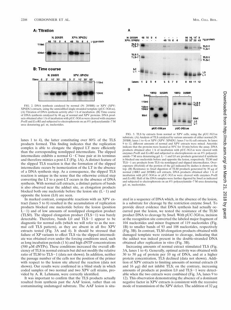
Primer extension using the unmodified substrate. The rep-lication activities of extracts from normal (205BR) and XPV(XP6DU) cells were tested by using a primed unmodified sin-gle-stranded template (pUC-3G0.ss). For both extracts, prod-uct yield increased with increasing amounts of protein (Fig.2A). In a typical time course experiment, radiolabeled 104-nucleotide products were detected after a 2-min reaction andreached a plateau at 10 min. Completion of DNA synthesisaround the whole template followed by subsequent ligationwas observed by the appearance of a band at 119 nucleotides(Fig. 2B). Quantitative analysis of the results indicates thatafter a 1-h incubation with 48 mg of either extract, about 20%of the primers were elongated. All of these data indicate thatXPV cell extracts have replication activity similar to that ofnormal cells on a lesion-free template, consistent with thenormal DNA replication observed in undamaged XPV cells invivo (17).
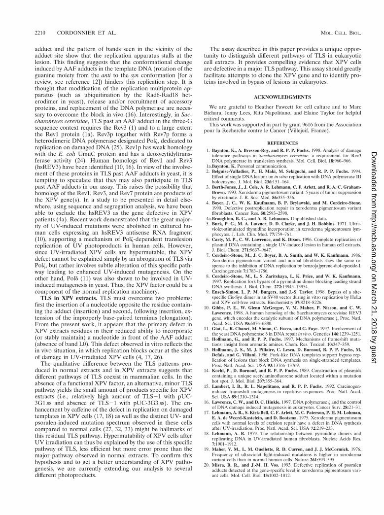
Differential TLS catalyzed by extracts from normal or XPVextracts. TLS can be described in terms of the different repli-cation intermediates that are involved. The replication inter-mediate in which the last nucleotide of the primer is locatedopposite the lesion in the template will be referred to as alesion terminus (LT). All replication intermediates precedingand succeeding this step will be referred to as prelesion termini(pre-LT) and postlesion termini (post-LT), respectively. Giventhese definitions, TLS can be viewed as a succession of at leasttwo reactions (pre-LT3LT3post-LT). The progression fromone step to the next normally requires a DNA synthesis step. Amarked difference between normal and XPV extracts was ob-served for TLS past the AAF adduct at the G3 position (Fig.3A). Despite the presence of the lesion on the modified sub-strate (pUC-3G.3ss), normal extracts (205BR) were able tocatalyze TLS efficiently as 40 to 60% of the elongated primerswere extended past the adduct site upon incubation with 48 mgof normal extract proteins. Both nonslipped (TLS0) andslipped (TLS21) elongation products were seen (Fig. 3A,
FIG. 1. AAF-induced 21 frameshift pathway. The primer terminus whenlocated opposite the lesion site is designated the lesion terminus (LT). The LTcan isomerize via a slippage mechanism into the slipped intermediate, which hasa paired primer terminus (post-LT). Elongation from the nonslipped and slippedintermediates lead to TLS0 and TLS21 products, respectively. (B) Diagram ofthe modified template, pUC-3G3.ss. A single AAF adduct is located with therecognition site of SmaI. The positions of PvuII and EcoRI restriction sites andlengths of the strands produced upon elongation of the labeled primer areindicated. L0, fragment elongated up the lesion site; nt, nucleotides.
VOL. 19, 1999 IN VITRO TRANSLESION SYNTHESIS IN EUKARYOTIC CELL EXTRACTS 2207
on March 21, 2018 by guest
http://mcb.asm
.org/D
ownloaded from
lanes 1 to 4), the latter constituting over 80% of the TLSproducts formed. This finding indicates that the replicationcomplex is able to elongate the slipped LT more efficientlythan the corresponding nonslipped intermediate. The slippedintermediate exhibits a normal G z C base pair at its terminusand therefore mimics a post-LT (Fig. 1A). A distinct feature ofthe slipped TLS reaction is that the formation of the slippedintermediate occurs by isomerization of the LT in the absenceof a DNA synthesis step. As a consequence, the slipped TLSreaction is unique in the sense that the otherwise critical stepconverting the LT to a post-LT occurs in the absence of DNAsynthesis. With normal cell extracts, a distinct pattern of bandsis also observed near the adduct site, as elongation productsblocked both one nucleotide before the lesion site (L21) andopposite the lesion (L0) are seen.
In marked contrast, comparable reactions with an XPV ex-tract (lanes 5 to 8) resulted in the accumulation of replicationproducts blocked one nucleotide before the lesion (positionL21) and of low amounts of nonslipped elongation product(TLS0). The slipped elongation product (TLS21) was barelydetectable. Therefore, bands L0 and TLS21 appear to bediagnostic for normal cells (which we will refer to as the nor-mal cell TLS pattern), as they are absent in all five XPVextracts tested (Fig. 3A and 4). It should be stressed thatfailure of XP variants to effect TLS via the slipped intermedi-ate was obtained even under the forcing conditions used, suchas long incubation periods (1 h) and high dNTP concentrations(500 mM dNTPs). These conditions increased the overall effi-ciency of TLS in normal extracts but did not modify the relativeratio of TLS0 to TLS21 (data not shown). In addition, neitherthe passage number of the cells nor the position of the primerwith respect to the lesion site altered the reaction (data notshown). Our results were confirmed in a blind study in whichcoded samples of two normal and two XPV cell strains, pro-vided by A. R. Lehmann, were correctly identified.
It was important to confirm that the TLS products actuallyresulted from synthesis past the AAF lesion, rather than oncontaminating undamaged substrate. The AAF lesion is situ-
ated in a sequence of DNA which, in the absence of the lesion,is a substrate for cleavage by the restriction enzyme SmaI. Toprovide direct evidence that DNA synthesis had actually oc-curred past the lesion, we tested the resistance of the TLS0product DNA to cleavage by SmaI. With pUC-3G0.ss, incisionat the recognition site converted the labeled major fragment of104 nucleotides and minor fragment of 119 nucleotides (Fig.1B) to smaller bands of 93 and 108 nucleotides, respectively(Fig. 3B). In contrast, TLS0 elongation products obtained withdamaged template were resistant to cleavage, indicating thatthe adduct was indeed present in the double-stranded DNAobtained after replication in vitro (Fig. 3B).
Increasing amounts of normal extract stimulated TLS (Fig.3A, lanes 1 to 4). Generally, optimal activity was obtained with30 to 50 mg of protein per 10 ng of DNA, and at a higherprotein concentration, TLS declined (data not shown). Addi-tion of XPV extracts to limiting amounts of normal extracts (8and 16 mg) did not inhibit TLS; on the contrary, increasingamounts of products at position L0 and TLS21 were detect-able when the two extracts were combined (Fig. 3A, lanes 9 to12). This observation demonstrating the absence of a dominantnegative factor in XPV extracts is consistent with the recessivemode of transmission of the XPV defect. The addition of 32 mg
FIG. 2. DNA synthesis catalyzed by normal (N: 205BR) or XPV (XPV:XP6DU) extracts, using the unmodified single-stranded template (pUC-3G0.ss).(A) Titration of DNA synthesis activity after 1 h of incubation. (B) Time courseof DNA synthesis catalyzed by 48 mg of normal and XPV proteins. DNA prod-ucts obtained after 1 h of incubation with pUC-3G0.ss were cleaved with enzymesPvuII and EcoRI and subjected to electrophoresis on an 8% polyacrylamide–7 Murea denaturing gel. nt, nucleotides.
FIG. 3. TLS by extracts from normal or XPV cells, using the pUC-3G3.sssubstrate. (A) Analysis of TLS catalyzed by various amounts of either normal (N:205BR; lanes 1 to 4) or XPV (XPV: XP6DU; lanes 5 to 8) cell extracts. In lanes9 to 12, different amounts of normal and XPV extracts were mixed. Asterisksindicate that the proteins were heated at 50°C for 10 min before the assay. DNAproducts obtained after 1 h of incubation with pUC-3G3.ss were cleaved withenzymes PvuII and EcoRI and subjected to electrophoresis on an 8% polyacryl-amide–7 M urea denaturing gel. L21 and L0 are products generated if synthesisis blocked one nucleotide before and opposite the lesion, respectively. TLS0 andTLS21 are products from TLS via nonslipped and slipped intermediates. Over-exposure (fivefold) of the portion of the gel indicated by dashes is shown at thetop. (B) Resistance to SmaI digestion of TLS0 products generated by 30 mg ofnormal (1BR3 and 205BR) cell extracts. DNA products obtained after 1 h ofincubation with pUC-3G0.ss or pUC-3G3.ss were cleaved with enzymes PvuIIand EcoRI. Half of the DNA samples were further digested by SmaI as indicatedand subjected to electrophoresis on an 8% polyacrylamide–7 M urea denaturinggel. nt, nucleotides.
2208 CORDONNIER ET AL. MOL. CELL. BIOL.
on March 21, 2018 by guest
http://mcb.asm
.org/D
ownloaded from

of XPV extract to a limiting amount (16 mg) of normal cellextract stimulates the normal cell TLS pattern (i.e., presence ofbands TLS-1 and L0) compared to the replication assay wherethe same quantity of heat-inactivated XPV extract is added tolimiting quantities of normal cell extract (Fig. 3A, lanes 9 and10). This finding demonstrates that it is not the XPV factorthat limits the TLS reaction with 16 mg of normal cell extractssince XPV extracts can stimulate the normal cell TLS patternunder these conditions. On the other hand, heat inactivation ofthe normal cell extract (lane 11) yields the TLS pattern typicalfor XPV cells (i.e., absence of both TLS21 and L0 bands).
Complementation studies. The assay described above pro-vides a complementation approach to isolate proteins fromnormal extracts that would allow XPV extracts to produceTLS21 efficiently with pUC-3G3.ss as a template. As a firststep, we tested complementation between extracts from fivedifferent XPV individuals (Fig. 4). Complementation was notobserved upon mixing equal amounts of any of the five ex-tracts, suggesting that these five XPV patients are mutated inthe same gene.
Analysis of TLS by using a substrate where the possibility ofslippage is reduced. The data obtained with the pUC-3G3.sssubstrate indicate that XPV extracts are less able than normalextracts to elongate a primer past an AAF adduct. However,this defect could be specific for the elongation of the post-LTgenerated by slippage (Fig. 1). Another possibility could bethat a misincorporation at this site prevents slippage fromoccurring. To further investigate these points, we analyzedTLS by using a substrate where the slippage process is mini-mized because the AAF adduct is located on the first G of therun (pUC-3G1.ss). We previously showed (15) that at thisposition, the frequency of induced 21 frameshift mutation wasreduced 100-fold in vivo in E. coli. Interestingly, cellular ex-tracts from normal cells were able to perform TLS with pUC-3G1.ss as efficiently as with pUC-3G3.ss (Fig. 5). In agreementwith the result obtained with E. coli, TLS using substrate pUC-3G1.ss resulted mainly in normal elongation products (TLS0).The TLS21 products could consist of either targeted deletions(-G within the run of guanine residues) or semitargeted dele-tions (-C in the 59 flanking repetitive cytosine sequence) asfound in E. coli (15). In the vicinity of the lesion site, the
presence of three bands at L21, L0, and L11 revealed that theadduct hindered the progression of DNA synthesis. Obviously,the absence of band L11 when pUC-3G3.ss was used as thetemplate indicates that this step was obviated by the slippageevent. Irrespective of the site of the lesion (G1 or G3), theefficiency of TLS was highly reduced with XPV extracts.
The high amount of TLS0 obtained in normal extracts withthe pUC-3G1.ss substrate indicates that efficient TLS can oc-cur independently of the slippage process. In XPV extracts, theefficiency of TLS was reduced 10-fold, providing evidence thatthe nonslipped TLS product formed with normal cell extractsupon incubation with pUC-3G1.ss arises by a mechanism thatrequired the XPV factor, similarly to the slipped TLS productformed with pUC-3G3.ss.
DISCUSSION
Complete replication past site-specific UV-induced lesions(6, 29) or AAF adducts (30, 31) in double-stranded DNAcarrying the simian virus 40 origin of replication has beenobserved in HeLa cell extracts, in the presence of T antigen.Recently, using a similar approach, three groups (8, 9, 28)demonstrated that in contrast to normal cell extracts, extractsfrom XPV cells were completely (8) or partially (9, 28) defi-cient in the bypass of a single cis-syn thymine dimer. In thesestudies, impaired replication fork bypass observed in XPVextracts could be due to a defect either in mechanisms broadlyreferred to as postreplication repair (such as polymerase tem-plate switching or recombinational strand transfer) or in asimple TLS reaction. However, in the present work, using asingle-stranded template with a single lesion, we show directlyand unequivocally that it is TLS that is impaired in XPVextracts. This result highlights the biological importance of afactor(s) involved in TLS in human cells, since a defect in thispathway leads to enhanced mutagenesis and to sunlight-in-duced skin cancer.
TLS in normal cell extracts. In normal cell extracts, the slowprocess of formation of complete TLS products past the AAF
FIG. 4. Absence of complementation between extracts from five differentXPV individuals. XPV extracts (15 mg of each) were mixed together as indicatedand incubated for 1 h with pUC-3G3.ss. In parallel, 30 mg of normal (N; 205BR)or XPV extracts was incubated with pUC-3G3.ss. DNA products were cleavedwith enzymes PvuII and EcoRI and subjected to electrophoresis on an 8%polyacrylamide–7 M urea denaturing gel. Overexposure (13-fold) of the portionof the gel indicated by dashes is shown at the top. XPV extracts: 1, XP11BR; 2,XP30R0; 3, XP7BR; 4, XP6DU; 5, XP7DU. All of these cell strains have thecellular defect in DNA synthesis after UV irradiation (unpublished data andreference 17) that is diagnostic for XP variants.
FIG. 5. TLS past and AAF adduct by extracts from normal or XPV cells.Analysis of TLS catalyzed by 30 mg of either normal (N; 205BR) or XPV(XP6DU) cell extracts. DNA products obtained after 1 h of incubation withpUC-3G1.ss or pUC-3G3.ss were cleaved with enzymes PvuII and EcoRI andsubjected to electrophoresis on an 10% polyacrylamide–7 M urea denaturing gel.L21, L0, and L11 are products generated if synthesis is blocked one nucleotidebefore, opposite, and one nucleotide after the lesion, respectively. TLS0 andTLS21 are products from TLS via nonslipped and slipped intermediates. As aninternal standard, an identical amount of a 120-nucleotide fragment (end labeledwith [g-32P]ATP by T4 polynucleotide kinase) was added to each reaction at theend of the replication step.
VOL. 19, 1999 IN VITRO TRANSLESION SYNTHESIS IN EUKARYOTIC CELL EXTRACTS 2209
on March 21, 2018 by guest
http://mcb.asm
.org/D
ownloaded from
adduct and the pattern of bands seen in the vicinity of theadduct site show that the replication apparatus stalls at thelesion. This finding suggests that the conformational changeinduced by AAF adducts in the template DNA (rotation of theguanine moiety from the anti to the syn conformation [for areview, see reference 12]) hinders this replication step. It isthought that modification of the replication multiprotein ap-paratus (such as ubiquitination by the Rad6-Rad18 het-erodimer in yeast), release and/or recruitment of accessoryproteins, and replacement of the DNA polymerase are neces-sary to overcome the block in vivo (16). Interestingly, in Sac-charomyces cerevisiae, TLS past an AAF adduct in the three-Gsequence context requires the Rev3 (1) and to a large extentthe Rev1 protein (1a). Rev3p together with Rev7p forms aheterodimeric DNA polymerase designated Polz, dedicated toreplication on damaged DNA (25). Rev1p has weak homologywith the E. coli UmuC protein and has a deoxycytidyltrans-ferase activity (24). Human homologs of Rev1 and Rev3(hsREV3) have been identified (10, 16). In view of the involve-ment of these proteins in TLS past AAF adducts in yeast, it istempting to speculate that they may also participate in TLSpast AAF adducts in our assay. This raises the possibility thathomologs of the Rev1, Rev3, and Rev7 protein are products ofthe XPV gene(s). In a study to be presented in detail else-where, using sequence and segregation analysis, we have beenable to exclude the hsREV3 as the gene defective in XPVpatients (4a). Recent work demonstrated that the great major-ity of UV-induced mutations were abolished in cultured hu-man cells expressing an hsREV3 antisense RNA fragment(10), supporting a mechanism of Polz-dependent translesionreplication of UV photoproducts in human cells. However,since UV-irradiated XPV cells are hypermutable, the XPVdefect cannot be explained simply by an abrogation of TLS viaPolz, but rather involves subtle alteration of this specific path-way leading to enhanced UV-induced mutagenesis. On theother hand, Pold (11) was also shown to be involved in UV-induced mutagenesis in yeast. Thus, the XPV factor could be acomponent of the normal replication machinery.
TLS in XPV extracts. TLS must overcome two problems:first the insertion of a nucleotide opposite the residue contain-ing the adduct (insertion) and second, following insertion, ex-tension of the improperly base-paired terminus (elongation).From the present work, it appears that the primary defect inXPV extracts residues in their reduced ability to incorporate(or stably maintain) a nucleotide in front of the AAF adduct(absence of band L0). This defect observed in vitro reflects thein vivo situation, in which replication blocks occur at the sitesof damage in UV-irradiated XPV cells (4, 17, 26).
The qualitative difference between the TLS patterns pro-duced in normal extracts and in XPV extracts suggests thatdifferent pathways of TLS coexist in mammalian cells. In theabsence of a functional XPV factor, an alternative, minor TLSpathway yields the small amount of products specific for XPVextracts (i.e., relatively high amount of TLS21 with pUC-3G1.ss and absence of TLS21 with pUC-3G3.ss). The en-hancement by caffeine of the defect in replication on damagedtemplates in XPV cells (17, 18) as well as the distinct UV- andpsoralen-induced mutation spectrum observed in these cellscompared to normal cells (27, 32, 33) might be hallmarks ofthis residual TLS pathway. Hypermutability of XPV cells afterUV irradiation can thus be explained by the use of this specificpathway of TLS, less efficient but more error prone than themajor pathway observed in normal extracts. To confirm thishypothesis and to get a better understanding of XPV patho-genesis, we are currently extending our analysis to severaldifferent photoproducts.
The assay described in this paper provides a unique oppor-tunity to distinguish different pathways of TLS in eukaryoticcell extracts. It provides compelling evidence that XPV cellsare defective in a major TLS pathway. This assay should greatlyfacilitate attempts to clone the XPV gene and to identify pro-teins involved in bypass of lesions in eukaryotes.
ACKNOWLEDGMENTS
We are grateful to Heather Fawcett for cell culture and to MarcBichara, Jenny Lees, Rita Napolitano, and Elaine Taylor for helpfulcritical comments.
This work was supported in part by grant 9616 from the Associationpour la Recherche contre le Cancer (Villejuif, France).
REFERENCES
1. Baynton, K., A. Bresson-Roy, and R. P. P. Fuchs. 1998. Analysis of damagetolerance pathways in Saccharomyces cerevisiae: a requirement for Rev3DNA polymerase in translesion synthesis. Mol. Cell. Biol. 18:960–966.
1a.Baynton, K. Personal communication.2. Belguise-Valladier, P., H. Maki, M. Sekiguchi, and R. P. P. Fuchs. 1994.
Effect of single DNA lesions on in vitro replication with DNA polymerase IIIholoenzyme. J. Mol. Biol. 236:151–164.
3. Berth-Jones, J., J. Cole, A. R. Lehmann, C. F. Arlett, and R. A. C. Graham-Brown. 1993. Xeroderma pigmentosum variant: 5 years of tumor suppressionby etretinate. J. R. Soc. Med. 86:355–356.
4. Boyer, J. C., W. K. Kaufmann, B. P. Brylawski, and M. Cordeiro-Stone.1990. Defective postreplication repair in xeroderma pigmentosum variantfibroblasts. Cancer Res. 50:2593–2598.
4a.Broughton, B. C., and A. R. Lehmann. Unpublished data.5. Burk, P. G., M. A. Lutzner, D. D. Clarke, and J. H. Robbins. 1971. Ultra-
violet-stimulated thymidine incorporation in xeroderma pigmentosum lym-phocytes. J. Lab. Clin. Med. 77:759–761.
6. Carty, M. P., C. W. Lawrence, and K. Dixon. 1996. Complete replication ofplasmid DNA containing a single UV-induced lesion in human cell extracts.J. Biol. Chem. 271:9637–9647.
7. Cordeiro-Stone, M., J. C. Boyer, B. A. Smith, and W. K. Kaufmann. 1986.Xeroderma pigmentosum variant and normal fibroblasts show the same re-sponse to the inhibition of DNA replication by benzo[a]pyrene-diol-epoxide-I.Carcinogenesis 7:1783–1786.
8. Cordeiro-Stone, M., L. S. Zaritskaya, L. K. Price, and W. K. Kaufmann.1997. Replication fork bypass of a pyrimidine dimer blocking leading strandDNA synthesis. J. Biol. Chem. 272:13945–13954.
9. Ensch-Simon, I., P. M. Burgers, and J.-S. Taylor. 1998. Bypass of a site-specific Cis-Syn dimer in an SV40 vector during in vitro replication by HeLaand XPV cell-free extracts. Biochemistry 37:8218–8226.
10. Gibbs, P. E., W. Glenn McGregor, V. M. Maher, P. Nisson, and C. W.Lawrence. 1998. A human homolog of the Saccharomyces cerevisiae REV3gene, which encodes the catalytic subunit of DNA polymerase z. Proc. Natl.Acad. Sci. USA 95:6876–6880.
11. Giot, L., R. Chanet, M. Simon, C. Facca, and G. Faye. 1997. Involvement ofthe yeast DNA polymerase d in DNA repair in vivo. Genetics 146:1239–1251.
12. Hoffmann, G., and R. P. P. Fuchs. 1997. Mechanisms of frameshift muta-tions: insight from aromatic amines. Chem. Res. Toxicol. 10:347–359.
13. Hoffmann, J. S., M. J. Pillaire, C. Lesca, D. Burnouf, R. P. P. Fuchs, M.Defais, and G. Villani. 1996. Fork-like DNA templates support bypass rep-lication of lesions that block DNA synthesis on single-stranded templates.Proc. Natl. Acad. Sci. USA 93:13766–13769.
14. Koehl, P., D. Burnouf, and R. P. P. Fuchs. 1989. Construction of plasmidscontaining a unique acetylaminofluorene adduct located within a mutationhot spot. J. Mol. Biol. 207:355–364.
15. Lambert, I. B., R. L. Napolitano, and R. P. P. Fuchs. 1992. Carcinogen-induced frameshift mutagenesis in repetitive sequences. Proc. Natl. Acad.Sci. USA 89:1310–1314.
16. Lawrence, C. W., and D. C. Hinkle. 1997. DNA polymerase z and the controlof DNA damage induced mutagenesis in eukaryotes. Cancer Surv. 28:21–31.
17. Lehmann, A. R., S. Kirk-Bell, C. F. Arlett, M. C. Paterson, P. H. M. Lohman,E. A. de Weerd-Kastelein, and D. Bootsma. 1975. Xeroderma pigmentosumcells with normal levels of excision repair have a defect in DNA synthesisafter UV-irradiation. Proc. Natl. Acad. Sci. USA 72:219–233.
18. Lehmann, A. R. 1979. The relationship between pyrimidine dimers andreplicating DNA in UV-irradiated human fibroblasts. Nucleic Acids Res.7:1901–1912.
19. Maher, V. M., L. M. Ouellette, R. D. Curren, and J. J. McCormick. 1976.Frequency of ultraviolet light-induced mutations is higher in xerodermavariant cells than in normal human cells. Nature 261:593–595.
20. Misra, R. R., and J.-M. H. Vos. 1993. Defective replication of psoralenadducts detected at the gene-specific level in xeroderma pigmentosum vari-ant cells. Mol. Cell. Biol. 13:1002–1012.
2210 CORDONNIER ET AL. MOL. CELL. BIOL.
on March 21, 2018 by guest
http://mcb.asm
.org/D
ownloaded from
21. Moore, P. D., K. K. Bose, S. D. Rabkin, and B. S. Strauss. 1981. Sites oftermination of in vitro DNA synthesis on ultraviolet and N-acetylaminoflu-orene treated FX 174 templates by prokaryotic and eukaryotic polymerases.Proc. Natl. Acad. Sci. USA 78:110–114.
22. Mozzherin, D. J., S. Shibutani, C. K. Tan, K. M. Downey, and P. A. Fisher.1997. Proliferating cell nuclear antigen promotes DNA synthesis past tem-plate lesions by mammalian DNA polymerase d. Proc. Natl. Acad. Sci. USA94:6126–6131.
23. Napolitano, R. L., and R. P. P. Fuchs. 1997. New strategy for the construc-tion of single-stranded plasmids with single mutagenic lesions. Chem. Res.Toxicol. 10:667–671.
24. Nelson, J. R., C. W. Lawrence, and D. C. Hinkle. 1996. Deoxycytidyl trans-ferase activity of yeast REV1 protein. Nature 382:729–731.
25. Nelson, J. R., C. W. Lawrence, and D. C. Hinkle. 1996. Thymine-thyminedimer bypass by yeast DNA polymerase z. Science 272:1646–1649.
26. Park, S. D., and J. E. Cleaver. 1979. Postreplication repair: question of itsdefinition and possible alteration in xeroderma pigmentosum cell strains.Proc. Natl. Acad. Sci. USA 76:3927–3931.
27. Raha, M., G. Wang, M. M. Seidman, and P. M. Glazer. 1996. Mutagenesisby third-strand-directed psoralen adducts in repair-deficient human cells:high frequency and altered spectrum in a xeroderma pigmentosum variant.Proc. Natl. Acad. Sci. USA 93:2941–2946.
28. Svoboda, D. L., L. P. Briley, and J.-M. H. Vos. 1998. Defective bypassreplication of a leading strand cyclobutane thymine dimer in xerodermapigmentosum variant cell extracts. Cancer Res. 58:2445–2448.
29. Svoboda, D. L., and J.-M. H. Vos. 1995. Differential replication of a single,UV-induced lesion in the leading or lagging strand by a human cell extract:fork uncoupling or gap formation. Proc. Natl. Acad. Sci. USA 92:11975–11979.
30. Thomas, D. C., X. Veaute, R. P. P. Fuchs, and T. A. Kunkel. 1995. Frequencyand fidelity of translesion synthesis of site-specific N-2-acetylaminofluoreneadducts during DNA replication in a human cell extract. J. Biol. Chem.270:21226–21233.
31. Thomas, D. C., X. Veaute, T. A. Kunkel, and R. P. P. Fuchs. 1994. Mutagenicreplication in human cell extracts of DNA containing site-specific N-2-acetylaminofluorene adducts. Proc. Natl. Acad. Sci. USA 91:7752–7756.
32. Wang, Y. C., V. M. Maher, and J. J. McCormick. 1991. Xeroderma pigmen-tosum variant cells are less likely than normal cells to incorporate dAMPopposite photoproducts during replication of UV-irradiated plasmids. Proc.Natl. Acad. Sci. USA 88:7810–7814.
33. Wang, Y. C., V. M. Maher, D. L. Mitchell, and J. J. McCormick. 1993.Evidence from mutation spectra that the UV hypermutability of xerodermapigmentosum variant cells reflects abnormal, error-prone replication on atemplate containing photoproducts. Mol. Cell. Biol. 13:4276–4283.
VOL. 19, 1999 IN VITRO TRANSLESION SYNTHESIS IN EUKARYOTIC CELL EXTRACTS 2211
on March 21, 2018 by guest
http://mcb.asm
.org/D
ownloaded from