implant surface vs osseointegration
TRANSCRIPT

GOOD MORNING
IMPLANT SURFACE TREATMENTS
vs
OSSEOINTEGRATION
Presented by
Dr. MAHINDER & PHANINDRA
PG- Dept. of Prosthodontics
Mamata Dental college and Hospital
Contents
• Introduction
• Classification of implant surfaces
• Methods to alter implant surfaces
• Evaluation of the interface
• Conclusion
• References
Introduction
Definition:
Osseointegration:
The apparent direct attachment or connection of osseous tissue to
an inert, alloplastic material without intervening connective tissue.
- GPT 8
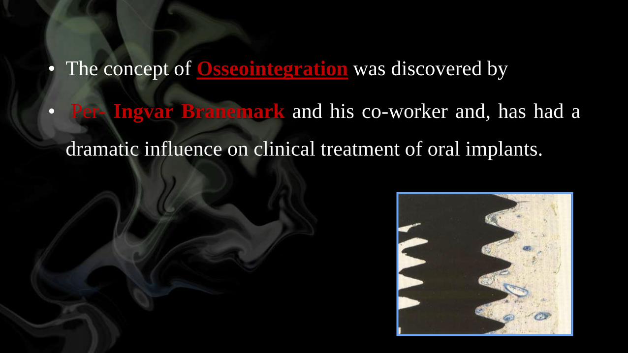
• The concept of Osseointegration was discovered by
• Per- Ingvar Branemark and his co-worker and, has had a
dramatic influence on clinical treatment of oral implants.

• The First generation titanium implants which were machined
with a smooth surface texture.
• Implant surfaces have been recognized to play an important
role in molecular interactions, cellular response and Osseo
integration.

• The Second generation implants with surface modification
can accelerate and improve implant osseointegration.
• Implants underwent mechanical blasting, acid etching,
bioactive coatings, more recently , laser modified surfaces.
• The main objective for the development of implant
surface modifications is to promote Osseo integration,
with faster and stronger bone formation.
• Furthermore, it accelerates the bone healing and thereby
allowing immediate or early loading .
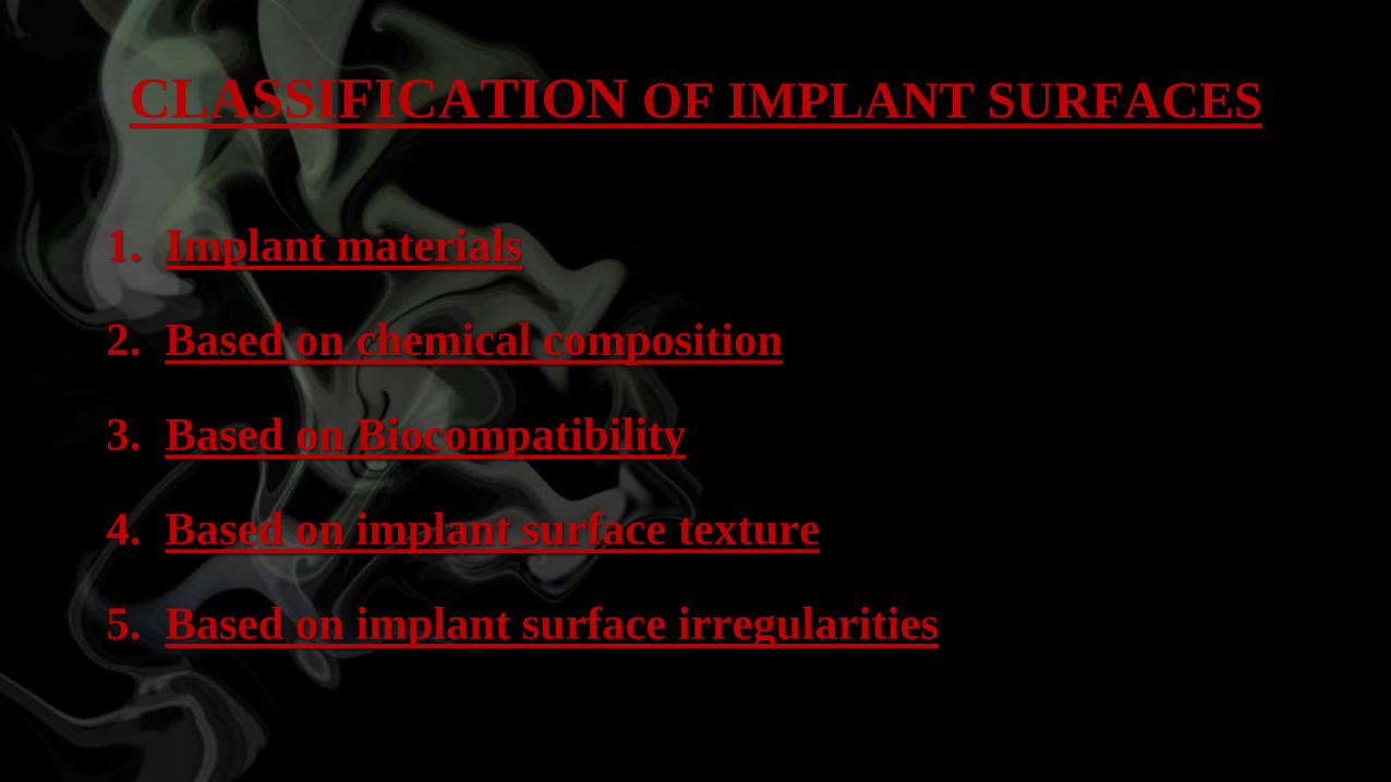
CLASSIFICATION OF IMPLANT SURFACES
1. Implant materials
2. Based on chemical composition
3. Based on Biocompatibility
4. Based on implant surface texture
5. Based on implant surface irregularities
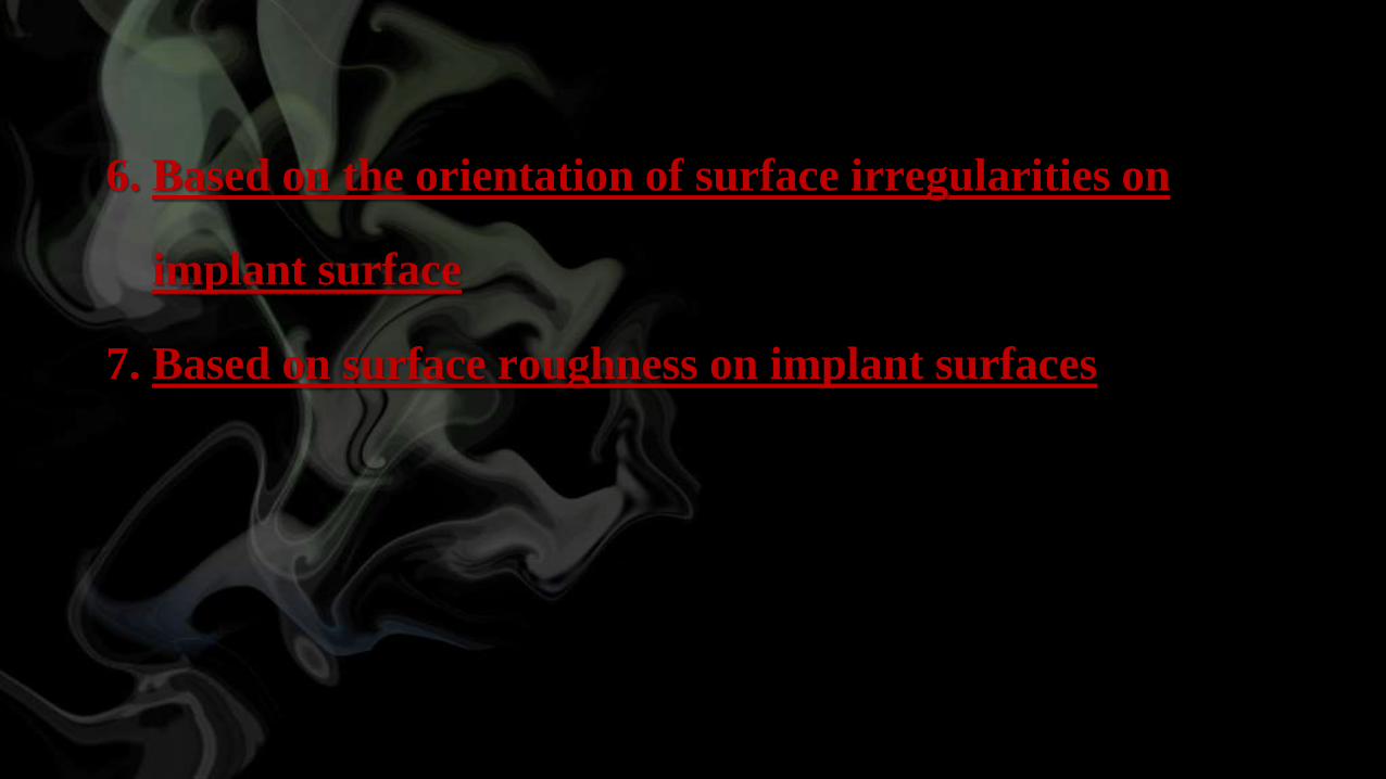
6. Based on the orientation of surface irregularities on
implant surface
7. Based on surface roughness on implant surfaces
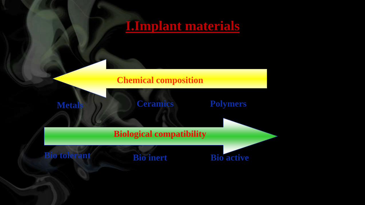
I.Implant materials
Metals Ceramics Polymers
Chemical composition
Biological compatibility
Bio inert Bio tolerant Bio active
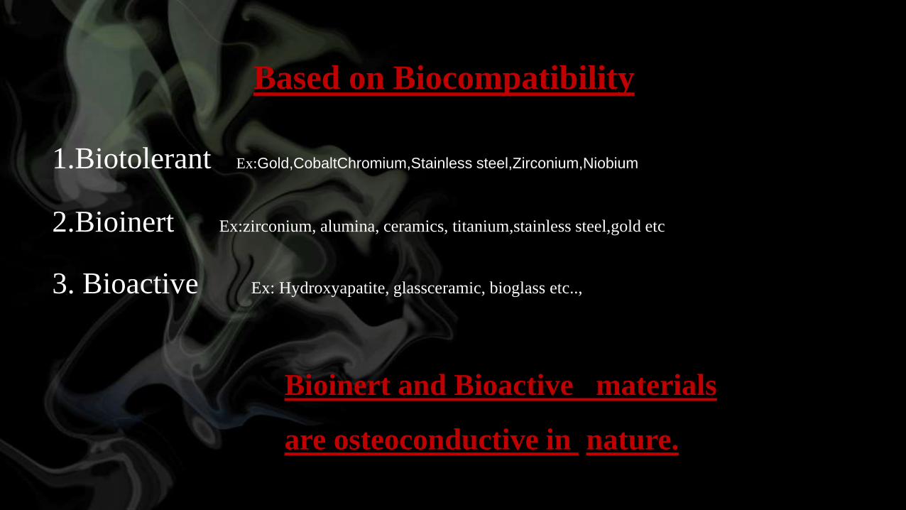
Based on Biocompatibility
1.Biotolerant Ex:Gold,CobaltChromium,Stainless steel,Zirconium,Niobium
2.Bioinert Ex:zirconium, alumina, ceramics, titanium,stainless steel,gold etc
3. Bioactive Ex: Hydroxyapatite, glassceramic, bioglass etc..,
Bioinert and Bioactive materials
are osteoconductive in nature.
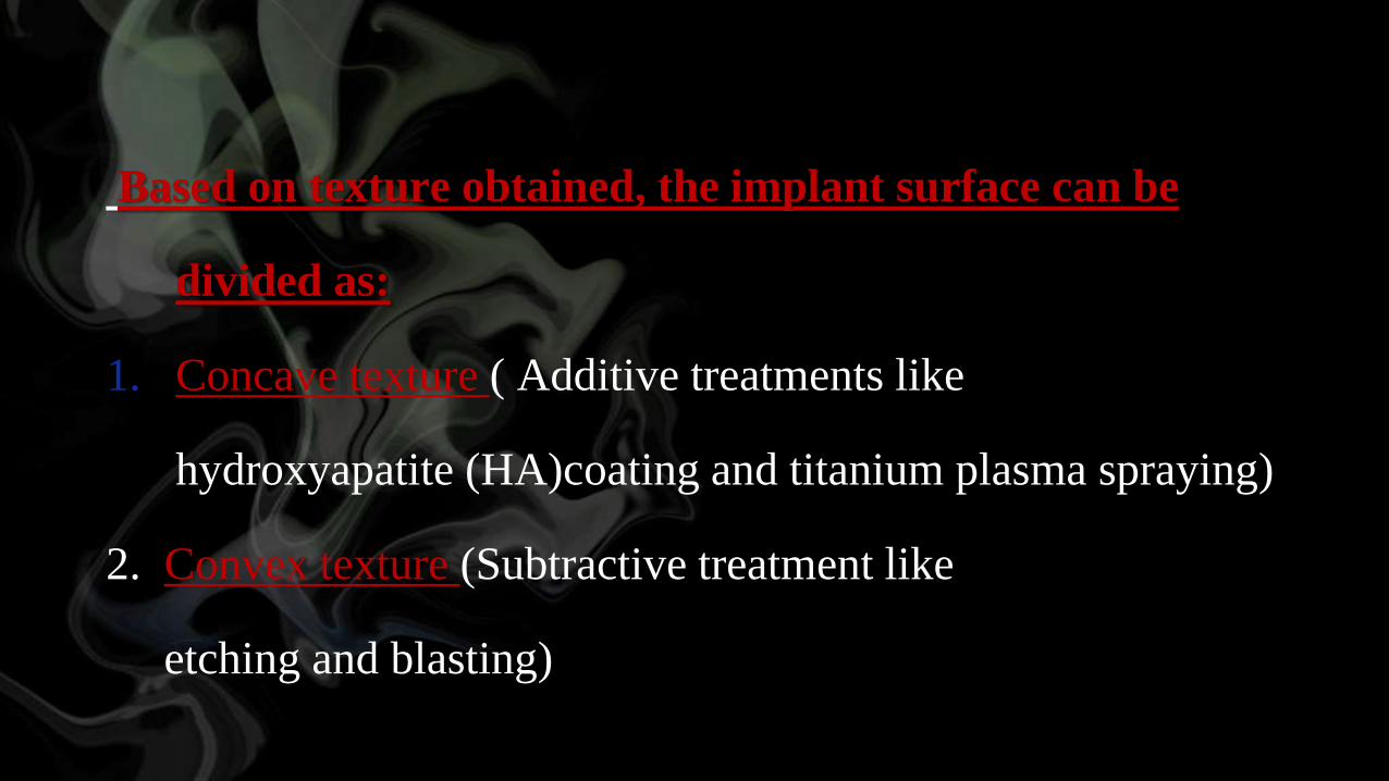
Based on texture obtained, the implant surface can be
divided as:
1. Concave texture ( Additive treatments like
hydroxyapatite (HA)coating and titanium plasma spraying)
2. Convex texture (Subtractive treatment like
etching and blasting)
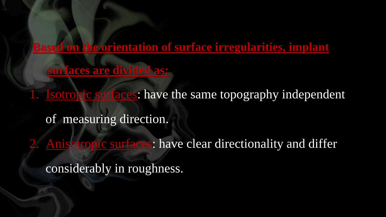
Based on the orientation of surface irregularities, implant
surfaces are divided as:
1. Isotropic surfaces: have the same topography independent
of measuring direction.
2. Anisotropic surfaces: have clear directionality and differ
considerably in roughness.
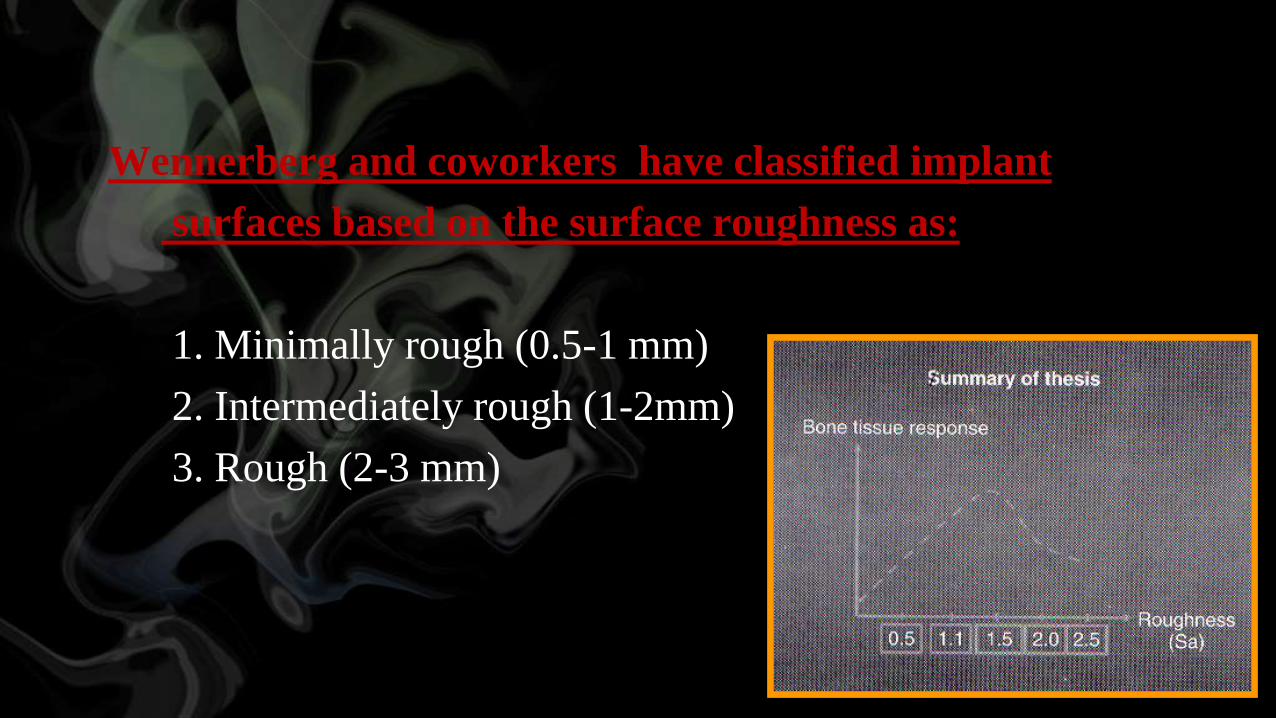
Wennerberg and coworkers have classified implant
surfaces based on the surface roughness as:
1. Minimally rough (0.5-1 mm)
2. Intermediately rough (1-2mm)
3. Rough (2-3 mm)
Methods to increase the surface roughness
1. Blasting
2. Chemical etching
3. Porous surfaces
4. Plasma-sprayed surfaces
5. Ion-sputtering coating
6. Anodized surface
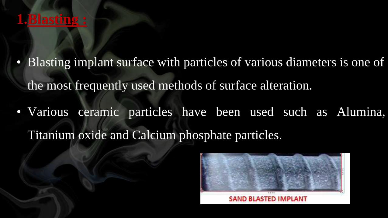
1.Blasting :
• Blasting implant surface with particles of various diameters is one of
the most frequently used methods of surface alteration.
• Various ceramic particles have been used such as Alumina,
Titanium oxide and Calcium phosphate particles.
2. Chemical etching
• Etching with strong acids such as HCl, H2SO4,HNO3 and
HF is used for roughening dental implants.
• Acid-etching produces micropits on implant surfaces with
sizes ranging from 0.5 to 2 μm in diameter. Acid- etching
has been shown to greatly enhance Osseointegration.

• Recently a new surface was introduced that was sandblasted
with large grit and acid-etched (SLA).
• This surface is produced with large grit (250-500 micro-
metres) blasting process and followed by Hydrochloric and
sulfuric acid.
3. Porous surfaces
• These are produced when spherical powder of the
metallic/ceramic material becomes a coherent mass within the
metallic core of the implant body.
• These are characterized by pore size, shape, volume and depth,
which are affected by the size of the spherical particles and the
temperature and pressure of the sintering chamber.
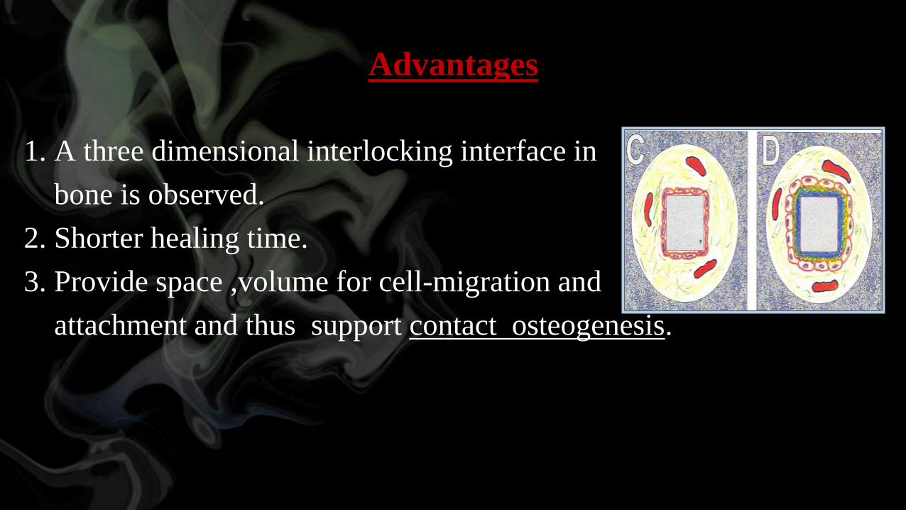
Advantages
1. A three dimensional interlocking interface in
bone is observed.
2. Shorter healing time.
3. Provide space ,volume for cell-migration and
attachment and thus support contact osteogenesis.
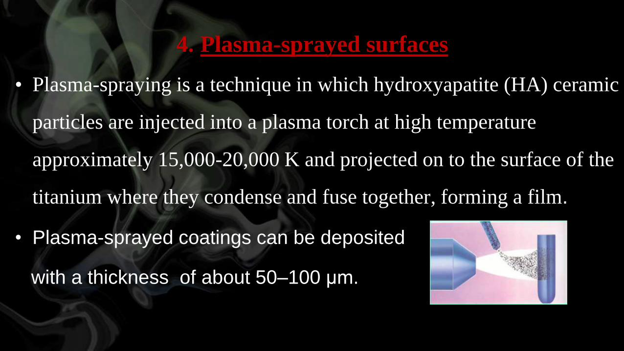
4. Plasma-sprayed surfaces
• Plasma-spraying is a technique in which hydroxyapatite (HA) ceramic
particles are injected into a plasma torch at high temperature
approximately 15,000-20,000 K and projected on to the surface of the
titanium where they condense and fuse together, forming a film.
• Plasma-sprayed coatings can be deposited
with a thickness of about 50–100 μm.

5. Ion-sputtering coating
• It is the process by which a thin layer of Hydroxyapatite can be
coated onto an implant substrate.
• This is performed by directing a beam of ion onto an HA block
that is vaporized to create plasma and then recondensing this
plasma onto the implant.
6.Anodized surface:
• Oxidation process can be used to change the characteristic of
the oxide layer and make it more biocompatible.
• This is carried out by applying a voltage on the titanium
implant immersed in the electrolyte.
• This results in a surface with micropores of
variable diameter and demonstrates lack of cytotoxicity
and increased cell attachment and proliferation.
Advantages of increased roughness:
1. Increased surface area of implant adjacent to bone.
2. Improved cell attachment to bone.
3. Increased bone present at implant interface.
4. Increased biochemical interaction of implant with bone.
Methods
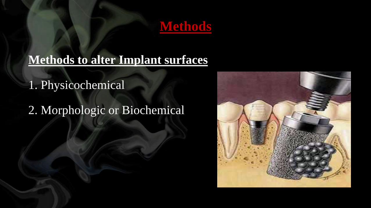
Methods to alter Implant surfaces
1. Physicochemical
2. Morphologic or Biochemical
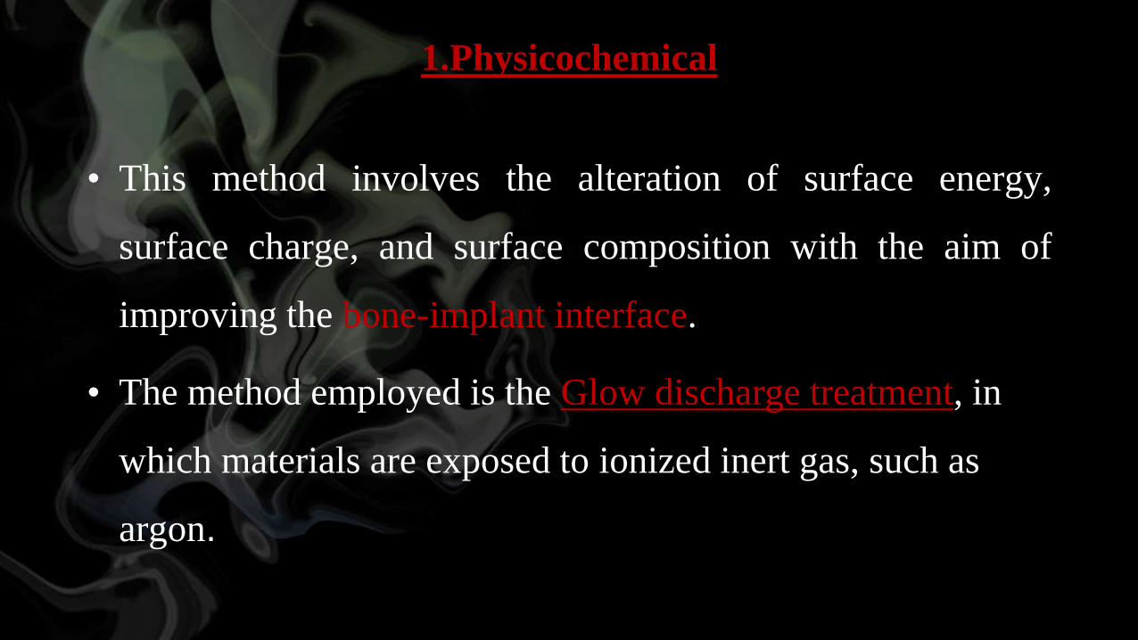
1.Physicochemical
• This method involves the alteration of surface energy,
surface charge, and surface composition with the aim of
improving the bone-implant interface.
• The method employed is the Glow discharge treatment, in
which materials are exposed to ionized inert gas, such as
argon.
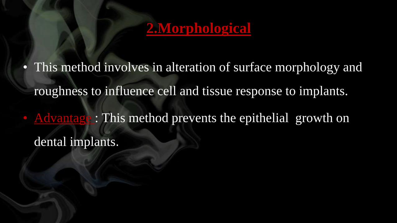
2.Morphological
• This method involves in alteration of surface morphology and
roughness to influence cell and tissue response to implants.
• Advantage : This method prevents the epithelial growth on
dental implants.
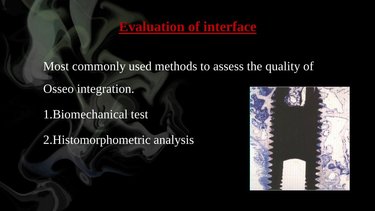
Evaluation of interface
Most commonly used methods to assess the quality of
Osseo integration.
1.Biomechanical test
2.Histomorphometric analysis
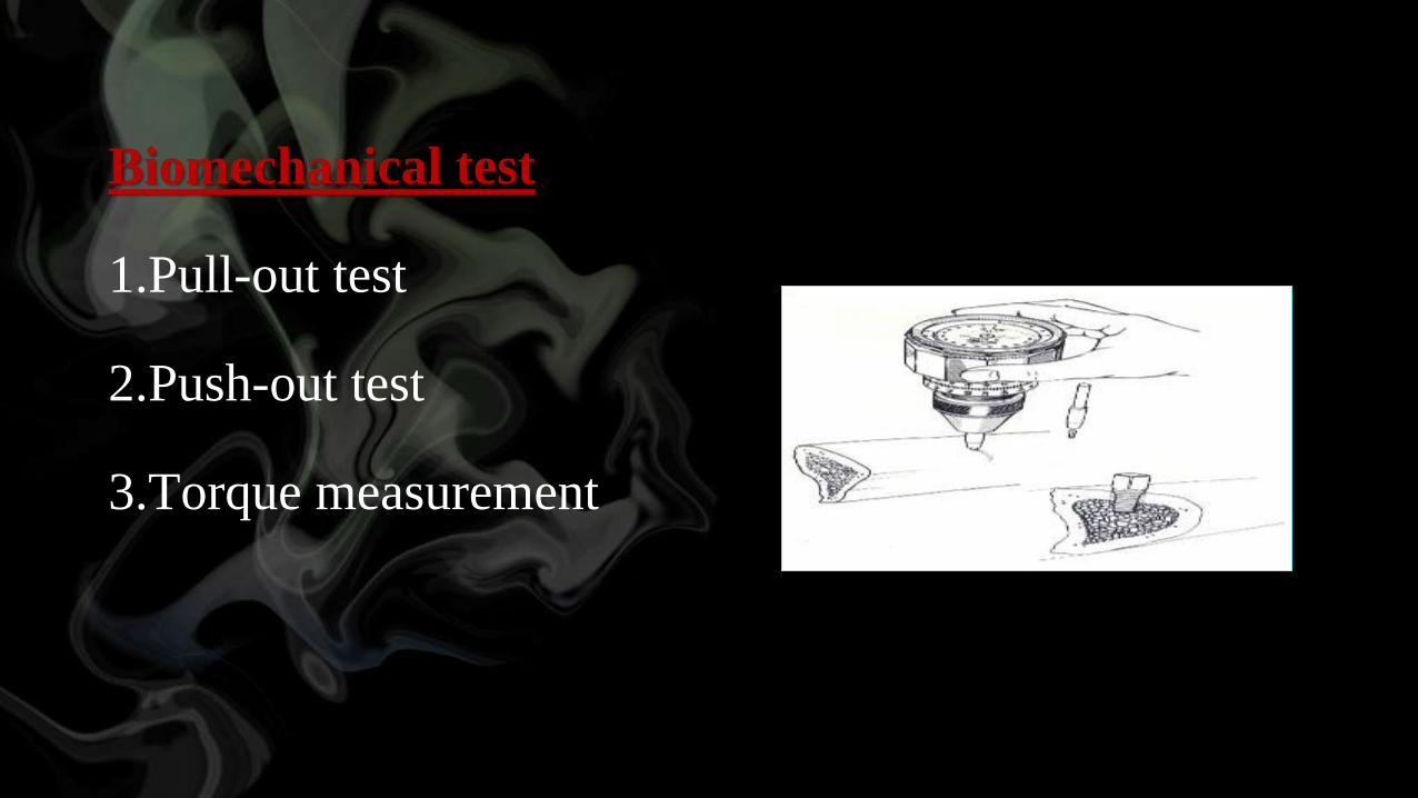
Biomechanical test
1.Pull-out test
2.Push-out test
3.Torque measurement
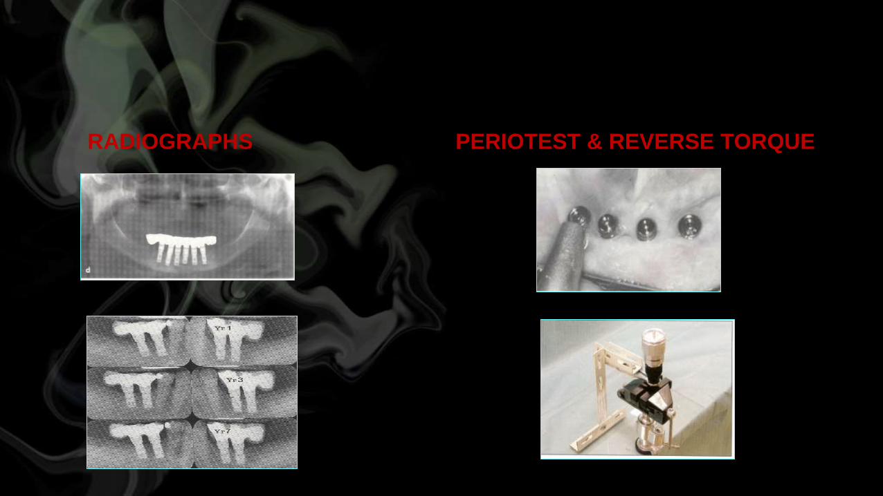
RADIOGRAPHS PERIOTEST & REVERSE TORQUE
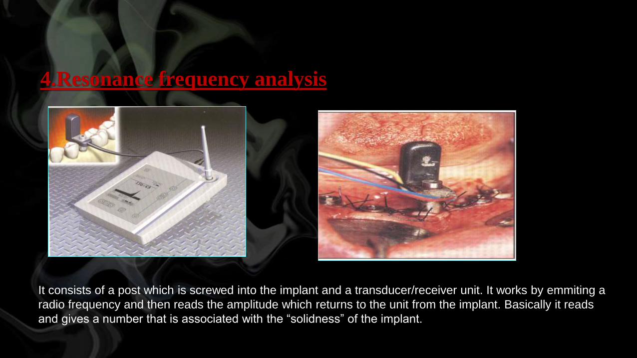
4.Resonance frequency analysis
It consists of a post which is screwed into the implant and a transducer/receiver unit. It works by emmiting a
radio frequency and then reads the amplitude which returns to the unit from the implant. Basically it reads
and gives a number that is associated with the “solidness” of the implant.

Conclusion
• There are number of surfaces commercially available for
dental implants.Various methods modifying the implant
surface have greatly influenced the quality of clinical
service in implant prosthodontics.
• Implant surface characterization and working knowledge
about how surface and bulk biomaterial properties inter
relate to implant osseo integration represent an important
area in implant based reconstructive surgery
REFERENCES:
1)INT J Oral Maxillofac Implants 2000;15:675-690
2)Indian Journal of Dental Sciences.(March 2012) 3) Wennerberg A, Albrektsson Suggested guidelines for the topographic evaluation of implant surfaces.
4)Int J Oral Maxillofac Implants 2000;15:331-44.
5) Brunette DM. The effects of implant surface topography on
the behavior of cells. Int J Oral Maxillofac Implants1988;3:231
6) Puleo DA, Thomas MV. ImplantSurfaces. Dent Clin North
Am 2006;50:323-338.

Thank you