implications of the intestinal microbiota in diagnosing...
TRANSCRIPT

Review ArticleImplications of the Intestinal Microbiota inDiagnosing the Progression of Diabetes and the Presence ofCardiovascular Complications
Alina Mihaela Leustean,1 Manuela Ciocoiu ,2 Anca Sava,3 Claudia Florida Costea,4
Mariana Floria ,5,6 Claudia Cristina Tarniceriu,7 and Daniela Maria Tanase 5,6
1Department of Gastroenterology, “Sf. Spiridon” County Clinical Emergency Hospital, “Grigore T. Popa” University of Medicineand Pharmacy, Iasi, Romania2Department of Pathophysiology, Faculty of Medicine, “Grigore T. Popa” University of Medicine and Pharmacy, Iasi, Romania3Department of Morpho-Functional Sciences I, Faculty of Medicine, “Grigore T. Popa” University of Medicine and Pharmacy,Iasi, Romania4Department of Ophthalmology, Faculty of Medicine, “Grigore T. Popa” University of Medicine and Pharmacy, Iasi, Romania5Department of Internal Medicine, “Grigore T. Popa” University of Medicine and Pharmacy, Iasi, Romania63rd Internal Medicine Clinic, “Sf. Spiridon” County Clinical Emergency Hospital, Iasi, Romania7Department of Morpho-Functional Sciences I, Discipline of Anatomy, Faculty of Medicine, “Grigore T. Popa” University of Medicineand Pharmacy, Iasi, Romania
Correspondence should be addressed to Mariana Floria; [email protected] Daniela Maria Tanase; [email protected]
Received 10 August 2018; Revised 6 October 2018; Accepted 21 October 2018; Published 12 November 2018
Guest Editor: Gaetano Santulli
Copyright © 2018 Alina Mihaela Leustean et al. This is an open access article distributed under the Creative Commons AttributionLicense, which permits unrestricted use, distribution, and reproduction in any medium, provided the original work isproperly cited.
The prevalence of diabetes is steadily rising, and once it occurs, it can cause multiple complications with a negative impact on thewhole organism. Complications of diabetes may be macrovascular: such as stroke and ischemic heart disease as well as peripheralvascular and microvascular diseases—retinopathy, nephropathy, and neuropathy. Key factors that cause cardiovascular disease inpeople with diabetes include hyperglycemia, dyslipidemia, obesity, insulin resistance, inflammation, hypertension, autonomicdysfunction, and decreased vascular response capacity. Microbes can be considered a complex endocrine system capable ofensuring the proper functioning of the body but are also responsible for the development of numerous pathologies (diabetes,coronary syndromes, peripheral arterial disease, neoplasia, Alzheimer’s disease, and hepatic steatosis). Changes in the intestinalmicrobiota may influence the host’s sensitivity to insulin, body weight, and lipid and carbohydrate metabolism. Dysbiosis causesactivation of proinflammatory mechanisms, metabolic toxicity, and insulin resistance. Trimethylamine N-oxide (TMAO) is amicrobial organic compound generated by the large intestine, and its concentration increases in the blood after ingestion offoods rich in L-carnitine and choline, such as red meat, eggs, and fish. The interest for TMAO in cardiometabolic research hasrecently emerged, given the preclinical evidence that reveals a link between TMAO, diabetes, and cardiovascular complications.Intestinal microbiota can be modulated by changing one’s lifestyle but also by antibiotic, probiotic, prebiotic, and fecaltransplantation. The purpose of this article is to highlight issues related to the involvement of microbiota and trimethylamineN-oxide in the pathogenesis of diabetes mellitus and cardiovascular disease. Better appreciation of the interactions between foodintake and intestinal floral-mediated metabolism can provide clinical insights into the definition of individuals with diabetic riskand cardiometabolic disease as well as potential therapeutic targets for reducing the risk of progression of the disease.
HindawiJournal of Diabetes ResearchVolume 2018, Article ID 5205126, 9 pageshttps://doi.org/10.1155/2018/5205126
1. Definition
Microbiota is part of a complex system that includes allmicro-organisms, cells, antimicrobial peptides, luminous com-pounds, and all interactions between them [1, 2].
The microbiota is involved in numerous activities suchas vitamin production, regulation of gene expression, fightagainst pathogenic bacteria, absorption of nutrients, and reg-ulation of metabolic disorders [3].
Intestinal microbiota is influenced by factors such asgenetics, lifestyle, diet, and antibiotherapy [2, 4, 5].
Microorganisms such as bacteria, viruses, and fungi sur-vive in the gastrointestinal tract. The intestinal microbiotais the result of the complex interaction between the environ-ment and host genetics; diet is the component that modulatesthe intestinal bacterial activity. An imbalance of intestinalhomeostasis causes the internal dispersion of bacterialfragments and promotes intestinal permeability and bacte-rial and circulating endotoxin translocation, which initiatesinflammation in tissues responsible for insulin metabolismthus causing insulin resistance [1, 6]. Dysbiosis also plays animportant role in the pathogenesis of cardiovascular andmetabolic diseases [7, 8].
The intestinal microbiota can be considered a gate thatmodulates the transition into cardiometabolic diseases thatinvolve the hepatobiliary tract [4, 7].
2. Microbiome Composition
Our body is colonized by a series of microbiota, primarilybacteria, which exist in a symbiotic relationship with the hostand play an important role in maintaining the homeostasis ofthe host [8]. The intestinal microbe is comprised of billions ofcells, of which the most important are Gram-positive bacteriabelonging to the phyla Firmicutes and Actinobacteria andalso to the genera Clostridium, Bifidobacterium, Lactobacil-lus, Ruminococcus, and Streptococcus and Gram-negativebacteria belonging to the genera Bacteroides, Prevotella,and Akkermansia [2].
The link between microbiota compounds and the host’simmune system is supported by a series of molecules and sig-naling processes that can affect the intestine, liver, brain, andother organs [9]. On the other hand, the intestinal immunesystem plays a significant role in the exposure of bacteriato host tissues, causing stratification of intestinal bacteriaon the lumbar side of the epithelial barrier, and controlsthe composition of the intestinal microbiota [4]. Residualbacteria provide signals that favor the development of anormal immune system and regulate the resulting immuneresponses. Changing these can cause significant repercus-sions on the host’s health [9]. The disruption of the intestinalmicrobe may also be associated with numerous pathologies:metabolic syndrome, obesity, diabetes, renal disease, cardio-vascular disease, neoplasia, and Alzheimer’s disease [2]. Clos-tridium species correlate negatively with glucose, HbA1c, andinsulin levels, whereas Lactobacillus species correlate posi-tively with glucose and HbA1c levels [10]. It has been demon-strated that a higher blood glucose concentration can bepredicted by reducing the proportion of anaerobes, especially
Bacteroides [11]. Markers of glucose metabolism disorders(e.g., insulin and insulin resistance-HOMA-IR) are com-monly associated with the microbial genotype, suggestingthat people with fewer genotypes are predisposed to meta-bolic disorders and secondarily to diabetes [11].
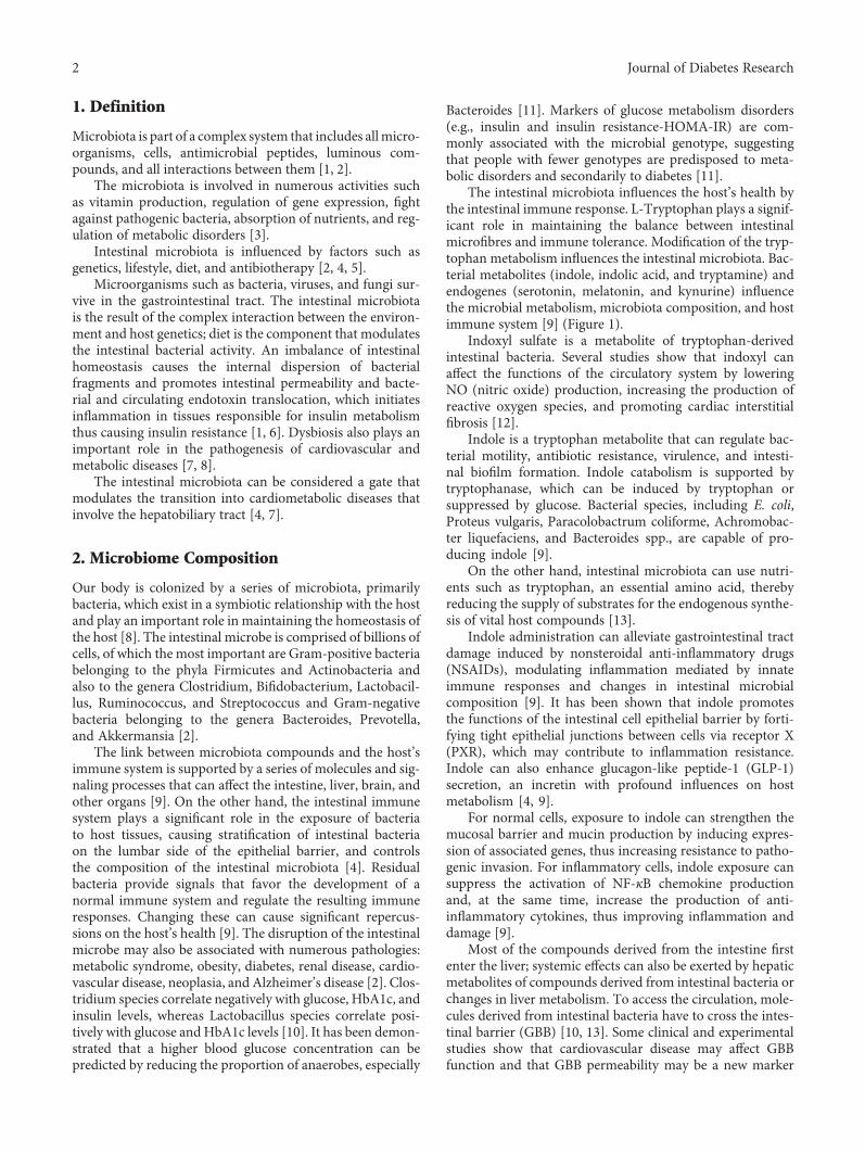
The intestinal microbiota influences the host’s health bythe intestinal immune response. L-Tryptophan plays a signif-icant role in maintaining the balance between intestinalmicrofibres and immune tolerance. Modification of the tryp-tophan metabolism influences the intestinal microbiota. Bac-terial metabolites (indole, indolic acid, and tryptamine) andendogenes (serotonin, melatonin, and kynurine) influencethe microbial metabolism, microbiota composition, and hostimmune system [9] (Figure 1).
Indoxyl sulfate is a metabolite of tryptophan-derivedintestinal bacteria. Several studies show that indoxyl canaffect the functions of the circulatory system by loweringNO (nitric oxide) production, increasing the production ofreactive oxygen species, and promoting cardiac interstitialfibrosis [12].
Indole is a tryptophan metabolite that can regulate bac-terial motility, antibiotic resistance, virulence, and intesti-nal biofilm formation. Indole catabolism is supported bytryptophanase, which can be induced by tryptophan orsuppressed by glucose. Bacterial species, including E. coli,Proteus vulgaris, Paracolobactrum coliforme, Achromobac-ter liquefaciens, and Bacteroides spp., are capable of pro-ducing indole [9].
On the other hand, intestinal microbiota can use nutri-ents such as tryptophan, an essential amino acid, therebyreducing the supply of substrates for the endogenous synthe-sis of vital host compounds [13].
Indole administration can alleviate gastrointestinal tractdamage induced by nonsteroidal anti-inflammatory drugs(NSAIDs), modulating inflammation mediated by innateimmune responses and changes in intestinal microbialcomposition [9]. It has been shown that indole promotesthe functions of the intestinal cell epithelial barrier by forti-fying tight epithelial junctions between cells via receptor X(PXR), which may contribute to inflammation resistance.Indole can also enhance glucagon-like peptide-1 (GLP-1)secretion, an incretin with profound influences on hostmetabolism [4, 9].
For normal cells, exposure to indole can strengthen themucosal barrier and mucin production by inducing expres-sion of associated genes, thus increasing resistance to patho-genic invasion. For inflammatory cells, indole exposure cansuppress the activation of NF-κB chemokine productionand, at the same time, increase the production of anti-inflammatory cytokines, thus improving inflammation anddamage [9].
Most of the compounds derived from the intestine firstenter the liver; systemic effects can also be exerted by hepaticmetabolites of compounds derived from intestinal bacteria orchanges in liver metabolism. To access the circulation, mole-cules derived from intestinal bacteria have to cross the intes-tinal barrier (GBB) [10, 13]. Some clinical and experimentalstudies show that cardiovascular disease may affect GBBfunction and that GBB permeability may be a new marker
2 Journal of Diabetes Research

in CVD. In this case, a key factor for the proper functioningof GBB is proper blood perfusion through the intestines [13].
Multiple studies suggest that intestinal microbiota couldproduce biologically active compounds that enter the circula-tion and affect circulatory system homeostasis. To enter thecirculation, intestinal bacterial metabolites have to passthrough the intestinal barrier (GBB). The integrity and per-meability of the intestinal barrier depend on numerous fac-tors, including intestinal blood flow [14].
The intestinal blood barrier is made up of several layers.The internal or mucosal layer prevents pathogens fromadhering to epithelial cells. The physical barrier consists ofa single layer of enterocytes connected by tight junctions,which play a crucial role in the selectivity of the intestinalbarrier [15].
Intestinal bacteria produce many vital nutrients forhuman homeostasis, such as vitamins K and B and SCFAwhich contribute to the transformation and degradation ofbiliary acids and steroids [13].
Intestinal metabolites such as hydrogen sulfide (H2S),SCFA, indole, or trimethylamine may exert effects on circula-tory system homeostasis and on nerve and humoral control.Short-chain fatty acids, including acetic, propionic, butyric,and valeric acids, are formed from carbohydrates by bacterialfermentation [14] (Figure 1). The amount of SCFA may havevasorelaxant effects on the arterial resistance in the colon,improving microcirculation. SCFA depends on the composi-tion of intestinal bacteria, diet, and intestinal transit time andplays a local role as an energy source for intestinal cells sup-pressing the growth of pathogens by reducing the pH in theintestines. There is also some evidence that SCFA derivedfrom intestinal bacteria can affect blood pressure [13].
Short-chain fatty acids (SCFA), produced by bacterialfermentation in the colon, contribute to a significant propor-tion of the daily energy requirement. SCFA, especially buty-rate and propionate, play an important role in differentiatingregulatory T cells and regulating immunity in the intestinaltract. Increased production of acetate by intestinal microbi-ota could lead to the activation of the parasympathetic ner-vous system, which promotes increased secretion of insulinstimulated by glucose, hyperphagia, and obesity [16].
Intestinal bacteria produce many biologically active mol-ecules, some of which play a role in regulating the circulatorysystem and energy balance. Besides numerous metabolites,intestinal flora produces methylamine, including TMA,dimethylamine, and monomethylamine. The intestinal bar-rier is considered a functional, immunological, and anatom-ical unit, separating the intestinal lumen from circulatingblood, preventing bacterial adherence and transport regula-tion. Methanolamine and other intestinal metabolites reachalmost all tissues as small molecules thus affecting both neu-rohormonal and peripheral regulatory mechanisms [17].
A fat-rich diet causes bowel dysbiosis and reduces intes-tinal integrity. Recent studies have shown that dysbiosismay contribute to the development of inflammation and sub-sequently the progression of cardiovascular disease (CVD) bypromoting two major risk factors—arterial hypertension andatherosclerosis [7, 10].
TMAO precursors such as choline, L-carnitine, γ-butyro-betaine, phosphatidylcholine, betaine, glycerophosphine, andcrotonobetaine are metabolized by the intestinal microbiotato produce trimethylamine which is then further metabolizedto TMAO by the monooxygenase 3 enzyme (FMO3) [18].Metabolizing TMAO by FMO3 was linked to insulin sensi-tivity and glucose metabolism [19]. Several studies have alsosuggested a significant role of choline in regulating insulinresistance and glucose metabolism. Diet significantly affectsthe intestinal microbiota and the production of TMAO [20].
FMO3 (flavin monooxygenase 3) is the preponderantenzyme in the liver, and flavin monooxygenase 1 and flavinmonooxygenase 2 (FMO1 and FMO2, respectively) can alsocause TMAO oxidation. In some patients with FMO3 genemutation, accumulation of trimethylamine (TMA) spreadsin the body and is released through sweating and breathing,resulting in fish smell syndrome, a genetic disease [20].
The plasma level of TMAO in the human organism is inthe range of 0.5-10μmol/L. Recently, a number of clinical tri-als have indicated a possible positive correlation betweenincreased plasma TMAO and an increased risk of cardiovas-cular disease [13].
Choline is an essential nutrient which is both synthesizedendogenously and obtained from various animal and plant
Gutmicrobiota
CholineL-Carnitine
Betaine
Trimethylamine
Tryptophan Indol
H2S
SCFA
Acetic acidPropionic acid
Butyric acid, valeric acid.
Figure 1: Gut microbiota metabolites.
3Journal of Diabetes Research

products [21, 22]. Food-derived choline is generally metabo-lized in the liver and is involved in various biological processes,synthesis of acetylcholine neurotransmitter, lipoprotein, andmembrane phospholipids [23]. Betaine is the direct oxidationproduct of choline, which is a metal donor in homocysteineremethylation, and plays an important role in maintainingstability and cellular volume [22]. Together, choline andbetaine have been recognized as achieving hepatoprotectionand improving insulin resistance [23].
The intestinal microbiota through enzyme activity canturn choline into trimethylamine (TMA), a harmful metabo-lite known for its strong ammonia smell. TMA is absorbedand transmitted to the liver, where it can be rapidly detoxifiedby monooxygenase 3 (FMO3) to be transformed into tri-methylamine N-oxide (TMAO) [23].
Significant conversion of choline into TMA by the intes-tinal microbiota may, however, reduce the bioavailability ofcholine, which could affect the secretion of very-low-densitylipoproteins, with increased accumulation of triglycerides inthe liver and could stimulate hepatic steatosis [22–24].
Blood TMAO levels depend on many factors, includingdiet, intestinal barrier permeability, liver enzyme activityand methylamine excretion rate, composition, and activityof the intestinal microfibres [25]. Changes in intestinalmicrobiota may influence the host’s sensitivity to insulin withthe onset of diabetes. Several studies have shown that thelevel of TMAO is significantly associated with the risk of type2 diabetes [26, 27].
Thus, FMO3 is suppressed by insulin, and also FMO3levels are elevated by glucagon secretion from pancreatic αcells to stimulate an increase in blood sugar [28]. Both gluca-gon suppression and insulin resistance are correlated withweight loss and act together to improve glucose homeostasisfollowing a dietary restriction with body mass loss [26, 29]. Ahigh-fat diet has led to changes in the intestinal microbialcomposition causing insulin resistance [30].
TMAO may cause inflammation of the adipose tissuewith disruption to the insulin signaling pathway. This mech-anism plays an important role in the emergence of insulinresistance and subsequently in the evolution towards diabetes[31–33]. The increase in TMAO levels may result from die-tary differences; intestinal microbiota plays an important rolein varying TMAO levels. L-Carnitine is essential for the mito-chondrial metabolism of long-chain fatty acids, and somestudies have provided evidence of glycemic control andplasma lipid control following L-carnitine administration intype 2 diabetes [29]. However, other studies demonstratethe increased risk of diabetic complications in patients withhigher circulating L-carnitine concentrations. The role ofL-carnitine in cardiovascular health was recognized afterdiscovering the proatherogenic nature of TMAO and itsrelationship to L-carnitine metabolism [26].
TMAO produces a proatherogenic macrophage pheno-type that affects the metabolism of cholesterol and sterol inmacrophages, intestines, and liver. Studies have shown thatdiabetes and body mass index (BMI) are associated withhigher levels of TMAO [27, 28]. After ingestion of phospha-tidylcholine or L-carnitine, circulating TMAO levels increasein 4 to 8 hours and normalize after 24 hours depending on
renal clearance [30]. Trimethylamine N-oxide (TMAO),which is derived from intestinal metabolite-derived metabo-lites, is possibly linked to diabetic, atherosclerotic, and car-diovascular risk [25]. The circulating TMAO levels areelevated and associated with the severity of the disease andwith patients with atherosclerosis, chronic kidney disease,and peripheral arterial disease [7, 8]. Previous studies haveshown that the bacterial species belonging to the familiesClostridiaceae and Peptostreptococcaceae have been associ-ated with increased blood levels of TMAO in humans [8].
TMAO generates atherosclerosis, perhaps by formingfoam cells in the arterial wall. High levels of TMAO affectlipid metabolism, and inflammatory response promotesendothelial dysfunction and exacerbation of platelet reactiva-tion and stimulates thrombosis. This highlights the impor-tance of this molecule for cardiovascular complications [28].
TMAO activates in vascular smooth muscle cells andendothelial cells MKKK (mitogen-activated protein kinases),and the isolation of nuclear factor-κB (NF-κB) leads toincreased expression of inflammatory genes and adhesionof endothelial cells to leukocytes. TMAO in vivo can increasethe receptor expressed on CD36 and SR-A1, leading to theformation of foam cells, by a greater absorption of modifiedmacrophage LDL. Furthermore, TMAO increases calciumconcentration in the endoplasmic reticulum in the platelets,which consequently leads to platelet aggregation and throm-bosis with increased risk of acute coronary syndromes [20](Figure 2).
TMAO activates prothrombotic pathways by rapidlyincreasing the release of calcium ions (Ca2+) resulting in theactivation of certain platelet stimuli. In endothelial cells andsmooth muscle cells, TMAO rapidly activates the mitogen-activated protein kinase and the activated B cell activatedkappa amplifier factor, which in turn favors the expressionof adhesion molecules such as E-selectin [20]. TMAO canregulate and differentiate monocytes into foam cells andmac-rophages. TMAO can initiate profibrotic processes in theheart and kidneys by transforming the phospho-SMAD3growth factor-β signaling axis [20, 34]. The association ofall these complex cellular mechanisms accelerates atheroscle-rosis and thrombotic vascular disease and leads to secondaryrenal insufficiency [35]. Therefore, understanding the molec-ular mechanism of action of TMAO and discovering newmechanisms and receptors by which TMAO may lead to itsadverse effects will have a much wider implication in clarify-ing its role in pathogenesis in humans [20, 35].
Increased levels of TMAO and choline are associatedwith low levels of HDL-cholesterol and plasma phospho-lipids [29]. Increased concentrations of TMAO in the bloodinfluence the activity of intestinal microbiota and the perme-ability of the intestinal barrier and determine the activity ofliver enzymes. Therewas a direct relationship between plasmaTMAO concentrations, diabetes mellitus, acute coronarysyndromes, and peripheral vascular disease [25, 36].
Furthermore, some experimental studies show thatTMAO should affect lipid and hormone homeostasis, pro-viding indirect evidence for the possible contribution ofTMAO to the development of CVD. It has been found thatTMAO could lead to decreased beta-oxidation of fatty acids
4 Journal of Diabetes Research

through cardiac muscle cells. In addition to TMA, othermetabolites have also been reported to play a role in thepathology of many diseases [16].
Indoxyl sulfate is produced by intestinal microbial tryp-tophanases that convert food tryptophan to indole, which isthen transformed into indoxyl and indoxyl sulfate in theliver. It has been shown that indoxyl sulfate may have proin-flammatory and prooxidant effects on cardiomyocytes andcardiac fibroblasts [16].
It is believed that complex molecules mediate intracellu-lar and extracellular signaling, but it also appears that gas-eous molecules, later referred to as “gas transmitters,” playan essential role in maintaining the body’s homeostasis.The gas transmitters include carbon monoxide (CO), hydro-gen sulfide (HS), and nitric oxide (NO), the cytotoxic mole-cule produced by phagocytic leukocytes [37].
The intestinal microbe uses sulfur-containing com-pounds to produce hydrogen sulfide. Hydrogen sulfide is animportant biological mediator that is involved in variousphysiological processes, including blood pressure regulation[13, 16]. Moreover, phenylacetylglutamine is a product thatis formed by the conjugation of phenylacetate and glutamine.Increased serum concentrations of phenylacetylglutamineshould be a strong and independent risk factor for overallmortality and cardiovascular disease. Further studies areneeded to elucidate the causal relationship between thesemetabolites and CVD [16].
3. Diabetes, Cardiovascular Diseases,and Microbiota
When the pancreas does not produce enough insulin or whenthe body cannot use insulin produced by the pancreas, diabe-tes can occur [38].
Diabetes mellitus is associated with an alteration of inter-dependent metabolic pathways (phospholipids, lipids, andmethylation) and also with diseases such as retinopathy,nephropathy, neuropathy, and heart failure [36, 39].
Diabetes mellitus is a major risk factor for cardiovasculardisease (CVD), which is the most common cause of deathamong adults with diabetes. The link between hyperglycemicstatus and microvascular disease is much more commonthan the link between hyperglycemic status and macrovascu-lar disease, with a 37% increase in risk of renal failure or ret-inopathy [40].
Prevalence of diabetes increases with age. Diabetespatients are at a higher risk of developing cardiovascular dis-ease. Inflammation and oxidative stress have a role in the
mechanisms underlying cardiovascular disease and othercomplications in the development of diabetes [41]. Diabeticshave a two to four times higher risk of developing cardiovas-cular complications and premature death. Myocardial ische-mia is frequently asymptomatic in patients with diabetes andis associated with an unfavorable prognosis [42].
Diabetes causes various microvascular complications,such as autonomic and peripheral neuropathy, nephropathy,and retinopathy, and these complications are correlated withadverse cardiovascular effects [43].
Atherosclerosis of the large arteries and coronary arteriesleads to macrovascular complications such as stroke, ische-mic heart disease, and peripheral vascular disease. Athero-sclerosis of small arteries causes diabetic nephropathy andis related to cardiovascular morbidity. Diabetes, regardlessof its effect on atherosclerosis, is associated with changes incardiac structure and function leading to myocardial dys-function, called “metabolic cardiomyopathy” [42].
The presence of a metabolic syndrome exposes patientsto an increased risk of cardiovascular complications. More-over, in addition to classical risk factors, there are otherunconventional factors that cause vasoconstriction andthrombosis, endothelial dysfunction, inflammation, oxida-tive stress, and vascular wall abnormalities that also contrib-ute to an increased cardiovascular risk [40]. When theglycemic level falls below 70mg/dL, autonomic nerve activa-tion occurs. This may produce symptoms such as tremor,tachycardia, diaphoresis, anxiety, hunger, and headache [39,40]. There are several mechanisms through which hypoglyce-mia could promote adverse cardiovascular effects in high-risk individuals. Hemodynamic changes following autono-mous self-induced hypoglycemia include increased systolicblood pressure, heart rate, myocardial contractility, and car-diac output [36].
These effects can exacerbate ischemia. Hypoglycemia, asa complication of long-term diabetes, has also been associ-ated with a prolonged QT interval. The relationship betweenhypoglycemia, autonomic neuropathy, and cardiac repolari-zation may contribute to arrhythmias and the risk of suddendeath in people with diabetes [38]. Finally, hypoglycemiamay have adverse effects on endothelial function, plateletreactivation, and coagulation cascade, and this increasesblood viscosity and decreases serum potassium levels [40].
Concerns about cardiovascular complications associatedwith type 2 diabetes have traditionally focused on athero-sclerotic vasculo-occlusive events such as myocardialinfarction, stroke, and limb ischemia [41]. However, oneof the most common and most serious cardiovascular
FMO3TMA → TMAO
(i)
(ii)
↑Ca → activate prothrombotic pathways → thrombosis strokemyocardial infarction
Differentiate monocyte into macrophages and foam cells → atherosclerosis
(iii) Myocardial fibrosis
(iv) Decreasing nitric oxide → ↑adverse cardiac remodeling
Figure 2: Effect of TMAO on cardiovascular disease.
5Journal of Diabetes Research

disorders in patients with diabetes is cardiac insufficiency.Cardiac insufficiency and diabetes are physiologicallyrelated [42, 43]. Type 2 diabetes and heart failure have acommon insulin resistance characteristic and are accompa-nied by the activation of the cascade of neurohormonalsystems: norepinephrine, angiotensin II, aldosterone, andneprilysin. The two diseases overlap; diabetes is present ina proportion of 35-45% of patients with chronic heart fail-ure, regardless of whether they have a reduced or conservedejection fraction [44].
Reduced blood flow from the intestinal endothelium inpatients with heart failure is due to a decreased cardiacoutput which causes intestinal wall ischemia, leading todisruption of the intestinal barrier function, increasing per-meability [40, 43].
Systemic congestion in patients with heart failure mayalso cause edema of the intestinal wall, resulting in increasedintestinal permeability. Thus, a translocation of endotoxins,microbial metabolites, and microbial components producedby Gram-negative bacteria entering the systemic circulationis determined at the intestinal level. These processes may fur-ther activate cytokines and may generate systemic inflamma-tion that contributes to the progression of heart failure [7].There is evidence of chronic heart failure (CHF) and gastro-intestinal (GI) involvement in this syndrome. It is knownthat cytokine activation occurs in patients with chronicNYHA class III-IV heart failure, with both clinical severityand prognosis. It has been suggested that endotoxin may bean important stimulant for the production of cytokines inpatients with chronic heart failure by its action on mononu-clear cells [43–45]. According to this hypothesis, endotoxinenters the circulation through bacterial translocation in theintestine. The two main factors are intestinal edema andhypoperfusion. The finding that patients with edematousheart failure decompensation have elevated levels of endo-toxin normalizing after diuretic treatment tends to suggestthat edema of the intestinal wall may contribute to endotox-emia. In addition, there is evidence that intestinal hypoperfu-sion may result in mucosal ischemia, which may lead toincreased intestinal wall permeability. Furthermore, theintestine may play an important role in the development ofcardiac cachexia [45].
Bowel dysfunction and dysbiosis may contribute to met-abolic disease. Diabetes mellitus and cardiovascular diseasecan compromise intestinal function by producing macro-and microangiopathy. In cardiac failure, there is centraliza-tion of circulation that further reduces intestinal perfusion.Intestinal ischemia leads to the progressive deterioration ofthe connections between enterocytes and an accelerated pas-sage through the blood barrier [15].
Excessive intake of salt is considered a cardiovascular riskfactor. Studies suggest that metabolites from intestinal bacte-ria, such as trimethylamine N-oxide (TMAO), are consideredto be a potential marker of cardiovascular diseases and mayaffect homeostasis [12, 16]. Increased evidence suggests thathomeostasis may very much depend on a mutualist relation-ship with intestinal bacteria and that CVD is associated withintestinal microbial dysbiosis. Research has shown that highsalt intake is associated with increased TMAO in plasma
and reduced urinary excretion of TMAO. Furthermore, ithas been found that increased salt intake affects the intestinalmicrobial composition [12].
High blood pressure is a major risk factor for heart fail-ure, coronary artery disease, and stroke, causing morbidityand high mortality. Hypertension is known to produce path-ological changes in the vasculature, such as microangiopathyin the retina, kidneys, and other organs. However, there isinsufficient data on the effect on hypertension in the intesti-nal vasculature [13, 14, 16].
A positive correlation between trimethylamine N-oxidein plasma at birth (TMAO) and the possible increased riskof major cardiovascular adverse events has been suggested;however, the value of diagnosing TMAO levels in blood incardiovascular diseases is questionable. However, if the nutri-ent concentration exceeds the transport capacity of the smallintestine, they reach the large intestine and are metabolizedby intestinal bacteria that produce trimethylamine (TMA).Therefore, the concentration of TMAO in the blood maydepend on a few factors, including diet, intestinal microbialactivity, GBB permeability to TMA, liver and TMA oxida-tion, and TMA and TMAO excretion [14].
Dysbiosis can contribute to the evolution of high bloodpressure, another risk factor for cardiovascular disease. Dys-biosis promotes hypertension by modifying vascular toneand developing vascular fibrosis [39]. Hypertension can bedefined as reducing the arterial lumen with increased periph-eral vascular resistance, resulting in increased blood pressure(BP). Also, intestinal dysbiosis contributes to hypertensionby vasoconstriction induced by LDL oxidation [8].
High blood pressure is a major factor contributing to anincreased risk of cardiovascular disease in patients with dia-betes. The presence of hypertension in patients with type 2diabetes increases the risk of myocardial infarction, stroke,and all-cause mortality. Combining both conditions increasesthe risk of heart failure, nephropathy, and other microvascu-lar events [30, 40].
There is an association between diabetes mellitus andatrial fibrillation. Both have common precursors of hyperten-sion, atherosclerosis, and obesity. Diabetes results fromdefects in insulin and glucose control. This, in turn, candirectly affect the atrial and ventricular myocardium. Theunderlying mechanism of atrial fibrillation may be linked toinflammation, with high levels of C-reactive protein and alsoatrial fibrosis. Diabetes is also associated with the formationof proinflammatory mediators. Left ventricular hypertrophy(LVH) is a common consequence of high blood pressure,and both are recognized risk factors for atrial fibrillation.Multiple studies have shown an association with left ventric-ular hypertrophy and low glucose tolerance and insulin resis-tance [46].
Microbial intestinal changes have been linked to changesin insulin sensitivity and in glucose metabolism and thedevelopment of metabolic syndrome with diabetes and sub-sequent cardiovascular complications [26]. Reduced globalmicrobial diversity in subjects with type 2 diabetes mellitus,diminution of Firmicutes bacteria (including Clostridia),and the occurrence of proteobacteria correlate with increasedplasma glucose in the oral glucose tolerance test [47]. In
6 Journal of Diabetes Research

diabetic patients, TMAO was found to be a significantmarker for cardiovascular events. In addition, L-carnitineplasma concentrations in patients with high TMAO concen-trations predicted an increased risk of cardiovascular diseaseand an increased incidence of major cardiac events [2, 6].
High plasma TMAO levels are associated with diastolicdysfunction and increased morbidity and mortality. TMAOmay also cause ventricular remodeling by fibrosis, subse-quent dilatation, thinning of the walls, and reduction of theejection fraction [21]. Similarly, elevated levels of cholineand betaine showed only an increased cardiovascular riskwhen associated with the concomitant increase in TMAO.These studies have enhanced the importance of dietary andantimicrobial therapy in cardiovascular health; TMAO levelis a possible target for therapeutic interventions [27].
4. Treatment
Changing one’s lifestyle can lead to a reduction in the risk ofchronic illness, including obesity and diabetes [48].
Cardiovascular disease, the leading cause of death world-wide, poses an interest in investigation of intestinal microbi-ota as an interventional mechanism which yields new andclinically relevant information for future research and has acomplex therapeutic potential [49].
Intervention on intestinal microbiota has already becomea new target for both the prevention and treatment of com-plex cardiometabolic diseases [4].
The intestinal microbiota can be modulated by theadministration of antibiotics, prebiotics, and probiotics orby fecal transplantation [26].
Prebiotics have protective effects and may reduce the riskof cardiovascular disease and diabetes by having a positiveimpact on the growth of beneficial microbial flora. Probioticsare new therapies for treating hypercholesterolemia [2, 50].
The administration of probiotics stimulates the immuneresponse, improves lactose tolerance, has anti-inflammatoryeffect, and even regulates intestinal disorders caused by obe-sity [1, 49]. Probiotics are living nonpathogenic microorgan-isms that provide benefits to the host [1, 21, 43]. Theintestinal microbiota may play an important role in the path-ogenesis of type 2 diabetes, influencing body weight, proin-flammatory activity, bile acid metabolism, insulin resistance,and intestinal hormone modulation [51].
Fecal transplantation may reduce the risk of obesity, type2 diabetes, insulin resistance, and increased BMI [1].
Modulation of intestinal microbiota by using probiotics,prebiotics, antibiotics, and fecal transplantation may havebenefits in improving glucose metabolism and insulin resis-tance [26]. Resveratrol (RSV) is a natural polyphenol withprebiotic benefits found mainly in grapes and berries. Fur-thermore, 3-dimethyl-1-butanol (DMB) is a structural ana-logue of choline and an inhibitor of TMA formation byinhibiting microbial enzymes [26, 51].
The interest in the study of resveratrol has started fromthe fact that the incidence of cardiovascular disease can bedecreased by consumption of 150-300mL/day of red wine.This has led to the extensive use of resveratrol in food supple-ments with doses ranging from 10-20mg [26]. Possible
mechanisms involve alteration of eicosanoid synthesis, lipidmetabolism, and platelet function, as well as inflammatoryresponse, downregulation of proinflammatory mediators,and inhibition of activated immune cells, mainly representedby neutrophils and macrophages. One of the possible mech-anisms involves reducing the regulation of inflammatoryresponse by inhibiting the synthesis and release of proinflam-matory mediators, modifying the synthesis of eicosanoidsand inhibiting activated immune cells [51]. Also, 3,3-dimethyl-1-butanol (DMB) is a choline analogue thatinhibits TMA-lyses, a family of bacterial enzymes that trans-forms multiple substrates into TMA. It is active against thesynthesis of TMA not only from choline but also from L-carnitine [52].
Moreover, 3,3-dimethyl-1-butanol is found in certainbalsamic vinegars, red wines, and some olive and grape seedoils [46]. DMB promotes microbial taxonomy reductionassociated with low plasma levels of TMA and TMAO andhas also reduced colonic dietary dependence in developingatherosclerotic lesions [29].
In addition, another drug has been described a few yearsago, already known to have cardioprotective clinical effects,by lowering the L-carnitine content in the body [46]. Thiscompound, meldonium (also called Mildronate), has beenshown to decrease TMAO by preventing the use of bacterialL-carnitine [52, 53].
5. Conclusions
Type 2 diabetes is a complex metabolic disease where con-comitant insulin resistance and beta cell damage lead tohyperglycemia. Its proliferation is rapidly and progressivelyincreasing due to an increase in prevalence of obesity andmaintaining a western lifestyle in developing countries. Asso-ciated complications that come about at some point arerelated to the major causes of morbidity, mortality, andexceptional healthcare costs. Nowadays, there are no inter-ventional clinical studies showing the beneficial effect ofmodulating microbiota in CVDs or diabetes.
All available clinical studies are only observational.Cardiovascular disease (CVD) is a major health problem
worldwide. Prospective studies should have demonstratedthat patients with diabetes have a two- or four-fold tendencyto develop heart failure and acute coronary syndromes,establishing that type 2 DM is an independent risk factorfor stroke and heart disease.
In this article, we highlighted aspects relating the involve-ment of themicrobiota and itsmetabolites to the pathogenesisof diabetes and cardiovascular complications. The profoundunderstanding of the mechanisms involved will allow theearly detection of diabetic patients with cardiovascular riskand the formulation of therapeutic regimens in order toreduce the risk of disease progression.
Conflicts of Interest
The authors declare that there are no conflicts of interestregarding the publication of this paper.
7Journal of Diabetes Research

References
[1] M. Le Barz, F. F. Anhê, T. V. Varin et al., “Probiotics as com-plementary treatment for metabolic disorders,” Diabetes &Metabolism Journal, vol. 39, no. 4, pp. 291–303, 2015.
[2] A. Chwalba and E. Otto-Buczkowska, “Participation of themicrobiome in the pathogenesis of diabetes mellitus,” ClinicalDiabetology, vol. 6, no. 5, pp. 178–181, 2017.
[3] J.-Y. Yang and M.-N. Kweon, “The gut microbiota: a keyregulator of metabolic diseases,” BMB Reports, vol. 49, no. 10,pp. 536–541, 2016.
[4] L. Miele, V. Giorgio, M. A. Alberelli, E. De Candia,A. Gasbarrini, and A. Grieco, “Impact of gut microbiota onobesity, diabetes, and cardiovascular disease risk,” CurrentCardiology Reports, vol. 17, no. 12, p. 120, 2015.
[5] P. Pokrzywnicka and J. Gumprecht, “Intestinal microbiota andits relationship with diabetes and obesity,” Clinical Diabetol-ogy, vol. 5, no. 5, pp. 164–172, 2016.
[6] C. L. Boulangé, A. L. Neves, J. Chilloux, J. K. Nicholson, andM.-E. Dumas, “Impact of the gut microbiota on inflammation,obesity, and metabolic disease,” Genome Medicine, vol. 8,no. 1, p. 42, 2016.
[7] T. Kitai andW. H.W. Tang, “Gut microbiota in cardiovasculardisease and heart failure,” Clinical Science, vol. 132, no. 1,pp. 85–91, 2018.
[8] K. Lau, V. Srivatsav, A. Rizwan et al., “Bridging the gapbetween gut microbial dysbiosis and cardiovascular diseases,”Nutrients, vol. 9, no. 8, p. 859, 2017.
[9] J. Gao, K. Xu, H. Liu et al., “Impact of the gut microbiota onintestinal immunity mediated by tryptophan metabolism,”Frontiers in Cellular and Infection Microbiology, vol. 8, p. 13,2018.
[10] L. J. Kasselman, N. A. Vernice, J. DeLeon, and A. B. Reiss, “Thegut microbiome and elevated cardiovascular risk in obesityand autoimmunity,” Atherosclerosis, vol. 271, pp. 203–213,2018.
[11] N. M. Delzenne, P. D. Cani, A. Everard, A. M. Neyrinck, andL. B. Bindels, “Gut microorganisms as promising targets forthe management of type 2 diabetes,” Diabetologia, vol. 58,no. 10, pp. 2206–2217, 2015.
[12] K. Bielinska, M. Radkowski, M. Grochowska et al., “High saltintake increases plasma trimethylamine N-oxide (TMAO)concentration and produces gut dysbiosis in rats,” Nutrition,vol. 54, pp. 33–39, 2018.
[13] M. Ufnal and A. Nowiński, “Gut bacteria-derived molecules asmediators and markers in cardiovascular diseases. The role ofthe gut-blood barrier,” Kardiologia Polska, vol. 76, no. 2,pp. 320–327, 2018.
[14] K. Jaworska, T. Huc, E. Samborowska et al., “Hypertension inrats is associated with an increased permeability of the colon toTMA, a gut bacteria metabolite,” PLoS One, vol. 12, no. 12,article e0189310, 2017.
[15] M. Ufnal and K. Pham, “The gut-blood barrier permeability –a new marker in cardiovascular and metabolic diseases?,”Medical Hypotheses, vol. 98, pp. 35–37, 2017.
[16] N. Yoshida, T. Yamashita, and K.-i. Hirata, “Gut microbiomeand cardiovascular diseases,”Diseases, vol. 6, no. 3, p. 56, 2018.
[17] Y. Heianza, D. Sun, X. Li et al., “Gut microbiota metabolites,amino acid metabolites and improvements in insulin sensitiv-ity and glucose metabolism: the POUNDS Lost trial,” BMJJournal-Gut, vol. 1, pp. 1–8, 2018.
[18] G. G. Schiattarella, A. Sannino, E. Toscano et al., “Gutmicrobe-generated metabolite trimethylamine-N-oxide ascardiovascular risk biomarker: a systematic review and dose-response metaanalysis,” European Heart Journal, vol. 38,no. 39, pp. 2948–2956, 2017.
[19] Z. Wang and Y. Zhao, “Gut microbiota derived metabolites incardiovascular health and disease,” Protein & Cell, vol. 9, no. 5,pp. 416–431, 2018.
[20] A. L. Komaroff, “The microbiome and risk for obesity anddiabetes,” The Journal of the American Medical Association,vol. 317, no. 4, pp. 355-356, 2017.
[21] G. M. Barlow, A. Yu, and R. Mathur, “Role of the gut micro-biome in obesity and diabetes mellitus,” Nutrition in ClinicalPractice, vol. 30, no. 6, pp. 787–797, 2015.
[22] W. H. W. Tang, Z. Wang, X. S. Li et al., “Increased trimethyla-mine N-oxide portends high mortality risk independent ofglycemic control in patients with type 2 diabetes mellitus,”Clinical Chemistry, vol. 63, no. 1, pp. 297–306, 2017.
[23] J. Sun and P. K. Dudeja, Mechanisms Underlying Host-Microbiome Interactions in Pathophysiology of Human Dis-eases, The American Physiological Society by Springer, 2018.
[24] S. Rath, B. Heidrich, D. H. Pieper, and M. Vital, “Uncoveringthe trimethylamine-producing bacteria of the human gutmicrobiota,” Microbiome, vol. 5, no. 1, p. 54, 2017.
[25] M. Trøseid, “Gut microbiota and acute coronary syndromes:ready for use in the emergency room?,” European Heart Jour-nal, vol. 38, no. 11, pp. 825–827, 2017.
[26] M. Dambrova, G. Latkovskis, J. Kuka et al., “Diabetes isassociated with higher trimethylamine N-oxide plasma levels,”Experimental and Clinical Endocrinology & Diabetes, vol. 124,no. 4, pp. 251–256, 2016.
[27] S. Upadhyaya and G. Banerjee, “Type 2 diabetes and gutmicrobiome: at the intersection of known and unknown,”Gut Microbes, vol. 6, no. 2, pp. 85–92, 2015.
[28] Y. Heianza, W. Ma, J. A. E. Manson, K. M. Rexrode, andL. Qi, “Gut microbiota metabolites and risk of major adversecardiovascular disease events and death: a systematic reviewand meta‐analysis of prospective studies,” Journal of theAmerican Heart Association, vol. 6, no. 7, article e004947,2017.
[29] J.-L. Han and H.-L. Lin, “Intestinal microbiota and type 2diabetes: from mechanism insights to therapeutic perspec-tive,” World Journal of Gastroenterology, vol. 20, no. 47,pp. 17737–17745, 2014.
[30] S. Ascher and C. Reinhardt, “The gut microbiota: an emergingrisk factor for cardiovascular and cerebrovascular disease,”European Journal of Immunology, vol. 48, no. 4, pp. 564–575,2018.
[31] A. Nowiński andM. Ufnal, “TrimethylamineN-oxide: a harm-ful, protective or diagnostic marker in lifestyle diseases?,”Nutrition, vol. 46, pp. 7–12, 2018.
[32] M. T. Velasquez, A. Ramezani, A. Manal, and D. Raj, “Tri-methylamine N-oxide: the good, the bad and the unknown,”Toxins, vol. 8, no. 11, p. 326, 2016.
[33] S. Sarkar, B. Das, and S. K. Banerjee, “Insights into the humangut microbiome and cardiovascular diseases,” Journal of thePractice of Cardiovascular Sciences, vol. 4, no. 1, pp. 10–14,2018.
[34] S. Subramaniam, “Trimethylamine oxide (TMAO): a newtoxic kid on the block,” Journal of Biomolecular Research &Therapeutics, vol. 7, no. 1, 2018.
8 Journal of Diabetes Research

[35] T. Arora and F. Backhed, “The gut microbiota and metabolicdisease: current understanding and future perspectives,” Jour-nal of Internal Medicine, vol. 280, no. 4, pp. 339–349, 2016.
[36] D. G. Hirst and T. Robson, “Nitric oxide physiology andpathology,” in Nitric Oxide, H. McCarthy and J. Coulter,Eds., vol. 704 of Methods in Molecular Biology, , pp. 1–13,Humana Press, 2011.
[37] R. Obeid, H. M. Awwad, Y. Rabagny, S. Graeber,W. Herrmann, and J. Geisel, “Plasma trimethylamine N-oxide concentration is associated with choline, phospholipids,and methyl metabolism,” The American Journal of ClinicalNutrition, vol. 103, no. 3, pp. 703–711, 2016.
[38] W. Zhu, Z. Wang, W. H. W. Tang, and S. L. Hazen, “Gutmicrobe-generated trimethylamine N-oxide from dietary cho-line is prothrombotic in subjects,” Circulation, vol. 135, no. 17,pp. 1671–1673, 2017.
[39] Y. Heianza, D. Sun, S. R. Smith, G. A. Bray, F. M. Sacks, andL. Qi, “Changes in gut microbiota–related metabolites andlong-term successful weight loss in response to weight-lossdiets: the POUNDS Lost trial,” Diabetes Care, vol. 41, no. 3,pp. 413–419, 2018.
[40] C. S. Fox, S. H. Golden, C. Anderson et al., “Update on preven-tion of cardiovascular disease in adults with type 2 diabetesmellitus in light of recent evidence: a scientific statement fromthe American Heart Association and the American DiabetesAssociation,” Diabetes Care, vol. 38, no. 9, pp. 1777–1803,2015.
[41] J. B. Halter, N. Musi, F. McFarland Horne et al., “Diabetes andcardiovascular disease in older adults: current status andfuture directions,” Diabetes, vol. 63, no. 8, pp. 2578–2589,2014.
[42] R. Haththotuwa, “Cardiovascular complications of diabetes,”InnovAiT: Education and inspiration for general practice,vol. 9, no. 11, pp. 694–701, 2016.
[43] P. B. Sandesara, W. T. O’Neal, H. M. Kelli et al., “The prognos-tic significance of diabetes and microvascular complications inpatients with heart failure with preserved ejection fraction,”Diabetes Care, vol. 41, no. 1, pp. 150–155, 2018.
[44] A. Krack, R. Sharma, H. R. Figulla, and S. D. Anker, “Theimportance of the gastrointestinal system in the pathogenesisof heart failure,” European Heart Journal, vol. 26, no. 22,pp. 2368–2374, 2005.
[45] S. Subramaniam and C. Fletcher, “Trimethylamine N-oxide:breathe new life,” British Journal of Pharmacology, vol. 175,no. 8, pp. 1344–1353, 2018.
[46] S. Dobbin, M. Fisher, and G. McKay, “Management of atrialfibrillation in diabetes,” Practical Diabetes, vol. 35, no. 1,pp. 27–31, 2018.
[47] M. Packer, “Heart failure: the most important, preventable,and treatable cardiovascular complication of type 2 diabetes,”Diabetes Care, vol. 41, no. 1, pp. 11–13, 2018.
[48] Y. Ke, D. Li, M. Zhao et al., “Gut flora-dependent metabolitetrimethylamine-N-oxide accelerates endothelial cell senes-cence and vascular aging through oxidative stress,” Free Radi-cal Biology and Medicine, vol. 116, pp. 88–100, 2018.
[49] D. Fennema, I. R. Phillips, and E. A. Shephard, “Trimethyla-mine and trimethylamine N-oxide, a flavin-containing mono-oxygenase 3 (FMO3)-mediated host-microbiome metabolicaxis implicated in health and disease,” Drug Metabolism andDisposition, vol. 44, no. 11, pp. 1839–1850, 2016.
[50] K. Y. Hur and M.-S. Lee, “Gut microbiota and metabolicdisorders,” Diabetes and Metabolism Journal, vol. 39, no. 3,pp. 198–203, 2015.
[51] C. Druart, M. Alligier, N. Salazar, A. M. Neyrinck, andN. M. Delzenne, “Modulation of the gut microbiota by nutri-ents with prebiotic and probiotic properties,” Advances inNutrition, vol. 5, no. 5, pp. 624S–633S, 2014.
[52] M.-L. Chen, L. Yi, Y. Zhang et al., “Resveratrol attenuatestrimethylamine-N-oxide (TMAO)-induced atherosclerosisby regulating TMAO synthesis and bile acid metabolismvia remodeling of the gut microbiota,” mBio, vol. 7, no. 2,pp. e02210–e02215, 2016.
[53] C. Alarcón de la Lastra and I. Villegas, “Resveratrol as an anti-inflammatory and anti-aging agent: mechanisms and clinicalimplications,” Molecular Nutrition & Food Research, vol. 49,no. 5, pp. 405–430, 2005.
9Journal of Diabetes Research

Stem Cells International
Hindawiwww.hindawi.com Volume 2018
Hindawiwww.hindawi.com Volume 2018
MEDIATORSINFLAMMATION
of
EndocrinologyInternational Journal of
Hindawiwww.hindawi.com Volume 2018
Hindawiwww.hindawi.com Volume 2018
Disease Markers
Hindawiwww.hindawi.com Volume 2018
BioMed Research International
OncologyJournal of
Hindawiwww.hindawi.com Volume 2013
Hindawiwww.hindawi.com Volume 2018
Oxidative Medicine and Cellular Longevity
Hindawiwww.hindawi.com Volume 2018
PPAR Research
Hindawi Publishing Corporation http://www.hindawi.com Volume 2013Hindawiwww.hindawi.com
The Scientific World Journal
Volume 2018
Immunology ResearchHindawiwww.hindawi.com Volume 2018
Journal of
ObesityJournal of
Hindawiwww.hindawi.com Volume 2018
Hindawiwww.hindawi.com Volume 2018
Computational and Mathematical Methods in Medicine
Hindawiwww.hindawi.com Volume 2018
Behavioural Neurology
OphthalmologyJournal of
Hindawiwww.hindawi.com Volume 2018
Diabetes ResearchJournal of
Hindawiwww.hindawi.com Volume 2018
Hindawiwww.hindawi.com Volume 2018
Research and TreatmentAIDS
Hindawiwww.hindawi.com Volume 2018
Gastroenterology Research and Practice
Hindawiwww.hindawi.com Volume 2018
Parkinson’s Disease
Evidence-Based Complementary andAlternative Medicine
Volume 2018Hindawiwww.hindawi.com
Submit your manuscripts atwww.hindawi.com