improved for detection of membrane frozenjcp.bmj.com/content/jclinpath/42/8/875.full.pdf ·...
TRANSCRIPT
J Clin Pathol 1989;42:875-880
Improved method for immunoperoxidase detection ofmembrane antigens in frozen sectionsM A GREEN, L SVILAND, A J MALCOLM, A D J PEARSON* Departments ofPathologyand * Child Health, University ofNewcastle upon Tyne
suMMARY The visualisation of membrane antigens in frozen sections using metal-enhanced3,3'diaminobenzidine (DAB) was investigated. Particular attention was paid to the degree ofreactionenhancement, the disruptive effect on morphological detail, and the ease of the techniques. The bestresult was obtained using nickel-modified DAB at pH 6-0 to develop the peroxidase enzyme, withfurther enhancement in cobalt chloride at neutral pH. Silver methenamine enhancement is alsopossible but can give rise to non-specific or background staining.
Immunoperoxidase is the most widely used method todetect antigens in tissue, and 3,3'diaminobenzidine(DAB) is the most common substrate for the visuali-sation of the enzyme. Attempts have been made toimprove the sensitivity of the detection and theintensity of the reaction. Alternatives to DAB, thoughsafer to handle, have not been found to be as good.'Most studies, therefore, have tried to improve theDAB reaction in paraffin wax sections, 2 3 plasticembedded tissue,4 or a model system, such asnitrocellulose strips.' These techniques have entailedeither modifying the DAB solution by the addition ofheavy metal ions to produce a more dense reaction, orby enhancement procedures which exploit the abilityof polymerised DAB to reduce metal ions.Some techniques require the use of unpleasant or
dangerous chemicals5 or are too time consuming to beuseful in a routine laboratory6; others produce anincrease in intensity (density of the reaction product)but no improvement in sensitivity (total number ofcells with visible reaction product). As many impor-tant antigens can be detected only in frozen tissue, aneffective DAB enhancement procedure is needed foruse in cryostat sections.
In this study techniques shown to produce goodresults in fixed tissue were developed for the demon-stration ofmembrane antigens in cryostat sections andthen compared for improved sensitivity, degree ofnuclear preservation, the ease of the procedure andtheir applicability to the routine laboratory.
Accepted for publication 16 February 1989
Material and methods
Sections oftonsil (4pm) were cut on a disposable bladeto reduce cutting artefacts, air dried overnight at roomtemperature, and fixed in acetone for 10 minutes atroom temperature before being subjected to theprimary and secondary antibody stages of a standardindirect immunoperoxidase technique7 for OKT6(CD I), LeuM2 (monocytes/macrophages), and TA-l(T-cell activation marker). These antibodies werechosen for their variable staining of different types ofcell (OKT6), weak staining (TA-1), and for the lownumber of positive cells in tonsil (LeuM2). Thestandard DAB procedure with imidazole was com-pared with the other techniques. Variations in resultswere minimised by using the same batch of DABthroughout (Isopac from Sigma), by cutting all frozensections at the same thickness, and by keeping incu-bation times and antibody dilutions constant. Thesections then received the following peroxidasedevelopment methods:(a) Standard DAB (sDAB): 50 mg% DAB (Sigma) inTris-buffered saline pH 7-6 (TBS) and 0-068%imidazole (Sigma).8 Sections were incubated for theoptimum time, determined microscopically.(b) Heavy metal modification ofDAB using
(i) Copper and cobalt2: 2 ml of a 1% solution ofcobalt chloride or copper sulphate in Tris buffer atneutral pH was added to 100 ml ofsDAB. The sectionswere incubated in both metal-DAB solutions for fiveminutes before hydrogen peroxide was added to a finalconcentration of 0-0I% and the reaction completed.
(ii) Nickel' : the solution comprises 1% nickel
875
on 5 June 2018 by guest. Protected by copyright.
http://jcp.bmj.com
/J C
lin Pathol: first published as 10.1136/jcp.42.8.875 on 1 A
ugust 1989. Dow
nloaded from
876sulphate, 0-068% imidazole, and 0-8% sodiumchloride in 0-1 M acetate buffer, pH 6-0. Hydrogenperoxide is added to a final concentration of 0-01%and the sections incubated until the reaction is com-plete.(c) Post-DAB enhancement with
(i) Heavy metals: after sDAB development thesections were washed briefly in TBS then immersed in0-5% solutions of cobalt chloride, nickel sulphate, orcopper sulphate'0 in 0-1 M Tris-HCl buffer, pH 7-4, forfour minutes at room temperature.
(ii) Ferric-ferricyanide3: after sDAB developmentsections were incubated at room temperature in a freshmixture of three parts 1% ferric chloride to one part0-1% potassium ferricyanide until background was
observable microscopically (about 10 minutes).(iii) Silver methenamine with and without subse-
quent gold toning4: after the sDAB stage sections werewashed well in distilled water and then incubated for10 minutes in a mixture of 20 parts 3% hexamine toone part 5% silver nitrate to two parts 2-5% sodiumtetraborate, preheated to 60°C. After washing indistilled water 0-05% gold chloride toning was appliedto some sections until the sections turned grey (about20 seconds).
(iv) Silver intensification of DAB-reduced goldchloride: after sDAB development sections wereimmersed in 01% gold chloride for four minutes,washed well in distilled water, and then subjected tothe silver methenamine method outlined in (iii).
(v) The Gallyas silver method6: after the sDAB stagethe sections were washed well in distilled water andthen immersed in a mixture of equal parts solution A(5% anhydrous sodium carbonate) and solution B(0-2% ammonium nitrate, 0-2% silver nitrate, 1%tungstosilicic acid and 0-5% formaldehyde) for 10minutes.
After the above procedures the sections werewashed in distilled water and counterstained with
Green, Sviland, Malcolm, Pearson
Mayer's haematoxylin for brown reaction products,and nuclear fast red for black or green-blue results.After alcohol dehydration and clearing in xylene thesections were mounted in DPX resin.(d) Hybrid DAB enhancement Sections were cut asabove and reacted with the antibodies OKT6 and TA-1, with a "no primary antibody" negative control.After peroxidase labelling, adjacent sections were
developed with nickel-modified DAB and sDAB forcomparison and then subjected to the followingenhancements: (i) silver methenamine (SM) only; (ii)0-5% cobalt chloride (Co) only; (iii) Co followed bySM; (iv) gold chloride followed by SM, and; (v) Cofollowed by gold followed by SM.Gold toning was applied where necessary to remove
background colouration.A summary of methods tested is found in table 1.
Results
Heavy metal modification of the DAB produced a
range of results, copper having little effect on intensityor sensitivity of the reaction; nickel giving a usefulimprovement in both areas. Nickel was also thesimplest procedure of the three as the solution can bemade up in large quantities, divided into aliquots,and stored at - 20°C for months. The cobalt-DABsolution auto-oxidises even at - 20°C. All three metalsgave adequate nuclear morphology and were simple touse.Post DAB enhancement withCopper, cobalt, and nickel produced a noticeableimprovement in the nuclear preservation, withnucleoli and chromatin patterns much more obvious,especially after the cobalt treatment. The colour of theDAB was slightly darkened by the metals but no
increase in sensitivity was observed. This step onlyadds a few minutes to the technique and the cobaltsolution can be re-used.
Table 1 Summary ofmethodologies
Modification ofDAB with: Buffer Post-DAB enhancement Buffer Subsequent steps
Cobalt chloride TrisCopper sulphate TrisNickel sulphate Acetate
- - CoC12 Tris- - CuSO4 Tris- - NiSO4 Tris- - Ferric-ferricyanide- - Silver methenamine- - Gold chloride - Silver methenamine- - Gallyas silver solution -
NiSO4 Acetate Silver methenamineNiSO4 Acetate CoC12 TrisNiSO4 Acetate CoC12 Tris Silver methenamineNiSO4 Acetate Gold chloride Silver methenamineNiSO4 Acetate CoC12 Tris Gold chloride then silver methenamine
on 5 June 2018 by guest. Protected by copyright.
http://jcp.bmj.com
/J C
lin Pathol: first published as 10.1136/jcp.42.8.875 on 1 A
ugust 1989. Dow
nloaded from
Improved methodfor immunoperoxidase detection ofmembrane antigens infrozen sections
0
4..
I'I i
t Xi
'pI
4
A
I
.D
-.4 10
¶44
J,i
2
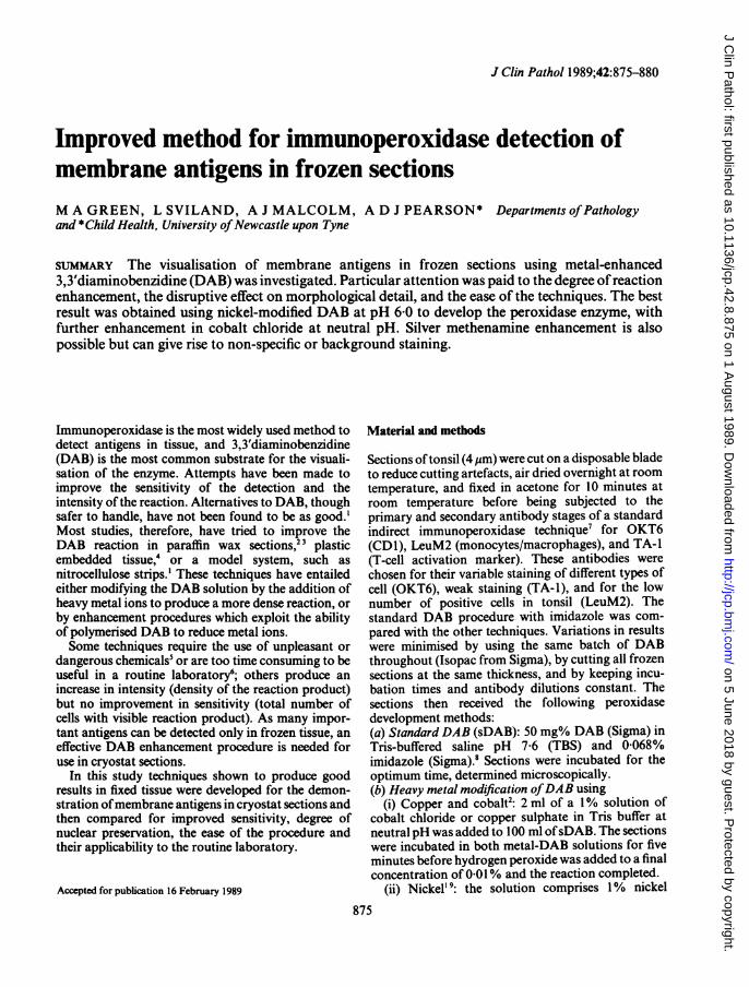
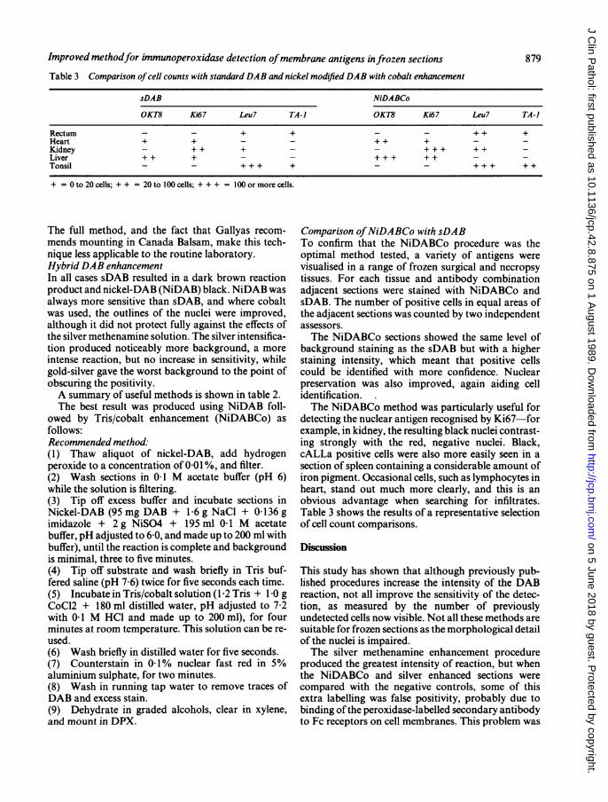
Figs 1 and 2 These sections show the increase in sensitivity of the NiDABCo methods. The same area ofadjacent frozensections ofrectum was stainedfor RFD7 (macrophage marker), and developed with sDAB in I and NiDABCo in 2.
Ferric-ferricyanide treatment caused a colour changefrom brown to blue-green with a concurrent increasein the intensity of the reaction. This did not improvethe sensitivity of the technique, however, and themorphological detail was seriously compromised.Silver methenamine intensification produced a
noticeable increase in both the intensity and thesensitivity of the technique. Cells not visible by sDAB
were light brown after silver enhancement. Unfortu-nately, the morphological detail was again poor.
The binding ofgold to the polymerised DAB producesnucleation sites for the precipitation of silver fromsubsequent solution. The result is a black reactionproduct and increased sensitivity. In common withother metal solutions, the gold chloride conferredsome protection to the nuclei, which are adequately
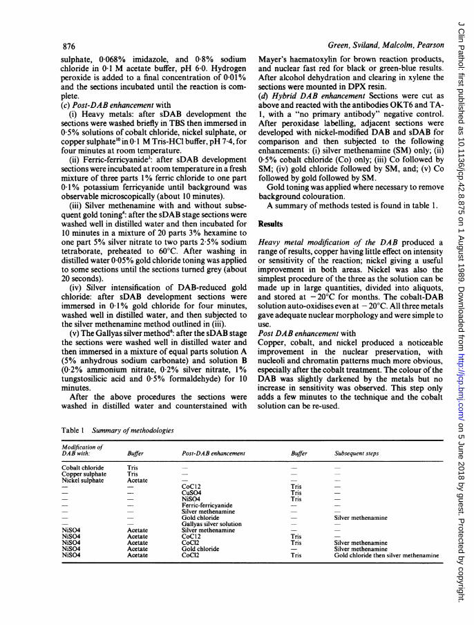
Table 2 Summary ofuseful methods
Method Nuclear morphology Reaction product Comments
sDAB Adequate BrownDAB-Co Very good nucleoli and chromatin Dark brown Tris-cobalt can be re-used
patternsNiDABCo Very good Intense, black, crisp Easy to perform. Solution keeps well at - 20°C.
Clean stainingNiDABCo-SM Adequate Intense, black Lengthy technique, background problemssDABCo-SM Good Intense, dark brown Good alternative, background less obvious
q
4
4
I ,
a.
h v ,.'
877
1*1
k . # A,4.-11
71.1.1:4
.0.".1.9 - -
I .+. v
I
on 5 June 2018 by guest. Protected by copyright.
http://jcp.bmj.com
/J C
lin Pathol: first published as 10.1136/jcp.42.8.875 on 1 A
ugust 1989. Dow
nloaded from
Green, Sviland, Malcolm, Pearson
...*.r
I 1'8.
*,$4-i; ..
.... ..
;....0..0 .¢
--::
*S 7;'\:t.6 v o }.
;> ... .t
.,f
3
Ir'
.....i.;.k 0
..
o'tw
*::f
.. 2.;F. ,.bJ:..
a.'.
:. =
-_ 6s
-_-:__.
4
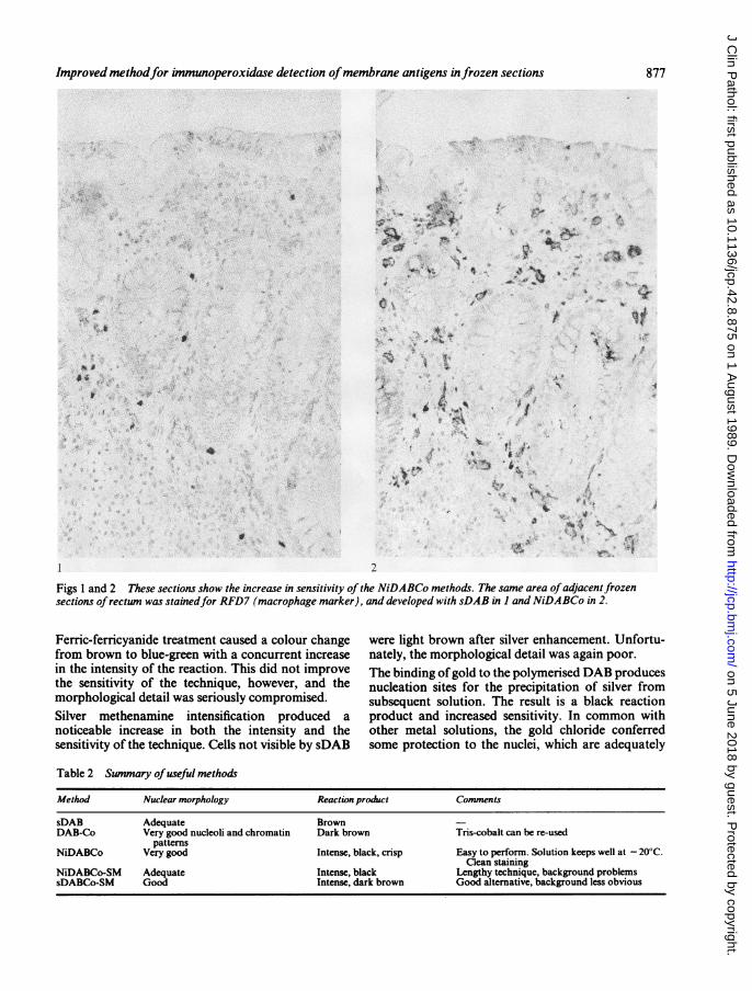
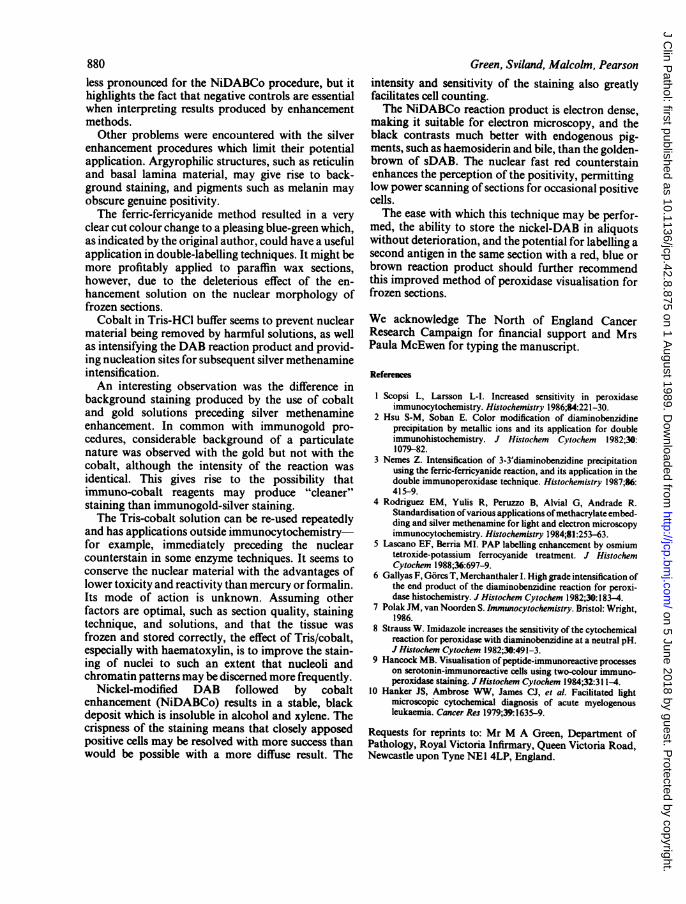
Figs 3 and 4 These sections show the effect ofthe cobalt chloride on the nuclear preservation. Adjacentfrozen sections oftonsil were stainedfor Leu4 (Pan T cells) and developed with sDAB in 3 andsDABCo in 4.
preserved in this method. Gold toning, if used, should good results, with poor morphological detail and onlybe as brief as possible to avoid replacement of the slight intensification of the DAB. The method follow-silver with gold. ed here omitted the four hour incubation step inThe Gallyas method, although simple, did not give thioglycollic acid to reduce background argyrophilia.
A.. I_
.aC.46 *A:4
..
l".s
878
*eX
",-,w-"-'.--.i.1 -.&X.
.. e.,. .... .!M
A,*
V.* ".A.
:0 ... 1.
m4w
on 5 June 2018 by guest. Protected by copyright.
http://jcp.bmj.com
/J C
lin Pathol: first published as 10.1136/jcp.42.8.875 on 1 A
ugust 1989. Dow
nloaded from
Improved methodfor immunoperoxidase detection ofmembrane antigens infrozen sections
Table 3 Comparison ofcell counts with standardDAB and nickel modifedDAB with cobalt enhancement
sDAB NiDABCo
OKT8 Ki67 Leu7 TA-I OKT8 Ki67 Leu7 TA-1
Rectum - - + + - - + + +Heart + + - - ++ + - -
Kidney - + + + - - + ++ ++ -
Liver ++ + - - +++ ++ - -
Tonsil - - +++ + - - +++ ++
+ = 0 to 20 cells; + + = 20 to 100 cells; + + + = 100 or more cells.
The full method, and the fact that Gallyas recom-mends mounting in Canada Balsam, make this tech-nique less applicable to the routine laboratory.Hybrid DAB enhancementIn all cases sDAB resulted in a dark brown reactionproduct and nickel-DAB (NiDAB) black. NiDAB wasalways more sensitive than sDAB, and where cobaltwas used, the outlines of the nuclei were improved,although it did not protect fully against the effects ofthe silver methenamine solution. The silver intensifica-tion produced noticeably more background, a moreintense reaction, but no increase in sensitivity, whilegold-silver gave the worst background to the point ofobscuring the positivity.A summary of useful methods is shown in table 2.The best result was produced using NiDAB foll-
owed by Tris/cobalt enhancement (NiDABCo) asfollows:Recommended method:(1) Thaw aliquot of nickel-DAB, add hydrogenperoxide to a concentration of 001%, and filter.(2) Wash sections in 01 M acetate buffer (pH 6)while the solution is filtering.(3) Tip off excess buffer and incubate sections inNickel-DAB (95 mg DAB + 1-6 g NaCl + 0-136 gimidazole + 2 g NiSO4 + 195 ml 0-1 M acetatebuffer, pH adjusted to 6-0, and made up to 200 ml withbuffer), until the reaction is complete and backgroundis minimal, three to five minutes.(4) Tip off substrate and wash briefly in Tris buf-fered saline (pH 7 6) twice for five seconds each time.(5) Incubate in Tris/cobalt solution (1 -2 Tris + 1-0 gCoC12 + 180 ml distilled water, pH adjusted to 7-2with 0- 1 M HCI and made up to 200 ml), for fourminutes at room temperature. This solution can be re-used.(6) Wash briefly in distilled water for five seconds.(7) Counterstain in 0-1% nuclear fast red in 5%aluminium sulphate, for two minutes.(8) Wash in running tap water to remove traces ofDAB and excess stain.(9) Dehydrate in graded alcohols, clear in xylene,and mount in DPX.
Comparison ofNiDABCo with sDABTo confirm that the NiDABCo procedure was theoptimal method tested, a variety of antigens werevisualised in a range of frozen surgical and necropsytissues. For each tissue and antibody combinationadjacent sections were stained with NiDABCo andsDAB. The number of positive cells in equal areas ofthe adjacent sections was counted by two independentassessors.The NiDABCo sections showed the same level of
background staining as the sDAB but with a higherstaining intensity, which meant that positive cellscould be identified with more confidence. Nuclearpreservation was also improved, again aiding cellidentification.The NiDABCo method was particularly useful for
detecting the nuclear antigen recognised by Ki67-forexample, in kidney, the resulting black nuclei contrast-ing strongly with the red, negative nuclei. Black,cALLa positive cells were also more easily seen in asection of spleen containing a considerable amount ofiron pigment. Occasional cells, such as lymphocytes inheart, stand out much more clearly, and this is anobvious advantage when searching for infiltrates.Table 3 shows the results of a representative selectionof cell count comparisons.
Discussion
This study has shown that although previously pub-lished procedures increase the intensity of the DABreaction, not all improve the sensitivity of the detec-tion, as measured by the number of previouslyundetected cells now visible. Not all these methods aresuitable for frozen sections as the morphological detailof the nuclei is impaired.The silver methenamine enhancement procedure
produced the greatest intensity of reaction, but whenthe NiDABCo and silver enhanced sections werecompared with the negative controls, some of thisextra labelling was false positivity, probably due tobinding ofthe peroxidase-labelled secondary antibodyto Fc receptors on cell membranes. This problem was
879
on 5 June 2018 by guest. Protected by copyright.
http://jcp.bmj.com
/J C
lin Pathol: first published as 10.1136/jcp.42.8.875 on 1 A
ugust 1989. Dow
nloaded from
880 Green, Sviland, Malcolm, Pearsonless pronounced for the NiDABCo procedure, but ithighlights the fact that negative controls are essentialwhen interpreting results produced by enhancementmethods.
Other problems were encountered with the silverenhancement procedures which limit their potentialapplication. Argyrophilic structures, such as reticulinand basal lamina material, may give rise to back-ground staining, and pigments such as melanin mayobscure genuine positivity.The ferric-ferricyanide method resulted in a very
clear cut colour change to a pleasing blue-green which,as indicated by the original author, could have a usefulapplication in double-labelling techniques. It might bemore profitably applied to paraffin wax sections,however, due to the deleterious effect of the en-hancement solution on the nuclear morphology offrozen sections.
Cobalt in Tris-HCI buffer seems to prevent nuclearmaterial being removed by harmful solutions, as wellas intensifying the DAB reaction product and provid-ing nucleation sites for subsequent silver methenamineintensification.An interesting observation was the difference in
background staining produced by the use of cobaltand gold solutions preceding silver methenamineenhancement. In common with immunogold pro-cedures, considerable background of a particulatenature was observed with the gold but not with thecobalt, although the intensity of the reaction wasidentical. This gives rise to the possibility thatimmuno-cobalt reagents may produce "cleaner"staining than immunogold-silver staining.The Tris-cobalt solution can be re-used repeatedly
and has applications outside immunocytochemistry-for example, immediately preceding the nuclearcounterstain in some enzyme techniques. It seems toconserve the nuclear material with the advantages oflower toxicity and reactivity than mercury or formalin.Its mode of action is unknown. Assuming otherfactors are optimal, such as section quality, stainingtechnique, and solutions, and that the tissue wasfrozen and stored correctly, the effect of Tris/cobalt,especially with haematoxylin, is to improve the stain-ing of nuclei to such an extent that nucleoli andchromatin patterns may be discerned more frequently.
Nickel-modified DAB followed by cobaltenhancement (NiDABCo) results in a stable, blackdeposit which is insoluble in alcohol and xylene. Thecrispness of the staining means that closely apposedpositive cells may be resolved with more success thanwould be possible with a more diffuse result. The
intensity and sensitivity of the staining also greatlyfacilitates cell counting.The NiDABCo reaction product is electron dense,
making it suitable for electron microscopy, and theblack contrasts much better with endogenous pig-ments, such as haemosiderin and bile, than the golden-brown of sDAB. The nuclear fast red counterstainenhances the perception of the positivity, permittinglow power scanning of sections for occasional positivecells.The ease with which this technique may be perfor-
med, the ability to store the nickel-DAB in aliquotswithout deterioration, and the potential for labelling asecond antigen in the same section with a red, blue orbrown reaction product should further recommendthis improved method of peroxidase visualisation forfrozen sections.
We acknowledge The North of England CancerResearch Campaign for financial support and MrsPaula McEwen for typing the manuscript.
Referenoes
I Scopsi L, Larsson L-I. Increased sensitivity in peroxidaseimmunocytochemistry. Histochemistry 1986;84:221-30.
2 Hsu S-M, Soban E. Color modification of diaminobenzidineprecipitation by metallic ions and its application for doubleimmunohistochemistry. J Histochem Cytochem 1982;30:1079-82.
3 Nemes Z. Intensification of 3-3'diaminobenzidine precipitationusing the ferric-ferricyanide reaction, and its application in thedouble immunoperoxidase technique. Histochemistry 1987;86:415-9.
4 Rodriguez EM, Yulis R, Peruzzo B, Alvial G, Andrade R.Standardisation ofvarious applications ofmethacrylate embed-ding and silver methenamine for light and electron microscopyimmunocytochemistry. Histochemistry 1984;81:253-63.
5 Lascano EF, Berria MI. PAP labelling enhancement by osmiumtetroxide-potassium ferrocyanide treatment. J HistochemCytochem 1988;36:697-9.
6 Gallyas F, G6rcs T, Merchanthaler I. High grade intensification ofthe end product of the diaminobenzidine reaction for peroxi-dase histochemistry. J Histochem Cytochem 1982;30:183-4.
7 Polak JM, van Noorden S. Immunocytochemistry. Bristol: Wright,1986.
8 Strauss W. Imidazole increases the sensitivity of the cytochemicalreaction for peroxidase with diaminobenzidine at a neutral pH.J Histochem Cytochem 1982;30:491-3.
9 Hancock MB. Visualisation of peptide-immunoreactive processeson serotonin-immunoreactive cells using two-colour immuno-peroxidase staining. JHistochem Cytochem 1984;32:311-4.
10 Hanker JS, Ambrose WW, James CJ, et al. Facilitated lightmicroscopic cytochemical diagnosis of acute myelogenousleukaemia. Cancer Res 1979;39:1635-9.
Requests for reprints to: Mr M A Green, Department ofPathology, Royal Victoria Infirmary, Queen Victoria Road,Newcastle upon Tyne NEI 4LP, England.
on 5 June 2018 by guest. Protected by copyright.
http://jcp.bmj.com
/J C
lin Pathol: first published as 10.1136/jcp.42.8.875 on 1 A
ugust 1989. Dow
nloaded from