in animal health and zoonoses control tufts …pdf.usaid.gov/pdf_docs/pdabt717.pdf · in animal...
TRANSCRIPT
...
Pb~ iPlI6T-'q-I-=J--10 qq 0"2-
SEVENTHSEMUANNUAL PROGRESS REPORT FOR THE
MIDDLE EAST REGIONAL COOPERATION PROGRAM
STRENGTHENING REGIONAL COLLABORATION IN
ANIMAL HEALTH AND ZOONOSES CONTROL
Tufts University School of Veterinary Medicine
In collaboration with
Ministry of Agriculture and Land Reclamation, Egypt Ministry of Agriculture, Israel
Ministry of Agriculture, Jordan CARE USA (CARE International, Office for the West Bank and Gaza)
Prepared for the United States Agency for International Development
By
George Saperstein, DVM
June, 2001
I

L Technical Progress A. Research Objectives - Overall objectives remain the same as reported in the previous progress
report due to the problems in the region. They are reiterated below. For brucellosis: Projects:
1. Field experiment for evaluation of Rev I vaccine for goats 2. Experiments to compare RE51 and S19 protection against B. melitensis field strains. 3. Continuation of serological standardization of the diagnosis of camel, swine, and
buffaloes 4. Isolation and biotyping of Brucella spp. And Rev I vaccine strains from field isolates 5. Training in the participating countries on molecular biology and modem serology 6. Evaluation of mass vaccination strategy in goats with Rev 1 vaccine (full dose,
ocular method) 7. Improvement of the surveillance of small ruminant brucellosis by milk analysis
Workshops: 1. Training on the production of Rev 1 vaccine (JOVAC) 2. Molecular analysis and biotyping of Brucella spp (Israel) 3. Applied mass vaccination strategy (West Bank) 4. Application of serological tests and Bmcella spp. By conventional methods (Egypt) 5. Epidemiological approach to the control of Brucellosis (Joint workshop between
PBCP and MERC, to be coordinated by Dr. Ashley Robinson)
ForFMD: 1. Control activities
Regnlar vaccination campaigns, especially in high risk areas and boundaries. New attention will be placed on previously unvaccinated animals, such as pigs.
2. Diagnostic activities Collection of samples for virus isolation and identification, along with information exchange.
3. Serosurveillance Continue in parallel with control
4. Exchange information via the regional website. 5. Complete short -term laboratory training. Begin long-term training according to the
interests of the parties. Include epidemiology and statistics. Train on-site after equipment is in place.
6. Reagents Produce standard reference reagents in a central laboratory and distribute to all parties. Use SOP.
7. Meetings Meet twice a year.
For Neonatal Mortality in Small Ruminants: 1. Do the epidemiological survey in each country and exchange the information among
the participants.
2. Train more skilled teams, including mutual visits to each others' facilities, to carry ont both field and laboratory work
3. Analyze results and draw conclusious as to the major causes of neonatal mortality in the region.
4. Develop control and preventive measures for neonatal losses, through changes in management and medically.
....
-

5. Present the results of collaborative studies to scientific and agricultural community worldwide.
B. Research Accomplishments Accomplishments for each participant are listed by country below. Collaboration was minimal for the period of this report.
C. Scientific Impact of Cooperation The work has suffered because of the inability of the participants to collaborate as a group. The annual scientific workshop, the highlight of the year, had to be postponed. It was planned for Aqaba, Jordan in November, 2000.
D. Description of Project Impact Some highly encouraging trends are beginning to appear as a result of collaborative work. The incidence of brucellosis in humans has been steadily declining over the period of the project. Rabies cases in domestic animals are down dramatically in 2001. Rapid communication between the parties concerning new and emerging diseases in the region has improved dramatica1!y.
E. Strengthening of Middle East Institutions Sufficient scientific eqnipment has been purchased in both Jordan and Palestine to bolster their diagnostic labs to levels less dependent on outside labs. Unfortunately, much of the equipment for Palestine was delivered the day before the problems began and is not in use.
F. Future Work Much of the collaborative work and nearly all of the work within Palestine has ceased since September, 2000. An extension of at least 6 months will be necessary to try to complete the plan of work.
ll. Project Management and Cooperation A. Managerial Issues
Research in Palestine has been seriously hampered. The goat study, planned for Jerico has been put on hold, as the researchers cannot get to the site. We are trying to move it to another site (Ramallah), but finding that task extremely difficult as well. In addition, administrators from CARE as well as the US PI are having great difficulties communicating with any of the participants by usual means such as email or phone.
B. Special Concerns None
C. Cooperation, Travel, Training and Publications Listed below by country.
C. Request for USAID Actions USAID could provide assistance in the region coordinating joint projects and meetings.
3

Country Reports
....
...
...
EGYPT

;;&& ,
.... ,
.iIi
...
...
Ministrv o(Agriculture And Land Reclamation
. Egypt
Strengthening of Regional Collaboration In Animal Disease and Zoonoses Control
In the Middle East (SRCAD)
( Annual Report)
April 2000 - April 2001
Coordinator: Prof. Dr. Hassan Aidaros Chairman of General Organization Of Veterinary Services (CVOS)
Principal Investigator: Prof. Dr. Ismael Mohamed Reda Prof. of Virology Faculty of Veterinary Medecine Cairo University
Co.PI: Prof. Dr. M.K. Refai Prof. ofMfcrobiology Faculty of Veterinary Medecine Cairo University
MinistJy of Agriculture And Land Reclamation Egypt "SRCAD"
Neonatal Loses of Small Ruminants Activity
( Annual Report)
April 2000 - April 2001
Principal Investigator: Prof. Dr. M. A. Shalaby """"":~~",,Chairman Dept. of Virology
Faculty of Veterinary Medecine Cairo University

Virology
...
'. '-_",,~T , .. ;~."",::"-_:...-., .• , .. ,,
1
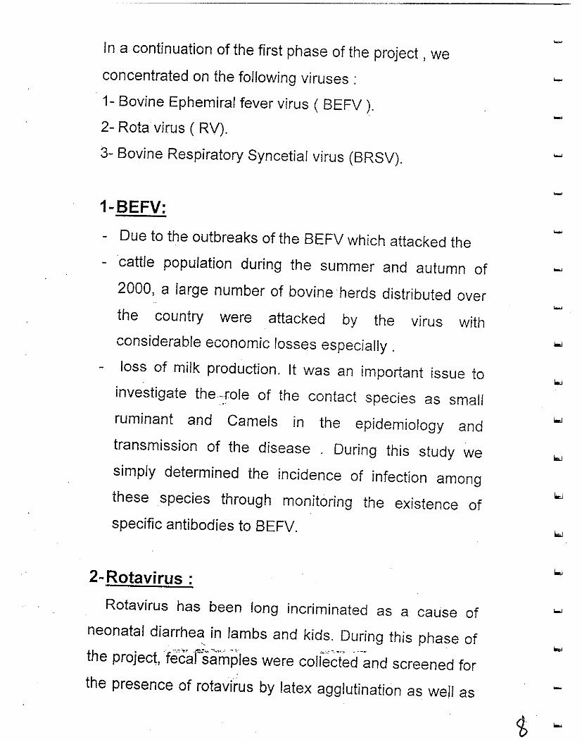
In a continuation of the first phase of the project, we
concentrated on the following viruses: 1- Bovine Ephemiral fever virus ( BEFV ).
2- Rota virus ( RV).
3- Bovine Respiratory Syncetial virus (BRSV).
1-BEFV:
- Due to the outbreaks of the BEFV which attacked the cattle population during the summer and autumn of 2000, a large number of bovine· herds distributed over the country were attacked by the virus with considerable economic losses especially. loss of milk production. It was an important issue to
investigate the-.role of the contact species as small ruminant and Camels in the epidemiology and transmission of the disease . During this study we simply determined the incidence of infection among these species through monitoring the existence of specific antibodies to BEFV.
2- Rotavirus :
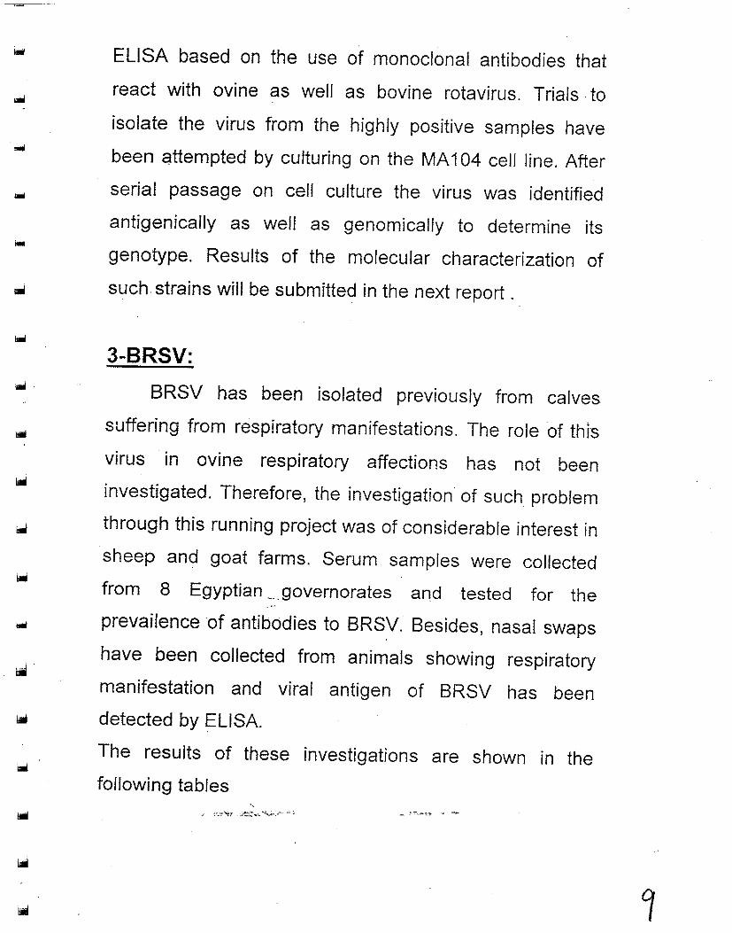
Rotavirus has been long incriminated as a cause of neonatal diarrhea in lambs and kids. During this phase of
"
the project, cfecat=saimpIes were colrec'ted-~nd screened for the presence of rotavirus by latex agglutination as well as
...
ELISA based on the use of monoclonal antibodies that
react with ovine as well as bovine rotavirus. Trials· to
isolate the virus from the highly positive samples have
been attempted by culturing on the MA 104 cell line. After
serial passage on cell culture the virus was identified
antigenically as well as genomically to determine its
genotype. Results of the molecular characterization of
such strains will be submitted in the next report .
3-BRSV:
BRSV has been isolated previously from calves
suffering from respiratory manifestations. The role of thIS
virus in ovine respiratory affections has not been
investigated. Therefore, the investigation of such problem
through this running project was of considerable interest in
sheep and goat farms. Serum samples were collected
from 8 Egyptian ___ governorates and tested for the ..
prevailenceof antibodies to BRSV. Besides, nasal swaps
have been collected from animals showing respiratory
manifestation and viral antigen of BRSV has been
detected by ELISA.
The results of these investigations are shown in the
following tables --
'·.,· .... r .~ .... "'->",;..:----;, .-~-" - --
The BEF study
.....
Materials and Methods
• Samples:
1 ) Camel Sera: Blood samples were collected ....
from·· Basaten abbatoir . Sera were obtained and
stored at - 20°C until used.
2 ) Sheep sera: Blood samples were obtained
from different farms.
• Virus Webester vaccinal strain, adapted to
Vera cells.
• Cells Vera cell cultures grown In Ml=M i'J
supplementedwith fetal calf serum.
" ..; '-.~'-:l ,~'""",.:"-_2- __ -" 0..,
1°
...
...
Total
Total 391
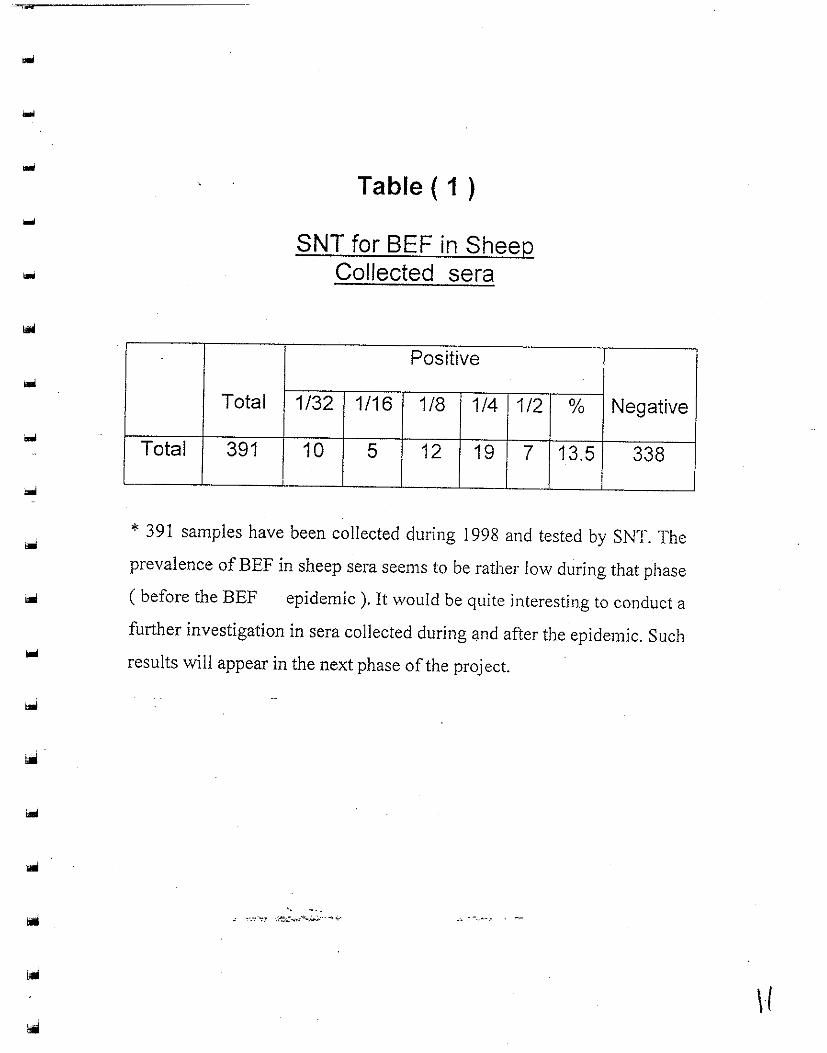
Table ( 1 )
SNT for BEF in Sheep Collected sera
Positive
1/32 1/16 1/8 1/4 1/2
10 5 12 19 7
% Negative
13.5 338
* 391 samples have been collected during 1998 and tested by SNT. The
prevalence ofBEF in sheep sera seems to be rather low during that phase
( before the BEF epidemic ). It would be quite interesting to conduct a
further investigation in sera collected during and after the epidemic. Such
results will appear in the next phase of the project.
'. ~ ' •. ~' '':7 .;~"",,:''''-.;'''';':''''' ~ .•
\1
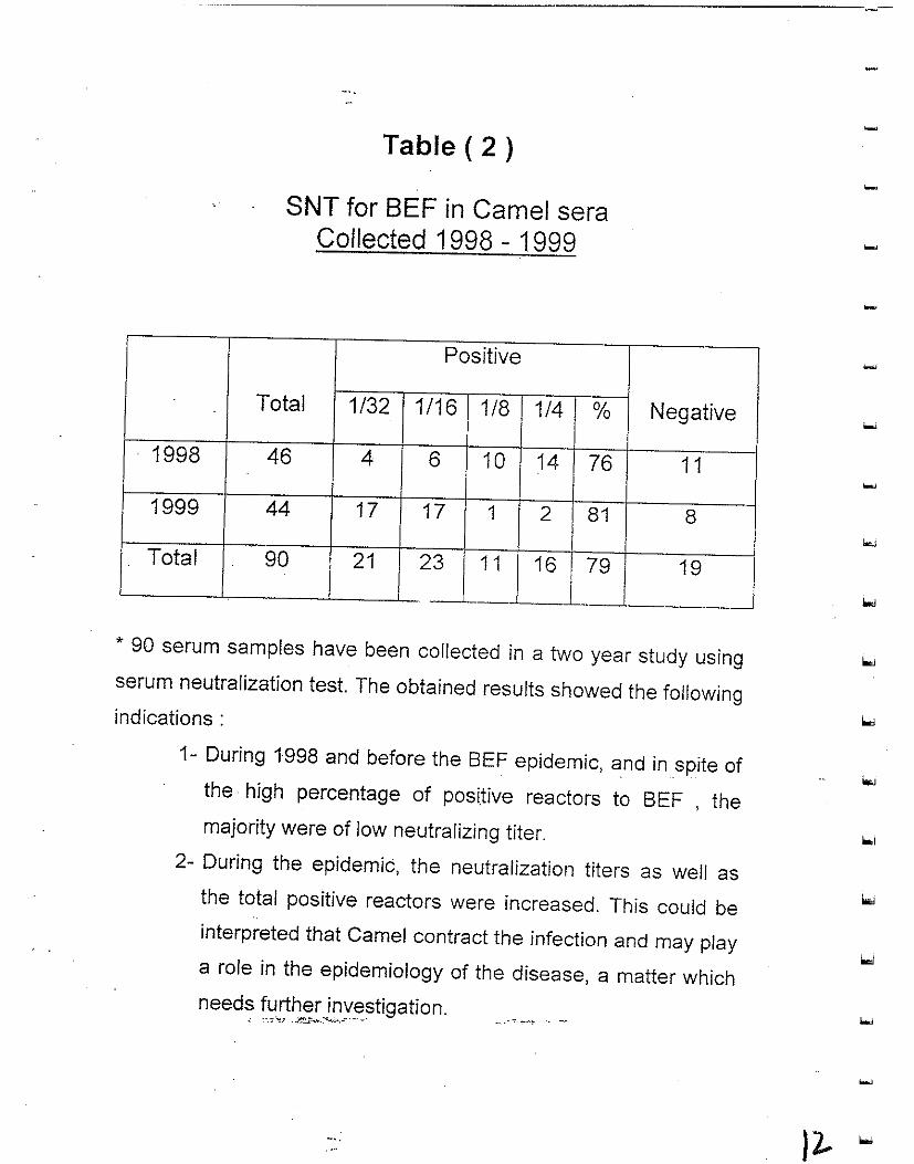
1998
1999
Total
Table ( 2 )
SNT for BEF in Camel sera Collected 1998 - 1999
Positive
Total 1/32 1/16 1/8 1/4 %
46 4 6 10 14 76
44 17 17 1 2 81
Negative
11
8
90 !
21 23 11 16
~-~, * 90 serum samples have been collected in a two year study using serum neutralization test. The obtained results showed the following indications :
1- During 1998 and before the BEF epidemic, and in spite of the· high percentage of positive reactors to BEF , the majority were of low neutralizing titer.
2- During the epidemic, the neutralization titers as well as the total positive reactors were increased. This could be interpreted that Camel contract the infection and may play a role in the epidemiology of the disease, a matter which needs further investigation.
~ --. -:: "-:, ,.~ .... ~.-:->:-.", ....... ~,,,,
12 ..
The Rotavirus work
Materials and Methods used
- ELISA : ( Using monoclonal antibodies against VF6 of
Bovine Rotavirus ).
- MA 104 cells: ( Monkey Kidney cells ).
- Tissue Culture Media : ( Growth and maintenance
media ).
- Fecal Samples : ( The samples which gave pm\:tive "i
results by the Latex agglutination test and
recorded in the last report ).
- Latex Agglutination: ( Virotect - Rota - Omega
. diagnostics - UK).
" " ._-.~"t! :..:e...::,...._ ... '-.~.,_A-.,'. """.--,,,"<.,. • . -

--------------------~~--------------.~
Trials for the isolation and identification of
rotavirus from small ruminants
Trials for isolation of.rotavirus from fecal samples obtained
from diarrheic small ruminants were conducted. Rota
virus ,strongly positive samples by latex agglutination and
ELISA were inoculated in MA 104 cell culture for virus
isolation. Following up the cytopathic effect in tissue
culture ,after 3 passages on MA 104 cells gave rise to the
isolation of 4 field strains from sheep samples. 'The
isolates were identified using ELISA based on monoclonal
antibody. Further characterization of the isolated viruses
antigenically and genomically are in progress.
'. ~ '-.~~.T ,.~:","":":.,,., ..... ";.

...
The BRSV work
Materials and Methods
o Samples:
1 ) Blood samples : were collected from sheep
suffering from different levels of respiratory
manifestations. The sera were tested by SNT (
Results are shown in table 3 ).
2 ) Nasal swabs : were also examined by dot
ELISA for detegtion of BRSV antigen.
o Virus : BRSV MDBK adapted reference strain
kindly supplied by Prof. Dr. Chase - South Dakota ,
USA.
o Cell Culture: MDBK.
o Serum: Positive reference serum - kindly supplied
. by Prof. Dr. Chase - South Dakota, USA.
o Dot ELISA:
using polyclonal antisera against BRSV prepared "
in rabbit' an,fAntirabbit conju'gatecfperoxidase.

i
I I I I I
] ] j j j ] ] j ] j ] ] ] ] ]
Table ( 3 )
Results of SNT for BRSV in sheep sera
No. of samples Breed Screening by SNT End titer Sites
+ % 1/2 1/4 1/8 1/16 1/32 .
Site ('1 ) 55 Balady 15 27 5 3 3 I 3 I 1 ". !
Site ( 2 ) 25 Balady 12 48 - 3 - ''7 2 Site ( 3 ) I 50 Oernamy 37 74 2 I 5 17 ~O 3 Site' (.4 ) I 48 Balady 32 , 67 2 I 6 10 7 7 Site (5 ) I 50 Balady 21 42 4 5 5 I 7 -
,Site ( 6 ) I 50
I Balady 9 I, 18 5 3 1 - -
Site ( 7 ) , I 63 Balady 11 17.5 7 4 - I - I -39 I Balady 3 8 1 I 2
Site ( 8 ) - ',- -•
"
.' II ~.; From the above table, it is clear that the percentage of sheep shown antibodies to BRSv in these farms ranged from 8 % to 74 % , The serum neutralizing titers ranged from 1/2 to 1/32 , and the majot.ity lies in the range of 1 /4 to 1/16. .
Successful detection of the viral antigen by dot ELISA was mainly in swabs obtained from animals showing low titers of neutralizing antibodies.
]
I
i
- d4
...
...
.. -Sites
.. Site ( 1:)
Site I 2.)
. Site ( 3 ) 0
Site ( 4 )
Total
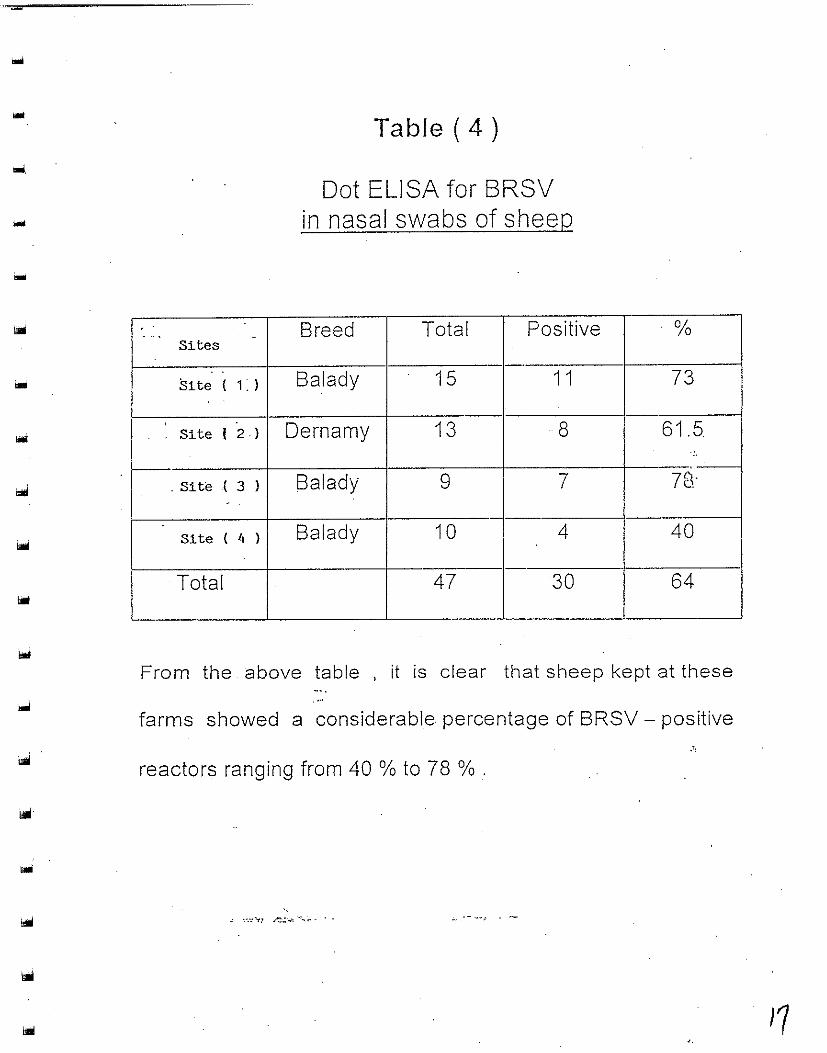
Table ( 4 )
Dot ELISA for BRSV in nasal swabs of sheep
Breed Total Positive
Balady 15 1 1
Dernamy 13 8
Balady 9 7
Balady 10 , 4 I
47 30
%
73
61.5 .'
7('(· Ie.
40
64
From the above table , it is clear that sheep kept at these
farms showed a considerable percentage of BRSV - positive
reactors ranging from 40 % to 78 % .
'. ",~~; .1':::":: .. ' ~'~;.- . .,. ~~~ .-
Strengthening of Regional Collaboration In Animal Disease and Zoonoses Control In the Middle East (SRCAD)
Bacteriology
Presented by :
Dr.AdeI F. Farid ' Emeritus Chief Researcher- Microbiology
Form Deputy Director, Animal Health Research Institute CO/PI of the project
PREFASE
Small ruminants are considered an important animal in our region,in Egypt and adjacent countries, and constitute a great economic value. Scanty information are available about the
-magnitude of losses-in these animals, specially newborn and growing ones. The success of any program, which could be designed for controlling such losses, is dependent on such
- information. Information and data about pathogenic bacterial agents that play a role in such losses are actually scanty. The plan of work which was pr¢sumed, in the project, was directed to clarify the following two main points: • Determination of the major bacterial causes of diarrhea and
_' ",·:·"u ,.~";:"","',,,,"':.""-" . _-.'-, .... , - -'-
-

...
neonatal mortalities (Aerobic, Anaerobic and Enteric in newly born and growing lambs and kids.
• Determination the incidence of each bacterial pathogen that plays a role as an etiological agent of the problem.
Activities • Field visits (trips) were continued according to the previous
programe. The visited Farms were belonging to 19 different governorates representing different field problems.
• . A full details about the total number of animals which were subjected for clinical examination are given in another section of this report. Generally The age of animals, form which samples were collected, ranged from birth date up to 6 months of age.
Bacteriological Studies
Salmonellosis
Introduction
Salmonellosis continued to be an important infectious disease for several reasons; changing patterns of infection and morbidity rate in virtually all domestic species of livestock and public health concerns regarding human illness from animal sources as well as the adverse publicity relating the zoonotic potential of the infection.
Several serotypes (serovars) of salmonella have been associated with deaths and abortion in sheep and goats all over the world. The number of Hocks affected annually may be small, but within thos'e;tlHflo~s'ses'may be catastrophic~ as many animals can be affected in anyone outbreak.
,1
and was incubated 42 C. for 6 hrs. The M.broth culture was then centrifuged at 1000 X gm for 20 minites.
The cell pellets were resuspended in 2 ml phosphate buffer saline (P.B.S.) (pH 7.4 ) and heated for 1 hr in boiling water, then stored at 4 C. until used for Eliza test. The optimum dilution test sample was 1: 1 00 in bovine serum albumin K.P.L.(USA) ..
Preparation of hyper immune serum was carried out according to Hartman and Minnich (1981). The antibody level was measured by tube agglutination test (Cheebrough, 1985). The obtained anti sera undergoes purification and fractionation to obtain IgG fraction used in Eliza by gel filtration technique (Fey, 1975). Optimum dilution of antisera was 1:50 in P.B.S.
Elisa procedure: The Antigen Capture Eliza was perfonned according to Rigby (1984) and Desmidt et al. (1994).
Results
Bacteriological; examination of faecal samples from diarrhoeic lambs showed 14 and 9 positive samples for salmonella infection out of 188 and 115 samples examined in a percentage of 7.5 % and 7.8 % respectively. Also it was found that 17 lambs sample and 3 kids samples were positive for Klebriella oxytoca (out of 188 lambs and 115 kids respectively) with a percentage of9.0 % and 2.6 % respectively.
Serologically, the isolated sallmonella strains revealed the following results:
Salmonellaa typltimurium from Salmonella dublin from
"
Salmonella enteritidis" from
10 lambs and 6 kids 3 lambs and 1 kid I-lamb -and 2 kids
20
.•
...
...
Material and Methods
A total of 303 faecal samples were collected from diarrhoeic lambs and kids suffering from profuse watery diarrhoea. Each sample was divided into 2 portions, the first was subjected to cultural method and the second portion was prepared and tested by enzyme immunoassay for the detection of Salmonella typhimurium.
Culturing method Approximately 1 gram of faecal samples was placed into 8 ml of tetrathionate broth for the enrichment and incubated at 43 C. for 24 hrs. Then, the incubated broth was streaked on Hekton enteric agar plates, and Salmonella - Shigella plates and incubated at 37 C for 24 hrs. (Pelton et al., 1994).
Biochemical identification The suspected growing colonies were identified according to Krieg and Holt (1984) by colonial characterization and biochemical reactions.
Serological identification Antigenic characterization was performed according to Edwards and Ewing (1972) and Kauffmann (1973) using slide agglutination test with monovelant "0" and "H" antisera against Salmonella typhimurium,obtained from Wellcome Diagnostics, . Dartford, England.
Enzyme immunoassay Faecal preparation: Each sample was suspended in 9 volumes oftetrathionate broth (Emswiler et al., 1984) at 42 C. for 24 hrs, 5 ml of broth was mixed with 5 ml ofRibozyme 41 (A commercial protease enzyme solution) and incubated at 37 C. for 1 hr. (Zierdt, 1982 and Rigby, 1984) to eliminate non specific reaction~:~-One ml of the incubated broth was mixed with 10 illl of M.broth (Difco) with 10 ug of Novobiocin to enhance flagellar production by bacteria (Desmidt et a!., 1994)

----------------~~--------------------
Anaerobic Bacteria
Introduction
Clostridia microorganisms are widely distributed in nature and are normally present every where such as in soil, dust, air, water, sewage and manure (EI Sayed, 1979). Clostridia are a normal inhabitant in the intestinal tract of different animals. It plays an important role in producing diseases as a result of an interplay between infection with such microorganism and many factors other than the bacteriological agent
Clostridia. perjringens is the predominant microorganism among clostridia species. It causes serious economic losses in young animals specially in lambs and kids. Harbola et al (1988) studied neonatal mortality due lamb dysentery.
The lethality of C. peTj'ringens is due to the secretion of the toxin in the intestinal tract causing sever damage and degeneration of the parenchymal tissues which may lead to· death within few hours postnatal (Yanny, 1989). Blackwell and butler (1992) described the clinical responses to treatment and vaccination in four goat herds in which a diagnosis of enterotoxoemia attributable to C.penjringens type D. This was confirmed on the bases of signs of diarrhoea ,sudden death ,isolation of c.perjringens and presence of epsilons toxin in the faeces at the time of admission.
Material and Methods
--A total of 187 faecal samples were collected from diarrhoeic newborn Iambs and kids (155 and 32 samples respectively). These animals were less than three months of age
-, '" ,-.-:-'"t.T ,'.~;",.:-<-'~.-- -, ~---.... -. ---~
Faecal material was cultured into two tubes of freshly prepared cooked meat broth. One of these tubes was incubated
......
22.

...
anaerobically at 37 C for 24 hours. Sample from each was then separately streaked on to the surface of 10% sheep blood agar with neomycin sulfate (200 ug / mI.). The other tube was heated at 80C for 10 minutes to kill all vegetative bacterial
. cells and then incubated anaerobically at 37C for 24 hours then, it was streaked on sheep blood agar for the diagnosis of other clostridia organisms. Plates were incubated anaerobically at 37C for 24 hours. Suspected colonies for clostridia were purified subcultured and kept for further identification (Carter and Cole, 1990). Biochemical identification was applied for each separate isolate according to Cruickshank et al. (1975) and Koneman et al.(1992).
Toxigenicity of C. peifringens isolates was detected by both lecithin's test (Smith and Holdman, 1968) and their pathogenicity to Guinea pigs (With, 1977). The typing of C. peifringens strains were determined by dermonecrotic test in Guinea pigs according to Stern and Batty (1975).
Results
The incidence of C. perjringens in diarrhoeic newborn lambs and kids showed that out of 155 faecal samples of lambs, 32 samples were positive for C. peifringens with an incidence of 20.7 %. Testing the toxigenisity of these isolates showed that 12 isolates (7.7 %) were non toxigenic while the rest 20 samples (12.9 %.) revealed toxigenic isolates.
Out of the 32 faecal samples collected from newborn kids, 10 samples were positive for C. peifringens micro organism with an incidence of 31.3 %. The non toxigenicisolates in kids samples were 6 samples (18.8 %), while the toxigenic isolates were 4 samples (12.5 %).
Typing of C.perjringens strains were- determined by dermonecrotic test in Guinea pigs. Results showed that the 20 positive toxigenic isolates of lambs fecal samples were typed as
C.perjringens type A (6 isolates), C. per/ringes types D (6 isolates). C. per/ringes type E (one isolate). C. per/rlnges types A & D as mixed infection (6 isolates) and C. perjringes types A & E as mixed infection (one isolate) ..
The four positive toxigenic isolates from kids faecal samples were typed as C. per/ringens type A (2 isolates) and 2 isolates were found to be C. per/ringens type D. No C. perjringes type E or mixed types were recorded in kids strains.
Enteropathogenic E. coli
Introduction
The transmissible enteropathogenic Escherichia. coli K antigen from calf and lamb was previously called kco. ( Orskov et al 1975). In neonatal Escherichia coli diarrhea enteropathogenic E coli strains, adhere to the villous epitheluim in the small intestine(Moon et al., 1977 and Evans et al., 1978 ). Adherence to the mucoid surface helps to overcome mechanical clearance by intestinal motility and thus facilitates colonization of the small intestine resulting in produce diarrhoea and dehydration.
Infected kids showed profuse watery diarrhoea following by dehydration and death. Infection by E.coli K99 is a significant cause of diarrhoea - in newborn kids up to 2-3 weeks of age (Nagy et al , 1983). Strains of enteropathagenic E coli cause acute infection scour in lambs and produce plasmid - encoded virulence deterrninans. fimbrial adhesin K99 (Acres, 1985).
The ability of E. coli to produce fimbrae confers on the bacterium ~ 9.Qp.~lg~r?-pJe advantage qfS.9ionization. Fimbriae are best expressed in vitro at 37c.(Smyth et al , 1994).
...
...
Material and Methods
A total of 145 faecal samples were collected from diarrheic newborn lambs and Kids (82 and 63 samples respectively), less than three weeks of age. Samples were collected before any treatment have been initiated. Fecal material was diluted in sterile saline, and one drop was streaked on the surface of Minca Isovitalex agar plate. Plates were incubation for 18 - 24 hours at 37 C. Five completely separate translucent colonies from each plate were selected and subcultured on slope agar tubes of the same medium.
Slide agglutination test was carried out to each isolate using diagnostic anti K99 sera locally prepared against E. coli strain B 41 (0101: K99) and B 85 (09: K99). Serological confirmation was carried out using commercial E. coli K99 diagnostic antiserum. Serologically positive strains were subjected for biochemical reactions and sugar fermentation for confirmation as E. coli.
Result
Results showed that out of 82 investigated diarrhoeic lambs and 63 kids, enteropathogenic E. coli isolates that harbouring k99 antigen were .isolated from 23 (28.1 %) lambs and from 16 (25.4 % ) kids. _ All k99 positive isolates gave typical biochemical reaction and sugar fermentation of E .coli .
General Comments The above data needed to be matched with similar data of the other neighboring and adjacent countries of the same region.
The common,patnogenic Bacterial agents, in Egypt and other countries of the region, should be faced and to be controlled by methods based on a scientific basis.
Parasitology
' . ..: ',-.• ':r .;~y'.,:"-.,;., .... ..,.;."
. ...
1011
The aim of the present investigation is to explore the prevalence of Cryptosporidium, Neospora caninum and Toxoplsma gondii in sheep and goats in different governorates in Egypt.
Cryptosporidium: Material and Methods
Faecal and serum samples were collected from each of226 sheep and 209 goats during the period from 1st November 1999 to 31st October 2000. These samples were collected from different Governorates in Delta region in Egypt. 1- Faecal examination:
Faecal smears from each of the collected faecal sample were prepared, fixed in absolute methanol and stained with modified Ziehl-Neelsen technique (Henriksen and Polzlenz, 1981) as follows:
I. The fixed faecal smears were stained with carbol fuchsin for 20 minutes. 2. The slides were washed with tap water for 2 minutes and decolorized with 7%
. sulfuric acid for 20 second. 3. The slides were washed in tap water for 2 minutes. 4. The slides were counter-stained with 5% malachite green for 5 minutes. 5. The slides were washed with tap water for 2 minutes, air dried and examined
microscopically using higher power (x 400) and (xl00) for identification of Cryptosporidium oocystes.
II- Serological eumination The collected serum samples were investigated for detection of
anticryptosporidium antibodies by the use of the following serological test: A. Indirect immunofluorescence antibody test (IFT) (Lorenzo-Lorenzo et al, 1993).·
The antigen slides were prepared from purified Cryptosporidium oocystes collected from infected sheep and goats and fixed in cold acetone for 10 minutes, wrapped in tissue paper and stored t -70°C until used. IFAT procedure:
1. The prepared IF A frozen slides were left at room temperature for one hour. 2. The collected sheep and goats sera were examined at predetermined dilution
1:40 in PBS. Positive and negative control sera were included in each slid. 3. Five III of each diluted serum was applied to each well. 4. The slides were incubated at 37°C in moist chamber for 30 minutes. 5. The slides were washed 3 times in distilled water for 5 minutes each. 6. Twenty five III fluorescein iso-thiocyanate anti-sheep IgG at dilution 1:40 in
1/10000 Evans blue were added to each sheep serum well. While 25111 of antigoat IgG FITC conjugate at 1:40 dilution in 1/10000 Evans blue were used for goats sera and the slides were incubated at 37°C for 30 minutes in moist chamber. ..
7. The slides were washed and dried as mentioned before then mounted with glycerol in PBS pH 9.0 (9:1), then examined under fluorescent microscope.
8. The sample was considered positive when yellow green fluorescence extends around the entire periphery of the oocytes. The negative sample appears dull.
B- Enzyme Iinkedlnin':l1Ilrosorbent assay (ELISA) (Ortiga-Mora et al, 19931: 1 Antigen preparation:. .
Crud antigen was prepared from purified Cryptosporidium oocystes by freezing and thawing for three times, then disrupted ultrasonically by sonication for
2 t.1
60 second and interupted for 60 second for total time 20 minutes at 795 disruption. The senicated suspension was centrifuged at 16000 rpm for 30 minutes at 4°C. The supernatant was recovered, aliquated and stored at -70°C until used. The antigen was used at 0.5 ~gm protein per well. 2. ELISA procedure:
The tested sera were used at predetermined dilution 1 :40 in ELISA buffer (0.4 M PBS, 0.05% Tween 20 pH 7.2).
The antigen was used at concentration ofO.5~g protein per well checker board manner (Catty and Raykundalia, 1989).
1. The micro ELISA 96-wells 200~1 plates (Immunoplate 2; Nune, Copenhagen, Denmark) were coated with antigen suspended in coating buffer (0.1 M carbonate buffer, pH 9.6) up to 100 j.lifwell and covered.
2. The plates were incubated at 37°C for 1 hour then at 4°C overnight. 3. The coating material was then discarded and the plates were rinsed three times
for 5 minutes each, with 200 ~l of 0.04 M PBS. pH 7.2. 4. The wells were blocked for 1 hour at 37°C with 200 III of carbonate buffer (pH
9.6) plus 1% bovine serum albumin, and stored at 4°C until used. 5. The plates were rinsed three times for 5 minutes each with washing buffer (0.04
M PBS pH 7.2 containing 0.05% Tween 20. 6. 100 III of each tested serum at predetermined dilution 1:40 in 0.4 M PBS, 0.05
Tween 20 pH 7.2 was added to duplicate wells, then incubated for 1 hour at 37°C. 7. The plates were rinsed three times with washed buffer for 5 minutes each. 8. For sheep sera samples:
A- 100 III of anti-sheep 19G alkaline phosphtase conjugate (A-5187, Sigma) diluted in PBS 0.05% Tween 20 at dilution 1: 20.000 were added to each well and incubated for 1 hour t 37°C, then the plates were washed three times for 5 minutes each with washing buffer.
B. 100 III of substrate solution (I mg ofP-nitrophenyl phosphate (Sigma-N-9389) in 10% diethanolamine buffer pH 9.8 were added and allowed to react at room temperature for 2 hours.
C. Enzyme hydrolysis of substrate was stopped by adding 50111 of 3M NaOH to each well.
9. For goat sera samples: A. 100 III of anti-goat IgG peroxidase conjugate (Sigma) was used at dilution of
1: 10.000. B. The substrate used was Ortho-phenylene diamine (Sigma chemical Company,
Sint Louis, USA) pH 5.9. C. Enzyme hydrolysis of substrate stopped by adding 50111 of 0.05% HzOz.
10. Absorbance was read at 405 nm for sheep sera and at 492 nm for goat sera with micro-ELISA reader system (Microplte Autoreader- El 311, Biotek, Instruments Ins., USA). _
11. Positive sera, negative sera and antigen without serum were included in each plate as controls.
Test reading The mean of absorbance reading of duplicate well equal to or more than twice
the mean value of. all negative control sera was considered positive (Iacona et aL, 1980). - '-.-" .. =v .. -~.~ ..... -.- .. - .-
3
-
...
>oIi ..
Neospora caninum: Serum samples were collected from· 202 sheep nd 78 goats from different
localities of Egypt (table 1). Each serum sample was examined for detection of antiNeospora caninum antibodies by the use of Neospora agglutination test (NAT). Detection of anti-Neospora caninum antibodies
Neospora agglutintion test (NAT) was used for detection ofanti-Neospor caninum antibodies. The antigen used was prepared from tachyzoites of the NC-l strain of Neospora cninum according to Hi/ali et aL (1998). The same serum samples of ewes and goats tested for T. gondii were also screened for Neospora.
The NAT was performed in 96 round-bottom-well microplates according to the method previously described for toxoplasmosis (Desmonts and Remington, 1980). In brief, SOIlI of 0.2 M 2-mercaptoethnol in PBS was distributed in each well and 2-fold serial dilutions of sera were performed, starting from either 1 :20, 1 :200, or 1: 500. Parasites were resuspended in alkaline buffer {7.02g NaCI, 03.09g H3B03, 24 ml of IN NOH, 4g bovine plasma albumin (fraction v), and enough distilled water to bring the volume to· 11; pH 8.7} and their concentration was adjusted at 2xl 04/1l1. After the sera had been diluted, SOIlI N. emlinum antigen suspension was distributed in each well. Plates were gently agitated to allow for complete mixing and were then incubated overnight at 30°C. A clear-cut button-shaped deposit of parasite suspension at the bottom of the well was interpreted as_negative reaction, and complete carpet of agglutination organisms was considered positive. Each assay included two negative controls and one positive control. The negative controls comprised antigen alone and one serum sample with a high titer of anti-T.gondii antibodies. A serum sample obtained from a rat with an experimental N. caninum infection was selected as the positive control. Toxoplasma gondii:
Serum samples obtained from 197 sheep and 66 goats. The samples were collected from different localities of Egypt (Table 1). The sera were screened for detection of anti-toxoplasma antibodies by the use of modified agglutination tes (MAT) the test was performed according to Desmonts and Rimengton (1980) as follows:
Antigen, positive and negative control sera were kindly supplied by Dr. J. P. Dubey (Department of Agriculture, Beltsville, Maryland, USA). The antigen was prepared from the RH strain of T. gomiii cultivated along with mouse TG 180 sarcoma cells in the peritoneal cavities of mice (fomalized whole tachyzoites).
Diluted Tris buffer (0.5 M and pH 8.5) was prepared as follows: Tris (hydroxmethyl) aminomethane 6.6 gm [NH2C(CH20H3) F.W. 121.14] and distilled water to 100 mI. Preparation of antigen mixture:
400 III antigen was mixed with 3.S III Tris buffer, 45 1l12-mercaptoethanol and 70 III of (1: 10) evans blue solution (10 III evans blue + 0.1 ml Tris buffer). Test procedure:
The sera were tested for detection of T. gondi antibodies using the formalized whole tachyzoites. The serum samples were examined at predetermined dilution 1: 25, 1: 50 and 1: 500 after titration using positive and negative control sera with Tris buffer (pH. 8.5). In the microtitratioh.'plafer·(U~shaped bottom 96-well:srthe diluted Tris buffer was added in a volume of 192 ml in all wells of the first raw (A), 100 III in all wells of the second raw (B) and 180 III in wells of the third raw (C).
4

Eight ~l of each tested serum was added in the first raw (each tested sample/well) and after good mixing 100 ~I of the diluted sera were transferred from first raw (A) to second raw (B). After good mixing 20 ~I were transferred to third raw (C). Hence the final dilution of each tested serum was 1.25, 1: 50 and 1: 500 in raw A, Band C respectively. The volume of the diluted serum in each well was adjusted to 50~1/wel1 by removing the extra amount of the diluted serum from each well. 50~1 antigen mixture was added to each well with good mixing. A positive and negative control sera were included in each plate. The plates were covered with sealing tape and incubated at 37°C cover night.
Reading the results: A blue button at the bottom of the well indicates a negative result. While a clear bottom indicates a positive one. Results: 1- Cryptospiridium:
A. Faecal examination: Cryptosporidium parvum was detected in 75 (33.19%) out of226 sheep faecal samples. While it was identified in 71 (34%) of209 goats faecal samples. B. Serodiagnosis: 1. IFAT: This investigation showed that 105 (46.46%) out of226 of sheep
had antibodies against Cryptosporidium. Also, the investigation revealed ... that 96 (45.43%) out of 209 goat sera had anti-Cryptosporidium
antibodies.
Titration of antibodies in the 105 IF AT positive sheep serum sampks showed that 70, 30, 3 and 2 samples had IFAT titer of 1: 40, 1: 80, 1: 160 and 1: 320 respectively. While the titer in the 96 IF AT goat positive samples was 1:40 (65), 1: 80 (20), 1: 60 (7), 1: 320 (3) and 1: 640 (1).
2. ELISA: Serological examination of 226 sheep and 209 goats serum samples by using
ELISA showed that 101 (44.69%) and 90 (43.1%) had anti Cryptosporidium antibodies respectively. Relation between faecal consistency and Crvptosporidium infection (using MZA stain technique, IFAT and ELISA for diagnosis
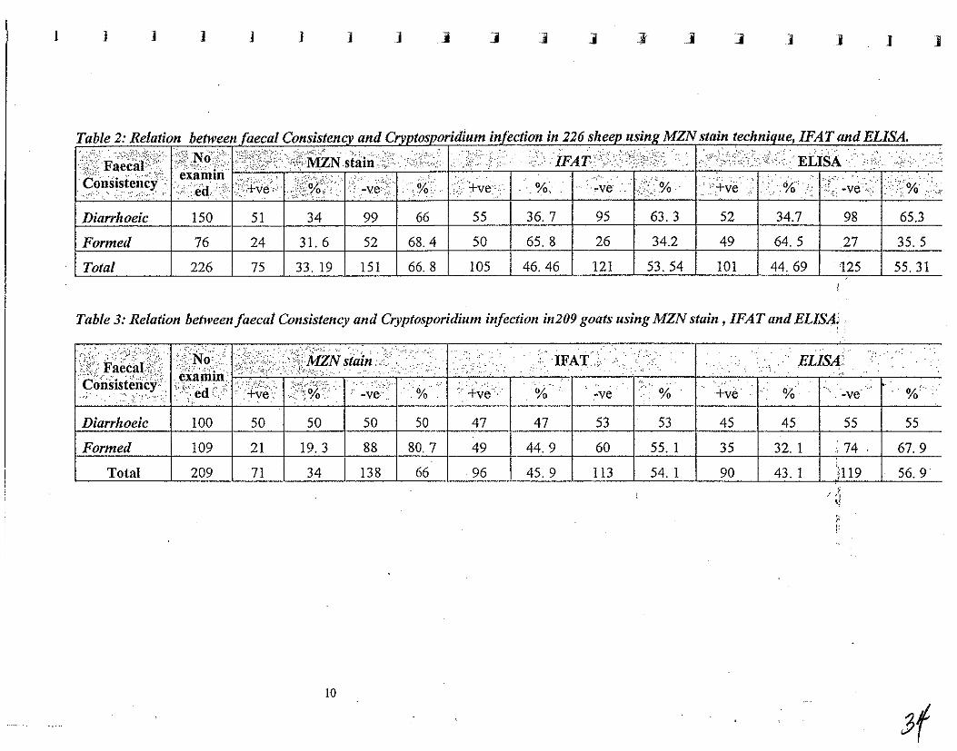
The present study showed that among 150 diarrhoeic sheep samples out of226 examined 51 (34%),55 (36.17%) and 52 (34.70%) were positivebyusingMZA technique, IFAT and ELISA respectively (Table 2). On the other hand out of76 formed faecal samples of total 226 sheep samples, 24 (31.60%), 50 (65.80%) and 49 (64.50%) were Cryptosporidium positive by the three tests respectively. Also, this investigation. revealed that out of 100 diarrhoeic goats out of209 goats samples 50 (50%) harboured Cryptosporidiumm oocystes by MZN, while 47 (47%) and 45 (45%) had Cryplosporidium antibodies by IFAT and ELISA respectively. Concerning the 109 formed goats faecal samples 21 (19.3%), 49 (44.9%) and 35 (32.1%) were Cryplosporidium positive by the.3 test respectively (Table 3). II- Neospora caninum:-
Serodiagnosis of 202 sheep sera and 78 goats sera by using Neospora agglutination test (NAT) revealed that 43 (36.14%) and 31 (39.73%) had anti Neospora caninum'antibodiestespectively (Table'l t ....
5
-

....
... .
..... III- Toxoplasma gondii: Serological examination. of 197 sheep serum samples by using modified . agglutination test (MAT) showed that 56 (28.43%) were T.gondii positive (Table J
.and.4). Also, this study revelaed that 25 (37.88%) of 66 goats had antitoxoplasma
, '.,'
6
3\
ConcIu,ion
"'1:'1,,-,, "--,:>\",,1- 1--"," ,.,_. " .. ,.t"'f)"·'':;': ':;,"':';<1'.:,[ l(')'l-I')I,~lrlc""'l (l()',",((',',i,i "~-H ,\i")(" ___ (_I'" 1 J:i.,... I)ll. .'all.:.;",...::: I.):' C.;.i;l~,)'~U;;:'_ (....:;::'--'.J" _"""/" .}" ~ (:> v. a,h~ .Il ).''1),/ u
C(,nilUn,li was l-:;:gh 2:::c';~g sh::ep \,,-j;i>: i;-:. goats the incidence ci'bo~h parasites \\'3S
eY-;:1J }:igl;e~. /\:t.-'G:-jc:; \.:c:c~Jre,d i:-: sh::e:.' 2.~:C: goals sho\ving high titers of [C)XOI'/ciSii?:2
go:';dii or :\-<]osr<.:ro (,2.:i:l~"J;n. H()\\'e\'~:: -.)::1;:; nc~ obertec! [en:2.)eS of shee-p &.r:d gcc~s J:2d high Ellers fJ;- :--:::~h p23.site5 (}::-.~;- ct-otrific.anl agent 3.:1d n~.ll;it;o;;al i''c,:tOiS s;;cujj C,t ccr:.sic(:;-t'::: ',:,"j'lf::: iT.,.e5:ig:::.::;-,~ 2.~-'_'E-ted sheep af:d go(~lS \vbich 2!e nc-g,<ive i'er a:il:-T),;:cipiasn;,:,; ?, ~,: 9_;~1:-.\'(',:)\:')o.'·:: ?.:::i'(c,dies,
Referen cc-s
('U-,-,iy', 1'), ""'I",'J"",'I"'/.'<,l:',':,!, ("" ('9('9 1. T=! :, A -::nd "PI'rlten e~,,""\.'·'~jt:> ;'~"";u'nO"SC;0:':' r' 07_ ~, • '"'~ '-" ~ v I' __ ~..)"_""' __ ."'a .... ,LC-..l"_,,,.u.,\, .,\.Q ~G.)'~1.J . ./,
152. J;-; D, (2.:1:-' (e.j). :le·s. 3 practical approad\ 'VoL 11. IRJ._:. Press. O:.-:.fc;-j.
Lh:Y.I?:o,'Z!(J! (,~ (:iiU/ R::'l.<:inf!i(JI1) IS (19S(I): Direct aggiu:in3tior11est [cillie di;:gnosis ::: ~',; ,'( t". ¥' ' '.' ?;-I : i:, fe,c~icl ~-j: ?~-,::<!.:) j ~or inc.re 2.sing sensiti " 'i1 Y and spe-ci j~ city, J. C::~l. \li,~:<'L-' 11. 56=-S~~S.
Ifcflr//..:scn,
()!-((!ga-.~\!ar(j~ L._'11.: Tu,!t'Zc·oS(.'. J/d.: Rr.jo- Va::.quc::::: F.A. a.nd C'(){J7e;:,-BcuiTis(([! ;\f.
(I:).9~~}: .~~;::""::·:l 2:-.:~::c,J~- ;"('::;--: . .'r:s..: in 1amLs nz.uJdd)~ c.nd e.\.pe~i!:lenla)jy ;r;fec::ed \\';::i C}~,p.t(J.,,!)()rj(./il;,'.'j _:X;~->·i:!i? Vet. Parasitol.~ 50 C!2): rp. 45-54.
Li..}.~e!1::O Lo"en:/,! .... !cj,J).: .·jl'es-.\fa~as-, E, Jnd Villacorta ,1ia!1ine;:. de f.JaturorrG, r (J 993j: De>:c::cl~ c:- .'.)C<:>.~:~- 2.;:0 19G c.i1t!~;.:),jjes :0 C}>l){Osj!oridi;nll
par~ :n72 ilJ s."~:·"lJ:pto:~1?::\:: ad:.:;; :z:".le. Vet. Parasito!., 47 pp. 9-15.
BEST AVAILABLE COpy
7
-

fUI"lhrr Research \VoI"k
"I' .'nl() <.j)() r;, ~'i ~!l?' 'iiI?"';';)) 1.-.- • .'1'~ _." .• (..-l""I).~ \d"I.
I\ i 01 eCl21af ch Ie (:1 eriZE, ('::'ii of sllee.p .
...
...
BEST AVAILABLE COPY
8
J ] ] j j j ] j j j j .1 1 j j ]
Table 2: Relation between faecal Consistencv and Crvvtosvoridium infection in 226 sheev usin/! MZN stain technique, IFAT and ELISA.
;E~F~~ilil~(":;:"\N0',< ;';'i;S:;';(')f/MZNst~i~·.L .· .• /:,.i.L<}}iFAT-",:; , .. /'; ..•.. : I')'ii,ii .•• ·.·•·•··· ELISA .•.... . .. >,; ...•........ '. So~Sisi~~~y, e~~:il~; +ve . ,'i,;.;o:I>-ve'; ,% . j'+ve '. %. ~~e ';:,.' % .' :',+~e r)<% .' ['J; ~~e.;'%\~ Diarrhoeic 150 51 34 99 66 55 36.7 95 63.3 52 34.7 98 65.3
Formed 76 24 31. 6 52 68.4 50 65. 8 26 34.2 49 64. 5 27 35. 5
Total 226 75 33. 19 151 66. 8 105 46.46 121 53. 54 101 44. 69 125 55.31
Table 3: Relation between faecal Consistency and Cryptosporidium infection in209 goats using MZN stain, IFAT and ELISA:' .
.... '.................... ..•. .... . .'." .IFAT; ' ... , ....... . . ELISA:'
. -ve .. % +~e .' .: . % -ve %' ...
Diarrhoeic 100 50 50 50 50 47 47 53 53 45 45 55 55
Formed 109 21 19.3 88 80. 7 49 44. 9 60 55. 1 35 32. 1 74 67. 9
Total 209 71 34 138 66 96 45. 9 113 54. 1 90 43. 1 h19 56.9 , .j " ;: !:
10

,-I I
...
Ministrv·ofAgricultlire And Land Reclamation Egypt -- - ,--
Diagnosis and Control of Brucellosis Melitensis Activity .
( Annual Report)
April 2000 - April 2001
Principal Investigator: Dr.Abdel Khalek M.Montasar Animal Health Research Institute
Agricultural Research Center
ANNUAL REPORT of MERe project
(OctobeL1999 to March.ZO,Ol) "."
Diagnosis and control of Brucella melitensis
Introduction
Brucellosis is a contagious bacterial diseases of animals and man
characterized by abortion, retained placenta, orchitis, epididymitis in its
principal animal hosts and by weakness, fever, chills, sweating, joint pain
and headaches in man.
It occurs in all closely-settled countries and is known to be
present in most parts of the world where there is any considerable cattle,
buffaloes, sheep, goats and camels population. The disea.se is
economically considered as one of the most important diseases affecting
animals, on account of causing great losses in calves, lambs and kids
through abortion, reduction in milk production and sterility among bulls
and rams which threat animal health. Besides losses in animals,
Brucellosis is hazard to public health since man can be infected with all
types of Brucella. However the definitive diagnosis for brucellosis
requires the isolation of the organism. Therefore, diagnosis has
been based mostly on the results of serological tests.
To support the capability for surveillance and control of
brucellosis, application of CFT as an accurate standard
confirmatory test to all animal samples was applied in
department of brucellosis in the central veterinary laboratory in
Dokki. Diagnosis of Brucellosis should be set too strong by
isolation and identification of the causative agents.
....
-....

r~· -
Objectives
According iowhat was settled~d.oWlLin Aswan in '.
November 99 the plane for the present reporting periods was
as follows:
1. Continuation . of national surveillance and control
programs.
2. Implementation of a generalized vaccination program
for brucellosis in cooperation ofGOVS.
3. A pilot laboratory experiment for choosing the suitable
vaccine (either Rev 1 vaccine or RB-51 vaccine) to be used
against brucellosis in different animal species in Egypt
4. Investigation of the dominant strains of Brucella by
isolation and typing from different localities all over the
country.
5. Other relevant research activities.
2
2-Activities
1. Continuation of surveillance program::",in,an infected
sheep flock:
During the period of this report we examined 1994 sheep
In an infected flock by using different screening tests (buffered
acidified plate antigen test and rose Bengal plate test), positive
samples were retested by confirmatory tests (complement
fixation test and Rivanol test). The obtained results indicated
that the incidence of infection was (0.6%). Previously, the
incidence was 3%. This finding pointed out that the control
program in this farm run successfully. The results were
illustrated in table (1)
Table (1): Results of serological examination in sheep flock
Results of different serological tests Animal Total BAPAT RoseB SAT Riv t CFT species No. +ve -ve +ve -ve +ve +ve -ve +ve -ve +ve
sheep 1994 28 1966 12 1982 12 2 1980 12 1982 10
BAPAT. Buffered acidified plate antigen test Rose B. rose Bengal plate test
SAT. Serum agglutination test Riv.T. Rivanol test
CFT. Complement fixation test
2. Vaccination activity:
During this period we vaccinated 452 female lambs 3-6
month of age with a dose 1-2 xl09 CFU sic. Before
vaccination, animals were serologically examined and it was
-.
-
I
+ve -ve
2 1982
found that two animals were positive, which were
slaughtered. The _ n~gative lambs :v"e.(~;-:;.' ~Epjected to
vaccination in March 2000.
After six months in September 2000 , 349 blood samples
from vaccinated animals were tested to follow up the level of
immune responses produced due to 'vaccination. It was found
that 26 animals were still have post vaccinal persistent titer to
serum agglutination test, buffered acidified, plate test and
four animals have week reaction with Rose Bengal plate test
and negative with other tests. This is mainly due to the
disappearance ofIgG in vaccinated animals earlier than IgM.
But in true field infection IgM is declined and disappeared in
chronic stage and IgG still gave positive reaction which
detected by confirmatory tests like Rose Bengal, and eFT
table (2).
Table (2) Results of serological examination of vaccinated animals
Titer of Total BAPAT Rose Bengal
SAT number +ve -ve +ve -ve,
-ve 323 0 323 0 322
1110 13 13 0 0 13
1120 11 10 1 2 0
1140 2 2 0 2 0
Total 349 25 324 4 345
4

3. Confirmation activity:
The Brucellol>is .D~partment Aniwal);:leq.lth. Research
Institute in Dokki was received a total of 4171 samples from
different animals for confirmation from different localities in
Egypt (1866 sheep, 450 goats, 1716 cattle and 139 buffalo).
The samples received after being examined by different
field screening tests namely Buffered Acidified Plate Antigen
Test (BAPAT) and Rose Bengal plate test (REPT). The reactor
samples were confirmed in Brucellosis Department Animal
Health Research Institute by using different confirmatory tests
which serum agglutination test (SAT), Rivanol test, and
complement· fixation test (eFT). The conclusion of the results
to reactors, SUSpICIOUS. and negative according to the
Brucellosis report number 7. The results were illustrated in
table (3).
Table 3 results of serological confirmation in central lab.
Animal Total Serological results
Species. number Reactors Suspicious Negative
Sheep 1866 70 3 1793
Goats 450 1 0 449
Cattle 1716 347 18 1351
Buffalo 139 69 2 68
Total 4171 487 23 3661
5
-
-
....
4. Examination of imported animals in quarantine:
From beginni_ng ,,9X _N ovember 1 ~,9~, 19. ~El1~ 2000, the
Brucellosis Department received a total number of samples
7347 from impOlied pregnant cattle heifers. These animals were
impOlied from countries as brucellosis free countries and they
were examined for brucellosis. One animal was positive and it .
was slaughtered immediately.
5. Isolation activities:
Isolation trials were done on a total of 153 samples (10
milk samples, 139 lymph nodes and tissues and 14 aborted
foetei from different animal species). It was succeeded to isolate
61 brucella straines (44 from cattle, 9 from buffalo, 6 from
sheep, and 2 from goats) as shown in Table (4). These samples
were collected from serologically positive animals. Biotyping of
isolated strains, which are shown in table' (5), indicated that all
the isolated strains are Brucella melitensis biovar 3.
Brucella melitensis biovar 3 is mostly dominant strain
recovered from different animal species' in Egypt in last ten
years. This finding reinsure the impOliance of using Rev 1
vaccine for vaccination of different animal species for proper
eradication of brucellosis in Egypt as previously recommended
through other reports.
6

J ) I ] J j ] J ] ] j ] ]
Table (4) Results of bacteriological cultures of different samples collected from different animal species.
-
J
Milk Lymph nodes* Aborted foeti * * Tissues*** SpeCies Total Total Total Total
~- : sample -ve +ve sample -ve +ve sample -ve +ve sample ;.
Cattle 6 5 1 102 62 40 8 5 3
buffalo 4 3 I 19 12 7 2 1 1
sheep 0 0 0 15 9 6 2 2 0
goals 0 0 0 3 2 1 2 1 1
Total 10 8 2 139 8S S4 14 9 5 I
Brucella mehtenslS bIOtype3
* Lymph nodes. Supramammry, Retropharyngeal, and internal iliac lymph nodes ** Aborted foeti. Fetal fluids and stomach content of foetus *** Tissues. Liver, spleen and udder tissues
102
19
15
3
139
-ve
62
12
9
2
85
J 1 j
+ve
40
7
6
1
54
1
• Table (5) Biotyping of isolated Brucella strains recovered from different animals.
:z > <= ;:I :3 Biochemical reaction S": cr
" ~ :i .., -,' 0 '" ' . .... "O~ :;::. g I~ .' 0 Co2 H2s Catalase Urease ..... ~ ., ~'t ~
" " Reg Pro '" , "
Cattle 44 - - + +
Buffalo 9 - - + +
Sheep 6 - - + +
Goats; 2 - - + +
Total I 61
Reference starins: Br. melitellsis 16M
M = monospecific antisera (mtlitensis)
Tb = Tblisi phage
I I I ]
Biotyping
Growth on dyes Mono.S Sera phage lysis
Thionin Fuchsin Tb
a b c B C A M RTD 10' RTD
- + + + + + + - -
- + + + + + + - -
- + + + + + + - -- + + + + + + - -
All isolated staines Brucella melitensis biovar 3
Br. suis 1330 Br. abortus 544
.a = 1:25000 b= 1:50000
RTD = routine test diluation
2
] J ) ] J I
A. = monospecific anrisera (abortus)
c=1:100000
104 RTD = 104X rotuine test diluation
11 I
6. Relevant activities:
6-1 Evaluation of Brucella abortus RB51"Ycaccine against '.
brucella melitensis field strain in Guinea pigs.
Experimental design
The laboratory animals were divided into two groups:
First group 160 animals were injected by RbSl vaccine.
Second group 40 animals left as control
Every .. week during the experiment the vaccinated animals
were sacrificed in fixed numbers (three guinea pigs) for detecting
.. the antibody titers by using conventional serological tests. Using
smooth antigen (Rose Bengal Plate Test, Buffered Acidified Plate
antigen test, Serum Agglutination Test and Rivanol test and
eFT), and serum agglutination test prepared from rough strains of
RESI (SATr)
Bacterial isolation was done weekly from internal organs
and spleen. After complete failure of recovery of vaccinal strains
and disappearance of any serological titers. The remaining
animals were challenged by Brucella melitensis biovar 3 (field
strains) and in the same time the control group was also injected
by the same strain. Then follows up the serological titer and trials
of isolation from the two groups weekly after challenge and up to
the end of experiment .The results of this experiment were
discussed in detail in the next report.
6-2 Evaluation of different serological tests on cattles III 8
governorates.
Materials:
1 * blood samples:
.,:,.._-- ... ~."",:-:;;. i.l...:.'_' .-
A total of 4357 blood samples were collected from cattle
fi'om different localities in Egypt during this period as explained
in Table (1).
Table (1): Blood Samples collected frol11 different localities
Sites Total No. of Cattle' S:Lte ( 1. ) 2566 Site ( 2 ) 66 Site ( 3 ) 19 ·Site ( 4 ) 775
. Site ( 5) 120 Site ( 6 ) 46 Site ( 7 ) 667 Site ( 8 ) 98
. Cumulative totals 4357
.2.Tissue specimens:
Supramammary, internal iliac, retropharyngeal, prescapular
and prefemoral lymph nodes as well as spleens, 6varies, livers
were collected from 80 slaughtered cows positively reacted in one
loll or more of the applied serological tests. The samples were
collected for bacteriological examination.
3. Aborted foeti
The stomach contents of 18 bovine aborted foeti from
individually raised aborted cows were collected by sterile syringe
and kept on ice during transportation to the laboratory and used
for bacteriological examination.

· Table (2) Number of blood samples, aborted foeti and
tissue specimens collected from different localitiesill Egypt. - -~-. - ."".', ,..,. .. -. "
No, of No, of No, of No, of No, of No, of No, of
blood aborted slaughtered lymph ovarre testicles spleen
sample foeti animals nodes s
4357 . 18 80 280 4 I 3
4. RESULTS
Prevalence of Brucella reactors among cattle in different
governorates,
Results of this investigation are presented in Table (3 ) which show
that out of 4357 blood samples collected from 8 ,Sites , the
percentage of positive rePtctors were 10.21,12,12,5.26,25,68,0,0,13,04,
0,0 and 0,0% for SHes (11) ,(2) , (3) .;(4) , (5) ,',(6) , (1) . , (8)
and (9) .respectively __
Table (3): Prevalence of Brucella reactors among cattle samples
collected frol11 different governorates under investigation.
No. of No. of % of No. of % of Sites
sam pIes Positive positive Negative negative
Site (1) 2566 262 10,21 2304 89.79
Site (2) 66 8 12.12 58 87.88 Site (3) 19 1 5.26 18 94.74 Site (4) 775 199 25.68 576 74.32 Site (5) 120 0.0 0.0 120 100
Site (6) 46 6 13.04 40 86.96
Site (1 ) 667 0.0 0.0 667 100 Site (8) 98 0.0 0.0 98 100
Total 4357 476 10.92 3881 89.08
Table (4):
Results of Tube Agglutination Test(TAT) for cattle sera collected from different governorates under investigation.
I:, No. of .. " Results of reactors at the following dilutions (1110 up to 1/X) Negative samples Positive samples .' ..
. Sites , samples 1/10 .% 1120 :% 1/40 % 1180 0/0 No. % No. % . ,. SHe~.(1 ):
, 2566 18 0.70 56 2.18 69 2.69 80 3.12 2343 91.31 149 5.81
Site (2) ,
66 1 0.02 ~ 4.55 4 6.06 0.0 0.0 58 87.88 4 6.06 .J
Site (3) 19 0.0 0.0 0.0 0.0 0.0 0.0 0.0 0.0 19 100 0 0.0 . Site .(4) 775 19 2.45 32 4.13 45 5.81 65 8.39 614 79.23 110 14.19'
Site (5) 120 .0.0 0.0 0.0 . 0.0 0.0 0.0 0.0 0.0 120 100 0 0.0
; Site. (6) 46 0.0 0.0 1 2.17 4 8.70 0.0 0.0 41 89.13 4 8.70
Site m I 667 0.0 0.0 0.0 0.0 0.0 0.0 0.0 0.0 667 100 0 0.0
Site (8) 9 0.0 0.0 0.0 0.0 0.0 0.0 0.0 0.0 98 100 0 0.0
~otal 4357 38 0.87 92 2.11 122 2.80 145 3.33 3960 90.89 267 6.13 ,
Titre of lOis considered as negative. Titre of 20 is considered as suspect Titre of 40 up to 1280 is considered as positive.
J I J 1 1 ] 11 1 1 1

J ] J j I 1 j j j ] ] ] ] j j j j
Table (5): Results of Rivanol Test (RivT) for cattle sera collected from different governorates under investigation .
. ,:.!. :.No. of' . _'.~' .. ·.~:Y·: ,:'. Results ofRivanol Tests atthe follo\ving dilution: ...7 Negative' . :'-,;
Sites .. . 1/25 % .1/50 %. 11100 % 11200 . 0/0 .
.
1/400 ,% No: ,. ..
No. '. ' .. ::."
%. "%
Site ( ,1 ) 2566 26 1.01 65 2.53 41 1.6 42 1.64 47 .1.83 2345 91.39 221 8.61 I, :l
Site. (,'2 ) 66 3 4.55 3 4.55 0.0 0.0 0.0 0.0 2 3.03 58 87.88 8 12.12 "
Site (i:3) 19 0.0 0.0 0.0 0.0 0.0 0.0 1 5.26 ,---~~,--;---------~---+--~-r----;-----r
0.0 0.0 18 94.74 1 5.26
Site (: 4 ) 775 37 4.77 45 5.81 10 1.29 25 3.23 27 3.48 631 81.42 144 18.58
.Site(5) 120 0.0 0.0 0.0 0.0 0.0 0.0 ·0.0 0.0 0.0 0.0 120 100 0 0.0
Site ( 6 ) 46 3 6.52 1 2.17 1 2.17 0.0 0.0 0.0 0.0 41 89.13 5 10.87
Site ( 7 ) 667 0.0 0.0 0.0 0.0 0.0 0.0 0.0 0.0 0.0 0.0 667 100 0 0.0
Site (, 8 ) 98 0.0 0.0 0.0 0.0 0.0 0.0 0.0 0.0 0.0 0.0 98 100 0 0.0 .
Total: 4357 69 1.58 114 2.62 52 1.19 68 1.56 76 1.74 3978 91.30 379 8.70

. -- ".-.-- .-. '-._ ...
Table (6): Results of complement fixation test for cattle blood samples collected from ciifferent governorates under.investigation. , ;~ ..
Results of complement fixation test at the following dilutions : ,
Positive '. Negati~e Sites I"i ...
" .. ,':.-'
" 114 % 1/8 % 1/16 0/0 1132 % 1164 % 1/128 % 11256 0/0 No. % ' No: % , ,
Site l'~ 'j 2566 10 0.39 78 3.04 51 1.99 37 1.44 32 .1.25 17 0.66 1 0.Q4 216 8.42 2340 91.1
Site ( 2 ) 66 0.0 0.0 0.0 0.0 0,0 0.0 0.0 0.0 1 1.52 0.0 0.0 0.0 0.0 1 1.52 65 98.4
Site ( 3 ) 19 0.0 0.0 0.0 0.0 0.0 0.0 0.0 0.0 0.0 0.0 0.0 0.0 0.0. 0.0 0.0 0.0 . . 19 101
Site ( 4 1 775 7 0.90 ·52 6.7 33 4.26 24 3.1 22 2.84 18 2.32 0.0 0.0 149 19.23 619 79.S , Site ( 5;) 120 0.0 0.0 0.0 0.0 0.0 0.0 0.0 0.0 0.0 0.0 0.0 0.0 0.0 0.0 0.0 0.0 120 101
Site ( 6') , 46 1 2.17 1 2.17 3 6.52 0.0 0.0 0.0 0.0 0.0 0.0 0.0 0.0 4 8.70 41 89.1
Site ( 7 ) 667 0.0 0.0 0.0 0.0 0.0 0.0 0.0 0.0 0.0 0.0 0.0 0.0 0.0 0.0 0.0 0.0 667 101
Site ( 8 ) 98 0.0 0.0 0.0 0.0 0.0 0.0 0.0 0.0 0.0 0.0 0.0 0.0 0.0 0.00 0.0 0.0 98 101
Totals 4357 18 0.41 131 3.0 87 2 61 1.4 55 1.26 35 0.8 1 0.02 370 849 3969 91.1
I 1 11 1 'I
SUMMARY This investig~tio~' -is concerned with"'th~'p;:ev~lence of Brucella
infection and evaluation of some serological tests used In Brucella
diagnosis among cattle in some §ites "under investigation
A total of 4357 cattle serum samples were collected from different
localities in these governorates under investigation during this period
Sera from these cattle are examined for brucellosis by different serological
tests inCluding Buffered Acidified Plate Antigen Test (BAP AT), Rose
Bengal Plate Test (RBPT), Tube. Agglutination test (TAT), Complement
Fixation Test (CFT) and Rivanol Test (Riv.T).
1. The infection was high among cattle of SiteC<;1 4 ) .
followed by Site (6) , Site ( 2) ,'Site ( 1 ) and Site ").
2. BAPAT, among all the tests used in the study, gave the highest
rate of positive cattle samples (10.63%) followed by RBPT
(10.19%) and Riv.T (8.70%), CFT and SAT reacted positively with
8.49% and 6.13% respectively.
3. The application of CFT as a confirmatory, quantitative test and
more specific test for Brucella antibodies which gives 100%
correlation with direct culturing.
4. Comparing the results of the different tests it seems advisable to
use the buffered acidified plate antigen test (BAPAT) as a
presumptive test due to its higher sensitivity, easy to be done and
to exClude negative cases. The use of Rose Bengal Plate test
(RBPT) as a screening test due to its higher sensitivity to confirm
doubtful cases by CFT and TAT test. ~D IooJ
...
...
...
.....
5. Collected tissues and lymph node samples from 80 slaughtered
cattle which proved to be positive by serological tests were 0_ ,.-,~:,· .• ·""-t;::;r <+,.':."0' .-
subjected to bacteriological examination which revealed the
isolation of 13 isolates which under biochemical tests typed as Br.
melitensis biovar 3 .
6. In addition, 18 stomach contents from aborted foeti obtained
from aborted cattle during the late stage of gestation (7-9 months)
were examined to isolate the cause of abortion using both animal
inoculation and direct culturing on specific media. This revealed
the isolation of 15 isolates. which were under biotyping .
7. The present data and those of previous authors indicate the
prevalence of brucellosis in Egypt; which undoubtedly points to a
problem of considerable magnitude not only due to its impact on
economical losses but also as disease of zoonotic importance. Thus the
disease must be overlooked as an international one of the most
dangerous and notifiable diseases. This implies the necessity of
directing all efforts to help in rapid and accurate diagnosis with valid
records to fulfill the necessary measures of control this disease .
2 51
Conclusion
Throughout fhcpedod of this projectwe'fi·y to cover all
objectives recommended in the previous report but we could
not cover some of these objectives. These objectives were
mainly:
The achieved objectives:
1. Evaluation of RBS1 vaccme m a pilot experiment m
laboratory animals against local field strain.
2. The successful use of Rev 1 vaccine on a small scale in
lambs in a flock of sheep with a history of Brucella infection
and followed up the imn1une responses after vaccination
3. Isolation and typing of Brucella strains from milk, lymph
nodes, aborted foeti and tissues of different animal specIes
from different localities.
3
....
Ministry of Agriculture And Land Reclamation Egvpt
Foot and Mouth Disease Activitv
(Annual Report)
April 2000 - April 2001
Principal Investigator: Dr. Ahmed Daoud Director of Serum & Vaccine Research Institute Agricultural Research Center
Introduction
Foot and mouth disease (FiVlDI is one of the most infectious diseases of
cloven footed animals. The disease assumes an enzootic form in Egypt and some
other countries sharing the same borders. It is characterized by relatively low
mortality (4% in cattle and buffaloes, 2% in sheep) and economical losses that may be
as high as 25%. Insufficient or deficient disease combat measures are the major
factors that influence its spread and reCUITence among livestock in these areas.
Accordingly, there is a fear among countries that have been declared free from
FMD for decades, that it may attack them back, as in case of the recent FMD outbreak
that took place in Taiwan (spring of 1977). This country had been free from the
disease for 68 years. The outbreak resulted in tbe death of approximately four million
swine, with multimillion dollar loss due to clean up costs and lost export income.
Current responses to disease incursion to free countries inciude the inhibition
of animal and product movement, slaughter, disinfection and vaccination. In enzootic
areas, however, serological surveys and detection of carriers and susceptible animals
are the main measures for control\i.ng the disease spread.
The purpose of the present project was to establish a collaborative effort among a
group of enzootic countries in the iVI iddle East to understand disease transmission,
and develop better ways for better diagnosis and control. We have discussed some of
our activities at the Abbassia FMD Department in earlier reports Uoint training,
coastal surveys, vaccine improvement and attempts to enhance disease diagnosis
using serological or molecular methods), and here, we will discuss our recent surveys
and research findings during the last few months.

....
"'"
"'"
~.
"",'
** The following three major topics have been agreed upon in Aswan workshop (18-
21 November, 1999);
1. Extension of FlVID survey in animals living in coastal areas in
Egypt:
Serological surveys on cattle, buffalo, sheep and goats from EI Areesh, Port
Said, Ismailia, Suez and Alexandria have been conducted and discussed in previous
reports. Results indicated that while many animals showed specific antibodies to
FMD (by SNT and ELISA), some were also positive for 3D or VIA antibodies.
Another group of animals harbored the virus in the nasal mucosa, as determined by _.
peR.
However, because of the lack of funding we were unable to continue visiting
the coastal provinces nor to collect more samples and analyze them. As such, the
ability to achieve the goals of the project is highly jeopardized. A number of projects
could have been started, and survey trips could have been paid to the borders of the
country. The lack of funding also reduced the interaction among the participating
countries.
This problem should receive considerable attention, if this projeCt was to
continue. As shown below, we have depended on our limited resources in assessing 'a
couple of FMD outbreaks that took place in our country, but much more analysis and
research elTorts are needed.
Recent outbreaks in Egvpt
A. On the 8th of January, 2000, FMD was reported in Menofya province among an
importedherd of Holstein cattle that entered the country through Alexandria on the
first day of the new year. Original number of imported animals was 1027, but 200 of
them were isolated (19.4%) upon the appearance of FMD signs. All herd was
vaccinated for FMD on the 13 th of January, but 97 animals succumbed afterwards
(9.4%). An investigation team found out that the disease was predisposed by siress
factors and unhealthy management during transportation (e.g. long trip, ship was not
2
disinfected after previous shipment). Symptoms were complicated by secondary
infections.
FMOV was detected in 3/5 eospophary;ig~a(('Op)samples and in 2/2'gingN3.f1'issue!;"
using peR and complement fixation test (positive with 0 group antisera).
B. Around the same time, another outbreak took place in EI-Tal EI-Kebir, Ismailia. Of
358 buffaloes, 43 developed symptoms of the disease (12%). These animals were
reported to have been vaccinated within the previous month. Samples were collected
for virus isolation and identi fication as mentioned before. However, it was noticed
that contact cattle had more severe symptoms than buffaloes and more than 900 sheep
in the neighborhood were sound and healthy. Virus identification tests showed it to be
of the 0 serogroup also.
C. Six months after Menofya's outbreak, a'survey on serum samples collected from
vaccinated animals in this province was conducted. Results are shown in the
following table:
Table I. Summary of serological results in 180 cattle sampled in Menofya
ArealFann Sample size Spp. +ve SNT% G:: 1.2) +ve 30%
27 Cattle 70.3 0.0
2 37 Cattle 83.0 5.0
3 36 Cattle 75.0 4.8
4 80 Cattle 82.5 0.0
Total 180 78.9 1.1
2. Further studies on the role of pigs in the epidemiology of FlVID in
Egypt:
Pigs constitute a major FMD threat to other animals. However, there are no
accurate statistics on the swine population in Egypt. Official figures (27.00 animals)
are probably way below the actual numbers as deduced from the slaughter house
figures.
3

Although these animals do not receive FMD vaccination, analysis of their random
... serum samples showed that the majority had SN titers. Also, tonsillar lymph nodes
collected from slaughterhouses carried Elv1Ds.icus-at low concentraiiops w_~lt.n.o. • __ ". _". _ _"~ ,,~.... lA._-,- ',C
... clinical signs. Isolated virus was proved ~xpcrimentally to be infectious for
susceptible pigs.
-.
Another experiment conducted at the FMD department showed that susceptible
pigs contracted the disease after 4 days when placed in contact with infected calves.
Antibodies were detected in pigs after the first week or exposure and were generally
of low-level as shown in the table2. Sows (especially when under pregnancy stress)
o.j developed the disease before males. The virus was isolated from the different lesions
of infected pigs and identified by the use of eFT, FA, baby mice and tissue culture
inoculations.
ioiI.
....
In a third experiment, cattle in contact with infected pigs developed mild FMD
signs on the 5th day post exposure (Table 3). This may agree with the famous
Taiwan's outbreak, where no clinical signs of FMD were observed among cattle and
sheep that lived in the vicinity of infected pigs. In our laboratory, contact sheep
showed no FMD symptoms, but VIA antibodies were dctectcd in most a'rYimals (Table
4).
Table 2. Summary of SN and VIA antibodies in 4 pigs infected by contact with cattle
Weeks post-exposure 2 3 4 5 6 7 8 9 10 II 12' 13
Mean SNT titer (loglO) 0.6 0.8 0.8 0.9 0.8 0.8 0.6 0.7 0.4 0.6 0.4 0.4 0.1
VIA antibodies o 2/4 Y..2/4 214 0 0 0 0 0 0 o 0
Table 3. Summary ofSN and VIA antibodies in 5 sheep living in contact with
infected pigs
Weeks post-exposure 2 3 4 5 6 7 8 9 10 11 12
Mean SNT titer (I0g10) 1.6 1.8 2.0 2.0 2.0 1.9 2.0 1.9 1.8 1.6 1.7 1.7
VIA antibodies 0 2/5 515 2/5 1/5 115 0 0 0 0 0 0
4
Table 4. Summary of SN and VIA antibodies in 5 cattle living in contact with infected
pigs
Weeks post-exposure 2 3 4 5 . _6 7 8 9 10 11 12 .".~ -,,-.~'.--.,,:-'~,:~~" ".',. . .:.".:
Mean SNT titer (logJO) OJ 1.1 0.8 0.7 0.4 0.3 0.4 0.2 0.1 0.1 0.2 0.1
VIA antibodies 0 4/5 4/5 4/5 2/5 2/5 liS 1/5 1/5 0 0 0
3. Quantitative relationships among ELISA, Dot ELISA and AGPT
in the detection of 3 D antigen:
The presence of a reliable serological technique for the detection of FMD in
infected animals is highly important in endemic areas. Recent research 00· FMDV
relies on the detection of serotype-independent, infection associated, nonstructural
(NS) proteins or enzymes that are released during virus replication, such as VIA, 3D,
2C or 3ABC antigens.
In the present srudy we have adapted a Dot ELISA technique for the detection
of3D antibodies in experimentally infected cattle. Specific 3D antigen was prepared
by cloning and transformation methods. as described elsewhere. Indirect plate ELISA
and AGPT were perfomled for control purposes. Sensitivity results of these methods
using reference and diseased sera (from experimentally infected animals) are shown
in tables 5 and 6:
Table 5. Minimal detectable level of3D antigen by Dot ELISA, plate ELISA and
AGPT
Assay
Sensitivity lIIO 1!20 lI40 1/80 1/160 t!320 1/640 1/1280
Dot ELISA + + + + + + + +
ELISA + + + T + +
AGPT + + +
5
I .. :
...
Table 6. Comparative evaluation of Dot ELISA, plate ELISA and AGPT using sera
from experimentally infected animals
Assay
Dot ELISA
ELISA
AGPT
6
No. tested samples
ISO
ISO
ISO
N o~'positive -
ISO
141
93
No. Negative.·.······Positive% '.
o 9
57
100
94
62
Proiect extension:
It has been claimed that more practical and less invasive methods are
required for sampling lesion~a'ld.TD.uc!Jus membranes of,~.njm!ll~Jn(~<;!ed with '.
FMD. More fast and reliable diagnostic methods would have to be developed.
Confirmation of disease-free and carrier animals assumes utmostimportance to
limit disease spread. Virus characterization and evaluation of antigenicity are
always needed for tracing the virus during outbreaks and to de felop more
efficient vaccines.
Since funding was not enough during previous term, th~ following may be
fulfilled with the extension:
I. Continue serological and virological (PCR) surveys on animals living at the
. borders of Egypt.
2. Explore the role of pigs in the epidemiology ofFMD in Egypt.
-.- 3. Develop and improve FMD quarantine and control measures among
collaborating countries (based on sufficient data).
4. Prepare and provide guinea pig and rabbit hyperimmune sera against the local
FMDV strain (0,1 Aga/9J). Those may be exchanged for reference antisera
against other serotypes (A, C & SAT).
5. Prepare and provide JCD antigen for use in the region.
6. Carry out antigenic variation studies on FMDV isolates (0 strains) recovered
from collaborating countries to determine their dominance (by serlogical and
molecular methods) and potential iclusion in the vaccine.
7. Enhance cooperation and exchange of expertise among collaborators.
Ii. Jj
-if !

Papers Submitted This Year
.--
~l

__ . , __ ~,_:-'-'r .-0 '" ."-:"""':,,-.~~i<-';;;:: i1<-."=".' _. '.
THE EGYPTIAN JOURNAL OF
IMMUNOLOGY .. "
~"-"'~'~"""""''''N~''''_~''''''''''_'' ,_. .
.....
....
Reprint
~-=~ ===' ===--Member tif the trttetitatiohat UtliOii
< I ••
(iSSN 1110 - 4902)

....
I nr:.. cu r r 111\!"1 JVUI',!'if\.1-. VI IjYijYIUI'iVLVV I Y-V"---I elj, _vvv
Page: 97- 103
Evaluation of Relationships Among ELISA, Dot ELISA and Agar Gel Precipitation' Tests in The "Dete~ti'On . of 3 CD Antigens of FMDV
M Shawky H EI-Watany, A Samira EI-Kilany.und OH.Roshdy
f'MD Research Department, Veterinary Serum & Vaccine Research Institute, Abbasia, Cairo . . --
Dot' ELISA was used for the detection of antibodies against protease and polymerase genes (3 CD) antigens in animals infected with foot and mouth disease virus (FMDY). Antigen antibody reactions were detected with an antibovine serum labeled with horseradish peroxidase. Using 4-ehloro-I-naphthol as a substrate, positive reactions appeared as blue dots and the results were compared with those of solid phase ELISA and agar gel precipitation. Dot ELISA appeared to have a potential for rapid and economical field test in the diagnosis of FMD in infected nllimafs. .
A vailability of a reliable serological. technique for the detection of f'MD infected animals assumes a
paramount importance in endemic areas, where the disease is mainly controlled by vaccination (Sorensen et.al., 1998). Identification of infected animals relies on the detection of serotype-independent, infection-associated non-structural proteins or enzymes released during virus replication. In case of FMDV, these mlly comprise the so-called virus infection associated antigen (VIA), 3D, 2C and 3 ABC protein moieties (Lubroth et aI., 1998). The first two are widely sought among infected animals or after thc use of multiple or concentrated doses ofFMD vaccines.
Several methods have been employed for the detection of antigens and antibodies following a specific infection. Enzyme linked immunosorbent assay (ELISA) has found its way in this field (Charan and Guatam, 1984) because of its simplicity, sensitivity and specificity over 1110st other serological assays. The technique elaborates a specificity similar to that of agar gel immunodiffusion, but is, by 'far, more efficient in detecting small amounts of VIA (3 D) (Alonso et aI., 1990).
Likewise, Dot ELISA proves applicability in the diagnosis of some animal diseases, such as goat pox (Sedhukhan et aI., 1998), blue tongue in sheep (Mishra et aI., 1998), bovine brucellosis (Batra et aI., 1998), mycoplasmosis (Egwu and Aliyu, 1997), rinder pest (Gurkirpal-Singh et aI., 1997), rabies (J ayakumar et aI., 1996), neonatal bovine infection with Rota virus (Kapoor et aI., 1996), Taenia saginata cysticerci (Draelants et aI., 1995) and Schistosomiasis (Ye-Ping et aI., 1994). The technique is easy, sensitive and does not require sophisticated equipment.
In this paper, we describe a rapid dot ELISA for the detection of antibodies to 3 CD antigens in animals experimentally infected with I'¥DV.
Material and Methods
Recombinant proteins:
Those have been prepared as explained before (Shnwky et aI., 1999). Brief1y, the sequence corresponding to 3CD region of FMDV type 0 was amplified by PCR and cloned into pET 21 a-d vector transformed hito expression host and induced by IPTG to express proteins of the insert. Molecular weight of the final product was confirmed by

98
polyacrylamide gel electrophoresis (PAGE)
and by I iquid phase blocking -""sandwich
ELISA using two speci fie reference antisera
(O'Donnel, et a!., 1996). Standardization
experiments were performed to determine the
optimal dilutions of 3CD antigens of FMDV
in dot ELISA, ELISA and agar gel
precipitatior. test (AGPT), using variable
concentrations of a reference serum.
Obse,"ved titers were 1/640 (6 ng protein),
lI:J2{i (i 2 ng protein) and 1/40 (96 ng
protein) for each technique, respectively.
Serum samples."
A total of 150 serum samples were obtained
from 10 experimentally infected cattle" They
were collected on a weekly basis, between 2-
16 weeks post-infection. The same reference
positive and negative FMD sera were utilized
for control purposes as mentioned above and
were also used to establish optimal
concentrations and sensitivity.
3. Dol ELISk
The procedure was carried out as described
ly (Hawkes et aI., 1982). Briefly,
11itrocellulosc membrane filters (0.45 Ilm
pore size, Millipore Co., Ireland) were placed
on top of a 96-well microtiter plate, pressed
to fOIDl impression circles and immersed in
distilled water for 5 minutes, then allowed to
dry at room temperature. A volume of 3 to 5
fll of optimal antigcIl dilutions (l/640) was
dropped on each circle and allowed to dry at
room temperature for 15 minutes to stabilize
the binding of proteins. The membrane filter
was then washed for 5 minutes in Tris
buffered saJine (TBS; Tris 50 mM, 200 mM
Nael and "Tween 80" 0.05%, pH 7.4). The
free protein binding sites were blncLcd by
immersing the membrane in TBS conlnining
2% of bovine serum albumin for 60 millutes
at room temperature. Inactivated serum
samples from experimentally infected
animals (including negative controls) were
added to the dotted antigens and- incubated at.
room temperature for 60 minutes. Unreacted
antibodies ~~Ie.,!emoy~!I by immersion in a
washing buffer (TBS pH 7.4 + 0.05% tween
80) for 30 minutes, over a shaker and
changing the buffer each 10 minutes. The
membrane filter was then incubated with
antibovine conjugate (conjugated with
horseradish peroxidase enzyme) for one hour
at room temperature. The specific antigen
antibody reaction was visually detected by
immersing the membrane in freshly prepared
solution of enzyme substrate (3 mg of 4-
chloro-l-naphthollml anhydrous methanol).
Just before use, 1.7 ml of the stock solution
was mixed with 8.3 ml of TBS and 30 III of
~h02 (3%). The enzyme reaction was
stopped after 15 minutes by washing the
membrane with tape water. Specific positive
reactions appeared as dark blue dots at the
individual site of the dotted antigens.
ELISA (indireci ELISA):
ELISA plates were coated by an optimum
dilution of 3 CD antigen (11320) dissolved in
carbonate bicarbonate buffer (pH 9.6) by
over night incubation at 4°C. This was
followed by washing with phosphate buffer
saline containing 0.05% tween 20. Non
specific proieIn binding sites were blocked
by incubation for one hour at 37°C with
blocking buffer (1 % bovine albumin in PBS
with "tween 20" 0.05%). Plates were then
washed 3 times (with PBS-Tween) and 50
Illiwell of each tested serum sample was
added in duplicates. The plates were
incubated for one hour at 37°C. At the same
time negative control sera were tested. After
washing 3 times with PBS-Tween, the
conjugate was added (peroxidase-labelled
antibovine conjugate) and incubated for one
hour at 37°C. Finally the reaction was
developed using 50 Illlwell orthophenyl
diamine (OPD) and 0.005% of 3% H202.
Finally, the reaction was stopped after 15
minutes by the addition of 50 ~d per well of

-------~. ,~.~,~ ... '_...;;;r_r.___.-"". ~"'_"'\.,.'\ ... ''''' "YI'''',JI~\/''''''\JV I 'i'l
...
...
...
.'
1.25 M H2SO.\ and lhe absorbance was read at 492 nm.
Agar gel precipitation test:
The test was· performcd llsing all optimum dilution of3 CD antigen (1/40) placed in ccntrnl wells, while sera (including positive and ,Jjegr.ti ve controls) were placed pcriplJCrally in I % agar plates. Reactions were incubatcd at room temperature in a humidity chamber and read after 48 hours (McVicar and Sutmoller, 1970).
Results
Results of quantitative sensitivity of dot ELlSA is shown in Table 1 and figure 1. Positive reactions appeared as blue dots with varying intensity, depending all the amount of antigen-antibody reactions. Tile least conccntration of 3CD antigen giving a
positive reaction was 'I: 640, Ncgative tests showed no visible colour, this means that the test is' 'hlgll1y~~~sejlshive and specific. Minimum sensitivity of plate ELlSA was I :320; the cutoff point is conventionally twice the standard deviation of the negative cutoff. In this study the cutoff point was statistically and practically found to be 0.6, and that of AGPT is I :40 Crable I).
A comparison of the relative sensitivity of dot ELISA and other techniques employed in this study is shown in (figure 2). In this experiment, known serum' samples li'olll experimentally infected cattle were used. Sensitivity of Dot ELISA, ELISA and AGI'T to 3 CD antigens were 100%, 94% and 62% respectively, as shown in Table (2).
All serological tests proved specificity and sensitivity using positive and negative control antisera.
3CD antigens dilution 1110 1/20 1140 1/80 1/160 11320 1/640 111280
* * * * * (*) Posit.ive reaction (blue dots).
(**) Negative reaction (no co]or).
* *
rigu!'c 1. Quantilative sensilivity of Ihe dol ELISA to 3 CD anligens of FMDV.
**
Tnblc I. Determination of the minimal detectable level of 3 CD antigens of FMDV by dot ELISA, ELISA and AGPT against optimum dilulion ofrerercnce hyperimmune serum
Assay sensitivity Antigens concentration
1110 1/20 1/40 1/80 1/160 1/320 1/640 111280
Dot ELISA + + + + + + +
ELISA + + + + + +
AGPT + + +

100
. ,' . -,' .-.
·,
I
I 1
.. 1. I t
Dark colored dots of positive samples. Negative reactions using negative control surnples
Figure 2. Sensitivity and specificity of dot ELISA to 3 CD antigens ofFMDV.
Table 2. Comparative evaluation of sensitivity of dot ELISA, ELISA and AGPT to 3 CD antigens of FMDV
Assay No. of tested No. of positive No. of negative
Percent samples samples
Dot ELISA 150
EUSA 150
AGPT 150
Discussion
Identification of infected ruminant herds and differentiation between infected and vaccinated animals is essential for the effective control of the disease in FMD free areas and rccoonition of active infection in , c
areas where vaccination has been conducted following an outbreak.
Detection of antibodies against nonstructural viral proteins, whieh are not contained in the vaccines, arc usually associated with viral replication. Therc arc at
150
141
93
samples
100%
9 94%
57 62%
least six non-structural proteins that antibodies may be formed against and which can be found in bovine post-infection convalescent sera (Hagai Yauin et aI., 1998).
The J D antigen has shown specific and scnsitive reactions in ELISA compared to the other method's, J t has been reported to react with and dctect antibodies against all FMDV serotypes (Sorensen et aI., J 998).
In the present work the relative sensitivity and specificity of the dot ELISA, ELISA and AGPT werc estimated by using optimul11
.....
...
l1li.
THE EGYPTIAN JOURNAL Of IMMUNOLOGY
diltitions of 3 CD antigens (by checkerboard reactions) with each technique. _.' ..•. _,~
AGPT detected 3CD antigens as shown in Table (I) at a concentration of 1/40, which is less than that of ELISA. This agrees with the previous findings that confirm the low sensitivity of the first technique (Alonso et aI., 1990; DeDiego et aI., 1996 and Mackay et aI., 1996). Also, antibody response to 3CD antigen by AGPT in sera of experimental animals was 62% as shown in Table (2).
In our study, ELISA detected 3 CD antigens (Table 1) at a concentration of 1/320 and 94% of employed sera were positive (Table 2). However, dot immunobinding showed an equal or greater sensitivity than ELISA over a wide range of antigen/antibody concentrations (Hawkes et aI., 1982; Paul et aI., 1981; Ahmed EI-Sanousi, 1990 and Gupta et aI., 1990).
The assay is reproducible, simple to perform and sensitive. It detected the 3 CD antigen at a concentraion of 1 :640 (Figure 1 and Table 1). Meanwhile positive reactivities of serum samples were 100% as shown in Table (2) and Figure (2). In the other hand; Dot ELISA sensitivity was. greater by one fold dilution than ELISA due to the immunobinding ability of nitrocellulose membrane is greater than that of the microplate. Furthermore, the polystyrene plates used in the ELISA are considered to be inefficient in immobilization of antigens which agrees with (Hawkes et aI., 1982; Afshar et aI., 1986; Chauhan and Singh 1992). Nanogram quantities of 3CD antigens were needed for spotting, and the detection of antibodies. An advantage of dot ELISA over ELISA is that the former does not need npprccinbie 1l1ll0ulltsor antigens and clllct'lnillS n high nntigen binding capacity due (0 the nitrocellulose membrane (l3eyer,
1984). It is recommended that dot ELISA be used
in the field or in smull laboratories. The technique is fast, sensitive, broad, and
.'
.~ ..
101
reliable and does not require sophisticated equipmentor.l?bo~ious.processing.
Acknowledgment
Sincere thanks are due to Prof.Dr.Ahmed M. Daoud Director of Veterinary Serum and Vaccine Research Institute, Abassia, Cairo for his helping assistance to complete this work. Also the authors wish to appreciate the help of Prof. Dr. Momtaz Mahmoud,' professor of microbiology, NAMRU 3.
References
l.
2.
3.
4.
5.
6.
7.
Ahmed Et-Sanousi (1990): "Dot-based enzyme immunoassay for detection of canine parvovirus antigen (S) in fecal samples of living dogs and organs of dead and' still birth puppies". Vet.Med.J.Giza. 38(1): 63-76.
Afshar,A., Wright,P.F. and Dulac,G.G. (1986): "Dot enzyme immunoassay for visuat detection of antibodies to pseudo rabies virus in swine serum". J.Clinical Microbilogy. 23: 563-567.
Alonso.A.; Gomes,M.P.D.; Martins,M.A. and Sondahl,M.S. (1990): "Detection of foot and mouth disease virus infection associated antigen antibodies: Comparison of the enzyme linked immunosorbent assay and agar get immunodiffusion test". Preventive Veterinary Medicine. 9(3): 233-240.
Batra·HV; Agarwat-GS; Kiron-Bala; Kaul-RK and Bala-K (1998): "Comparison of dot enzyme linked immunosorbent assay (dot·ELISA) kit with other serotogical tests for the detection of Brucelta antibodies in bovines". Indian Journat of Comparative Microbiotogy. Immunology and Infectious Diseases, 19:2, 104-407.
Beyer,C.F.( 1984): "A dot immunobinding assay. on nitroc'eltulose membrane for the determination of immunoglobulin class of mouse monoclonal antibodies". J.ImmunoI.Methods. 79-87.
Charon,S. and Guatmn,O.I'. (1984): "Applic"lioo of ELISA in Veterinary Medicioc' Vet.Re'.Commun.8:255-267.
Chauhan-RS and Sigh-NP (1992): "Rapid diognosis of rota virus infeclion i~ calves by dot immunobinding assay". Veterinary Record.
t992, 130: 17,381.
.102
8. DeDiego,M.; Brocchi,E.; Mackay,D. and DeSimone,F. (1996): "The us~. of.. the. non structural polyprotein 3 ABC of 6v!D ~;rl;s 'as a diagnostic antigen in ELISA to differentiate infected from vaccinated cattle". 'Session of research group of Standing Technical Committee of The European COnlmision for the control of foot and mouth disease, Intel 2-6 September 1996,126-131.
9 Droelants,E.; Hofkens,E.; Harding,E.; I3rundt,!. and Geerts,S. (1995): "Development of a dot enzyme immunoassay for the detection of circulating antigen in cattle infected with Taenia saginata cysticerci". Veterinary Science, 1995, 58:1,99-100.
10. Egwu-GO and Aliyu-MM (1997): "Preliminary comparative studies on complement fixation, dot enzyme immunoassay and Western blotting for the detection of antibodies to Mycoplasma mycoides subspecies rnycoides SC in Nigerian Camel". Acta-Veterinaria-Hungarica 1997, 45: 2, 111-118.
II. Gupta,Y.; Chand,P.; Singh,A. and Jain N C (1990): "Dot irnmunobinding assay" i~ comparison with enzyme linked immunosorbent assay for thc detection of blue tongue virus antibodies in sheep". VeLMicrobiol. 22(4): 365-371.
12. Hagai Yadin; Dalia Chai; Boris Ge-iman; Marge Forsyth and D.Mackay (1998): :' A Geld survey of 3 ABC and 2 C antibodies in different sheep herds in Israel". European Commision for the control of foot and mouth disease. Pirbright England, September 14-18, 1998.
13. Hawkes,R.; Niday,E. and Gordon,J (1982): "A dot imrnunobinding assay for monoclonal and other antibodies". Analytical Biochcmistry, 119: 142-147.
14. Gurkirpal-Singh; Inderpal-Singh; Singh-G and Singh-I (1997): "Use of dot immunobinding assay for visual detection of rinderpest antibodies in vaccinated cattle". Tropical-Animal-Health and Production 29:2, 73-76.
IS. Jayakumar-R; Thirumurugan-G; Nachimuthu-K and Padmanaban-VD (1996): "Detection of rabies antigen in animals by avidin biotin dot ELISA". Zcntralblall-fur-bakteriologic.285: I 82-85. '
16. Kapoor-S; Singh-A and Pandey-R (1996): "A dot immunobinding assay for detection of rota virus
.•. ~
in faeces of neonatal bovine calves". Indian Journal of Virology. 12:2, 119-121.
.• ~- •.• -.-,~:<..", .. ,~; ....... :-.., -
17. Lubroth.!.; '. Lopez.A.; Ramalho.A.K.; Meyer.R.F.; Darsie.G.C. (1998): "Cattle response to foot and mouth disease virus non~ structural proteins as antigens within vacCines produced using different concentrations", Vet Q; 20 Suppl 2:S 13-7. .
18. Mackay,D.K.!.; Forsyth,M. and Davies,P.( 1996): . "A profiling ELISA using a panel of
recombinant, nOll structural proteins to differentiate infection from vaccination in toot and mouth disease". European Commision for the control of foot and mouth disease. Israel, 2-6 September 1996,99-107.
19. McVicar,J.W. and Sutmoller,P, (1970): "Foot and mouth disease: the agar gel diff',sion precipitin test for antibody to virus infection associated (VIA) antigen as a tool for epizootiologic surveys", American J. of Epidemiology.92.4: 273-278.
20. Mishra-N; Das-SC; Mishra-S and Bhagwan-PSK (1998): "Use of indirect and dot ELISA in detecting prevalence of blue tonOlle virus
,antibodies in ,heep". Indian Journal of Virology. 14:2,151-153.
21. M.Shawky; M.S.Saber; A.O.Abdel Rahman and A.EI-Sonousi (1999): "Cloning and expression of 3CD genes of FMDV". VeI.MedJ., Giza. Vol.47, No.3: 289-294.
22. O'Donnell,V.K.; Boyle,D.B.; Sproat,K.; Fondevila,N.A.; Forman,A.; SchueI,A.A. and Smitssaart,E.N. (1996): "Detection of antibodies against. FMDV using a liquid phase blocking sandwich ELISA (LPBE) with a bioengineered 3 D protein". J.VeI.Diagn.lnvesl. 8: 143-150.
23. Paul Herbrink; Fraus.!.; Van Bussel and Sven O. Wamaar (1982): "The antigen spot test (AST): A highly sensitive assay for the detection of antibodies". J.lmmunoI.Meth. 48:293-298.
24. Sedhukhan-T; Nag-NC; Ray-JP; Nadi-S and DaseR (1998): "Application of dot ELISA in the diagnosis of goat pox outbreak". Indian Veterinary Journal. 75:9,841-842.
25. Sorensen,K.G; Madsen,E.S; .Madsen,J.S.; Salt,LNqindi and D.K.! .Mackey (1998): uDifferentiation of infection from vaccination in foot and mouth disease by the detection of antibodies to the non structural proteins 3D, 3 AB and 3 ABC in ELISA using antigen
i
...
expJ'essed in baculovirus", Archives or Virology
143: 1641-1476.
26. Ye-Ping: Fu-Hui; Shell-Wei; Tian:r::' Gng' JinoJinoi Zh:lI1g~Zhcng On; Shi-Ynojun; Ling
Bung Fa; Qian-Chcngu; Zhallg-XucjulIn; LingJJ' Zhang-ZD; Sili-YJ: Ling-BF; Qian-CG and
7..hallg-Xl (i99'1): "Application of direct McAb
dot ELISA for measurement 01' circulating
untigen III sera from lal'ln cntt!c with
Schistosomiasis niter therapy". Chincsl..! JOllrnal
of Veterinary Science and Technology 1994,
24:7 .
. ~ ..

. i.:' .
, .. II" . , .'
.:; l(€\~l~': ~/
' ..
\ \ Y
j \ \\/ .
" \. ,J, \" \12-14 Se'pt'2000 Slum/.:£1 Shei'klh "South SiMi wAR EGYPT
."!.
. ' . " -, .... ' .J~'.L.' .'.' ..
,
I". Y \ \::/: \ 1\ \1,1 .
Iod'
, , ;. /. \ " ,
: \':"". ~ ",'I
, "
(. ,.\,.1 ,f , .. ..., . \ (~' / \ v) \ ,\:J>, "
,
./ .. " .i ..... ~'" ._.~
....
-.I,
BEST AVAILABLE COpy
5,1• Vel. ~·1cd. Z.1g. Confcn..'llcc
12-1·1 Sep!. 2000. Shil(lI1 EI-Shcikh -- -.----------------------------.-.. --------------- }2
Icl'lDEMlOl.OCICAI. STUDIES OF I'M!) IN BOIWEIt GOVImNOHATES IN H;YI'T
Sh:IWk)" 1\1.; D<:.gliaidy. \V.; EI.\Vafall)', II.; Ahdcl Hahman, A,O, :md Daoud, A.M .
.,. Vderinary Scr\llil alld Vaccine Rc:>can . .:h InstiHltc, Abba5ia. Cairo.
ABSTRACT
This study was plm1l1cd to li)llow up the cpizootiologicul situation of Ft-.fD in some representative E~ypljun coastal governorates. '}SC) serum samples wen! colkctcd from different ages alld sex of cattle, bul'fnlocs. sheep and goats. tv\ost f1nimnls were vaccinated against Ft-.'fD)- ._ 4 lllonths before and were apparently in good health. These s.ullplcs were undergo more fast and reliable serological dingnoslic 1I1«.:tllo<l5 would have to be developed by using serum IlClltr<'llizCltioll {cst (SNT) and sandwich EI.lSA techniqucs. Confirmation of tile disC<lSC free and carrier unimals assumes utmost importai1l to limit disease spread by usfng J CD ELifiA kchniqUl:: followed by PCR tecillliql1c to the nasal sl,\'ab-s orthc positive animals.
INTIWDliCTION
Fool Z'lflU (>"'\oull1 nist.:<1.'1~ O:1\'ID) rCIlHlin.) a mqjof threat [0 thc animals v.,:calth in Europe lind mosl countries in the MidJIc East. II has been noticed thallargc nHj·ve;:.~·!;.:-; of live .stock occur within the M iddl~ [:ns( area due to (.Ill; Iligh c.lcmamJ or IllCtlt <111d dairy products (lJuoll.d eI al., 1988).
Fool and r--,,1oulh Disease has become practically world-wide in extent ;'lIlt! Ilwjor panlootics have developed in some Pl!!ts or the world cvery few years. Also, indivitlua.! countries have sum.:rcd severe outbreaks. FMD is considered (0 be 011(,,: or the OlOSl important vi~i.ll anilllals diseuse in the world (AIIOII, 1978).
fn Egypt, FMD assumes an enzootic form and uttacks SUscc!)'tible animals causing high loS'es inll1iik and menl produclion (Mollssa el al., 1984).
The enlootic nature or thc major fadars lhat influencc FMD spread and recurrencc among lives·toek. Ihis is OLlt: to improper vaccinatiolJ progrmlls insufficient vaccines, poor disease contact measures and the presence of carrier animals.
Due to the danger or the disease and its economic losses, systemic vac'cinalion and quarantine measures are usually applied specially in enzootic area, as an etTcclivc control IllClisure, (Slta//(/II, 1962).
So, the aim or lhis work is lo follow lip the epizoolioiogieai silualion of FMD in sOllle representative Egyptian border governorates lo delecl i r lhcr~ is circulaling FMDV in Egypt.
MATEHIAL AND METHODS
I. Anim:d5:
21<) l;iltl[e, JX bulTaloes. 7f.) sheep Clnd 23 g()tll:-: Wl:r(..: investigated where nrc. clinically healthy.
2. Serum s:unplcs:
359 serum sflmples werc collected from diflcrclll ag.cs and sex or cattle, bumllo, sheep anu goals from diJ'lcrenl locaiitil:s. EI~I\Il:csh. Pori Snid. ismacli;l, Suez mHI Alexandria. The scnllll samples \vert len 10 clot at 3?uC for unl.' 110ur thell placed in the rd-rigcralor ovcrnight. Arter

,I'hflU'k,l·. /If .. t'l (/1. .. _________ _ ._. __ ...... ___ .... ___ . __ . ____ .. __ ._. ___________ . :u
retraction of the dot, serum wag dr::.cnnltd find centrifuged ilt :\000 r.p.rn. for 10 minutes. The
scm WCre inactivated al S6"C for]O minute's hcforc usc.
3. PrcjJara(ju(\ of I"Ccol1lhinal1t protei.lls:
The scquenec corresponding to Ihe 3ell region of" I'MD Iype "0" was a"'plilied by I'CR and
cloned inlo PET 21 a-d veclor and translorl1lcd to expression host inclIeec by IPTG 10 cxpreos
prolein of inserted seqllcoee (Slwwky <f af., /999)
Thc molecular weighl or lhe linnl products was conflrlllcd by pOlyacryltllllide gel
cleclrophoresis·(PAGE) und by liqllid phase hloeking Silf\dwich ELISA using Iwo rerercnce
polyclonal anlibodies (O'Do1llwll ef al., 1996).
4. Serulll nClllr'lJiI.:ltion te.lt (SNT):
The lest was pt:rf(lrllled using micflltr:chni'll!C as descrihed by Fcrrci~a (t97(1).
5. Sandwich Enzyme Linked InlTlltlllo:wrhcllf Assny (ELISA):
Tlli::;: lcst was carricJ out nccordil\g to IIalllblin el a!. (19H6) forquanlilication ofFMD type
spccilic ilnlibodic~.
6. ELISA for two of non struetur.1 proteins of FMDV (I'rotease and polymerase)
designated as (3 ~D) (Indirect ELISA):
This technique was developed according to Vollar ct al. (l976), for the Jdcction of
antibodies to FMD ·3 CD antigens in sera, coating or [USA plate with antigen using carbonate
bicarbonate buffer, pll 9.6 by overnight incubation at 4'C, tben washing with phosphate buffer
saline containiog 0.05% tween 20, non-specific protein binding sites wcre blocked uy incubation
for one hour at 37'C, blocking buffer (I % lactalbumin in PBS with tween 20 0.05%). 501-'1 per
well, 50 fli of each test seruln was added two wells and l~e plates were incubated for a further
hour al· 37"C. Guinea pig reference hypcrimmunc sertim was added to each well at the same time
negalive control with negative serum was made, incubated for one hour, the used conjugate was
Guine" pig conjugated with horse raddish pcroxid"sc enzyme for a rurther hour at 37'C. Finally.
the reaction was devdoped using 50 fL! pcr well orthophcnylin diamine (OPD) and 0.005%
ll,(h The reaction was stopped arter 15 minutes by addition or 50 fL! per well 1.25 M H,SO .•
and the absorbance or wells was read at 492 nm.
The cutoff point is conventionally twice the standard deviation orthe negative cutorr. In this
sttldy. the etltofr point was statistically and prac.lieally round to be 0.6.
7. Polymerase chnin renction:
RT-l'eR was carried Ollt as dcs"ibcd by 8//"(//11 ef al. (/995) for detectioll or FMIlY ill.""ol
swubs of suspecled animals.
-'
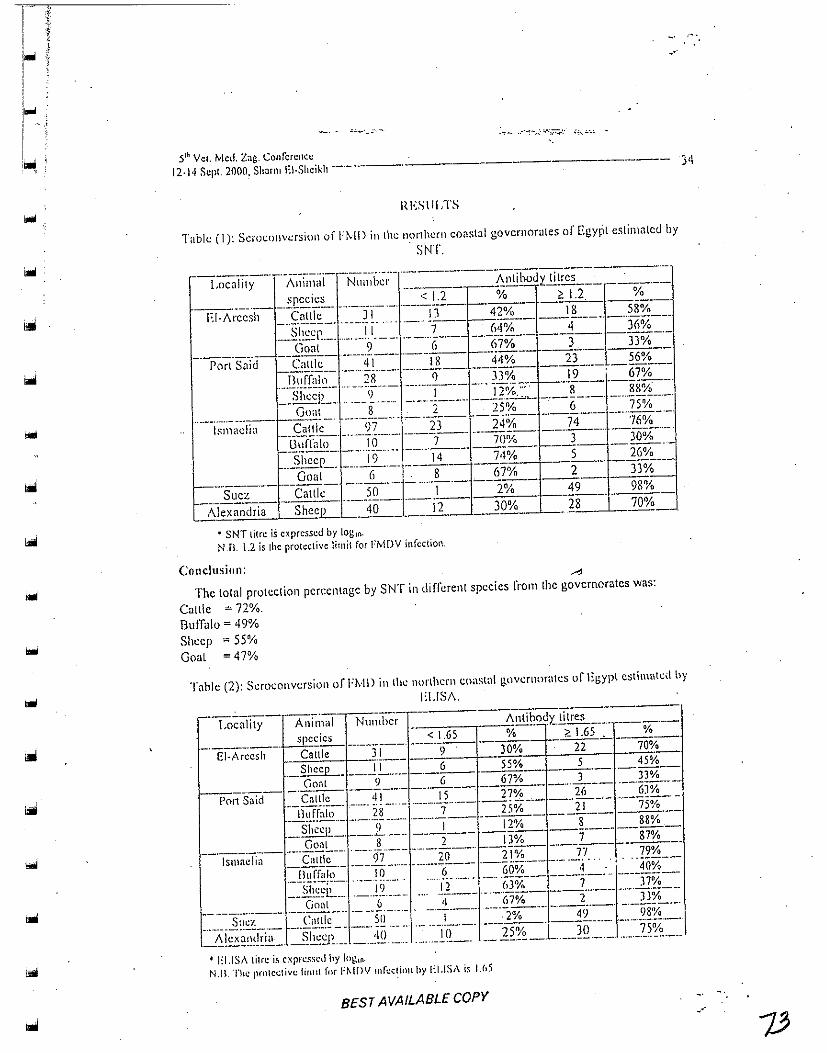
·.'
5th Vet. ivied. Zag. Conference
12·1<1 Sept. 2000, Shnflll EI·,Shcikh ~-.-----.• --- .. ---- 34
IU;SlILTS
Table..: (I): Scroc()llvcrsion of J-"l\.ID in the n~rlhcrll cO<lstal govcrnon.lles of Egypt estimated by
SNT.
--f,oc<liity -- "-;\~~~n;;i- - N\l~~'i):I:'- ·-·---------~Il~~dl: litrcs _=_ __._._ species < 1.2 % ~ 1.2. %
EI·J\I:cc~il- -'-CnllT;,-' ·-·-·:ii--·--·i·:i- 42% 18 5S;X;-- .. --.. ----.. ---.- -....... --.. ----r-----;'---. ------ - .. -----_~!~~1'._ ... _. __ 1.1. __ .. __ .. ~ ___ __ ..!~~!'!I~ __ _ ....'!__ )~y~_._
Gonl 9 (, 67% 3 33% -'-'-'--C'~ ---------.-.-.... --.--f---.----- --'-'-
Pori Snid Callie 41 IS 44% 23 56% ~:----- .. _ .... _ ... _ .. _ .. --... -- -----_. ------_.r}I!f[~I~ __ .... _.~~. ___ ..... '!. ______ . ,~~__ 19 '_Q?% __ __ She,!'!: __ ... __ l~. __ ... _. ___ ! ___ ... ___ Il~::':" _. __ ~ ____ .. !!~~_._
Goal 8 2 25% (, 75% ... - .. -... _-_._- ----,-- -----_._.-- .. -.-.. -- --_ .. _- ------- _ .... ----_.-
I.,maelia Callie 97 23 24% 74 76% --U:;-rr~iO' '---'10"-'--- -----7--- ---'700/0--- ----3----'· -300/;:-'"'' ---.. ---.-._-.... --.. -. _._-.. _. __ ._---1--- ---.-
.. ..:%ccp_. ___ J2. ______ 14___ 74% 5 _ 26°/~ __ _
Goal 6 ,8 67% 2 33% ------- ----- _ .. _-_._. - ._--_ .. - ~------------
__ ~~::..._. Callk . . ___ ~Q._ .. __ .! 2% 49 _~% _
Alexandria Sheep 40 12 30~ ___ ~8 70%
• SNT litre is expressed by 101;1(1'
N.n. \,2 is Ihe protective limil for FMDV infection.
Conclusion: . .., The total protection percentage by SNT in different species rrom the governorates was:
Callie = 72%. BufTalo = 49% Sheep = 55% Goal = 47%
T;:th)c (2): Scroconvcrsioll of Fl'vlD ill thl: nonhcrn coaslal g.nvemor<llcs of Egypt estimated by
I:LlS,\.
BEST AVAILABLE COpy .. '

" 1 , "
E£SqAVAIL48L£COPY
~_. _. ,-c ... '_: ,_.~
Shmdy . .41 .. cI (/1 __
Conclusion:
The protection pCrCCnl<1gc by l:l.IS/\ in di,llcrclll spcL'ics {'rom the governorates WflS:
C"lllc "77,5%, Bufralo ~ 57,57% Sheep = 61.5% (ioal =51%
Tahle (3): SerocollYcrsion al\d carrier s(atc or FM 0 in the northem coastal governorates in Egypl.
~-.. ----- r-------- -- .-- - ... _- -- ------ ---._-----------~---- ---- -LoealilY A Il! l11.a I Numbl: SN'I (~L2) EI.1SA • 3 CD Nasal
r <: I ,65 swab
'-- . species_ ----- El:!~A ---.lI'CR) ._---------22 (70'/0 EI-Arcesh Callie ' 31 f-J!i58%) o 2.21112~
-Sh';-en II _4_Q6%)_ 5 (45o/!2 o Nol done Goat :-9 3 (33%) f(33o/.,) o Not done
Pori Said ~ . .£:~ ~_I_. _23 (56o/~ 26 (63%) -.ll7%) 1(2%) ~falo 28 _~7%) __ ;21175%) 0 Notdr~~ \-----
Sheep 9 8 (88%) 8 (88%) 0 Not done L _____ Goal - 8 __ ,§"Q5%L ~ 'irS7%) 0 Not done ----- ---
Ismaelia Callie 97 74 (76%) 77 (79%) 5 (5%) 1(1%) PCR
Buffalo f--!-O - __ l.Q!!~!2 __ 4 (40%) 0 Not done -SI~en 19 5 (26%) 7(J?%) 0 Nol don,,_
Goal (, 2 P3%) _.lQ3%2. 0 NOI done ---8 (160/0
_. __ ._--Suez Callie 50 49 (98%) _ 49 (98%)_ Not done ,-- --
Alexandri Sheep 40 28 (70%) 30 (75%) 0 Not done a --- ,------- ------
-) CD (Protease <lnd polymerase).
35
It appears from Ihe abuve resulls Ihal some animals had probably conlracled the disease in the field (positive for 3 CD), bllt only a few number of animals was found to harbollr the virus in the nasalillueosa (as delermined by peR), .
J)JSClissrON
Fool and Mouth Disease is one of Ihe mosl devaslaling diseases affecting farm w\imals, Cattle, sheep, pigs and goals are all "flee led, although the disease rarely leads 10 dealh and productivity losses are llslially estimated 10 be as high [IS 25% (BroHIIl, 1985).
Krugli~·(}v e( al. (/989) n;porleu 111<11 within infect cd areas. Ihe best time to vaccillcllC illlilllais ill over 4 Illnllills of ilge and n:vaccill<1lio/l is an<:r g;X mOllths from prilllilry v(lccinaliofl.
In Egypt. HvlD HSStllllCS :1Il cni'.oolic 1"('l"In and attacks sus{;cpliblc animHls causil1g'high losses ill Illilk and !llcal proul1l;(ion (AfollsJa lIt a/., /98.f). Duc to the dallgcr ortlle disc:ase alld ils cl.:ollolllic InSgC3, pcriodic v<I!.:(.;inalinll .:lIld qU<lranlillc measures lire llsun!ly applied .sJl~ci;dly ill ellzootic In':<l as an dlcclive cuntrol Illc[i:)Urcs (Still/ifill, /1)62). The CllZDIJlic nature urlhl.! discas~ is Oil!.: of' the m<~or fal:lors Lilal inflllcilct.: FtvlD spread and rc!.:urrcncc among livcstlld.
'-;'.-
....
.--
r f}! " ~. .. i I ! ::; ,
,." iJ i l-
~;
;, '. ',! ,.
I I :~ ,\ai I
.<i,I. Vet. Mell. i'.;lg. Cnnfcn:ncc
-.
\2-\4 St.·p!. 2000. Shimll EI-Shcikh -."------.---.----------
This is dllC In il\lpropcr vilc.~illillil.lll proUr;lI11S, insunicicnl viH.:cincs poor disease combat
Il1COSlIrl..!S and the presence 0 r carrier <l\lima!::;.
The obtained'results I'eg:<,rding the s~lId)' of .the humoral immune response oftC!'>lcd
survcycu animals estimated by SNT revealed tl101l the percentage of pro\Ccti{;il ~··.·~rc 72%,49%,
;>50/0 ,lild 47% in cattle, buffaloes. sheep and gouts respectively. These antibody tilrcs were equaJ
or more than 1.2 !aglo and this is the prolcclivt.: antibody titre.
On the other halld, the result::; of illll1ll1nl.! response cslimntct.i by El ,]SA revealed llml the
percentage of protection were 77.5%, 57.5<\10, 6! .5% and 51 % in callie, hu'Talocs, shcl:p and
gonls, respectively in surveyed animals of different species ill various governorales \vith
salisfaclory proledi vc anlibody lilres equal 10 or more Ihan 1.65 log, •. These rcsulls agreed wilh
Ihose rcporled by !lulIlMi/l el /II. (1986) who menlioned Ihal a posilive correlalion between
ELISA and virus neulralization tilres oFI'~·IDV. The overaliregression belween EI.ISAISN dala
shuwed thai I in IG by SN was equivalenllo I in 40 by UJSA.
Als(l, Ihe resulis oblaind by [ie/l~elsd(}rll (1989) go in hand with Ihe oblained dala were
mort; l!l:m qS<~';, of vnt:cill':ltcd callic with S('>1 titres or Illore I.1Hl1I 1.2 !og\l\ \\'(;r~ rnlll:cled from
gencra/i;«d I:M I).
Also, the recorded results Wert.: ill agreement with lh:ll obtninco by Lombard e( al. (1982)
who foulld that ELISA techniq\Je and SNT gave similnr reslIIL<;.
The above mcntioned resull!) agreed with lhat obtained by Abd E::l-Samae ([988) who
reported that when study ek .... alion ofFMD vaccines prepared from type Oln172 Sharquia whkh
used in animals vaccinution during 1987 in Egypt. The mt.:ull SNT titrcs after 15,30,60 and 90
days post vaccination were 0.9, \ .6, 1.6 and 1.2. rcspcclively .
. Regarding the d(;l~Clion of <lntihodics to non-structural proteins (3 CD) (protease and
polymcrase) in the tested cillile SCflIll1 S<1Il1p/es wcr~ 0%, 7%, 5%, 16% ana 0% in E1~Arccsh,
Port Said, Ismadia, SUCl and Alexandria gQyernoratcs, respeclively. These results showcd
detection ofantibodics to nOIl-5trllclura! pl"Okins of foot and mOllth disease virus (fMDV) which
have the rotenliallo differenliale animals lilul have been infected wilh virus from Ihose thai have
been' vaccinated (Neilzerl el al., 1991, lierg/l/(//I el al., /993 nlld Lubrolll "wi Broll'll, /995).
A JlOIl structural illltigcn associa\cd with FMDV replication.. The virus infeclion
associaled anligen (VIA!\) i, composed maillly oflhe RNA polymerase. The anligenieily oflhis
prolein is conserved amongsl nil scroll' pes (Vil/ellg"r etal., 1989). To confilln lhc~e (csnlls of]
CO EI.ISA RT-PCR WilS done to nasa! swab.<; of the same animals using two specific primers l)i'
J j) gt:I\L' to J positive sa!1lp!c~ of Purl ,said, S positivI..: ~al11rlcs ofIsmadlil ilnd 2 ncg(ltive
"Illplcs or EI-Areos".
The ahnvc IlItlllinlll;d' data rC(';()l"llcd l!wl gT.Pflt was positive ill 011(' (Hit of \111"ce. tlllC
oul 01" live and 0 ntll of"lwo {i'om I'()rl Said. ISlllilcliil and I':I-Arccsh givt.'l"llurnh':s, rcspccliv(.;ly.
These agreed ",ilh resullS· ublained hI' Callell.l· el 11'1. (1998), DOIiIl el al. (199/) '!II(~.
StrO/1l ef (f/. (/995) \\lho .sillW .. 'cd thill thL' P( '!{ \I,"as ~\lpL'rior ror detecting the carrier slale in tisslll; .• -'
s:llllpks, moulh .'>wall=->, I\<I:-;al sc\.:n.:lioll and sera.
In cOllclusion, ii' nppenrs I"rolll the pn.:vinus I"l:S\llts thaI some normals h;ul proh;.\hly
contnlckti lhc disease in the fldd (pusitivc f\H" 3 CD), b\l\ only a lew !lumber of <.lnin)uls was
found to h':lrbor the virus in thc llasillllHICOS<.1 as dClcrminc;u by RT-PCR This means that FI'v1DV
stilt Clllklllic LInd t:ircul:lling ill Ft!fP1.
BEST AVAILABLE COpy

,.,
SlwlI'ky. M.; cI al. ---------- 37
REFEHlINCES
Abd EI-Sall/ae, M,Il[. (1988): Further studies 01\ Fool and Moulh Disease in Egypt. Ph.D.
Thesis. Inreclious Diseases, Fae. Vet. Med., Cairo Univ.
Alloa (1978): Ilisiory o[ [oat and moulh disease. Vet. Rec., 102: 184-185.
Benge/.,dorf/' ILl. (1989): Potency lesl or Cool and mouth disease vaccines correlation between
response to clmllcnge and corresponding neutralizing nntihooy titp.!s ofvaccinnlcd cattle.
Ber!. Munch Tiernrzl. Wsehr. 102: 193·198.
BrII'C"WIt, I.E.; Auge tit! A-Ie/lo, I'. (Iml j'ldlzerl, E. (/993): Diclgnosis nfpcrsislclll apillhovirlls
infection and its'di!Tcrcntiatio[1 from vaccination response in cattle hy lise ofcnzYli,c
linked imnHlllockclrntl'a!l~r('r hint <1nalysis with hiocng.inrcrcd non slructmnl viral
anlige"s. Am . .l..Vd. I(es .. 5 (6): Xl)· X.11.
Browl1, E (1 Y85): Ilnyc pcplidt,;s n future tlS ront alld mouth di$c,ISC vaccines. 4111 lVicelillg 0[" ihe
Europc,lI1 GrollP of M(!iccular Biology ofPic{)rna Viru!;cs, Septembcr, 1-7.
Caliells, Mo; De-C1ercq, K.; (imin, M. nIHI Dalles, M. (/998): Delection uCfool alld mouth
disease by reverse transcription polymerase chain reaction and virus isolation ill cOt\\acl
sheep wilhout clinical signs of Cool and mou!h disease. Vel. Q. 10 StiPP!', 2: 537 - 540.
Daol/d, A.; Olllar, A.: EI-Rar'r)', M.; Melll'ally, N.; EI-Mekkawi, M. alld EI-Kilm,y, S. (1988):
Strains of FMD virus roeovered Crom 1987 oulbrcakjll Egypt. J. Egypt. Vel. Med, Ass ..
48 (I): 63-7\.'·
D,?JlIt, A.,' Alartin, L.A. all~l DOllaltlsoll, A.I. (1994); improved detection offlcrsistcnt or foot
and mouth disease infection in C!lutc by the polymcra~.c.ch.~in reaction. J. Viral. Mdh(1d~i •
. 49 (2): 179 . 63. "-
Ferreira., MEV. (1976); Microlilr" neutralizalion lesl for the sludy ofF!vID alllibodie,. Bo!.
Cenlro ran Americana de Fiebre t\liosa, 21: 22-23.
/la/llbli/1; Barnell, T.R allil Hedger, R.S. (1986): A new enzyme linked immunosorbenl assay
(ELISA) Cor the deleelion oCantibodies against FMDV: I. Development and method or
ELISA. J. hnnlllllo!. Melhods. 93: lIS -121.
Krllglikal', 8.A.;Nalivaiko, V, G.; Me/llik, R.I. (lilt! Tarasenko, T. Y.A. (/989): OrganizatIon of
progral11~ rQr imlllunizillg farm nnimals against FMD. Vt:tcrinariya, Moscow, USSR; 22:
)) . 35. .
L011lburti, A1- amI PI.!/ermmlll, II. C. (1982): Correlation between ELiS/\ and serum
lIculnllizfltioll lesls for tile detcction oCnculralizing antibodies to FMOV ill SCHlIll cattle.
Tiem,-zl Helle Umsehau, 37 (5): 354-J 56.
Lubrollt. 1. (lilt! Browl/, F. (1995): ldcnti(icatiol1 of natiy.c fool <lnd mo'lllb ;.E~.::"sc virus nOll
structurai protein 2 C 1.1S n sCf0iogicai indiciltOf to dl+lcrcnlialC Ii\fccl~d from vuccinated
liveslock. Res. Vet. Sci .. 59: 70·&0. . . .
A4otfJSfl, A.A.J1,~ Daoud, A.; lIusseill, K.; Ifassllll, NA.; Fahmy, P.,: Az.nb, /i. {lwl El-
Sltahawy, l.. (1984): Prevalence ofFMD in Egypt. AgriC. Res. Rev., 62 (513): 55-58.
Neitzert, H.; Beck, E.; illIge de Hella, P.; GOllles, f. 11111/ lIerglll(lII, I.E. (1991): Expression or
"phihovinlS RNA polyrncnrse gene in Escheri'chia coli and its USc together with olher hio:·,
engineered nOl\·:-;\ructural <Hlligt:lls in detectioll oflate persislclll infection. Viro!.,· '1 S·(
7<}l)·K09.
....
): I
/-. I
i~
...
jllo Vel. Met!. l.n~ ('ot\rcrcncc
11·1 ,t S~·pl. lOOO, Shar III f:,I·Sllciklt .. -. --.. ... ,.'-_ ... _- --,.-------.-- ----- 3R
U' /JollltC!ll, ,.: 1(,; DH1'id R Hoy/e; Knllly Sprol/t, Norberta, A.,' FOl1df!vifn, An/harry ForJ1tn~r,
AI"jallllr(!, .. I.: Sci/fidei, Elinfla, tV. alld Smitsaarf (1996): Detcction of antibodies
ngnillsl foot and l1lomh··dj'~cb:~fc-viru.~ \Ising a liquid rhnsc",blqc}.ing ~.~n!1wich ELISA.
(l.I'I1E) lVith hincnginecred .11) protein. J. Vel. Ding. Invcst, 8: 143-150.
Shalr,,", M.S. (I 962):Thc vims nf' FMD. Agri. [\es. SCI". U.S. Dcpmhncnt of Agri. Greenport.
I..!.N Y .. p. 444-,151.
,,!tal~'/{.I', ,It.,.,' SalN!r, 11I.S.: t16de/ RalllTtall, A.n. a/HI EI-SarJoll,fj;, A. (/999): Clnning and
c"pressio" of Jell genes of FMIJV. Vet. Med. J .. (;i7.;1, 47 (Jl: 289-294 ..
. ~ .. StmJ11. r.o' Cfrai, D.; Fnwl~r, !I.H.: fifo/ad, 1:; ft.·feiri, N.,· Jlall /Jam Af., EI-Kifn11)\ S.; Fa/tltly,
/.:,. ,Ho/(s.\'{/, A.A. and Ymlill, If. (/995): Molcculnr epidemiology offootnml mouth
disc;l!)C (FMD) ill Israel in \994 <llld in olher Middle-Eastern coul1i.rk,~:!\ iile year 1992-
tQ94. Arch. Vim!.. HO(tO): t791-1797.
l'illillgCf, I-~; Mlil/er, If.K.; Timclwer, L.; .'lexa",all, M. nlld KillII/, U. (/989): Antibodies tr,
root nod IllOtllh di~casc virus illrcclion lIssocintcd (VIA) antigen: usc ofbioengineercd
VIA protein U$ "ntigen in all ELISA. Vel. Microbiol., 2(}: 235-246 .
. Voila, ;1.; Bidwell, 1). alld Bartlett, A. (1976): Enzymc immuno-assay in diagnostic mcdicine,
theory nnd practice. Rull. World lleallh Organ .. 53: 55-65.
JJI.l .lY':"'4 ..\..ul J r)A=>-.>n ¥- ~ JJ~ , u-ih.;.ll '+ ' r.S¥J ~lL 2Y; J..o.:>,...o
.~l01 - :"".;h;;JI wG.\illI J JI...."..':II w~ .lp..o •
:I,!G.L...JI wlNG...JI ~ c."; :..,.c)Wt ,r<-..JI u-'> yJ :..,.;t,.}1 ;JWI ~G.J U ,;.>ll ,1> UJ.J'?-\
.~y.ll .>'-"-" "-!,;~
jo.UI J ?llt.':II ... U"}"'l;JI) ,;",':11 L.ro ~ JlAc.':I ?fr" ¥ \"o~ ,)c l..."I.J.Il1 UJ.J'?-I.ll ...
. '-..r.";l.i\1 __ II' -"-'" ·~\·\I~· I: ... • . ..:, '.11 • .....- L!"" yo ,; J~ <....,; ..,., (j4 ~) >.s"'
r'L......,..~1 0" w.:&U 1.)'11 ),,:,,,1) JJw.Ji ?~I tS)l{,;.,1 J.,.:1.p..h.>I..JI y)~1 UJ.J'?-\
.:Ufo..ll ~WI
r~-:' oi. J ~)WI cr--JI U" J~"~ ,-!Lo )'1 0" w.:&U tS JStJl 3-CI) EUSA ,;\;>;.I .I.»l ~ l..S
wl,....,.,..Jl(PCR) ~I cSy..J!A!lyll Jcli:Jl )\;>;.I .Ift)'; ,;\;>;.'i\ I~J ~~'il uu,.J1 L.ro ,s1:J\
. .>'-"-" ~ <.,ut.,.J '.J.""""'! :..,.c;ull ~I U"J.JI-i Jy;.. ... ,si L... ,-!L......JI wlil~1 ,ylill.p.;\;1
BEST AVAILABLE COpy
11 ,

...
ISRAEL
BEST AVAILABLE COpy

Controlling Important Livestock Diseases without Borders -The Team Approach in the Middle East
2nd Annual Report, 2001
Projects of the Israeli Veterinary Services / Kimron Veterinary
Institute / Koret School of Veterinary Medicine
1. Project: Brucellosis
Principal Investigator - Dr M. Banai, National and OlE Reference Laboratory
for Brucellosis, Kimron Veteriuary Institute, P. O. Box 12, Bet Dagan 50250,
Israel.
Technical Progress
Research Objectives and Accomplishments
1. To strengthen the activity of the Israeli laboratory as a reference laboratory in the
region
Since the establishment of the Brucella web site, at the domain:
http://www.moag.gov.illbrunet.Dr.Banaihasactedasmoderatorofthesite.mainly
at the administrative level. The new B. melitensis genome web-site has been posted in
Brunet's "Forum Summary". The Discussion Group has communicated via Brunet,
mainly as a Sub-Committee on Brucella taxonomy. The Forum addressed the question
whether or not the genus Brucella should be divided into various species, compared to
accepting a single species taxa and dividing it to sub-species. The problem was raised
due to finding in GenBank the latter taxonomical form, whereas classical taxonomy
has long supported the division of the genus into 6 species.
This demonstrates the importance of Brunet as an international denominator in the
understanding of the Brucella organisms.
...
...
2. To train personnel from the participating countries in laboratory techniques for
brucellosis
Unfortunately, political events have hampered any meaningful collaboration.
Therefore, we cannot report on any training activity, or visits to the Israeli laboratory .
Scientific Impact of Cooperation
Dr. Banai has maintained a continuous contact with Dr. Randa Akashe from Jordan
by e-mail. This contact was aimed at keeping the warm relationships developed
between the partners prior to the political events. Using this route, Dr. Banai has been
kept updated with the scientific advancements among the other collaborating centers.
Unfortunately, no contact could be made with the Palestinian colleagues.
Description of Project Impact
At the local level, the laboratory was involved in diagnosis of a unique event of S 19
infection. in sheep. Following serological diagnosis of seroconversion among ewes,
suspected to be infertile, Brucella was isolated from a milk sample of one of the
positive reactors. Conventional biotyping revealed it was B. abortus S 19.
Subsequently, the strain was isolated from two more ewes. The biotyping was
confirmed by specific PCR test, and the strains were sent to Weybridge for final
confirmation.
This episode demonstrated the strength of the laboratory III diagnosis of an
unexpected event, using conventional and up to date PCR techniques. Specific
questions were also discussed in Brunet, facilitating the understanding of the
outbreak. This is precisely the role that a central reference laboratory should play in
the region .
. Strengthening of Middle Eastern Institutions
The Israeli P.I. conducted field visits to the veterinary district offices, to evaluate
implementation of the brucellosis control activity. The laboratory has acted as a
reference center for diagnosis of complicated human cases. We have produced

------------------------------------------------------------
diagnostic antigens for RBT and SAT tests. These antigens were given to hospital
laboratories and clinical centers to facilitate human diagnosis.
Future Work
Lack of collaboration at the political level among the counterparts has suspended any
practical plans for the immediate future. Nevertheless, there have been several
interesting field cases in Israel that required further attention.
The introduction by an unknown route of S 19 into a sheep flock has raised the
possibility for involvement of unrecognized Brucellae organisms in several field
cases. To be prepared for such cases, the Israeli laboratory should strengthen its
professional capacity regarding bacteriological and serological diagnosis of
brucellosis. We should be prepared for the introduction of B. abortus and B. canis into
the region, as well as for development of currently unknown atypical strains.
Work with the credited ocular Rev 1 vaccine should be reinforced and accepted as a
regional vaccine. The quality of the vaccine should be tested as a basic routine and the
newly developed peR method (manuscript in preparation, supported by BARD)
should be applied to distinguish between Rev1 like isolates and virulent field isolates.
The Israeli PI has submitted a project proposal for testing RB51 as a new candidate
vaccine in cattle. The proposal was submitted to an Israeli granting organization.
Project Management and Cooperation
Managerial Issues
No managerial problems were encountered. There is a budgetary concern since the
Israeli laboratory was supported with a limited budget ($45,000 for the whole three
years).
Special Concerns
None.
=
....
Cooperation, Travel, Training and Pnblications
Dr. Banai wrote a pre-proposal draft for a continued project. This was submitted to
the Jordanian counterpart for their response .
Reqnest for USAID Actions
USAID should intervene between the parties to remove political constraints and assist
the parties to convene together.
2. Project: Improvement of Foot and Mouth Disease (FMD)
Control in the Middle East
Principal Investigator - Dr H. Yadin, Foot and Mouth Disease Laboratory,
Kimron Veterinary Institute, P. O. Box 12, Bet Dagan 50250, Israel.
Technical Progress
Research Objectives and Accomplishments
Progress in project activities were crucially affected by the politics in the area and
postponing of the activities of the peace process. Despite the political problems,
during April 2001, specimens from sheep in the PA area, suspect for FMD, were sent
to the KVI - those samples were found negative.
All FMD suspect specimens in Israel (8 in number) were negative, which means that
the country has been free ofFMD for nearly two years.
1. Research ObjeCtives
1.1. Standardization afreagents
The FMD Laboratory of the KVI participates in the FAO Collaborative study of
diagnosis procedures for establishing reference standards for FMD serology. In this
study our serology tests were calibrated by using WRL reference sera and panel of
unknown "test" sera in our calibrated test systems.
Another collaborative study is with the F ADL, Plum Island, test kit for evaluation of
antibodies against non-structural protein 2C, started in April 2001. Reagents were
provided by F ADL. Other parties are invited to participate in this study which should
differentiate between field infection and vaccination.
I.2.Monitoring the annual FMD vaccination program in dairy farms
Six dairy farms distributed over different regions were monitored. Ten blood samples
from each of 4 age groups were collected at 6 and 12 months after the annual
vaccination.

-i.= .. -------------~~~~~~~~~-
...
...
1.3.Monitoring the annual vaccination program in sheep farms
Nine sheep herds located in 6 different regions participated III the monitoring
program. Blood samples were collected from 15 lambs and 10 ewes from each herd
on three occasions during the last year. Sera were checked for antibody titers against
01 geshur.
l.4.Production of FMD antigen for DOE vaccine
This part of the project was continued as planned.
2. Research Accomplishments
2.1 Standardization of reagents
The FAO collaborative study for this year is under evaluation. The collaboration
study with F ADL is in progress.
2.2.Monitoring the annual vaccination program in dairy farms
Results shows that 12 month after vaccination, full protection was achieved in
Group C which represent young cattle with three vaccinations.
2.3.Monitoring the annual vaccination program in sheep farms
The serological survey revealed variable results 5 and 10 months after vaccination
with the Al(0H)3 vaccine .
2. 4. Serological survey of DOE vaccine in dairy cattle and sheep herds
During the report period a 270 liter bio-reactor was purchased and installed in the
new FMD high containment facility and the function capabilities for cell growth
were tested with positive results. In addition, the existing 70 liter bio-reactor was
replaced to its new location in the KVI campus, the laboratory for cell production in
the new FMD high containment facility. Both bio-reactors were checked for all
parameters that should function perfectly, such as sterilization, control, registration
of temperature, pH, and levels of CO2 and O2 .

------------------------------------------------------------
It is expected that this year all systems of the production facility will function
properly and local produced :vaccine will be available for emergency vaccination in
the region.
3. Scientific Impact of Cooperation
During the the report period no training courses were conducted.
4. Description of Project Impact
The impact of the project activities can reveal its effects on different levels, in the
short and long terms. In the short term, the awareness of the veterinary staff who were
in the training was improved and this awareness is also expected to be transferred to
the colleagues in the Regional Veterinary Service stations and to the farmers. The
dispatching of FMD suspect specimens for diagnosis to the KVI, followed by a rapid
answer, improves relations between the participating countries on the one hand and
stimulates the sender to take precautions to control the outbreak onthe other hand.
In the long term, continuous training of extension service and laboratory staff will
improve the awareness and the control activities such as surveillance, serosurveillance
and vaccinations.
Unfortunately, due to the problematic political situation of the last few months, all
project activities were effectively ceased due to lack of communication.
5. Strengthening of Middle East Institntions
In the project budget the improvements in facilities and equipment in Jordan and P.A.
was funded. For realization of those plans the cooperation between the participating ,
parties must be improved. The training courses were expected to improve the
profesional and laboratory skills of the staff. Additional training courses are on the
program upon request of the participating parties.

6. Future Work
The stage of transfer of laboratory skills from the training level to self supporting
level is still to be effectively realized. Assistance in these subjects can be given upon
request.
A contingency plan for the region and for each of the participating parties should be
designed.
Project Management and Cooperation
Managerial Issues
No special problems.
Special Concerns
Due to the problematic political situation of the last few months, all cooperative
project activities have ceased.
Cooperation, Travel, Training and Publications
1. Cooperation
1.1. The regional Website is functioning and provide information regarding the
project activities and FMD situation in the region. The information should be updated
more often, close in time to the actual field incidents.
1.2. A standard outbreak investigation protocol was discussed and a draft proposal
was submitted.
1.4.An advisory visit to Jordan is still planned for evaluation of laboratory demands
and for training ofFMD diagnosis under local conditions.
2. Meetings
Dr. Yadin traveled to the European Veterinary Virology Congress in Brescia, Italy.

3. Publications
Experimental vaccination against FMD with immunocomplexes. H. Yadin, Dalia
Chai, A. Bril, B. Gelman, and M. A.madori. Submitted at the European Commission
for the Control of FMD, Session of the Research Goup, Borovets, Bulgaria, 5-8
September 2000.
Req~est for USAID
A coordinator between the participating parties may stimulate a better cooperation
and making the project more productive.
~----
3. Project: Neonatal Diseases of Small Ruminants
Principal Investigator - Prof. K Perk, Koret School of Veterinary Medicine,
Hehrew University of Jerusalem, Israel.
Technical Progress
Research Objectives and Accomplishments
1. Research Objectives
1.1. To utilize a unified epidemiological survey of neonatal small ruminant
morbidity and mortality.
1.2. To develop and exchange current knowledge on neonatal diseases among the
scientists and field personnel of all the participating parties.
1.3. To advance research on "slow virus" diseases in small ruminants that are
characterized by infection in the neonatal period, with disease only appearing after a
very long "latent" period, inflicting significant losses in milking breeds.
1.4. To establish a communication network among the project personnel of the 4
parties with regard to topics of neonatal losses. Accomplishments and selected topics
of husbandry and disease in small ruminants can so be disseminated in meetings and
symposIa.
2. Research Accomplishments
2.1. The epidemiological survey was continued, using the unified questionnaire
worked out and agreed upon by all participating parties. The survey also includes
frequent farm visits by project personnel and local veterinarians. As the work is still
in progress, results will be presented at a later stage.
2.2. The planned training laboratory courses and field studies in Israel, as agreed with
the teams of the participating countries (see last report) could not be implemented,
due to the political situation. We did however prepare and select lecturers, handout
material, laboratory and field material, and we hope that these important training
courses can be held in the near future.
2.3. As reported in the last report, we continued our studies on "slow virus" infections
(maedi-visna and caprine-arthritis-encephalitis viruses). The slow virus infections are
of special interest in the neonatal stage. The infection usually occurs shortly after birth
through consumption of colostral milk and has a very long initial period of latency.
Furthermore, the infection rate in Israel is very high. As reported previously, we could
demonstrate early development stages of immature lentivirus particles only in cells of
the CNS in clinically healthy adult sheep, which were infected at the neonatal stage.
However, neither mature virus particles nor clinical signs pertaining to the nervous
system were present. We now report that ganglia cells, obtained from one of the older
clinically healthy ewes, showed morphogenetic immature virus particles. The mature
virus could be induced by co-cultivation of the ganglia cells with a lentivirus
susceptible cell line (Thar cells) and stimulated by 5-bromo-21 deoxyuridine (Brdu).
Experiment 1. Tha, cells (Himalayan mountain goat ovary cells) were
cultivated with the ewe ganglia cells and tested for mature virus by electron
microscopy and by the presence of reverse transcriptase - revealed no virus
maturation. The ganglia cells degenerated while the Thar cells took over the tissue
culture plate.
Experiment 2. Induction. Co-cultivation of the Thar cells with the ewe's
ganglia cells (as in Experiment 1), but 2-4 days foll~wing the addition of 25-40 /lg/ml
of Brdu to the medium, the co-cultivated cell cultures revealed mature virus particles
in the Thar cells while the ganglia cells degenerated. This indicates that stress factors
may induce maturation of the virus, which may also infect susceptible cells in situ.
The induction of latent immature lentivirus in addition to its concealed location is of
interest in epidemiology and veterinary medicine, but may be analogous to human
immunodeficiency virus (HIV) which belongs to the same subfamily. After additional
work this will be submitted for publication shortly.
-

-
2.4, Additional communication equipment was purchased and installed, Constant and
frequent personal communications (e-mail and telephone) among the 4 parties, on
matters dealing with the project, were of greater importance since personal
professional visits could not be arranged, due to the political situation,
Two local professional meetings - symposia on sheep neonatal diseases and
husbandry were organized during the report period,
Future Work
I. Continue the epidemiological survey,
2, Continue the lentivirus study,
3, We hope that the political situation will allow the planned courses and the
professiorial visits to take place soon,
4, The results of the epidemiological survey will be processed in order to
establish measures to control neonatal mortality and morbidity under field
conditions,

....
-....
JORDAN

1. Gathering information from different districts about the number of vaccinated animals against Brucellosis, Foot and mouth disease, and other diseases. The percentage of vaccinated sheep against Brucella rose from 34% in 1999 to 53% in2000,and the percentage of vaccinated goat rose from 35% in 1999 to 42% in 2000
2. Frequent meeting with Msc students and their . supervisors to assess their work and to help . in _ purchasing biological and chemicals nee~e(r"for their research.;
3. Workshop held in the Veterinary Department for .... one week in the field of Animal Health with special reference to neonatal losses, Brucellosis, and "'" F.M.D, for 13 field vets from different districts.
4. With regard to neonatal losses, more data will be collected through questionnaires that have been circulated to vets whom participated .. in the said workshop.
5. Arrangement for participation of 11 vets in training courses to be held in Cairo May/June 2001 on the following subjects.
2 vets training on PCR technique and on differentiation between infection and vaccination
2 vets training on epidemiological studies with special reference to FMD.
5 vets training on neonatal losses

2 vets training on the diagnosis of FMD virus by using ELISA test. 6. Preparing a guideline extension information for the
sheep and goats farmers about the vaccination calendar for different diseases including Br. FMD, PPR, etc
7. Strengthen a good collaboration with the medical authorities in the field of zoonotic diseases especially Brucellosis.
Due to annual vaccination, extension there were dramatic human Brucellosis as follow:-In 1997 1998
953 684
health education and decrease In cases of
1999 2000 432 268
8.There were no reports of FMD outbreak in Jordan for the last seventeen months. 9.Necropsy finding were conducted on 53 lamb and kids that died during the neonatal period and will be analyzed soon. 1O.FMD kits were purchased and the staff in the diagnostic lab started the practical work
The following equipment were purchased
1. Horizontal laminar flow • Hot plate stirrers • Distiller • Water bath • Pcr thermo cycler • TV and Video
• Frigidair • Normal Microscope
2. Deep freezer GFL • Incubator • Air oven • Laminar flow
....

...
PALESTINE
...

.5,---------------------
PROGRESS REPORT MERCm.;FFT V:-;'VERSITY
During lhe last seven months. the project faced a lot of obsracles due to the curr~nt
situation. in. the Pal.e_stirua~ territories"cJos~re. and brea~i!H.~_ofthe \Ye,sl,Bank, and",Gaza
strip into 90 s.eperated areas beside the difficulties in mo\~ment between Inc cities and
villages. despite that some progress was achie\'ed but to certain limit.
The Experimental Farm in Jericho'
a. During the stan of this activity, and after we received the \'erhal 31.\reement from the
lands'; o\\'oer .• he board of directc:"s sent a lct!er asking more-details abom the
experiment a..'1d during which they asked to stop temporarily the rehabili:S!ion
acti"ities After a long bureaucratic correspondence with the Board. we got final
agreement to proceed \\ith the farm, and by the ti.-ne \' ... e got this agreement, the
Intifada staned and were unable to reach the place of activity because of the security
risks and the prcyemion of the Israeli anny to enter Jericho.
b. After seyeral meetings with CARE International it was agreed that we should use the
goat races and other needed materials that were purchased to rehabilitate another farm
near Ramallah. The reasons that our choice was limited to RamaJlah are:
• Dr. Shua'ibi: the Co·PI for the Brucellosis component of the project resides in
Ramallah and iI will be easier for him to visit the farm under the current securi~y
situation and the over whelming restrictions on movement forced by the Israeli army.
• The Yle is located in Ramallah and samples can be transferred easily from the
experimental farm to the \ lC.
• Other VIC Staff can access the Farm in case Dr. Shuaibj was unable to reach it and
that v.il! prevent the death of animals due to in sufficient food or water.
'Cntil nov., we were not able to locate a single farm that is willing to host our project and
we are still trying to find a place v.here we can rent and refurbish to suit our needs.
2. Training Courses:
Since .. the beginning of the Intifada movement restriction were imposed on all ten1lOri.es
and staff were unable to reach their workplace. In addition the situation created a big risk
for staff to move from one place to another due to Jack ofr.ecurity on roads.
All of the planned training workshops were cancelled whether it was nationally or
regionally or internationally. For example, the epidemiology course that was organized
by CARE had to be canncel1ed because the consultants were unable to come to the
country also staff would have been unable to reach the training place also because of
security reasons and restriction on movement.
3. Equipment for the labs:
Most of the purchased equipment were received and stored in Ramallah, Unfortunately these equipment was received only one day before the intifada started and we have been delayed in distributing it to the area labs and even we were unable to install them in the VIC. Recently we were able with a lot of difficulty "to send pan of tile equipment to Nablus area laboratOJY and to Hebron. After the delivery the technicians will be sent to install the equipment and operationalize them.
4. Project c.omponents:
All of the project activities have been delayed and continually postponed, field visits are nearly impossible due to the security situation, and inability to access the rural areas, where the farms are located. This has great implication on the project since the Co-PIs are unable to pay visits to the field on regular basis and samples can not reach the "le. As a cooping strategy the area laboratories will be equipped to receive samples and test them adequately.
GENERAL CONCERN:
It is clear that continuation oftbe current situation and acceleration of violence and mass punishments wm be a very serious problem to implement the project according to the work plan, but we hope that peace wHi dominate and we will be able to gain fruits of peace soon.
30.03.2001