in-lab exercises
TRANSCRIPT
1
2
IN-LAB EXERCISES
Use the following modules in Visible Body’s Anatomy & Physiology app to guide your exploration of the brain. Be sure to select the book icon under the structure names to learn more about the structures you are exploring.
You are responsible for the identification of all bold terms and all answers to the questions.
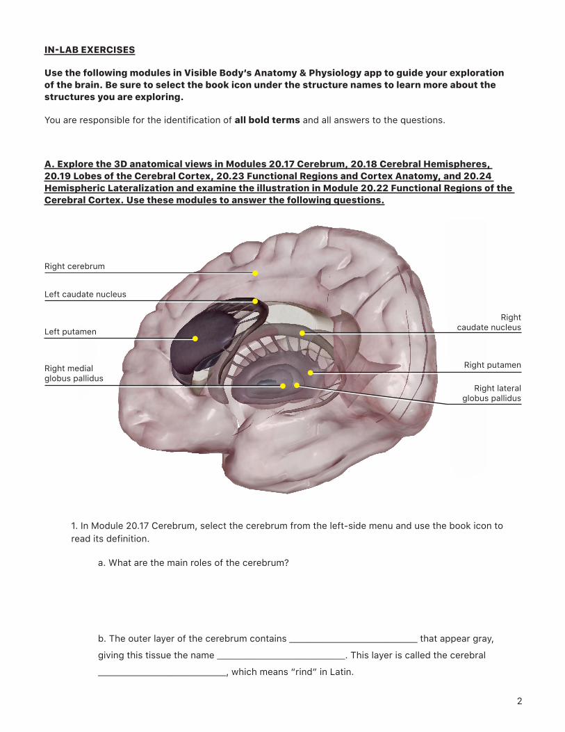
A. Explore the 3D anatomical views in Modules 20.17 Cerebrum, 20.18 Cerebral Hemispheres, 20.19 Lobes of the Cerebral Cortex, 20.23 Functional Regions and Cortex Anatomy, and 20.24 Hemispheric Lateralization and examine the illustration in Module 20.22 Functional Regions of the Cerebral Cortex. Use these modules to answer the following questions.
1. In Module 20.17 Cerebrum, select the cerebrum from the left-side menu and use the book icon to read its definition.
a. What are the main roles of the cerebrum?
b. The outer layer of the cerebrum contains _______________________________ that appear gray,
giving this tissue the name _______________________________. This layer is called the cerebral
_______________________________, which means “rind” in Latin.
Right cerebrum
Left caudate nucleus
Left putamen
Right medial globus pallidus
Right caudate nucleus
Right putamen
Right lateral globus pallidus
3
c. The inner layer of the cerebrum contains mainly _____________________________________________
that appear white, giving this tissue the name _______________________________. This layer is called
the cerebral _______________________________, which means “middle” in Latin.
2. Select the basal ganglia from the left-side menu and use the book icon to read their definition. The basal ganglia include structures from the telencephalon, diencephalon, and mesencephalon. (Note: The term “ganglia,” normally reserved for groups of neurons in the PNS, is used here for historical reasons. Some people use the term “basal nuclei.” For a different view of the basal ganglia, you can refer to Module 20.21 Basal Ganglia.)
a. These paired structures are composed of groups of _______________________________ surrounding
the _______________________________.
b. The basal ganglia regulate ____________________________________ by processing input from the
_____________________________________.
c. Select the upper, C-shaped part of one of the basal ganglia. What is this structure called?
d. Select the lower, oval-shaped part of one of the basal ganglia. What is this structure called?
e. Note the many bridges that connect the upper and lower parts of the basal ganglia. Select
the basal ganglia from the left-side menu again and use the Hide Others tool to remove the
other brain structures from the view. Then, rotate the view to see the inner side of one of the
ganglia and select one of the two oval structures on the medial side of the putamen. These two
structures are called the lateral and medial _______________________________.
4
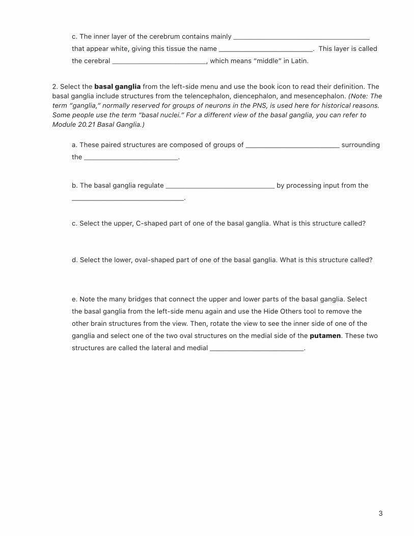
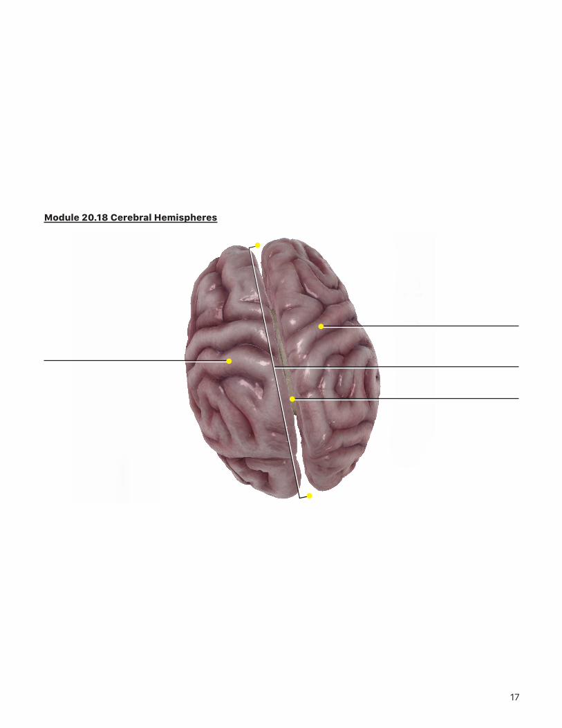
3. Use the right arrow at the bottom of the left-side menu to open Module 20.18 Cerebral Hemispheres.
Select the right or left cerebral hemisphere from the left-side menu and use the book icon to review
the cerebrum definition.
a. What space separates the right and left hemispheres?
b. Rotate the view to examine the inferior portion of the brain and observe how the two
hemispheres are connected in the center of the brain. Select the connecting structure, which is
called the _______________________________. It is a white commissure, meaning that it is made up
of _______________________________ connecting the two hemispheres.
Left cerebral hemisphere
Right cerebral hemisphere
Corpus callosum
Longitudinal fissure
5
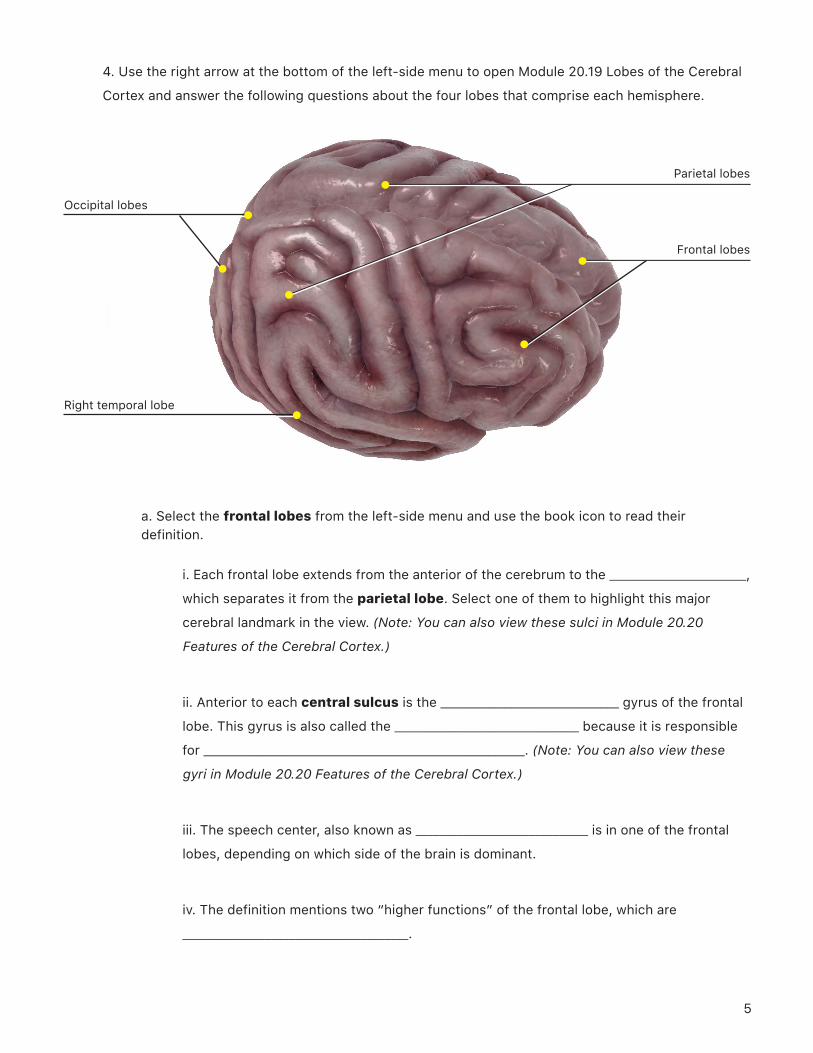
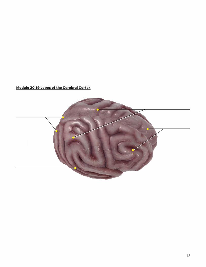
4. Use the right arrow at the bottom of the left-side menu to open Module 20.19 Lobes of the Cerebral
Cortex and answer the following questions about the four lobes that comprise each hemisphere.
Occipital lobes
Right temporal lobe
Frontal lobes
Parietal lobes
a. Select the frontal lobes from the left-side menu and use the book icon to read their definition.
i. Each frontal lobe extends from the anterior of the cerebrum to the _______________________,
which separates it from the parietal lobe. Select one of them to highlight this major
cerebral landmark in the view. (Note: You can also view these sulci in Module 20.20
Features of the Cerebral Cortex.)
ii. Anterior to each central sulcus is the ______________________________ gyrus of the frontal
lobe. This gyrus is also called the _______________________________ because it is responsible
for ______________________________________________________. (Note: You can also view these
gyri in Module 20.20 Features of the Cerebral Cortex.)
iii. The speech center, also known as _____________________________ is in one of the frontal
lobes, depending on which side of the brain is dominant.
iv. The definition mentions two “higher functions” of the frontal lobe, which are
______________________________________.
6
b. Select the parietal lobes from the left-side menu and use the book icon to read their definition.
i. Each parietal lobe extends from the central sulcus anteriorly to the
_______________________________ sulcus posteriorly, which separates the parietal lobe from
the _______________________________ lobe.
ii. The parietal lobes integrate _______________________________ and play a role in _____________
__________________.
iii. The gyrus of the parietal lobe that is adjacent to each central sulcus is the
__________________________________, also known as the ______________________________.
Its role is to receive ___________________________________ and produce the sensation of
______________________________. (Note: You can also view these gyri in Module 20.20
Features of the Cerebral Cortex.)
c. Select the temporal lobes from the left-side menu and use the book icon to read their definition.
i. Separating the superior surface of each temporal lobe from the frontal and anterior
parietal lobes is the _______________________________ sulcus, another important landmark.
Select one of these sulci to highlight it in the view and learn its location. (Note: You can
also view these sulci in Module 20.20 Features of the Cerebral Cortex.)
ii. Each temporal lobe contains an _auditory cortex_ that receives input from the
_______________________________ nerve and association areas that integrate
_____________________________________________________________.
d. Select the occipital lobes from the left-side menu and use the book icon to read their
definition. The occipital lobes receive input from the _______________________________ and process
_______________________________.
7
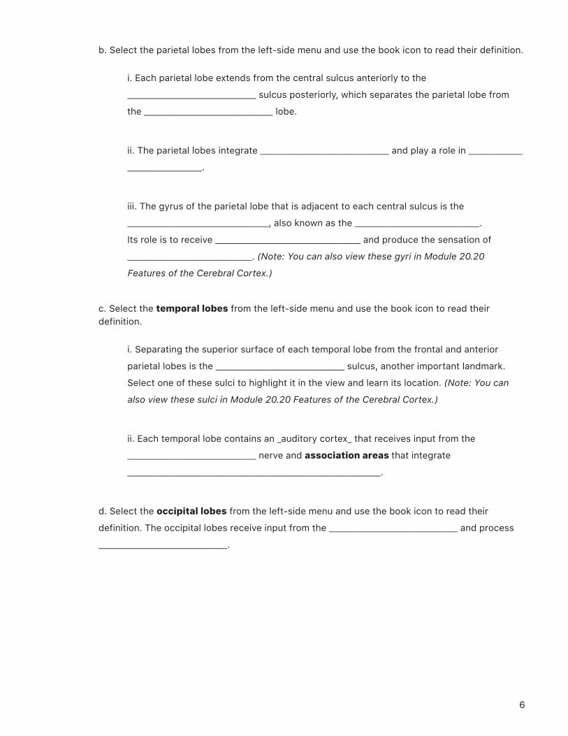
5. Examine the illustration in Module 20.22 Functional Regions of the Cerebral Cortex to learn about the four functional areas of the brain.
a. Which lobes are mainly responsible for the transmission of motor signals?
b. Which lobes are mainly responsible for processing the sensations of touch and taste?
c. Which lobes are mainly responsible for processing auditory signals?
d. Which lobes are mainly responsible for processing visual signals?
e. What is the role of association areas?
8
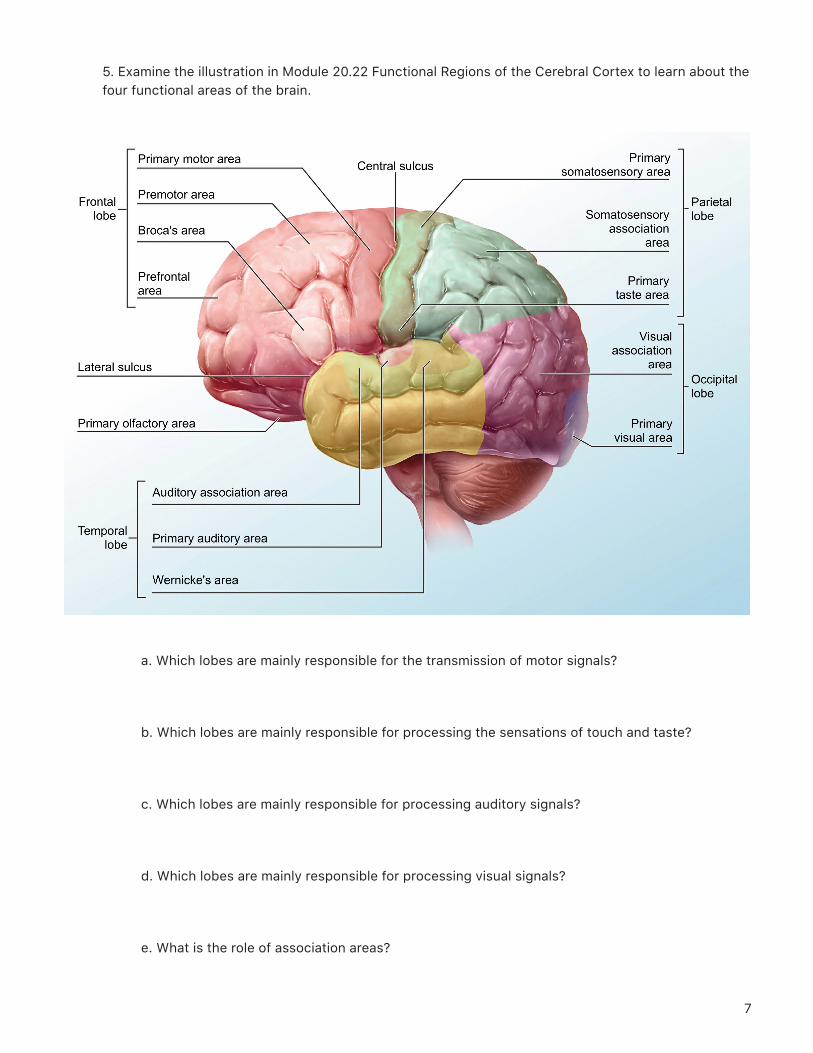
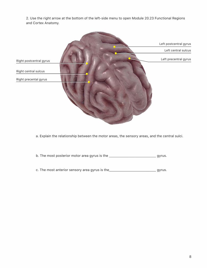
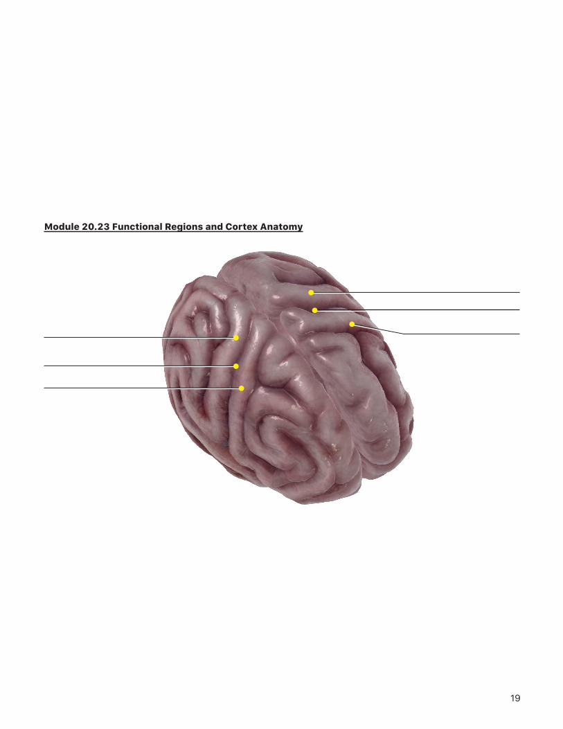
2. Use the right arrow at the bottom of the left-side menu to open Module 20.23 Functional Regions and Cortex Anatomy.
Right precental gyrus
Right central sulcus
Right postcentral gyrus
Left postcentral gyrus
Left central sulcus
Left precentral gyrus
a. Explain the relationship between the motor areas, the sensory areas, and the central sulci.
b. The most posterior motor area gyrus is the _______________________________ gyrus.
c. The most anterior sensory area gyrus is the_______________________________ gyrus.
9
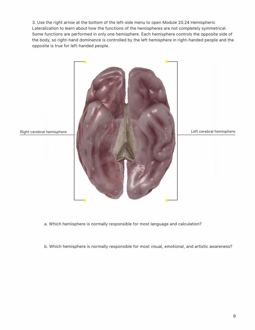
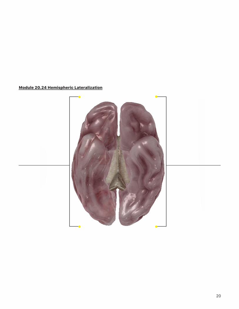
3. Use the right arrow at the bottom of the left-side menu to open Module 20.24 Hemispheric Lateralization to learn about how the functions of the hemispheres are not completely symmetrical. Some functions are performed in only one hemisphere. Each hemisphere controls the opposite side of the body, so right-hand dominance is controlled by the left hemisphere in right-handed people and the opposite is true for left-handed people.
a. Which hemisphere is normally responsible for most language and calculation?
b. Which hemisphere is normally responsible for most visual, emotional, and artistic awareness?
Right cerebral hemisphere Left cerebral hemisphere
10
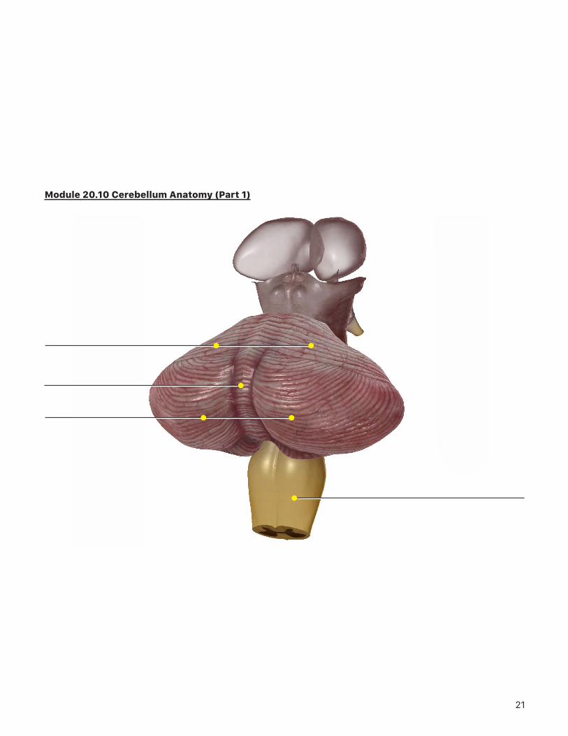
B. Explore the 3D anatomical views in Modules 20.10 Cerebellum Anatomy and 20.11 Cerebellum Function, and then answer the following questions.
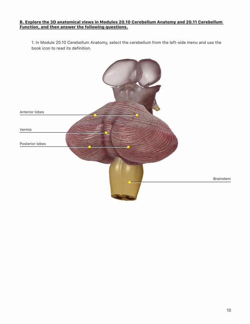
1. In Module 20.10 Cerebellum Anatomy, select the cerebellum from the left-side menu and use the book icon to read its definition.
Posterior lobes
Vermis
Anterior lobes
Brainstem
11
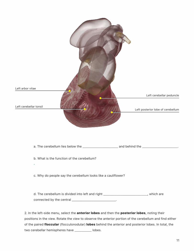
a. The cerebellum lies below the _________________________ and behind the _________________________.
b. What is the function of the cerebellum? .
c. Why do people say the cerebellum looks like a cauliflower?
d. The cerebellum is divided into left and right _______________________________, which are
connected by the central _______________________________.
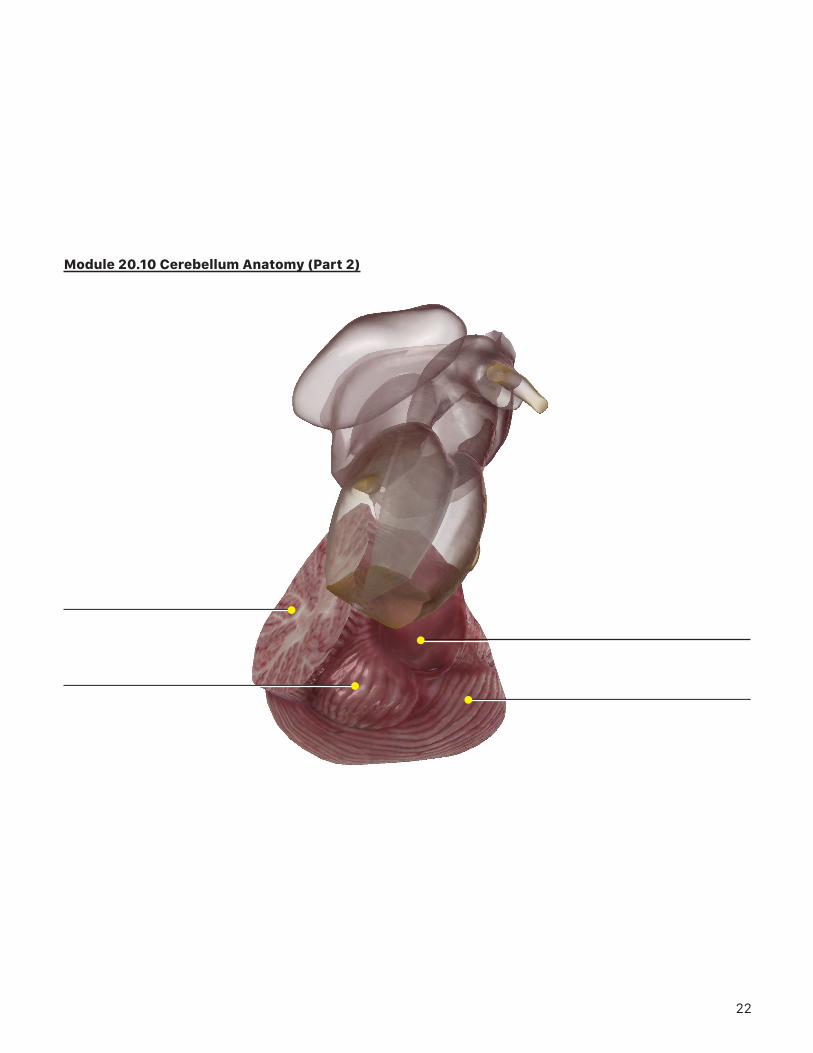
2. In the left-side menu, select the anterior lobes and then the posterior lobes, noting their
positions in the view. Rotate the view to observe the anterior portion of the cerebellum and find either
of the paired floccular (flocculonodular) lobes behind the anterior and posterior lobes. In total, the
two cerebellar hemispheres have ____________ lobes.
Left arbor vitae
Left cerebellar tonsil
Left cerebellar peduncle
Left posterior lobe of cerebellum
12
3. Select the vermis from the left-side menu. The vermis lies between the two cerebellar
_______________________________. (Note: The word vermis means “worm” in Latin; it refers to the shape of
the structure.)
4. Select the arbor vitae from the left-side menu and use the book icon to review the cerebellum definition. Then, tap anywhere outside the view to remove the highlighting. In the unhighlighted view, you can see the light-colored branches of the arbor vitae and its surrounding cortex of gray matter.
a. The arbor vitae is composed of tracts of _______________________________, which consist of
_______________________________.
b. Arbor vitae means “living tree” in Latin. How does this name relate to its structure?
c. Select cerebellar peduncles from the left-side menu and use the book icon to review the cerebellum definition. What is the role of the cerebellar peduncles?
13
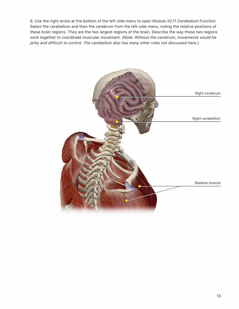
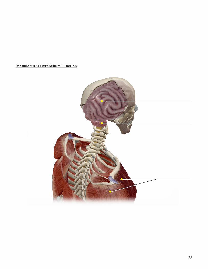
6. Use the right arrow at the bottom of the left-side menu to open Module 20.11 Cerebellum Function. Select the cerebellum and then the cerebrum from the left-side menu, noting the relative positions of these brain regions. They are the two largest regions of the brain. Describe the way these two regions work together to coordinate muscular movement. (Note: Without the cerebrum, movements would be jerky and difficult to control. The cerebellum also has many other roles not discussed here.)
Right cerebellum
Right cerebrum
Skeletal muscle
14
PUTTING IT ALL TOGETHER
1. The cerebellum is the second largest region of the brain. Describe the main role of the cerebellum.
2. The cerebrum is divided into left and right ________________________ by the ___________________________.
3. The cerebral cortex is folded into _______________________ that are separated by
__________________________. The cortex consists of _______________________________ matter, consisting
mostly of _______________________________. This tissue is responsible for most “higher” brain functions.
4. Motor processing areas are found anterior to the _______________________________ and
_______________________________ processing areas are found posterior to them.
5. Each cerebral hemisphere is divided into four _______________________________, each responsible for
different cortical functions. What are the four lobes?
6. Under the cortex is the cerebral _______________________________, which consists mostly of myelinated
neuronal processes that make it appear _______________________________.
7. The basal ganglia, which consist of clusters of neuronal cell bodies called ____________________________,
are found deeper in the cerebrum. The basal ganglia are associated with _______________________________.
8. The left cerebral hemisphere generally controls the _______________________________ side of the body
and is usually associated with _______________________________. The right cerebral hemisphere controls
the ________________________ side of the body and is usually responsible for ______________________________.
15
16
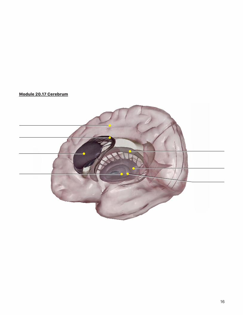
Module 20.17 Cerebrum
17
Module 20.18 Cerebral Hemispheres
18
Module 20.19 Lobes of the Cerebral Cortex
19
Module 20.23 Functional Regions and Cortex Anatomy
20
Module 20.24 Hemispheric Lateralization
21
Module 20.10 Cerebellum Anatomy (Part 1)
22
Module 20.10 Cerebellum Anatomy (Part 2)
23
Module 20.11 Cerebellum Function