in silico: 3d animation and simulation of cell biology ... · pdf fileaxs biomedical animation...
TRANSCRIPT
In Silico: 3D Animation and Simulation of Cell Biology with
Maya and MEL
PRELIMS-P373655.indd iPRELIMS-P373655.indd i 4/12/2008 10:00:26 AM4/12/2008 10:00:26 AM

PRELIMS-P373655.indd iiPRELIMS-P373655.indd ii 4/12/2008 10:00:26 AM4/12/2008 10:00:26 AM

In Silico: 3D Animation and Simulation of Cell Biology with
Maya and MEL
Jason Sharpe AXS Biomedical Animation Studio
Charles John Lumsden University of Toronto
Nicholas Woolridge University of Toronto
PRELIMS-P373655.indd iiiPRELIMS-P373655.indd iii 4/12/2008 10:00:26 AM4/12/2008 10:00:26 AM

Acquisitions Editor: Tiff any GasbarriniPublishing Services Manager: George MorrisonProject Manager: Mónica González de MendozaAssistant Editor: Matt CaterCover Design: Jason Sharpe / Alisa AndreolaCover Illustration: Jason Sharpe
Morgan Kaufmann Publishers is an imprint of Elsevier.30 Corporate Drive, Suite 400, Burlington, MA 01803, USA
Th is book is printed on acid-free paper.
© 2008 Jason Sharpe, Charles Lumsden, Nicholas Woolridge. Published by Elsevier, Inc. All rights reserved.
Designations used by companies to distinguish their products are often claimed as trademarks or registered trademarks. In all instances in which Morgan Kaufmann Publishers is aware of a claim, the product names appear in initial capital or all capital letters. All trademarks that appear or are otherwise referred to in this work belong to their respective owners. Neither Morgan Kaufmann Publishers nor the authors and other contributors of this work have any relationship or affi liation with such trademark owners nor do such trademark owners confi rm, endorse or approve the contents of this work. Readers, however, should contact the appropriate companies for more information regarding trademarks and any related registrations.
No part of this publication may be reproduced, stored in a retrieval system, or transmitted in any form or by any means—electronic, mechanical, photocopying, scanning, or otherwise—without prior written permission of the publisher.
All images © the authors unless otherwise stated in the text. Certain images and materials contained in this publication were reproduced with the permission of Autodesk, Inc. © 2007. All rights reserved. Autodesk and Maya are registered trademarks of Autodesk, Inc., in the U.S.A. and certain other countries.
Th e information in this book and accompanying CD-ROM disk is distributed on an “as is” basis, without warranty. Although due precaution has been taken in the preparation of this work, neither the authors nor the publisher shall have any liability to any person or entity with respect to any loss or damage caused or alleged to be caused directly or indirectly by the information contained in this book and accompanying CD-ROM disk, including, without limitation, any software, whether in object code or source code format.
Permissions may be sought directly from Elsevier’s Science & Technology Rights Department in Oxford, UK: phone: (+44) 1865 843830, fax: (+44) 1865 853333, E-mail: [email protected]. You may also complete your request online via the Elsevier homepage (http://elsevier.com), by selecting
“Support & Contact” then “Copyright and Permission” and then “Obtaining Permissions.”
Library of Congress Cataloging-in-Publication DataSharpe, Jason. In Silico: 3D Animation and Simulation of Cell Biology with Maya and MEL / Jason Sharpe, Charles John Lumsden, Nicholas Woolridge. p. ; cm. Includes bibliographical references and index. ISBN-13: 978-0-12-373655-0 (pbk. : alk. paper) 1. Cytology—Computer simulation. 2. Maya (Computer fi le) 3. Computer animation. 4. Computer graphics. 5. Th ree-dimensional display systems. I. Lumsden, Charles J., 1949– II. Woolridge, Nicholas. III. Title. IV. Title: Cell biology art and science with Maya and MEL. [DNLM: 1. Cells—Programmed Instruction. 2. Computational Biology—Programmed Instruction. 3. Models, Biological—Programmed Instruction. 4. Motion Pictures as Topic—Programmed Instruction. 5. Programming Languages—Programmed Instruction. QU 18.2 S532s 2008] QH585.5.D38S53 2008 571.601�13—dc22 2007053013
ISBN: 978-0-12-373655-0
For information on all Morgan Kaufmann publications,visit our Web site at www.mkp.com or www.books.elsevier.com
08 09 10 11 12 13 10 9 8 7 6 5 4 3 2 1
Printed in China
Working together to grow libraries in developing countries
www.elsevier.com | www.bookaid.org | www.sabre.org
PRELIMS-P373655.indd ivPRELIMS-P373655.indd iv 4/12/2008 10:00:29 AM4/12/2008 10:00:29 AM
CONTENTS Preface xiii
Who is this book for? xiv
Why Maya? xiv
What the book offers xv
Computer hardware and software xxi
About the authors xxii
Acknowledgments xxiii
Part 1 Setting the stage 1
01 Introduction 3
The challenge 4
Wetware for seeing 5
Visualization in science 6
Organizational hierarchy: Keys to biology in vivoand in silico 8
Enter Maya 13
Endless possibilities 19
References 19
02 Computers and the organism 21
Introduction 22
Information and process 22
Language and program 23
High and low 26
Interpret or compile? 27
The Backus watershed 28
Stored programs 30
PRELIMS-P373655.indd vPRELIMS-P373655.indd v 4/12/2008 10:00:30 AM4/12/2008 10:00:30 AM

vi
Conditional control 33
The computed organism 35
The computational organism 36
OOPs and agents 39
Summary 41
References 43
03 Animating biology 45
Introduction 46
Animation and fi lm perception 46
The animator ’ s workfl ow 49
The three-stage workfl ow 51
Putting it all together 67
References 67
Part 2 A foundation in Maya 69
04 Maya basics 71
Getting started 72
How Maya works (briefl y) 78
Maya ’ s UI 82
Summary 99
05 Modeling geometry 101
Introduction 102
NURBS modeling 103
Polygonal modeling 107
Tutorial 05.01: NURBS primitive modeling 109
Tutorial 05.02: Deform the sphere using components 117
Tutorial 05.03: Make and deform a polygon primitive 119
Tutorial 05.04: Construction history 122
CONTENTS
PRELIMS-P373655.indd viPRELIMS-P373655.indd vi 4/12/2008 10:00:30 AM4/12/2008 10:00:30 AM
Tutorial 05.05: Create a NURBS “ fi ber ” 129
Summary 134
References 135
06 Animation 137
Introduction 138
Animation 138
Tutorial 06.01: A keyframe animation 145
Animation nodes in the Hypergraph and Attribute Editor 151
Tutorial 06.02: A simple procedural animation 151
Summary 154
07 Dynamics 157
Introduction 158
The Dynamics module 160
Tutorial 07.01: Rigid body dynamics 166
Tutorial 07.02: Particles in a container 173
Tutorial 07.03: Create a playblast 184
Summary 185
08 Shading 187
Introduction 188
The Render menu set 190
Shading 191
Tutorial 08.01: Shading 203
Summary 214
09 Cameras 215
Maya Cameras 217
Tutorial 09.01: A camera on hemoglobin 222
Summary 230
viiCONTENTS
PRELIMS-P373655.indd viiPRELIMS-P373655.indd vii 4/12/2008 10:00:30 AM4/12/2008 10:00:30 AM
viii
10 Lighting 231
Lighting 232
Tutorial 10.01: Lighting the hemoglobin scene 235
Summary 241
11 Action! Maya rendering 243
Rendering 244
Advanced rendering techniques with the mentalray for Maya renderer 249
Tutorial 11.01: Batch rendering 252
Tutorial 11.02: Playback using fCheck 257
Summary 259
12 MEL scripting 261
Introduction 262
The origins of MEL 263
In a word: Scripting 264
Getting started 266
MEL syntax 269
Values 270
Variables 271
Mathematical and logical expressions 277
The MEL command 280
Attributes in MEL 286
Conditional statements 288
Loops 289
Procedures 291
Animation expressions 292
Putting it all together: The MEL script 301
Tutorial 12.01: Building a MEL script 302
CONTENTS
PRELIMS-P373655.indd viiiPRELIMS-P373655.indd viii 4/12/2008 10:00:30 AM4/12/2008 10:00:30 AM
Debugging your scripts 306
Random number generation in Maya 308
Summary 309
13 Data input/output 311
Introduction 312
Translators 313
Reading and writing fi les with MEL 315
Tutorial 13.01: Visualizing cell migration 322
Summary 337
Part 3 Biology in silico—Maya in action 339
14 Building a protein 341
Introduction 342
Problem overview 346
Methods: Algorithm design 354
Methods: Encoding the algorithm 354
Results: Running the script 368
Results: Rendering your molecule 372
Summary 380
References 381
15 Self-assembly 383
Introduction 384
Problem overview 385
Methods: Actin geometry 394
Methods: Diffusion and reaction events 399
Methods: Reaction rates and probabilities 403
Methods: Algorithm design 409
ixCONTENTS
PRELIMS-P373655.indd ixPRELIMS-P373655.indd ix 4/12/2008 10:00:31 AM4/12/2008 10:00:31 AM
x
Methods: Encoding the algorithm 412
Results: Running your simulation 437
Summary 441
References 442
16 Modeling a mobile cell 443
Introduction 444
Problem overview 445
Model defi nition 449
Methods: Generating pseudopods 451
Methods: Algorithm design 453
Methods: A cell locomotion engine 454
Methods: Encoding the algorithm 466
Methods: Loading the script 475
Results: Running the script 476
Summary 477
References 477
17 Growing an ECM scaffold 479
Introduction 480
Problem overview 481
Model defi nition 483
Methods: Algorithm design 486
Methods: Encoding the algorithm 494
Methods: Grow your scaffold! 512
Results: Parameter effects 516
Summary 517
References 517
CONTENTS
PRELIMS-P373655.indd xPRELIMS-P373655.indd x 4/12/2008 10:00:31 AM4/12/2008 10:00:31 AM
18 Scaffold invasions: Modeling 3D populations of mobile cells 519
Introduction 520
Problem overview 521
Model defi nition 525
Methods: Model design 528
Methods: Encoding the algorithm 538
Methods: Running the simulation 565
Results: Data output 572
Summary 573
References 573
19 Conclusion: A new kind of seeing 575
Explanations, simulations, speculations 576
Maya ’ s role 578
The path so far 578
The future 579
References 582
Further reading 585
Glossary 593
Index 607
xiCONTENTS
PRELIMS-P373655.indd xiPRELIMS-P373655.indd xi 4/12/2008 10:00:31 AM4/12/2008 10:00:31 AM

PRELIMS-P373655.indd xiiPRELIMS-P373655.indd xii 4/12/2008 10:00:32 AM4/12/2008 10:00:32 AM
Preface
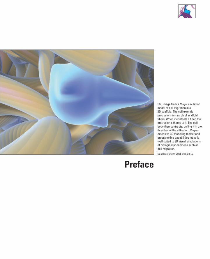
Still image from a Maya simulation model of cell migration in a 3D scaffold. The cell extends protrusions in search of scaffold fi bers. When it contacts a fi ber, the protrusion adheres to it. The cell body then contracts, pulling it in the direction of the adhesion. Maya’s extensive 3D modeling toolset and programming capabilities make it well suited to 3D visual simulations of biological phenomena such as cell migration.
Courtesy and © 2006 Donald Ly.
PRE-P373655.indd xiiiPRE-P373655.indd xiii 4/12/2008 9:59:16 AM4/12/2008 9:59:16 AM
Who is this book for? If, like us, you are involved with the study of cells and cell biology, or if your work takes inspiration from the organic world, this book is for you. We have written In Silico for the diverse creative community—scientists, artists, media designers, stu-dents, and hobbyists—now deeply involved with the living cell as a key to unlocking the complexity of organic matter and a gateway to powerful new understanding of disease. In the scientifi c area, cell and molecular biologists and their research part-ners today have little time to spare developing complex computer programs from the ground up. High-end three-dimensional (3D) computer programs like Autodesk Maya provide the busy scientist with a robust, fl exible development environment in which state-of-the-art computer methods can be used to analyze, model, and visualize cell data. Equipped with deeply customizable user and application programming inter-faces, Maya and other top-tier 3D animation programs aff ord rapid prototyping of data analysis and models through advanced graphics, physics, and rendering systems. Output capability embraces both crisp numerical data and polished 3D dynamic visu-alizations of cell physiology. Th ese tools have enough programming fl exibility that the working researcher can concentrate on the functional aspects of the data mapping or simulation capability they wish to create.
In the communications fi eld are individuals and groups immersed in the burgeon-ing marketplace of biocommunications, especially medical and scientifi c animation. Th e telling of stories is a human universal, common to all peoples and cultures. Th e increasingly complex world enabled by science and technology makes the accurate, compelling telling of scientifi c stories more important than ever. Constantly, anima-tors of medical and scientifi c subjects are called on to present ever more intricate, unusual phenomena involved in understanding how cells work and what goes wrong with them to cause devastating illnesses like cancer and heart disease. At the same time, the expectations of a media-savvy public for concise, truthful, entertaining visual stories rise even higher. Taking control of a program like Maya can empower the media artist to better interpret and visualize wonderfully intricate cellular phe-nomena—such as the crowded molecular landscapes of the cell interior, the cell waves coursing through the embryo ’ s interior, or the skein of blood vessels healing a wound—that would be impractically tedious or impossible to animate by hand.
And too numerous to count, surely, are the artists and citizens everywhere who draw inspiration from biology and the natural world, and who dream of imparting some facet of organic vitality and complexity to their creative work or personal appre-ciation of nature. Th e ideas and methods of this book will, we believe, inform and inspire everyone with such interests. Although the focus of our applications is the exciting realm of the living cell, those whose interests embrace other parts of liv-ing nature will fi nd the knowledge and techniques they learn here of useful in manydiff erent ways.
Why Maya? Although Maya is a top-tier product used worldwide for 3D animation in entertain-ment, gaming, and manufacturing, this Academy Award® winning program does not stand alone in representing the cutting edge of high-end 3D. Superb tools such as SoftImage XSI, Maxon Cinema 4D, NewTek LightWave 3D, Autodesk 3ds Max, and
xiv PREFACE
PRE-P373655.indd xivPRE-P373655.indd xiv 4/12/2008 9:59:20 AM4/12/2008 9:59:20 AM

Side Eff ects Software ’ s Houdini, stand alongside Maya to defi ne the state of the art in 3D animation capability. Maya is our subject in this book for three reasons. First, despite the excellence of alternative tools Maya currently enjoys a pre-eminent sta-tus in top-end 3D animation work. Second, the Maya programming interfaces—accessed through a C�� application toolset (the API—which we plan to deal with in a subsequent book), via scripting in the Python language, and through Maya ’ s own scripting language MEL, which we treat in this book—allow enormous power and fl ex-ibility in customizing Maya for scientifi c applications. Th ird, the academic outreachinitiatives supported by Autodesk, the fi rm that makes and sells Maya, haveenabled us to test Maya and some of its predecessors (such as Alias PowerAnimator) in demanding real-world science projects in cell and medical science. As a threesome, we have between us accumulated roughly 40 person-years of experience across awide range of such applications. We fi nd Maya worthy of close attention whenever there is a need to model and visualize 3D cell biology using a computer. Since our origins trace back to the early days, in which such computer methods were lab-writ-ten custom jobs in languages like Fortran, C��, and OpenGL, Maya for us means shorter time to software completion while increasing the power of the animated visualization.
If you are already a user of a 3D animation package other than Maya, you will still fi nd considerable useful material in the pages to follow. Th e book is going to show you how to approach complex biological problems eff ectively, by means of a workfl ow in 3D visual computing. We have developed this workfl ow over the years of our medical and biocommunications research and use it daily in our teaching and scientifi c inves-tigation. By working through the book ’ s projects and case studies, you will be able to adapt our workfl ow to other 3D animation products as well as take them much further in Maya itself.
What the book offers In the world of computer graphics software, Maya is a relatively complicated applica-tion. Learning and, eventually, some degree of genuine mastery, take time, but don ’ t despair. Page by page, the learning map we have set up will take you from one pro-ductive result to the next. You will deal throughout with learning content that has genuine interest and signifi cance in the world of science and cell biology. In Part 1 you will meet the key ideas and terms from scientifi c computer graphics needed to dive into Maya while assessing its historic relevance to leading edge visualization. In Part 2 , you will receive a self-contained introduction to Maya and to our workfl ow that will take you from starting the program through to a polished animation ren-dering of a complex protein. With this foundation you are ready to meet MEL, the programming language by which you will harness Maya ’ s ability to model and render complex events. Th en in Part 3 , we put this all to work. You will develop a portfolio of case studies ranging from the single biological molecule to populations of inter-acting macromolecules, and then on to mobile cells as they move through their tis-sue environment. As you complete each element in the portfolio, you will have taken command of powerful new strategies for using MEL to control Maya ’ s numerical and visual rendering activity.
Here ’ s what you can expect in the rest of the book.
xvPREFACE
PRE-P373655.indd xvPRE-P373655.indd xv 4/12/2008 9:59:20 AM4/12/2008 9:59:20 AM
Part 1: Setting the stage
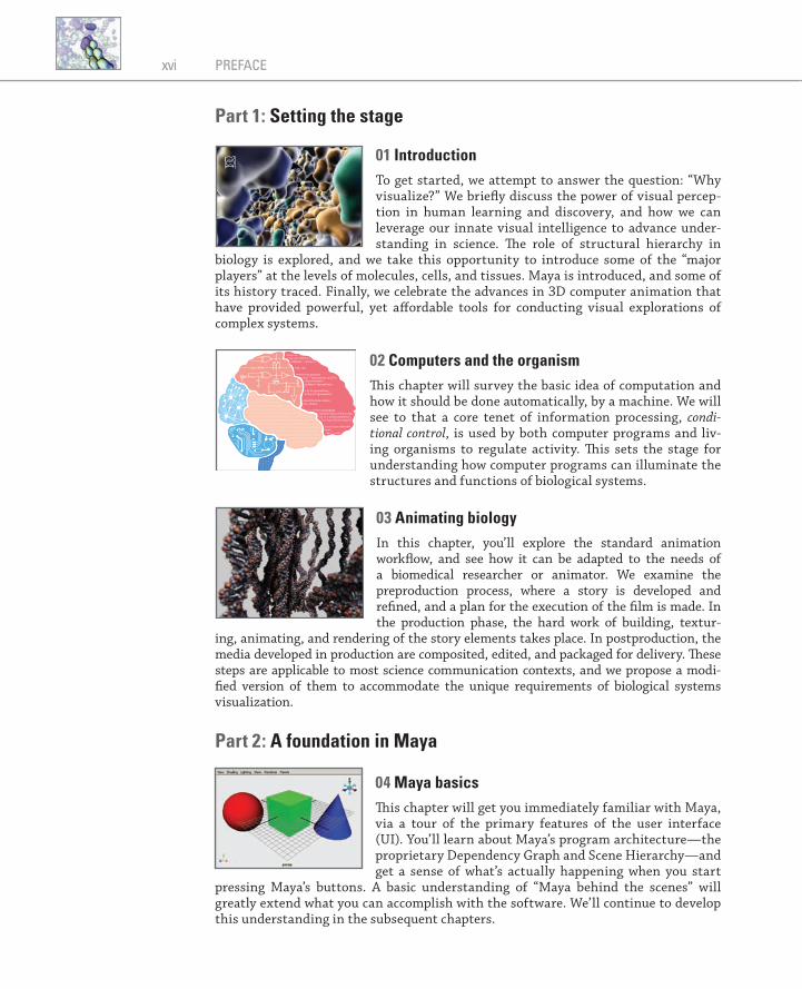
01 Introduction To get started, we attempt to answer the question: “ Why visualize? ” We briefl y discuss the power of visual percep-tion in human learning and discovery, and how we can leverage our innate visual intelligence to advance under-standing in science. Th e role of structural hierarchy in
biology is explored, and we take this opportunity to introduce some of the “ major players ” at the levels of molecules, cells, and tissues. Maya is introduced, and some of its history traced. Finally, we celebrate the advances in 3D computer animation that have provided powerful, yet aff ordable tools for conducting visual explorations of complex systems.
02 Computers and the organism Th is chapter will survey the basic idea of computation and how it should be done automatically, by a machine. We will see to that a core tenet of information processing, condi-tional control , is used by both computer programs and liv-ing organisms to regulate activity. Th is sets the stage for understanding how computer programs can illuminate the structures and functions of biological systems.
03 Animating biology In this chapter, you ’ ll explore the standard animation workfl ow, and see how it can be adapted to the needs of a biomedical researcher or animator. We examine the preproduction process, where a story is developed and refi ned, and a plan for the execution of the fi lm is made. In the production phase, the hard work of building, textur-
ing, animating, and rendering of the story elements takes place. In postproduction, the media developed in production are composited, edited, and packaged for delivery. Th ese steps are applicable to most science communication contexts, and we propose a modi-fi ed version of them to accommodate the unique requirements of biological systems visualization.
Part 2: A foundation in Maya
04 Maya basics Th is chapter will get you immediately familiar with Maya, via a tour of the primary features of the user interface (UI). You ’ ll learn about Maya ’ s program architecture—the proprietary Dependency Graph and Scene Hierarchy—and get a sense of what ’ s actually happening when you start
pressing Maya ’ s buttons. A basic understanding of “ Maya behind the scenes ” will greatly extend what you can accomplish with the software. We ’ ll continue to develop this understanding in the subsequent chapters.
60Å
xvi PREFACE
PRE-P373655.indd xviPRE-P373655.indd xvi 4/12/2008 9:59:20 AM4/12/2008 9:59:20 AM
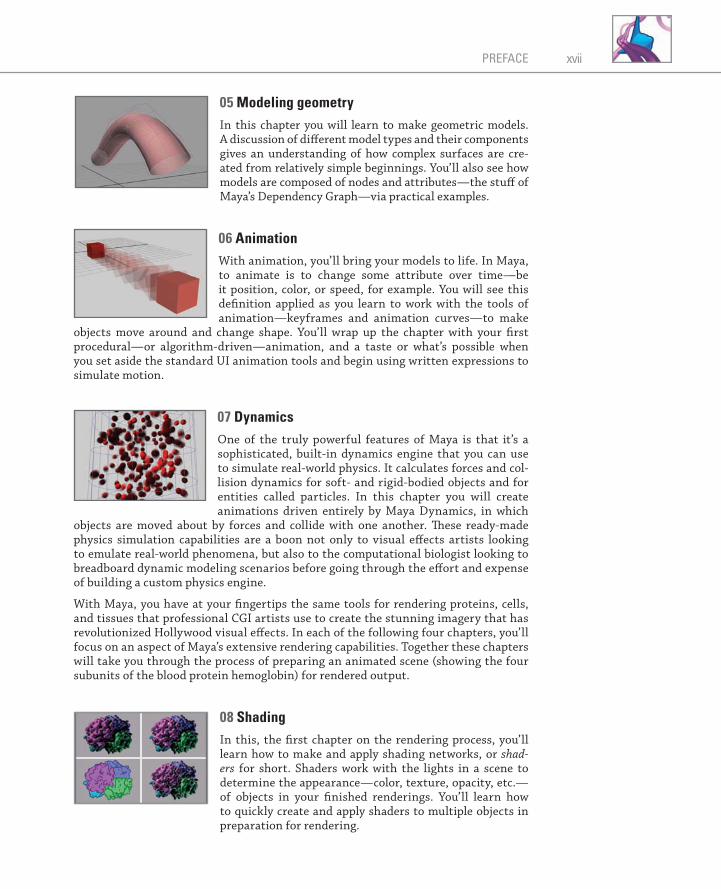
05 Modeling geometry In this chapter you will learn to make geometric models.A discussion of diff erent model types and their components gives an understanding of how complex surfaces are cre-ated from relatively simple beginnings. You ’ ll also see how models are composed of nodes and attributes—the stuff of Maya ’ s Dependency Graph—via practical examples.
06 Animation With animation, you ’ ll bring your models to life. In Maya, to animate is to change some attribute over time—be it position, color, or speed, for example. You will see this defi nition applied as you learn to work with the tools of animation—keyframes and animation curves—to make
objects move around and change shape. You ’ ll wrap up the chapter with your fi rst procedural—or algorithm-driven—animation, and a taste or what ’ s possible when you set aside the standard UI animation tools and begin using written expressions to simulate motion.
07 Dynamics One of the truly powerful features of Maya is that it ’ s a sophisticated, built-in dynamics engine that you can use to simulate real-world physics. It calculates forces and col-lision dynamics for soft- and rigid-bodied objects and for entities called particles. In this chapter you will create animations driven entirely by Maya Dynamics, in which
objects are moved about by forces and collide with one another. Th ese ready-made physics simulation capabilities are a boon not only to visual eff ects artists looking to emulate real-world phenomena, but also to the computational biologist looking to breadboard dynamic modeling scenarios before going through the eff ort and expense of building a custom physics engine.
With Maya, you have at your fi ngertips the same tools for rendering proteins, cells, and tissues that professional CGI artists use to create the stunning imagery that has revolutionized Hollywood visual eff ects. In each of the following four chapters, you ’ ll focus on an aspect of Maya ’ s extensive rendering capabilities. Together these chapters will take you through the process of preparing an animated scene (showing the four subunits of the blood protein hemoglobin) for rendered output.
08 Shading In this, the fi rst chapter on the rendering process, you ’ ll learn how to make and apply shading networks, or shad-ers for short. Shaders work with the lights in a scene to determine the appearance—color, texture, opacity, etc.—of objects in your fi nished renderings. You ’ ll learn how to quickly create and apply shaders to multiple objects in preparation for rendering.
xviiPREFACE
PRE-P373655.indd xviiPRE-P373655.indd xvii 4/12/2008 9:59:29 AM4/12/2008 9:59:29 AM

09 Cameras Like a real movie camera, a Maya camera defi nes what your audience will see. Many features are available with a real camera are embodied in the Maya version, allowing you to set up and record shots in virtual 3D space much as you would in the real world. Th e Maya camera also defi nes your view of the 3D scene as you work with it, and
is therefore an indispensable tool, whether or not you plan to make fi nished (ren-dered) movies with Maya. By the end of this chapter, you ’ ll know how to set up and animate a camera along a track called a motion path—much the way a movie camera is set up on a track to move as it records the action.
10 Lighting If the camera is a cinematographer ’ s brush , then light is the paint. Just like in the real world, light defi nes what is visible in your Maya scenes, and the quality of its appearance. We ’ ll show you how to achieve professional illumination with minimal eff ort in order to get the most out of your images.
11 Action! Maya rendering In this fi nal chapter on the rendering process, you ’ ll see how Maya integrates shaders, camera view, and lights to produce one or more image fi les. We ’ ll explore the diff er-ent render “ engines ” available in Maya and their relative advantages.
12 Mel scripting At this point in the book, you ’ ll know your way around the UI and be familiar with the concepts and terminology involved in modeling, animating, and rendering in Maya. You ’ ll be ready to depart somewhat from the standard UI tools and start exploring Maya ’ s scripting capabilities.
Th is chapter introduces Maya ’ s scripting (or programming) language, MEL (short for Maya Embedded Language). You ’ ll learn how to run individual MEL commands and how to compose a script—or short computer program—out of multiple MEL state-ments in order to automate tasks in Maya. Readers new to computer programming will learn the basic concepts—syntax, variables, operators, fl ow control, etc.—in the context of MEL. Th ose with previous programming experience can scan the chapter to pick up the MEL basics. In either case, plentiful examples and a short tutorial will have you coding Maya tasks using MEL in no time.
13 Data input/output Ready-made software plug-ins are available for porting some of the more common 3D data formats to and from Maya. However, if you ’ re working with a format for which no plug-in exists, such as experimental data formatted in a spread sheet, you may want to create your own importer
move -a 0 ($H/2) 0 $name1;move -a 0 (-$H/2) 0 $name2;$groupName = `group $name1 $name2`;
if ($j==0){
// Create the first peptides. $locatorName1 = `spaceLocator -p 0 0 0`; move $W 0 0 $locatorName1; parent $locatorName1 $groupName;
move -r $x $y $z $groupName; rotate -r $rx $ry $rz $groupName; // Increment the helix rotation. $rx = ($rx + $helix);
}else { // Create the next peptide.
// Store the translate values of the locator. $xyz1 = `xform -q -t -ws $locatorName1`; $x = $xyz1[0]; $y = $xyz1[1]; $z = $xyz1[2]; // delete the previous locator and make a ne delete $locatorName1; $locatorName1 = `spaceLocator -p 0 0 0`;
xviii PREFACE
PRE-P373655.indd xviiiPRE-P373655.indd xviii 4/12/2008 9:59:36 AM4/12/2008 9:59:36 AM
or exporter. Th is chapter shows you how to do just that using a suite of MEL com-mands for reading and writing external fi les. You ’ ll also learn the MEL commands useful for formatting the text that you read and write. In the chapter ’ s tutorial, you ’ ll extract 3D coordinates from a cell migration data fi le, use them to visualize the mov-ing cells, and then save out a report summarizing key migration statistics.
Part 3: Biology in silico—Maya in action In this part of the book, you ’ ll explore and use a workfl ow for in silico modeling and simulation that builds on your knowledge of Maya ’ s UI and scripting capabilities. We present fi ve tutorial-style projects, each dealing with a diff erent level of biological organization—from a single protein up to a population of cells in a tissue matrix. In each project we ’ ll guide you, step by step, through the composition of custom MEL scripts that automate the model building and/or dynamic simulation. Whether you ’ re a scientist looking to explore Maya techniques in 3D computation or an artist visual-izing topics in cell science, you ’ ll learn a range of useful techniques that can subse-quently be applied to your own projects.
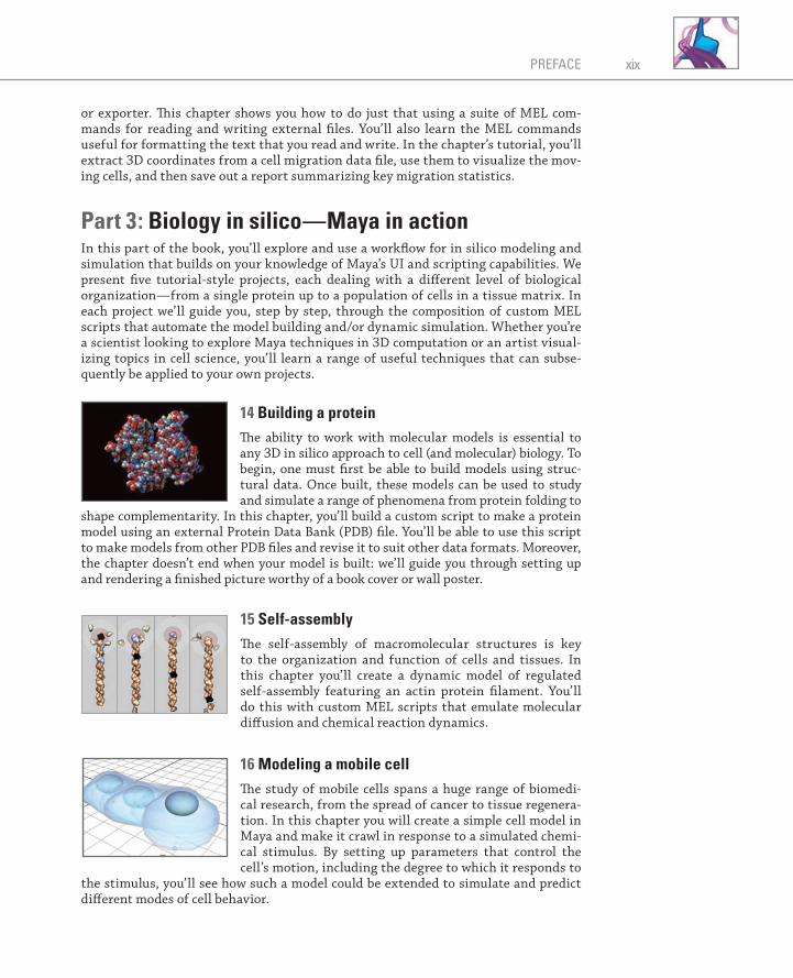
14 Building a protein Th e ability to work with molecular models is essential to any 3D in silico approach to cell (and molecular) biology. To begin, one must fi rst be able to build models using struc-tural data. Once built, these models can be used to study and simulate a range of phenomena from protein folding to
shape complementarity. In this chapter, you ’ ll build a custom script to make a protein model using an external Protein Data Bank (PDB) fi le. You ’ ll be able to use this script to make models from other PDB fi les and revise it to suit other data formats. Moreover, the chapter doesn ’ t end when your model is built: we ’ ll guide you through setting up and rendering a fi nished picture worthy of a book cover or wall poster.
15 Self-assembly Th e self-assembly of macromolecular structures is key to the organization and function of cells and tissues. In this chapter you ’ ll create a dynamic model of regulated self-assembly featuring an actin protein fi lament. You ’ ll do this with custom MEL scripts that emulate molecular diff usion and chemical reaction dynamics.
16 Modeling a mobile cell Th e study of mobile cells spans a huge range of biomedi-cal research, from the spread of cancer to tissue regenera-tion. In this chapter you will create a simple cell model in Maya and make it crawl in response to a simulated chemi-cal stimulus. By setting up parameters that control the cell ’ s motion, including the degree to which it responds to
the stimulus, you ’ ll see how such a model could be extended to simulate and predict diff erent modes of cell behavior.
xixPREFACE
PRE-P373655.indd xixPRE-P373655.indd xix 4/12/2008 9:59:53 AM4/12/2008 9:59:53 AM
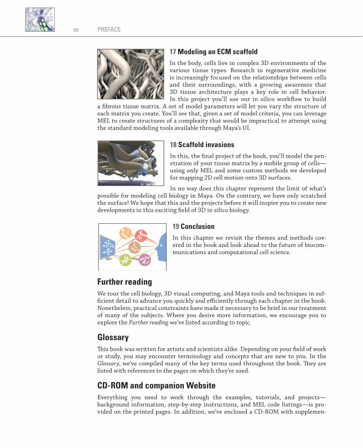
17 Modeling an ECM scaffold In the body, cells live in complex 3D environments of the various tissue types. Research in regenerative medicine is increasingly focused on the relationships between cells and their surroundings, with a growing awareness that 3D tissue architecture plays a key role in cell behavior. In this project you ’ ll use our in silico workfl ow to build
a fi brous tissue matrix. A set of model parameters will let you vary the structure of each matrix you create. You ’ ll see that, given a set of model criteria, you can leverage MEL to create structures of a complexity that would be impractical to attempt using the standard modeling tools available through Maya ’ s UI.
18 Scaffold invasions In this, the fi nal project of the book, you ’ ll model the pen-etration of your tissue matrix by a mobile group of cells—using only MEL and some custom methods we developed for mapping 2D cell motion onto 3D surfaces.
In no way does this chapter represent the limit of what ’ s possible for modeling cell biology in Maya. On the contrary, we have only scratched the surface! We hope that this and the projects before it will inspire you to create new developments in this exciting fi eld of 3D in silico biology.
19 Conclusion In this chapter we revisit the themes and methods cov-ered in the book and look ahead to the future of biocom-munications and computational cell science.
Further reading We tour the cell biology, 3D visual computing, and Maya tools and techniques in suf-fi cient detail to advance you quickly and effi ciently through each chapter in the book. Nonetheless, practical constraints have made it necessary to be brief in our treatment of many of the subjects. Where you desire more information, we encourage you to explore the Further reading we ’ ve listed according to topic.
Glossary Th is book was written for artists and scientists alike. Depending on your fi eld of work or study, you may encounter terminology and concepts that are new to you. In the Glossary , we ’ ve compiled many of the key terms used throughout the book. Th ey are listed with references to the pages on which they ’ re used.
CD-ROM and companion Website Everything you need to work through the examples, tutorials, and projects—background information, step-by-step instructions, and MEL code listings—is pro-vided on the printed pages. In addition, we ’ ve enclosed a CD-ROM with supplemen-
xx PREFACE
PRE-P373655.indd xxPRE-P373655.indd xx 4/12/2008 10:00:00 AM4/12/2008 10:00:00 AM
tary material. It includes MEL scripts, Maya fi les, and rendered animations from various chapters. Th e read_me.txt fi le in the root directory of the CD-ROM includes an index of the enclosed computer fi les.
On the books’s companion Website you’ll fi nd updates and corrections (when neces-sary) to the fi les provided on the CD-ROM.
www.insilico.book.net.
Computer hardware and software Th e Maya fi les and MEL scripts listed in this book and included on the CD-ROM were created and tested on a mid-range consumer-level PC with the following specifi cations:
Software Maya 8.5 for Windows OS Windows XP Professional 2002 (Service Pack 2) PC Dell Dimension 8300
CPU Pentium 4, 3.20 GHz RAM 1 GB
Graphics adapter ATI Radeon 9800 XT, 256 MB DDR
Th e book ’ s tutorials and projects have been developed over a number of versions of Maya, both in Windows and Mac OS. Th ey have been tested to work in Maya 8.5 for Windows . Users of older versions of Maya may have to look around for commands whose names have changed, but the MEL code will probably work largely unaltered. As this book went to press, a new version was announced (Maya 2008). Although we have not had the opportunity to test our projects against Maya 2008, we have no reason to believe that the techniques we rely on would have altered enough to have broken them.
Similarly, the instructions for accessing Maya menus and tools, along with references to the Maya Help Library, are specifi c to Maya 8.5 for Windows. With a little adaptation they can readily be applied to learning Maya in other environments, namely Mac OS and Linux.
If you are considering purchasing Maya, we strongly recommend you ensure its com-patibility with your hardware and software confi guration by consulting the system requirements and qualifi ed hardware specifi cations available via Autodesk ’ s website:
www.autodesk.com/fo-products-maya
xxiPREFACE
PRE-P373655.indd xxiPRE-P373655.indd xxi 4/12/2008 10:00:08 AM4/12/2008 10:00:08 AM

About the authors
Jason Sharpe is a cofounder of the award-winning AXS Biomedical Animation Studio in Toronto. Trained in mechanical engineering at Queen’s University, fi ne arts at Emily Carr Institute of Art and Design and biomedical communications at the University of Toronto, he has worked on a wide range of Maya-based 3D animation projects for research, education, and entertainment.
Charles J Lumsden is Professor of Medicine at the University of Toronto. Trained as a theoretical physicist, he studies the mathematical logic behind illnesses such as Alzheimer’s disease and cancer. He and his students have explored and championed a variety of 3D graphics software as aids to biomedical discovery, including top-tier commercial tools such as Maya and MEL.
Nicholas Woolridge , Associate Professor of Biomedical Communications at the University of Toronto, has played a major role in the development of the visu-alization design fi eld in the university’s renowned Master’s Degree in Biomedical Communications. His current research focuses on the optimization of visual media for medical research and education.
PRE-P373655.indd xxiiPRE-P373655.indd xxii 4/12/2008 10:00:09 AM4/12/2008 10:00:09 AM

Acknowledgments
Th e splendid staff at Morgan Kaufmann, our publisher, has given us essential aid—mixed with clearheaded expertise and unquenched enthusiasm—as In Silico found its way through the press and into your hands. Tim Cox, then a senior editor at Morgan Kaufmann, saw sense in our idea that time was right for a richly cross-disciplinary book exploring Maya and its programming language, MEL, as tools for adventure and discovery in biology and medicine. Tim also got behind our conviction that such a book would be at its best if written for a use by a diverse audience of artists, scien-tists, and highly motivated private citizens. Morgan Kaufmann is a world leader in producing texts that map the subtle intricacies of MEL programming; we were, and remain, honored to have In Silico at home in this distinguished setting. Once Tim had the project launched, our Editor, Tiff any Gasbarrini, and Assistant Editors Michele Cronin and Matt Cater, helped us survive the twists and turns of bringing the book to life. Th rough our publisher we benefi ted from the comments of expert readers, who responded to drafts of In Silico either in whole or in part. Our thanks to these hard-working colleagues for their generous allotment of time and attention: Prof. Klaus Mueller of Stony Brook University; David F. Wiley, President and CEO of Stratovan Corporation; Azam Khan, research scientist at Autodesk Corporation; and fi veanonymous reviewers. Th eir input, uniformly deft and relevant, has helped In Silico complete its journey with enhanced strength.
In addition, two student reviewers—Lori Waters (of the Biomedical Communications graduate program) and Tatiana Lomasko (PhD candidate in the Institute of Medical Science), both at the University of Toronto—completed many of the tutorials, provid-ing valuable feedback that helped us to hone our approach.
Th roughout their history, Maya and MEL were invented and advanced by a commu-nity of brilliant computer graphics innovators principally located in Toronto, Canada (with colleagues in offi ces in Paris and California). Th e software was originally devel-oped by Alias, Inc., and is now under the banner of the Autodesk Corporation. We cannot overstate our appreciation to Autodesk and to its staff of Maya and MEL experts in assisting us on occasional technical questions and allowing us to present the many illustrations in which Maya ’ s user interface is depicted. As well, In Silico takes the view that infl uential inventions like Maya are what they are not only through the genius of their creators, but also because they appear at a specifi c time and place in human history. Th erefore, appreciating historic trends in computer tech-nology, computer programming, and 3D computer animation gives us better under-standing of Maya and MEL. Th e history of Maya and MEL has not been written up extensively, and what sources exist we found to be occasional and widely scattered. We are therefore most grateful to Autodesk for granting us discussions with mem-bers of its staff , who number among the original inventors of Maya and MEL. Th ese incredibly busy people answered our questions about origins and inspirations with patience, grace, and good humor. We are delighted to be able to incorporate the gist of those discussions here, by way of introducing you to the depths of Maya and MEL. In particular we must thank Joyce Janczyn, lead designer of MEL, as well Mike Taylor,
ACK-P373655.indd xxiiiACK-P373655.indd xxiii 4/12/2008 10:00:55 AM4/12/2008 10:00:55 AM
Duncan Brinsmead, and Jos Stam for talks that opened our eyes to the inner life Maya.
Ravi Jagannadhan gave considerably of his own time to review and test the many MEL scripts published here. And, during this entire time Azam Khan (research scien-tist at Autodesk) never tired of his informal role as our advisor and principal facilita-tor amidst the elite world of those charged with inventing the latest versions of Maya and Maya programming.
Since this book hopes to be useful to readers who are new to computers, computer programming, or 3D animation—as well as an effi cient self-contained resource for experienced science researchers and computer artists—we have used key moments from computer history and animation history to lay newcomers a congenial path to MEL programming. It is a pleasure to thank all the computer historians, collectors, and archivists who helped us with information, recollections, and photographs. In particular we must note the extended assistance generously given our history frame by: portraitist Louis Fabian Bachrach III for his photograph of programming language pioneer John Backus, lead inventor of the Fortran language; computer scientist John Bennett (Sydney, Australia) for his assistance and support in presenting his early com-puter graphic of structure pattern data for the protein myoglobin; Deirdre Bryden, Queen ’ s University (Kingston, Ontario) archivist, and Marnee Gamble, University of Toronto archivist, for mainframe history and photographs at these Canadian research centers; Martin Campbell-Kelly, University of Warwick (Coventry, UK), for early computer history and photographs, especially the EDSAC; Annette Faux, archi-vist at Cambridge University ’ s Molecular Biology Laboratory, for early 3D models of the myoglobin protein; PDP-8 microcomputer collector and archivist David Gesswein, his wife Janet Walz, and their cats Khym and Py for the PDP-8 microcomputer pho-tograph shot specially for the book; Calvin Gotlieb, University of Toronto, for access to his archives on that institution ’ s computer center history; Bonnie Ludt, California Institute of Technology Archives, for her help with the Linus Pauling photographs; Dawn Stanford of the IBM Corporate Archives for assistance with IBM mainframe history; Peter Strickland, Managing Editor of the Acta Crystallographica journals, for his assistance with early computer visualizations of protein structure; Bjarne Stroustrup, inventor of the universally used C�� programming language, for his photograph; Marcia Tucker, Institute for Advanced Study Archives (Princeton, NJ) for assistance with the John von Neumann photograph; and Martin Zwick, Portland State University (Portland, OR), for information and photographs on key early work in molecular computer graphics. Our photo editor, Jane Affl eck, also gave us strong assistance in sourcing hard-to-fi nd images.
In Silico celebrates as well creative work by many of our colleagues who advance the visual interpretation of cell structure and dynamics through 3D computer graphics and animation. We especially thank Drew Barry, Marc Dryer, Stephen Ellis of Ellis Entertainment, David Goodsell, and Jenn Platt for letting us include their work here; Eddy Xuan and Sonya Amin of AXS Studio for their tremendous support and gener-ous contributions to the book ’ s illustrations; and Christina Jennings of Shaftesbury Films for letting us include animation stills from her pioneering dramatic series, Regenesis. Stunning visualizations in biology and medicine of course use technol-ogy other than computer graphics, such as photographic microscopy and video cap-ture. We are indebted to: Peter Friedl and Katarina Wolf, University of Würzburg, Germany; Sylvia Papp and Michal Opas, University of Toronto; and Alexis Armour, Hôpital Hôtel-Dieu du CHUM, Université de Montréal, for their help and consent in
ACKNOWLEDGMENTSxxiv
ACK-P373655.indd xxivACK-P373655.indd xxiv 4/12/2008 10:00:56 AM4/12/2008 10:00:56 AM
using their micrographs and/or video capture of cellular and tissue engineering mate-rials. A special thanks goes to John Semple of Sunnybrook Health Sciences Centre for his expertise and guidance in regenerative medicine that helped shape the book ’ s two fi nal projects. John, who is both an artist and a scientist, also provided feedback at an early stage that helped us craft the book for researchers and artists alike.
Th is book would not exist without the support we received from NSERC, the Natural Sciences and Engineering Research Council of Canada, in the form of a three-year grant under NSERC ’ s Collaborative Research and Development (CRD) program. Th e CRD program brings University-based researchers in Canada together with compa-nies that share common interests in science and technology—in this case the idea that a top-tier 3D animation package like Maya (itself a Canadian invention) can be a powerful tool in the hands of biomedical scientists and teachers. Th e NSERC-CRD initiative seeks outcomes with broad relevance to the advanced training needs and research application requirements of citizens in Canada and indeed worldwide. It has therefore been a special pleasure to design our work under this grant pro-gram, through NSERC-CRD Grant Number CRD 270158-03, entitled “ Interpretive Visualization: Understanding cell systems dynamics through computer animation ” ; so that our fi ndings can communicated in a book for working professionals and for trainees in both the sciences and the digital media arts. Our corporate partners, Bell Canada Enterprises in grant year 1 via the Bell University Grants Program at the University of Toronto, and Alias (now Autodesk) in grant years 2 and 3, were essen-tial to the success of our CRD project and we are deeply grateful for their participa-tion. At each step NSERC personnel at various levels—Eileen Jessop, Pamela Moss, Anne-Marie Monteith, Sylvie Boucher, and Lise Desforges—assisted us with practical guidance and patient advice.
No book large or small gets done without evenings, late nights, and weekends nipped from time otherwise owed to kith and kin. So we must end with our deepest thanks to our families, who have put up with all the stolen hours and steadfastly supported us throughout the book ’ s creation.
xxvACKNOWLEDGMENTS
ACK-P373655.indd xxvACK-P373655.indd xxv 4/12/2008 10:00:56 AM4/12/2008 10:00:56 AM