in situ sequencing for rna analysis in tissue sections
TRANSCRIPT

Feb 02, 2020
In situ sequencing for RNA analysis in tissue sectionsV.2
Nucleic Acids Research
Chika Yokota , Daniel Gyllborg , Mats NilssonScience for Life Laboratory, Department of Biochemistry and Biophysics, Stockholm University, Stockholm, Sweden
dx.doi.org/10.17504/protocols.io.bb2giqbw
Molecular Diagnostics - Mats Nilsson Group
Daniel GyllborgStockholm University
21 1 1
1
1
In situ sequencing method for parallel targeted analysis of short RNA fragments in morphologically preserved tissue. This protocol can be used to detect RNA molecules at the single cell level to aid in the identification of cell types according to their gene expression. The technique uses padlock probes to target desired genes of interest and rolling circle amplification to amplify signal for a high throughput methodolgy of spatial transcriptomics. With the use of barcode sequencing, identification of numerous genes is possible through multiplexing.
Version 2 Update: Only minor mistakes were corrected for version 2 of this protocol, no major changes to protocol were done. Some references were also updated.
nmeth.2563.pdf
DOI
dx.doi.org/10.17504/protocols.io.bb2giqbw
https://www.nature.com/articles/nmeth.2563
Chika Yokota, Daniel Gyllborg, Mats Nilsson 2020. In situ sequencing for RNA analysis in tissue sections.protocols.ioprotocols.iohttps://dx.doi.org/10.17504/protocols.io.bb2giqbw
protocol
Ke R, Mignardi M, Pacureanu A, Svedlund J, Botling J, Wählby C, Nilsson M. In situ sequencing for RNA analysis in preserved tissue and cells. Nat Methods. 2013 Sep;10(9):857-60. doi: 10.1038/nmeth.2563
in situ sequencing, iss, spatial transcriptomics, padlock probes, sequence by ligation, single cell, rolling circleamplification, RCA
protocol ,
Jan 31, 2020
Feb 02, 2020
1
Citation:Citation: Chika Yokota, Daniel Gyllborg, Mats Nilsson In situ sequencing for RNA analysis in tissue sectionshttps://dx.doi.org/10.17504/protocols.io.bb2giqbw
This is an open access protocol distributed under the terms of the Creative Commons Attribution LicenseCreative Commons Attribution License(https://creativecommons.org/licenses/by/4.0/), which permits unrestricted use, distribution, and reproduction in any medium,provided the original author and source are credited

32552
The following protocol is based off of In situ sequencing for RNA analysis in preserved tissue and cells. (Ke R et al., Nat. Methods, 2013) with some modifications and focus on fresh frozen tissue sections.
See also following publications for additional references:
Probabilistic cell typing enables fine mapping of closely related cell types in situQian X, Harris KD, Hauling T, Nicoloutsopoulos D, Munoz-Manchado A, Skene N, Hjerling-Leffler J, Nilsson M.Nat Methods. 2020 Jan;17(1):101-106.doi: 10.1038/s41592-019-0631-4
Spatial and temporal localization of immune transcripts defines hallmarks and diversity in the tuberculosis granulomaCarow B, Hauling T, Qian X, Kramnik I, Nilsson M, Rottenberg ME.Nat Commun. 2019;10(1):1823.doi:10.1038/s41467-019-09816-4
Padlock Probes to Detect Single Nucleotide PolymorphismsKrzywkowski T, Nilsson M.Methods Mol Biol. 2018;1649:209-229. doi: 10.1007/978-1-4939-7213-5_14.
In situ detection and genotyping of individual mRNA moleculesLarsson C, Grundberg I, Söderberg O, Nilsson M.Nat Methods. 2010 May;7(5):395-7. doi: 10.1038/nmeth.1448.
Protocol Workflow OverviewProtocol Workflow Overview
2
Citation:Citation: Chika Yokota, Daniel Gyllborg, Mats Nilsson In situ sequencing for RNA analysis in tissue sectionshttps://dx.doi.org/10.17504/protocols.io.bb2giqbw
This is an open access protocol distributed under the terms of the Creative Commons Attribution LicenseCreative Commons Attribution License(https://creativecommons.org/licenses/by/4.0/), which permits unrestricted use, distribution, and reproduction in any medium,provided the original author and source are credited

Padlock Probe (PLP) DesignPadlock Probe (PLP) Design
Overview of the general workflow of the protocol. Depending on incubation times, the days stated are approximations.
3
Citation:Citation: Chika Yokota, Daniel Gyllborg, Mats Nilsson In situ sequencing for RNA analysis in tissue sectionshttps://dx.doi.org/10.17504/protocols.io.bb2giqbw
This is an open access protocol distributed under the terms of the Creative Commons Attribution LicenseCreative Commons Attribution License(https://creativecommons.org/licenses/by/4.0/), which permits unrestricted use, distribution, and reproduction in any medium,provided the original author and source are credited

EquipmentEquipment
Hydrophobic pen (see Note 1Note 1)Forceps30°C, 37°C, and 45°C incubatorHumidity chamber for slide incubation (see Note 2Note 2)Secure-Seal hybridization chambers (Grace Bio-Labs) (see Note 3Note 3)Coverslips (see Note 4Note 4)Coplin jars or similar for washing of slides
Visualization of how PLP works and direction of synthesis and design considerations.
4
Citation:Citation: Chika Yokota, Daniel Gyllborg, Mats Nilsson In situ sequencing for RNA analysis in tissue sectionshttps://dx.doi.org/10.17504/protocols.io.bb2giqbw
This is an open access protocol distributed under the terms of the Creative Commons Attribution LicenseCreative Commons Attribution License(https://creativecommons.org/licenses/by/4.0/), which permits unrestricted use, distribution, and reproduction in any medium,provided the original author and source are credited

Adhesive microscopy slides (e.g., Menzel Gläser SuperFrost®).Wide-field epifluorescence microscope (6-channel) (see Note 5Note 5)
General Guidelines and ControlsGeneral Guidelines and Controls
1. This protocol has been optimized for fresh frozen mouse brain sections. However, other tissues have been shown to work robustly with this protocol as well. Optimization for specific tissues may be required such as fixation and pretreatment conditions.
2. Enzymes and other reagents included in this protocol can be purchased from several well-known vendors like NEB or Thermo Fisher Scientific and perform equally well in our hands.
3. Stock concentrations of reagents could vary depending on vendor used. Adjust tables so that final concentration of reagents is the same.
4. This protocol involves RNA work and special care needs to be taken to prevent RNases. It is recommended to have designated space and equipment for RNA work and should be treated with commercially available RNase and DNAse inactivating agents and then wiping with 100% ethanol after treatment.
5. Using sterile, disposable, RNase-free plasticware (pipette tips, slide boxes, tubes, and flasks) is recommended.
6. Synthetic DNA targets can be used to validate specificity of padlock probes.7. Rolling circle amplification (RCA) can be monitored in vitro by staining rolling circle products (RCPs) with
either intercalating dyes (SYBR dyes) or decorator probes and visualized under a microscope or qPCR system.
8. This protocol assumes correct design of padlock probes, anchors, and base library for sequencing. See publications for further details on probe design to target genes of interest. (see Note 6, 7Note 6, 7)
9. This protocol does not go into detail on padlock probe design and analysis of data. See publications for further detail and image analysis.
NotesNotes
1. We use ImmEdge Hydrophobic Barrier PAP Pen by Vector Laboratories (Cat. No: H-4000). Some other hydrophobic pens have shown to impede the in situ sequencing visualization.
2. Any container to hold slides in place flat and allow moisture retention (i.e. through wet whatman paper) will suffice.
3. Secure-Seal chambers come in different sizes, shapes and depths. Small chambers support ~50 μL chambers (round, 9 mm diameter, and 0.8 mm deep, enough for half a coronal section of a mouse brain). For larger tissue specimens, larger chambers and shapes can be used and volumes in protocol should be adjusted.
4. To achieve optimal optical resolution, cover glass thickness needs to be adjusted for the microscope setup used.
5. We use a Zeiss Axioimager.Z2 Epifluorescence microscope equipped with either a metal halide lamp or 6 LED light source and a Hamamatsu CCD camera. The following filter setup provides good wavelength separation and minimal crosstalk between different channels. 38HE (Zeiss) for imaging GFP/FITC/FAM dyes; SP102v2 (Chroma) for imaging Cy3 (minimal crass talk with 38HE filter); SP103v2 (Chroma) for imaging Cy3.5/TexasRed; SP104v2 (Chroma) for imaging Cy5; 49007 (Chroma) for imaging Cy7/Alexa 7.5 dyes.
6. Oligonucleotides for padlock probes, anchors, and base libraries were ordered through Integrated DNA Technologies (IDT). Upon arrival, desalted oligonucleotides are resuspended to a 100 μM stock in TE buffer (pH 8.0, IDTE) and stored at -20°C. Sequences can be checked for secondary structure using any web-based secondary structure prediction tools (OligoAnalyzer 3.1 tool from IDT).
7. Padlock probe (PLP) design software is available such as ProbeMaker at http://probemaker.sourceforge.net/. Program allows importing single or batch cDNA targets in FASTA format for automated PLP design. User defines parameters for PLPs. Current in-house Python software package utilizes ClustalW and BLAST+ using mouse transcriptome sequences from NCBI RefSeq database.
8. Keep 0.1% (v/v) DEPC in PBS or ddH2O for at least 1 hour at 37°C (or overnight at RT), followed by
autoclaving to break down DEPC residue. DEPC inhibits RNases present in water, buffers or labware.
5
Citation:Citation: Chika Yokota, Daniel Gyllborg, Mats Nilsson In situ sequencing for RNA analysis in tissue sectionshttps://dx.doi.org/10.17504/protocols.io.bb2giqbw
This is an open access protocol distributed under the terms of the Creative Commons Attribution LicenseCreative Commons Attribution License(https://creativecommons.org/licenses/by/4.0/), which permits unrestricted use, distribution, and reproduction in any medium,provided the original author and source are credited

9. Any slides that enhance adhesion of tissue sections can be used. (Menzel Gläser SuperFrost® work very well in our hands). We commonly use 10 μm thick sections and sections should not be more than 20 μm.
10. It is recommended to use freshly prepared formaldehyde solutions in DEPC-PBS. Working solutions can be prepared form either higher concentration methanol-stabilized stock solution or from paraformaldehyde powder. Aliquots can be stored at −20°C. Do not freeze and thaw.
11. Tween-20 coats the chambers, facilitates buffer exchange and prevents formation of ”dead spaces” inside the chamber. We recommend adding buffers and solutions into a chamber when slide is slightly tilted to prevent bubble formation.
12. RNaseH has the highest activity at 37°C. It degrades RNA from mRNA/cDNA heteroduplex during the first 37°C incubation. After 30 min, sample is transferred to 45°C which is the optimal temperature for the Amp ligase. Addition of formamide into the mix lowers dsDNA stability (Tm of PLP arms/cDNAduplex). This
enables extension of PLP arms that strengthens probe “locking” on cDNA and gives a good balance between arms melting and specific binding.
13. The optimal temperature for phi29 polymerase is 37°C. If RCA is performed for several hours (or over-night) at 37°C, RCPs may start to fragment what could interfere with accurate signal counting. If large RCPs are desired (thick tissue sections or those with high autofluorescence), we advise doing RCA at RT over-night. Such approach will generate very large but compact RCPs.
14. A double edge razor or forceps can be used to facilitate complete removal of the Secure-Seal chamber.15. We typically apply <10 μL of mounting medium for single, 50 μL Secure-Seal chamber. Remove excess of
the medium by gently pressing the slide against a coverslip (excess of medium will be absorbed by the
paper towel). Far-red dyes are more susceptible to photobleaching. SlowFade® Gold Antifade Mountant works best in our experience.
16. CellProfiler is a great, user-friendly tool to aid biologists in image processing and analysis. With respect to presented protocol, CellProfiler offers scripts for cell segmentation (definition of the nucleus and the cytoplasm), RCP segmentation, and assignment of RCPs to individual cells or fluorescence measurements. All scripts can be implemented in automated pipeline, allowing for batch image processing. An example script for cell and RCP identification is available at CellProfiler website http://www.cellprofiler.org. Briefly, gray scale TIFF images (offering highest resolution, JPEG images are processed faster and can also be used) from individual fluorescence channels are loaded into the pipeline. Cells are segmented to nuclei and cytoplasm and RCPs are identified and related to neighboring cells. Finally, number of RCPs for each cell is exported as a .csv file, which can be used for post-analysis processing.
17. 18.
MATERIALS
BSA-Molecular Biology Grade - 12 mg New EnglandNew England
BiolabsBiolabs Catalog #Catalog #B9000SB9000S
Nuclease-free Water Contributed by usersContributed by users
Absolute Ethanol Contributed by usersContributed by users
PBS Contributed by usersContributed by users
Ethanol 70% Contributed by usersContributed by users
Diethyl pyrocarbonate SigmaSigma
AldrichAldrich Catalog #Catalog #D5758D5758
6
Citation:Citation: Chika Yokota, Daniel Gyllborg, Mats Nilsson In situ sequencing for RNA analysis in tissue sectionshttps://dx.doi.org/10.17504/protocols.io.bb2giqbw
This is an open access protocol distributed under the terms of the Creative Commons Attribution LicenseCreative Commons Attribution License(https://creativecommons.org/licenses/by/4.0/), which permits unrestricted use, distribution, and reproduction in any medium,provided the original author and source are credited
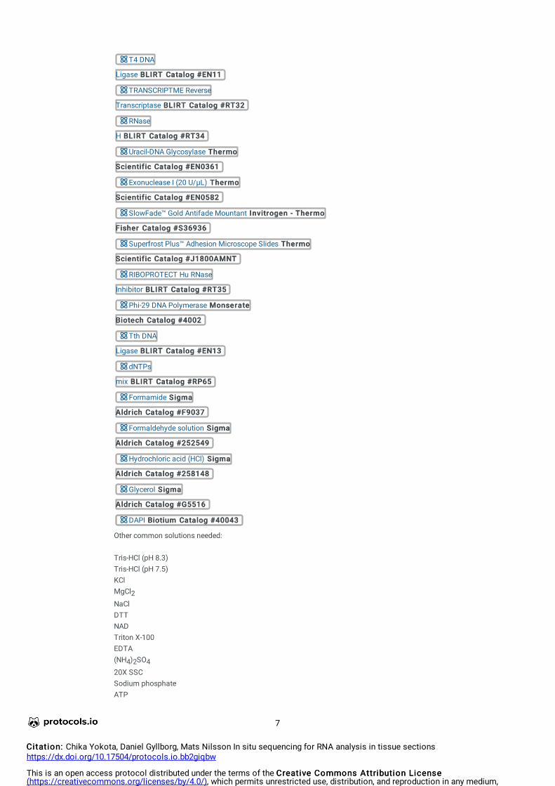
T4 DNA
Ligase BLIRTBLIRT Catalog #Catalog #EN11EN11
TRANSCRIPTME Reverse
Transcriptase BLIRTBLIRT Catalog #Catalog #RT32RT32
RNase
H BLIRTBLIRT Catalog #Catalog #RT34RT34
Uracil-DNA Glycosylase ThermoThermo
ScientificScientific Catalog #Catalog #EN0361EN0361
Exonuclease I (20 U/µL) ThermoThermo
ScientificScientific Catalog #Catalog #EN0582EN0582
SlowFade™ Gold Antifade Mountant Invitrogen - ThermoInvitrogen - Thermo
FisherFisher Catalog #Catalog #S36936S36936
Superfrost Plus™ Adhesion Microscope Slides ThermoThermo
ScientificScientific Catalog #Catalog #J1800AMNTJ1800AMNT
RIBOPROTECT Hu RNase
Inhibitor BLIRTBLIRT Catalog #Catalog #RT35RT35
Phi-29 DNA Polymerase MonserateMonserate
BiotechBiotech Catalog #Catalog #40024002
Tth DNA
Ligase BLIRTBLIRT Catalog #Catalog #EN13EN13
dNTPs
mix BLIRTBLIRT Catalog #Catalog #RP65RP65
Formamide SigmaSigma
AldrichAldrich Catalog #Catalog #F9037F9037
Formaldehyde solution SigmaSigma
AldrichAldrich Catalog #Catalog #252549252549
Hydrochloric acid (HCl) SigmaSigma
AldrichAldrich Catalog #Catalog #258148258148
Glycerol SigmaSigma
AldrichAldrich Catalog #Catalog #G5516G5516
DAPI BiotiumBiotium Catalog #Catalog #4004340043
Other common solutions needed:
Tris-HCl (pH 8.3)Tris-HCl (pH 7.5)KClMgCl2NaClDTTNADTriton X-100EDTA(NH4)2SO420X SSCSodium phosphateATP
7
Citation:Citation: Chika Yokota, Daniel Gyllborg, Mats Nilsson In situ sequencing for RNA analysis in tissue sectionshttps://dx.doi.org/10.17504/protocols.io.bb2giqbw
This is an open access protocol distributed under the terms of the Creative Commons Attribution LicenseCreative Commons Attribution License(https://creativecommons.org/licenses/by/4.0/), which permits unrestricted use, distribution, and reproduction in any medium,provided the original author and source are credited

See safety data sheets for proper chemical handling and waste disposal.
FormamideHandle with proper attire including gloves and eye protection. Work under fume hood when handling solution.
Hydrochloric acid (HCl)Highly corrosive. Handle with proper attire including gloves and eye protection. Work under fume hood when handling solution.
Formaldehyde/paraformaldehyde (PFA)Known carcinogen. Handle with proper attire including gloves and eye protection. Work under fume hood when handling solution.
DEPC (diethyl pyrocarbonate)Harmful, use personal protective equipment.
This working protocol has been setup to work on fresh frozen mouse brain tissue. Other tissue and other species might require different pretreatment conditions.
Enzyme Buffer solutions:Enzyme buffer solutions can be prepared prior to experiment and stored at -20°C.
Reverse Transcriptase Buffer (10x):500 mM Tris-HCl (pH 8.3)750 mM KCl30 mM MgCl2100 mM DTT
Tth Ligase Buffer (10x):200 mM Tris-HCl (pH 8.3)250 mM KCl100 mM MgCl25 mM NAD0.1% Triton X-100
Phi29 Buffer (10x):500 mM Tris-HCl (pH 8.3)100 mM MgCl2100 mM (NH4)2 SO4
T4 Ligase Buffer500 mM Tris-HCl (pH 7.5)100 mM MgCl2100 mM DTT
UNG Buffer (10x)200 mM Tris-HCl pH 8.010 mM EDTA100 mM NaCl
Other Buffers and solutions:
Hybridization Buffer (2x):
8
Citation:Citation: Chika Yokota, Daniel Gyllborg, Mats Nilsson In situ sequencing for RNA analysis in tissue sectionshttps://dx.doi.org/10.17504/protocols.io.bb2giqbw
This is an open access protocol distributed under the terms of the Creative Commons Attribution LicenseCreative Commons Attribution License(https://creativecommons.org/licenses/by/4.0/), which permits unrestricted use, distribution, and reproduction in any medium,provided the original author and source are credited

4x SSC40% FormamideNuclease-free water
(Note: Small stock can be made and stored at room temperature in the dark)
Phosphate buffered saline (1x PBS)137 mM NaCl10 mM sodium phosphate2.7 mM KClDEPC-ddH2O pH 7.4
DEPC-PBS-Tween (DEPC-PBS-T) (see Note 8Note 8)1x PBS0.05% Tween-20
DEPC-PBS (DEPC-PBS) (see Note 8Note 8)1x PBS
Tissue section sample preparation
1 Fresh frozen tissue samples embedded in OCT compound and stored at -80°C. Tissue is cryosectioned at 10 μm and collected on Superfrost Slides and can be stored at -80°C until fixation and start of procedure.(see Note 9Note 9)
2 Slides are taken from -80°C and left at room temperature (RT) for 3 min to air dry.
00:03:0000:03:00 Thawing Thawing
Fixation performed with freshly prepared 3% (w/v) paraformaldehyde in DEPC-PBS for 5 min at RT.PFA solution is applied directly on top of the section.(see Note 10Note 10)
00:05:0000:05:00 Fixation Fixation
Safety precautions: paraformaldehyde
3 Sections rinsed with DEPC-PBS two times (x2).
4 The tissue is permeabilized with 0.1 M HCl (in H2O) at RT for 5 min.
Glass slide is dipped in a ~20 ml solution of 0.1 M HCl.
Safety precautions: HCl
9
Citation:Citation: Chika Yokota, Daniel Gyllborg, Mats Nilsson In situ sequencing for RNA analysis in tissue sectionshttps://dx.doi.org/10.17504/protocols.io.bb2giqbw
This is an open access protocol distributed under the terms of the Creative Commons Attribution LicenseCreative Commons Attribution License(https://creativecommons.org/licenses/by/4.0/), which permits unrestricted use, distribution, and reproduction in any medium,provided the original author and source are credited
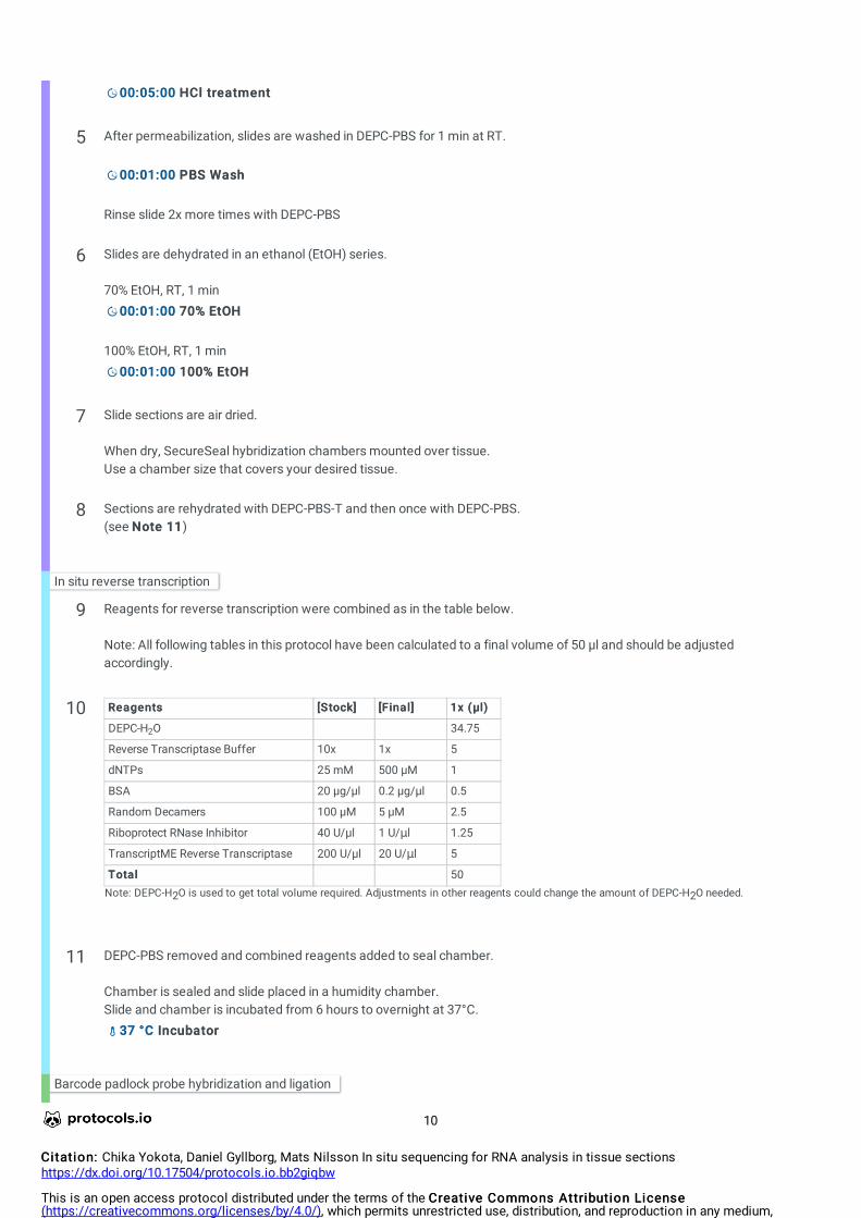
00:05:0000:05:00 HCl treatment HCl treatment
5 After permeabilization, slides are washed in DEPC-PBS for 1 min at RT.
00:01:0000:01:00 PBS Wash PBS Wash
Rinse slide 2x more times with DEPC-PBS
6 Slides are dehydrated in an ethanol (EtOH) series.
70% EtOH, RT, 1 min00:01:0000:01:00 70% EtOH 70% EtOH
100% EtOH, RT, 1 min00:01:0000:01:00 100% EtOH 100% EtOH
7 Slide sections are air dried.
When dry, SecureSeal hybridization chambers mounted over tissue. Use a chamber size that covers your desired tissue.
8 Sections are rehydrated with DEPC-PBS-T and then once with DEPC-PBS.(see Note 11Note 11)
In situ reverse transcription
9 Reagents for reverse transcription were combined as in the table below.
Note: All following tables in this protocol have been calculated to a final volume of 50 µl and should be adjusted accordingly.
10 ReagentsReagents [Stock][Stock] [Final][Final] 1x (µl)1x (µl)
DEPC-H O 34.75
Reverse Transcriptase Buffer 10x 1x 5
dNTPs 25 mM 500 µM 1
BSA 20 µg/µl 0.2 µg/µl 0.5
Random Decamers 100 µM 5 µM 2.5
Riboprotect RNase Inhibitor 40 U/µl 1 U/μl 1.25
TranscriptME Reverse Transcriptase 200 U/µl 20 U/μl 5
TotalTotal 50
2
Note: DEPC-H2O is used to get total volume required. Adjustments in other reagents could change the amount of DEPC-H2O needed.
11 DEPC-PBS removed and combined reagents added to seal chamber.
Chamber is sealed and slide placed in a humidity chamber.Slide and chamber is incubated from 6 hours to overnight at 37°C.
37 °C37 °C Incubator Incubator
Barcode padlock probe hybridization and ligation
10
Citation:Citation: Chika Yokota, Daniel Gyllborg, Mats Nilsson In situ sequencing for RNA analysis in tissue sectionshttps://dx.doi.org/10.17504/protocols.io.bb2giqbw
This is an open access protocol distributed under the terms of the Creative Commons Attribution LicenseCreative Commons Attribution License(https://creativecommons.org/licenses/by/4.0/), which permits unrestricted use, distribution, and reproduction in any medium,provided the original author and source are credited

12 Reverse transcription reagents are removed and postfixation performed with 3% (w/v) paraformaldehyde in DEPC-PBS for 30 min at RT.
00:30:0000:30:00 PFA fixation PFA fixation
13 Wash twice with DEPC-PBS-T.
14 Reagents for padlock hybridization are combined as in the table below.
15ReagentReagent [Stock][Stock] [Final][Final] 1x (µl)1x (µl)
DEPC-H O 22
Tth Ligase buffer 10x 1x 5
KCl 1M 0.05 M 2.5
Formamide 100% 20% 10
Padlock Probe 0.5 µM 10 nM each 1
BSA 20 µg/µl 0.2 µg/µl 0.5
Tth Ligase 5 U/µl 0.5 U/µl 5
RNaseH 5 U/µl 0.4 U/µl 4
TotalTotal 50
2
Note: DEPC-H2O is used to get total volume required. Adjustments in other reagents could change the amount of DEPC-H2O needed.
Safety precautions: formamide
16 DEPC-PBS-T removed and combined reagents added to seal chamber.
Chamber is sealed and placed in a humidity chamber.
Slide and chamber is incubated for 30 min at 37°C.Enzymes and temperature (see Note 12Note 12)
37 °C37 °C
00:30:0000:30:00
Then switched to incubation at 45°C for 1 hour.45 °C45 °C
01:00:0001:00:00
17 Then the slide is washed twice with DEPC-PBS-T.
Rolling circle amplification (RCA)
11
Citation:Citation: Chika Yokota, Daniel Gyllborg, Mats Nilsson In situ sequencing for RNA analysis in tissue sectionshttps://dx.doi.org/10.17504/protocols.io.bb2giqbw
This is an open access protocol distributed under the terms of the Creative Commons Attribution LicenseCreative Commons Attribution License(https://creativecommons.org/licenses/by/4.0/), which permits unrestricted use, distribution, and reproduction in any medium,provided the original author and source are credited
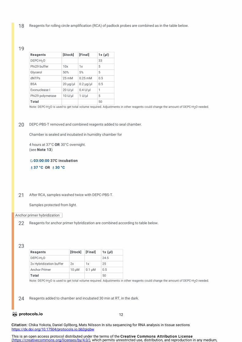
18 Reagents for rolling circle amplification (RCA) of padlock probes are combined as in the table below.
19ReagentsReagents [Stock][Stock] [Final][Final] 1x (µl)1x (µl)
DEPC-H O 33
Phi29 buffer 10x 1x 5
Glycerol 50% 5% 5
dNTPs 25 mM 0.25 mM 0.5
BSA 20 µg/µl 0.2 µg/µl 0.5
Exonuclease I 20 U/µl 0.4 U/µl 1
Phi29 polymerase 10 U/µl 1 U/µl 5
TotalTotal 50
2
Note: DEPC-H2O is used to get total volume required. Adjustments in other reagents could change the amount of DEPC-H2O needed.
20 DEPC-PBS-T removed and combined reagents added to seal chamber.
Chamber is sealed and incubated in humidity chamber for
4 hours at 37°C OR OR 30°C overnight.(see Note 13Note 13)
03:00:0003:00:00 37C incubation 37C incubation
37 °C37 °C OROR 30 °C30 °C
21 After RCA, samples washed twice with DEPC-PBS-T.
Samples protected from light.
Anchor primer hybridization
22 Reagents for anchor primer hybridization are combined according to table below.
23ReagentsReagents [Stock][Stock] [Final][Final] 1x (µl)1x (µl)
DEPC-H O 24.5
2x Hybridization buffer 2x 1x 25
Anchor Primer 10 µM 0.1 µM 0.5
TotalTotal 50
2
Note: DEPC-H2O is used to get total volume required. Adjustments in other reagents could change the amount of DEPC-H2O needed.
24 Reagents added to chamber and incubated 30 min at RT, in the dark.
12
Citation:Citation: Chika Yokota, Daniel Gyllborg, Mats Nilsson In situ sequencing for RNA analysis in tissue sectionshttps://dx.doi.org/10.17504/protocols.io.bb2giqbw
This is an open access protocol distributed under the terms of the Creative Commons Attribution LicenseCreative Commons Attribution License(https://creativecommons.org/licenses/by/4.0/), which permits unrestricted use, distribution, and reproduction in any medium,provided the original author and source are credited

00:30:0000:30:00 Anchor Incubation Anchor Incubation
25 Slide is washed twice with DEPC-PBS-T.
Sequence by ligation (SBL)
26 Reagents for base library hybridization and ligation are combined according to table below.
27ReagentsReagents [Stock][Stock] [Final][Final] 1x (11x (1
µl)µl)
DEPC-H O 39.25
T4 Ligase buffer 10x 1x 5
ATP 25 mM 1 mM 2
Base #A 10 µM 0.1 µM 0.5
Base #C 10 µM 0.1 µM 0.5
Base #T 10 µM 0.1 µM 0.5
Base #G 10 µM 0.1 µM 0.5
DAPI 100 μg/ml 0.5 μg/ml 0.25
BSA 20 µg/µl 0.2 µg/µl 0.5
T4 DNA Ligase 5 U/µl 0.1 U/µl 1
TotalTotal 50
2
Note: DEPC-H2O is used to get total volume required. Adjustments in other reagents could change the amount of DEPC-H2O needed.
28 DEPC-PBS-T removed and combined reagents added to seal chamber.
Slide is incubated 1 hour at RT in the dark.
01:00:0001:00:00 SBL SBL
29 Slide is washed twice with DEPC-PBS-T.
30 Dip slides into 70% EtOH solution and incubate for 1 min.
00:01:0000:01:00 70% EtOH 70% EtOH
31 Remove the SecureSeal chamber and dip slides in 100% for 1 min(see Note 14Note 14)
00:01:0000:01:00 100% EtOH 100% EtOH
13
Citation:Citation: Chika Yokota, Daniel Gyllborg, Mats Nilsson In situ sequencing for RNA analysis in tissue sectionshttps://dx.doi.org/10.17504/protocols.io.bb2giqbw
This is an open access protocol distributed under the terms of the Creative Commons Attribution LicenseCreative Commons Attribution License(https://creativecommons.org/licenses/by/4.0/), which permits unrestricted use, distribution, and reproduction in any medium,provided the original author and source are credited

Air dry the slides.
32 Apply small amount (~10 µL) of mounting medium (Slowfade or similar) and apply a cover slip.(see Note 15Note 15)
Slide can be stored in the dark and at +4°C for longer term storage before imaging.
Imaging
33 Use a standard epifluorescent microscope with 20x objective.
Use DAPI staining to focus on section as a reference point to visualize RCA products.
For details of microscope setup, check referenced publications.
34 Acquire images as needed using reference points that can be used for sequential imaging of other bases later on.
RCPs are located in different focal planes, therefore, a series of images should be captured at different focal depths. The stacks of images are then merged to a single image using the maximum-intensity projection.
Stripping of anchor and base
35 After every round of base imaging, fluorophores and probes need to be stripped of the rolling circle products in order to hybridize the next base.
Remove coverslip by dipping slide into 70% EtOH until it slips off.
Note: After second and all subsequent rounds of imaging, cover slip can be removed by dipping slide into PBS, dehydration step is not needed if hydrophobic pen is used.
36 Dip slide into 100% EtOH for 1 min and then air dry the samples.
00:01:0000:01:00 100% EtOH 100% EtOH
37 Use a hydrophobic pen to draw a barrier around section.
Alternatively, mount another SecureSeal chamber.
38 Rehydrate with DEPC-PBS-T.
39 Combine reagents in the table below for the stripping of anchor and sequence primer.
40
14
Citation:Citation: Chika Yokota, Daniel Gyllborg, Mats Nilsson In situ sequencing for RNA analysis in tissue sectionshttps://dx.doi.org/10.17504/protocols.io.bb2giqbw
This is an open access protocol distributed under the terms of the Creative Commons Attribution LicenseCreative Commons Attribution License(https://creativecommons.org/licenses/by/4.0/), which permits unrestricted use, distribution, and reproduction in any medium,provided the original author and source are credited

ReagentReagent [Stock][Stock] [Final][Final] 1x1x(µl)(µl)
DEPC-H O 43.5
UNG buffer 10x 1x 5
BSA 20 µg/µl 0.2 µg/µl 0.5
UNG 1 U/µl 0.02 U/µl 1
TotalTotal 50
2
Note: DEPC-H2O is used to get total volume required. Adjustments in other reagents could change the amount of DEPC-H2O needed.
41 Reagents added to section and incubated at RT for 30 min.
00:30:0000:30:00
42 Sections washed once with DEPC-PBS-T
43 Sections washed with 100% formamide at RT for 3 min.
00:03:0000:03:00 1st Wash 1st Wash
Repeat an additional two times for a total of 3 times.
00:03:0000:03:00 2nd Wash 2nd Wash
00:03:0000:03:00 3rd Wash 3rd Wash
44Sections washed twice with DEPC-PBS-T at RT
Repeat hybridization of anchor and next base.
45 Repeat Steps 22-44Steps 22-44 for additional required bases.
Note: If using hydrophobic pen, chamber will not be used and solutions can just be applied on top of section during incubations.
Image analysis
46 For most up to date methodolgy on image analysis, we refer to a recent publication from our group:
Probabilistic cell typing enables fine mapping of closely related cell types in situQian X, Harris KD, Hauling T, Nicoloutsopoulos D, Muñoz-Manchado AB, Skene N, Hjerling-Leffler J, Nilsson MNat Methods. 2020 Jan;17(1):101-106.doi: 10.1038/s41592-019-0631-4
We also refer to the publication this protocol is based on for image analysis:
In situ sequencing for RNA analysis in preserved tissue and cells.
15
Citation:Citation: Chika Yokota, Daniel Gyllborg, Mats Nilsson In situ sequencing for RNA analysis in tissue sectionshttps://dx.doi.org/10.17504/protocols.io.bb2giqbw
This is an open access protocol distributed under the terms of the Creative Commons Attribution LicenseCreative Commons Attribution License(https://creativecommons.org/licenses/by/4.0/), which permits unrestricted use, distribution, and reproduction in any medium,provided the original author and source are credited

Ke R, Mignardi M, Pacureanu A, Svedlund J, Botling J, Wählby C, Nilsson M. Nat Methods. 2013 Sep;10(9):857-60.doi: 10.1038/nmeth.2563
(Cell profiler: see Note16Note16)
16
Citation:Citation: Chika Yokota, Daniel Gyllborg, Mats Nilsson In situ sequencing for RNA analysis in tissue sectionshttps://dx.doi.org/10.17504/protocols.io.bb2giqbw
This is an open access protocol distributed under the terms of the Creative Commons Attribution LicenseCreative Commons Attribution License(https://creativecommons.org/licenses/by/4.0/), which permits unrestricted use, distribution, and reproduction in any medium,provided the original author and source are credited