in situ ultra-high vacuum transmission electron...
TRANSCRIPT
It
Ja
b
a
ARRA
KOTICCC
1
fsr(htoosasJ1castt
g
0d
Micron 43 (2012) 1195–1210
Contents lists available at SciVerse ScienceDirect
Micron
j our na l ho me p age: www.elsev ier .com/ locate /micron
n situ ultra-high vacuum transmission electron microscopy studies of theransient oxidation stage of Cu and Cu alloy thin films
udith C. Yanga,∗, Guangwen Zhoub
Department of Chemical and Petroleum Engineering, Department of Physics, University of Pittsburgh, 1249 Benedum Hall, 3700 O’Hara St., Pittsburgh, PA 15261, USADepartment of Mechanical Engineering, State University of New York, PO Box 6000, Binghamton, NY 13902-6000, USA
r t i c l e i n f o
rticle history:eceived 31 July 2011eceived in revised form 9 February 2012ccepted 9 February 2012
eywords:xidation
a b s t r a c t
Because environmental stability is an essential property of most engineered materials, many theoriesexist to explain oxidation mechanisms. Yet, nearly all classical oxidation theories assume a uniformgrowing film, where structural changes were not considered because of the previous lack of experimentalprocedure to visualize this non-uniform growth in conditions that allowed for highly controlled surfacesand impurities. With the advent of vacuum technologies and advances in microcopy techniques, espe-cially in situ, one can now see structural changes under controlled surface conditions. Here, we present
EMn situuu–Niu–Au
a review of our systematic studies on the transient oxidation stages of a model metal system, Cu, andits alloys, Cu–Au and Cu–Ni, by in situ ultra-high vacuum transmission electron microscopy (UHV-TEM).The dependence of the oxidation behavior on the crystal orientation, oxygen pressure, temperature andalloying is attributed to the structures of the oxygen-chemisorbed layer, oxygen surface diffusion, sur-face energy and the interfacial strain energy. Heteroepitaxial concepts, developed to explain thin filmformation on a dissimilar substrate material (e.g., Ge on Si), described well these initial oxidation stages.
. Introduction
Oxygen in the Earth’s atmosphere reacts with metal surfacesorming oxides that are brittle and spall off, leaving bare metalurface for further deterioration. Addressing the problems of cor-osion costs the US a few percent of its gross national productGNP) per year. Fundamental understanding of metal oxidationas therefore received extensive interest owning to its significantechnical importance. The general sequence of metal oxidation isxygen chemisorption, nucleation and growth of oxide, and bulkxide growth. The oxygen surface chemisorption has been exten-ively studied by surface science methods that mostly examine thedsorption of up to ∼1 monolayer of oxygen with particular empha-is on the structures of adsorbed phases (Besenbacher, 1993, 1996;enkins, 2006; Qin et al., 2008; Yagyu et al., 2009; Zhukov et al.,999). On the other hand, the growth of continuous oxide layerontrolled by parabolic, logarithmic, or other kinetic mechanismst the later stages of oxidation has been observed on many metals
ystems, and models based on the transport of ionic species throughhe thicker and continuous oxide film has been proposed to explainhe observed trends (Birks and Meier, 1983). Hence, the least∗ Corresponding author. Tel.: +1 412 624 8613; fax: +1 412 624 9639.E-mail addresses: [email protected], [email protected] (J.C. Yang),
[email protected] (G. Zhou).
968-4328/$ – see front matter © 2012 Elsevier Ltd. All rights reserved.oi:10.1016/j.micron.2012.02.007
© 2012 Elsevier Ltd. All rights reserved.
well-understood regime in metal oxidation is the transient oxida-tion stage, i.e., from the nucleation to the growth of metal oxides.Some simple pure metals such as Ni, Al, Cu, and Pb have beenselected as model systems to understand the reaction mecha-nism in the early stages of oxidation by different techniques overa wide range of pressures and temperatures (Brune et al., 1992;Christensen et al., 1986; Holloway and Hudson, 1974; Jacobsenet al., 1995; Kennett and Lee, 1975; Orr, 1962; Thurmer et al.,2002; Vaquila et al., 1993; Yang et al., 1998a,b). The report fromthese studies that the initial stages of metal oxidation typicallyinvolve the nucleation and growth of oxide islands, rather thanthe formation and thickening of a continuous film, representsa critical departure from previously held assumptions in clas-sic Cabrera–Mott and Wagner theories regarding metal oxidation(Cabrera and Mott, 1948; Wagner, 1933).
To exemplify the structural changes of the initial stages ofoxidation and its sensitivity to the environment, we studied theinitial oxidation stages of Cu and its alloys by in situ UHV-TEM.We selected Cu as the model system, since it has a long historyof being studied as a model metal system for oxidation (Cabreraand Mott, 1948; Lawless and Mitchell, 1965; Lawless, 1974; Milneand Howie, 1984; Young et al., 1956). Our study of Cu thin film
oxidation was later extended to the cases of Cu–Au(1 0 0) andCu–Ni(1 0 0) surfaces in order to seek the effects of alloying on theoxide formation (Bharadwaj and Yang, 2001; Luo et al., 2011; Yanget al., 1998a,b,c, 1999,2009, 2002; Yang and Yeadon, 1997; Zhou,
1 icron 4
22tasCdodggSteotbfptoktdfop
2
fiecpstCfiNwisbttitogouipiotn
sob3eZ
196 J.C. Yang, G. Zhou / M
009a,b,c, 2010; Zhou et al., 2005a,b, 2006,2008; Zhou and Yang,002, 2003a,b, 2004, 2005). In this review paper, we first presenthe oxidation kinetics and oxide morphology from Cu(1 0 0), (1 1 0)nd (1 1 1) oxidation as function of temperature and oxidizing pres-ures (including up to 1 atm), and then compare to the oxidation ofu–Au(1 0 0) and Cu–Ni(1 0 0). We find these initial stages of oxi-ation to be a very rich and elegant regime to study. Our studiesn the oxidation of Cu thin films demonstrate that oxygen surfaceiffusion is the primary mechanism of transport, nucleation androwth, in contrast to, for example, bulk diffusion of Cu or oxy-en diffusion into the Cu film and oxide nucleation within the film.ince initial stage of oxidation is a surface process, factors that affecthe surface impact the oxidation rate and the oxide morphologyvolution. A significant effect of temperature is to create differentxide nanostructures, due to temperature-dependent changes inhe interfacial strain and properties. This bears a striking resem-lance to heteroepitaxy where interfacial strain is the influentialactor in thin film growth and nano-shapes. Increasing oxidationressure shows increased oxidation kinetics, as well as a transi-ion from epitaxial to polycrystalline oxide formation at a criticalxidizing pressure. Alloying dramatically affects both the oxidationinetics and the morphology of the oxide islands. For Cu–Au oxida-ion, the oxidation mechanisms change where the Cu2O reveals aendritic growth. For Cu–Ni oxidation, the addition of Ni causes theormation Cu2O and/or NiO where the oxide type(s) and its relativerientation with the film depend on the Ni concentration, oxygenartial pressure and temperature.
. Materials and methods
Our in situ oxidation experiments were carried out in a modi-ed JEOL 200CX TEM (McDonald et al., 1989). This microscope isquipped with a UHV chamber with base pressure ∼10−8 Torr. Aontrolled leak valve attached to the column of the microscopeermits introduction of gases directly into the microscope. Pres-ure was monitored by an ion gauge mounted inside the column ofhe microscope near the sample region. Single crystals of Cu(1 0 0),u(1 1 0), Cu(1 1 1) as well as Cu–Au(1 0 0) and Cu–Ni(1 0 0) thinlms with thickness of 600–1000 A were grown on single crystalaCl by sputtering deposition or e-beam evaporation. The filmsere removed from the NaCl substrate by flotation in deion-
zed water, washed and mounted on a specially designed TEMample holder that allows for resistive heating at temperaturesetween room temperature and 1000 ◦C. The relationship betweenhe power input to the Si TEM sample holder and the tempera-ure was calibrated using a pyrometer. Oxygen gas can be admittednto the column of the microscope through the leak valve at a par-ial pressure (pO2) between 5 × 10⁻⁵ Torr and 760 Torr. Real timebservations can be made at pressures ≤8 × 10⁻⁴ Torr. To investi-ate the effects of the electron, the electron beam was shut off, thexidation reaction was carried out for a few minutes, the TEM col-mn was evacuated, and then the electron beam was turned on for
maging. This process was repeated to create a sequential oxidationrocedure in order to study the effect of the electron beam on the
n situ oxidation. The effect of the electron beam is to accelerate thexidation process but not the morphological developments, thushe mechanisms developed from in situ observations are valid andot an electron beam effect.
The Cu, Cu–Au and Cu–Ni films formed a native oxide on theurface due to air exposure. Before oxidation in situ, the nativexide of the Cu and the Cu–Au films was reduced inside the TEM
y annealing in methanol vapor or Ar plus 2% H2 environment at50 ◦C at a pressure of 5 × 10⁻⁴ Torr, as confirmed by selected arealectron diffraction (SAD) (Wang et al., 2006; Yang et al., 1998a,b;hou et al., 2007a). The Cu–Ni film was reduced by annealing the3 (2012) 1195–1210
Cu–Ni films at 750 ◦C within an Ar plus 2% H2 atmosphere at a pres-sure of ∼1 × 10⁻³ Torr as was confirmed by SAD (Yang et al., 2009).To oxidize the Cu and Cu alloy films, scientific grade oxygen gas(99.999% purity) was introduced into the TEM chamber.
The Cu–Ni oxidation study also used a controlled environmentsystem constructed for use at the synchrotron X-ray scatteringtechniques that provide complementary information over a widerange of pO2 and temperature. Oxidizing and reducing environ-ments are created by mixing purified gases Ar, O2, CO, CO2, H2and the temperature can achieve up to 1000 ◦C (Eastman et al.,2005). Epitaxial (0 0 1)CuxNi1−x thin film samples with thicknessesof 110–200 nm were grown by electron-beam evaporation ontoSrTiO3 single crystal (0 0 1) substrates. The samples were annealedtypically for 1 h at 840 ◦C in 5 × 10⁻⁴ Torr of Ar–2% H2 and then werecooled to the desired temperature for oxidation studies in situ.
3. Results and discussion
3.1. Oxidation of Cu
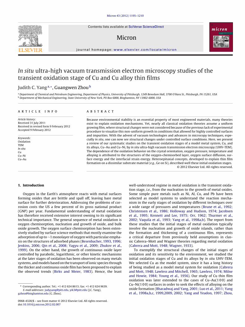
3.1.1. Nucleation and growth of Cu2O islands on Cu(1 0 0)Our studies of Cu(1 0 0) oxidation reveal an oxygen surface dif-
fusion as opposed to a Cu or oxygen bulk diffusion mechanism fortransport, nucleation and growth of the oxide island. Fig. 1a is asequence of TEM dark-field (DF) images taken from Cu2O(1 1 0)reflection, showing the nucleation events of Cu2O islands in theoxidation of Cu(1 0 0) surfaces at 350 ◦C and the oxygen pressure of5 × 10−4 Torr (see Supplemental information for the correspondingin situ video). It should be noted that Cu2O, not CuO, was observedto form under all oxidation conditions studied in this work. Ther-modynamically, CuO could only form after a continuous Cu2O iscreated, i.e., CuO cannot form directly on Cu; since our focus is on theinitial oxidation prior to the oxide scale formation, only Cu2O wasobserved in agreement with thermodynamics. After the introduc-tion of oxygen gas, no oxide islands appear within the first coupleof minutes; the smallest oxide nuclei observable was ∼1 nm. Theoxide islands then nucleate rapidly, reach a saturation number den-sity and followed by the island growth. The observation that thenumber density of oxide nuclei saturates suggests that the nucle-ation process is limited by oxygen surface diffusion (Yang et al.,1998b), i.e., an active zone of oxygen capture exists around eachisland, and the radius of this capture zone is proportional to theoxygen surface diffusion rate. The probability of a nucleation eventis proportional to the fraction of available surface area outside thesezones of oxygen capture. The saturation island density is reachedwhen the zones of oxygen capture of neighboring islands impingeon each other. The equation describing this oxygen surface diffusionbehavior for the nucleation of copper oxides:
N = 1
L2d
(1 − e−kL2d
t) (1)
The initial nucleation rate is equal to k, and the saturation islanddensity is 1/L2
d. Fig. 1b shows the number density of the oxide
islands with respect to the oxidation time, where the solid linecorresponds to the fitting to the oxygen surface diffusion limitednucleation processes.
Due to larger surface mobility of oxygen at higher temperatures,the radius of oxygen capture zone increases with temperature andthe attachment of oxygen to existing islands is more favorable thannucleation of new nuclei. Therefore, the dependence of the satura-
tion island density, NS, on oxidation temperature should also followan Arrhenius relationship (Yang et al., 1998b):Ns∼eEN /kT (2)
J.C. Yang, G. Zhou / Micron 43 (2012) 1195–1210 1197
Fig. 1. (a) Cu(2 0 0) dark field image after oxygen is introduced into the microscope at 5 × 10⁻⁴ Torr oxygen and 350 ◦C, (b) nucleation density as function of oxidation timeand the solid line is the theoretical fit to an oxygen surface diffusion model, and (c) cross-sectional area of oxide islands which fits a t0.33 growth rate where the solid linec
wanros2taFte
faafipdtfwHsagi
orresponds to the oxygen surface diffusion/direct impingement model.
here k is Boltzmann constant, T is the temperature, and EN is thectivation energy for the nucleation of oxide islands. It should beoted that this is a homogeneous nucleation model that assumesandom selection of nucleation sites. Nearly all of the in situ Cuxidation experiments seem to agree with this assumption, whereurface steps (Yang et al., 1999) and dislocations (Zhou et al.,005b) were not clearly preferential nucleation sites. However ifhe defects were significant, such as surface faceting, grain bound-ries or edge of a hole, then the oxides formed along the defects.or example, Cu(1 1 0) forms surface facets above T � 750 ◦C, wherehe oxide grew as nanorods along the valleys of the facets (Zhout al., 2005b).
The evolution of individual islands during growth can also beollowed in situ. Fig. 1c shows the quantitatively measured islandreas as a function of oxidation time when Cu(1 0 0) is exposed ton oxygen pressure of 5 × 10⁻⁴ Torr at 350 ◦C. The best power lawt to the island growth is t1.3, which is slightly higher than t—theredicted power law dependence for 3D growth by oxygen surfaceiffusion (Yang et al., 1997). To account for the slight deviation fromhe linear growth, we included the direct impingement or bulk dif-usion of oxygen that contributes to the island growth, which fitsith the experimental data (Yang et al., 1997), as shown in Fig. 1c.ence, these in situ studies of Cu oxidation revealed that oxygen
urface diffusion is the primary mechanism of transport, nucleationnd growth during the initial oxidation stage; as the oxide islandsrow larger, the oxygen atoms that land on the surface of the oxidesland also contribute to its growth.
3.1.2. Effect of surface orientation on the oxide formation:oxidation of Cu(1 1 0) and Cu(1 1 1)
Previous investigators have elegantly demonstrated thatCu(1 0 0) and (1 1 0) surfaces are unreconstructed, and then trans-form into “missing-row” or “adding-row” reconstruction whenexposed to oxygen (Jacobsen and Norskov, 1990; Jensen et al.,1990; Robinson et al., 1990). After reconstruction, oxygen diffuseson the reconstructed surface, and nucleation occurs on the recon-structed Cu–O surface. Further arriving oxygen can either nucleatenew oxide islands by reacting with copper atoms or attach to anexisting island, causing growth. Therefore, the surface diffusioncoefficient of oxygen may determine the outcome of the com-petition between nucleation and growth, and, hence, determinesthe number density of stable islands. Qualitatively a larger diffu-sion coefficient for oxygen should yield a lower number densityof stable islands. Since the path length of oxygen surface diffusiondepends on the atomic structure of the substrate plane, differentnucleation behaviors of Cu2O islands is therefore expected for dif-ferent orientations of the Cu. The Cu(1 0 0) has a more close-packedstructure, and is smoother than the corrugated Cu(1 1 0) surface.Similarly, the reconstructed (
√2 × 2
√2)R45◦ O–Cu(1 0 0) surface
has a more compact oxygen chemisorption than (2 × 1)O–Cu(1 1 0)surface which has a corrugated structure. Therefore, it is expected
the activation barrier of surface diffusion of the dissociated oxy-gen be higher on the Cu(1 1 0) surface, and thus have a shorterpath length. The shorter diffusion path length leads to a smallerzone of oxygen capture. If an adatom lands within the zone of
1198 J.C. Yang, G. Zhou / Micron 43 (2012) 1195–1210
Table 1Comparison of the nucleation kinetics in the oxidation of Cu(1 0 0) and Cu(1 1 0) at 350 ◦C and oxygen pressure of 5 × 10⁻⁴ Torr.
Initial nucleation rate(k: �m−2 min−1)
Radius of oxygencapture zone (Ld: �m)
Saturation islanddensity (1/L2
d: �m−2)
Island densitysaturation time (min)
Nucleation activationenergy (Ea: eV)
0.83
9.01
otgoTct1
odt“ciicstocot(
M
HscEssaDdsooitpabaotiisddm
sTeo
Cu(1 0 0) 0.17 1.09
Cu(1 1 0) 1.743 0.33
xygen capture, then adatom will diffuse on the surface and attacho an existing island; an adatom landing outside this zone of oxy-en capture could create its own nucleation site. The smaller zone ofxygen captures results in a higher number density of oxide nuclei.his is confirmed by our results where the active zone of oxygenapture around each island on Cu(1 1 0) is 0.3331 �m for the oxida-ion at 350 ◦C, which is significantly smaller than that on Cu(1 0 0),.09 �m, as shown in Table 1.
Rather than following the process of nucleation and growth ofxide islands as described earlier for Cu(1 0 0) and Cu(1 1 1), the oxi-ation of Cu(1 1 1) shows a very different behavior. As seen in Fig. 2,he oxidation of Cu(1 1 1) surface is via the nucleation and growth ofdiscontinuous-branched” Cu2O overlayer under similar oxidationondition (T = 450 ◦C pO2 = 5 × 10⁻⁴ Torr). Further oxidation resultsn growth of these Cu2O islands where they coalesce to createrregularly connected oxide clusters. With use of a connectivity-hecking algorithm, individual Cu2O clusters can be isolated fortatistical analysis, as shown in Fig. 2a–c (Zhou et al., 2008). Underhe scaling hypothesis, the infinite cluster at the percolation thresh-ld pc has the property of statistical self-similarity, which can behecked by measuring the mass M(L) of the infinite cluster and thatf the backbone with different length scale L. At the percolationhreshold, p = pc, the mass of the spanning cluster scales with L asFender, 1988; Stauffer, 1979):
(L) = LDf(
L
�
)→
{LD for L �LE for L �
(3)
ere � is the correlation length defined as the average root meanquare distance between occupied sites that belong to the sameluster, D is the fractal dimension of the cluster, and E is theuclidean dimension where E = 2 for a two-dimension surface. Ashown in Fig. 2d, the mass densities for the infinite cluster arecale dependent, follow the power law in Eq. (3) with the L �,nd have the fractal dimension of D = 1.71. The fractal dimension
= 1.71 of the infinite oxide cluster is inconsistent with the pre-iction by random percolation, which gives D = 1.9. We speculateuch a random site occupation mechanism does not apply to metalxidation, where the oxide nuclei attract oxygen atoms for thexide growth. This implies that the probability of site occupations neighbor dependent for surface oxidation, and sites adjacent tohe periphery of existing oxide have larger probability to be occu-ied. We use kinetic Monte-Carlo (KMC) simulations to verify thebove speculation (Fig. 2e), where the probability for oxygen atomseing captured by existing oxide islands is larger than the nucle-tion of new islands due to the smaller activation energy for anxygen atom sticking to an existing oxide island. Our KMC simula-ions reveal a fractal dimension of D = 1.75 with L � (Fig. 2f), whichs very close to the experimental fractal dimension (D = 1.71) of thenfinite oxide cluster observed experimentally. Their agreementupports our speculation that the percolating oxide film growthuring Cu(1 1 1) oxidation is related to the processes of neighbor-ependent site occupation of oxygen and supports our nucleationodel of an oxygen capture zone around existing islands.One might expect that the mechanism of neighbor-dependent
ite occupation is typical for oxide growth during metal oxidation.his implies that percolating oxide film growth could be a gen-ral phenomenon for metal oxidation. However, our in situ TEMbservations of the oxidation of Cu(1 0 0), Cu(1 1 0) and Cu(1 1 1)
22 1.4 ± 0.217 1.1 ± 0.2
under similar oxidation conditions reveal that the oxidationof Cu(1 0 0) and (1 1 0) leads to the formation of 3D compactCu2O islands (Zhou and Yang, 2002, 2003a,b, 2004; Zhou et al.,2005a), which obviously deviates from the fashion of percolat-ing oxide growth. These different oxidation behaviors can beattributed to their different oxygen chemisorbed structures. TheO-chemisorption on Cu(1 1 1) results in ordered ‘29’ (
√13R46.1◦ ×
7R21.8◦) and ‘44’ (√
73R5.8◦ ×√
21R − 10.9◦) lattice structures,which comprise distorted hexagonal arrays of O atoms with unitcell areas 29 and 44 times larger than that of the substrate Cu(1 1 1)(Besenbacher and Norskov, 1993; Jensen et al., 1991, 1991, 1992;Matsumoto et al., 2001). The oxygen atoms in the 29 and 44structures have more than one well-defined height with corru-gation up to ∼3.1 A with respect to the Cu surface. Compared tothe chemisorbed structures of (
√2 × 2
√2) R45◦ O–Cu(1 0 0) and
(2 × 1)O–Cu(1 1 0), the oxygen-chemisorbed ‘29’ and ‘44’ structureson Cu(1 1 1) surfaces have much larger surface corrugation, andsuch enhanced surface roughness can greatly inhibit surface diffu-sion of oxygen atoms. Efficient surface and edge diffusion usuallyleads to compact island growth, while sluggish surface diffusioncauses ramified island morphologies. The percolating oxide growthduring Cu(1 1 1) oxidation can be attributed to the restricted surfacediffusion of oxygen due to the highly corrugated O–Cu(1 1 1) surfacestructure, and the compact oxide island growth observed duringthe oxidation of O–Cu(1 0 0) and O–Cu(1 1 0) surfaces is related tothe efficient oxygen surface diffusion due to their smooth surfacestructures.
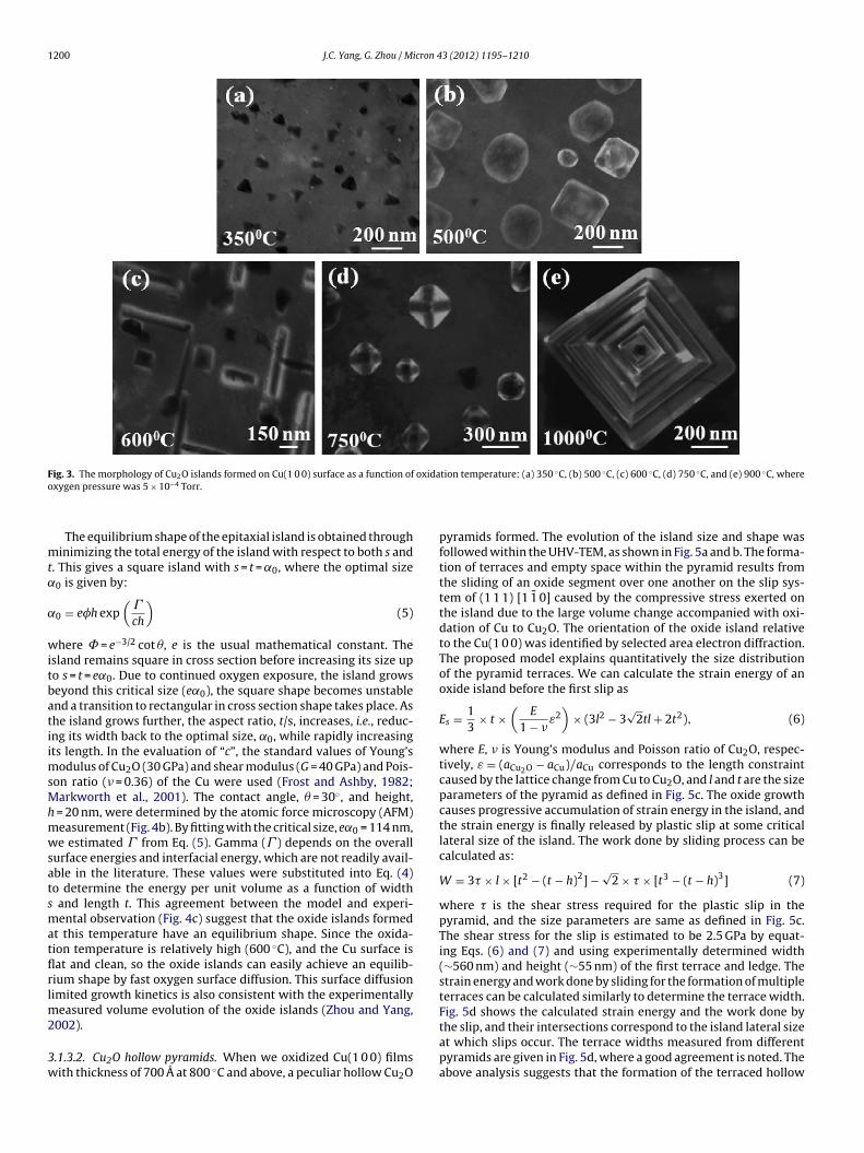
3.1.3. Effect of oxidizing temperature on the oxide morphologyWe systematically investigated the role of oxidizing tempera-
ture and pressure, where we discovered an unusually wide varietyof oxide nanostructures, from nanorods to pyramids to a nearlyuniform oxide layer, by simply changing the oxidation temperature(Fig. 3). These in situ studies also provide insights into nano-oxidesynthesis. Similar to heteroepitaxy, the lattice mismatch causes dif-ferent nanostructures; different shapes are noted including thinfilms, domes or nanorods where the common explanation is thestrain between the thin film and support material (Ross et al., 1998,1999; Tersoff and Tromp, 1993; Tromp and Ross, 2000). We find inthis case that strain does indeed play a critical role in defining theseshapes. The temperature impacts surface and interface energies aswell as mechanical properties, which affect the oxide morphology.Two of the unusual shapes—the nanorod (Zhou and Yang, 2002) andthe hollow pyramid (Zhou et al., 2005a)—were of particular interestand studied in detail.
3.1.3.1. Cu2O nanorods. Elongated Cu2O islands were observed toform on Cu(1 0 0) surfaces from the in situ oxidation in a narrowtemperature regime near 600 ◦C (Zhou and Yang, 2002, 2003b).The island initially has a square-in-cross-section shape and whena critical size (∼110 nm), is reached, the island transitioned to anelongated structure (Fig. 4a). These shapes bear a striking resem-
blance to nanorods observed in a few heteroepitaxial systems suchas Ge nanorods on Si. Tersoff and Tromp (1993) have proposedan energetic model to describe square-elongation shape transitionof epitaxial islands. According to their model, the energy per unit
J.C. Yang, G. Zhou / Micron 43 (2012) 1195–1210 1199
Fig. 2. The Cu2O formed on the Cu(1 1 1) film at the percolation threshold: (a) identification of oxide clusters by a connectivity analysis of the discontinuous oxide film,different clusters are shown by different colors, and the largest one (infinite cluster) has the brightest contrast and spans over the whole image; (b) the infinite cluster singledo 2
L for tb lengthf or-dec is the
v{
wri(
ut from (a). (c) Scaling analysis of the infinite cluster, (d) log–log plot of M(L)/L vs.e 1.71. Note that L is the number of pixels of the image and can be converted into
ormed at the percolation threshold from the KMC simulations based on the neighbluster, the fraction dimension D for the infinite cluster is determined to be 1.75 (L
olume (E/V) of a strained epitaxial island is:
E
V= 2�
(1s
+ 1t
)+ 1
h(ri + rt − rs)
−2ch
[1s
ln
(se3/2
h cot �
)+ 1
tln
(te3/2
h cot �
)],
}(4)
here s, t, and h are the width, length, and height of the island,espectively; � being the contact angle. � contains the surface andnterface energies and contact angle, � = �e csc� − (� t + �s − � i)cot �units J/m2); � t, �s, and �e are the surface energies (per unit area) of
he infinite cluster, the fraction dimension D for the infinite cluster is determined to scale by L(nm) = L(pixel number) × (image size(nm)/1024). (e) The infinite cluster
pendent site occupation mechanism; (f) log–log plot of M(L)/L2 vs. L for the infinitenumber of pixels).
the island’s top, the substrate, and the island’s edge facet, respec-tively; � i is the island-substrate interface energy. The first twoterms in Eq. (4) give the change in surface and interface ener-gies when an island forms on the surface. The substrate-islandlattice mismatch causes the island to exert a force on the substrate,which elastically distorts the substrate. The third term describes theenergy relaxation of the island by the cost of some strain in the sub-
strate, and this relaxation energy is related to c, c = �2b(1 − �)/2�G,
where � and G are the Poisson ratio and shear modulus of the sub-strate, and �b is the island bulk stress (Tersoff and LeGoues, 1994;Tersoff and Tromp, 1993).
1200 J.C. Yang, G. Zhou / Micron 43 (2012) 1195–1210
F oxida ◦ ◦ ◦ ◦ ◦
o
mt˛
˛
witbatiimsMhmwsatsmatflrlm2
3w
ig. 3. The morphology of Cu2O islands formed on Cu(1 0 0) surface as a function ofxygen pressure was 5 × 10⁻⁴ Torr.
The equilibrium shape of the epitaxial island is obtained throughinimizing the total energy of the island with respect to both s and
. This gives a square island with s = t = ˛0, where the optimal size0 is given by:
0 = eh exp(
�
ch
)(5)
here = e−3/2 cot �, e is the usual mathematical constant. Thesland remains square in cross section before increasing its size upo s = t = e˛0. Due to continued oxygen exposure, the island growseyond this critical size (e˛0), the square shape becomes unstablend a transition to rectangular in cross section shape takes place. Ashe island grows further, the aspect ratio, t/s, increases, i.e., reduc-ng its width back to the optimal size, ˛0, while rapidly increasingts length. In the evaluation of “c”, the standard values of Young’s
odulus of Cu2O (30 GPa) and shear modulus (G = 40 GPa) and Pois-on ratio (� = 0.36) of the Cu were used (Frost and Ashby, 1982;arkworth et al., 2001). The contact angle, � = 30◦, and height,
= 20 nm, were determined by the atomic force microscopy (AFM)easurement (Fig. 4b). By fitting with the critical size, e˛0 = 114 nm,e estimated � from Eq. (5). Gamma (� ) depends on the overall
urface energies and interfacial energy, which are not readily avail-ble in the literature. These values were substituted into Eq. (4)o determine the energy per unit volume as a function of width
and length t. This agreement between the model and experi-ental observation (Fig. 4c) suggest that the oxide islands formed
t this temperature have an equilibrium shape. Since the oxida-ion temperature is relatively high (600 ◦C), and the Cu surface isat and clean, so the oxide islands can easily achieve an equilib-ium shape by fast oxygen surface diffusion. This surface diffusionimited growth kinetics is also consistent with the experimentally
easured volume evolution of the oxide islands (Zhou and Yang,
002)..1.3.2. Cu2O hollow pyramids. When we oxidized Cu(1 0 0) filmsith thickness of 700 A at 800 ◦C and above, a peculiar hollow Cu2O
tion temperature: (a) 350 C, (b) 500 C, (c) 600 C, (d) 750 C, and (e) 900 C, where
pyramids formed. The evolution of the island size and shape wasfollowed within the UHV-TEM, as shown in Fig. 5a and b. The forma-tion of terraces and empty space within the pyramid results fromthe sliding of an oxide segment over one another on the slip sys-tem of (1 1 1) [1 1 0] caused by the compressive stress exerted onthe island due to the large volume change accompanied with oxi-dation of Cu to Cu2O. The orientation of the oxide island relativeto the Cu(1 0 0) was identified by selected area electron diffraction.The proposed model explains quantitatively the size distributionof the pyramid terraces. We can calculate the strain energy of anoxide island before the first slip as
Es = 13
× t ×(
E
1 − �ε2
)× (3l2 − 3
√2tl + 2t2), (6)
where E, � is Young’s modulus and Poisson ratio of Cu2O, respec-tively, ε = (aCu2O − aCu)/aCu corresponds to the length constraintcaused by the lattice change from Cu to Cu2O, and l and t are the sizeparameters of the pyramid as defined in Fig. 5c. The oxide growthcauses progressive accumulation of strain energy in the island, andthe strain energy is finally released by plastic slip at some criticallateral size of the island. The work done by sliding process can becalculated as:
W = 3� × l × [t2 − (t − h)2] −√
2 × � × [t3 − (t − h)3] (7)
where � is the shear stress required for the plastic slip in thepyramid, and the size parameters are same as defined in Fig. 5c.The shear stress for the slip is estimated to be 2.5 GPa by equat-ing Eqs. (6) and (7) and using experimentally determined width(∼560 nm) and height (∼55 nm) of the first terrace and ledge. Thestrain energy and work done by sliding for the formation of multipleterraces can be calculated similarly to determine the terrace width.Fig. 5d shows the calculated strain energy and the work done by
the slip, and their intersections correspond to the island lateral sizeat which slips occur. The terrace widths measured from differentpyramids are given in Fig. 5d, where a good agreement is noted. Theabove analysis suggests that the formation of the terraced hollow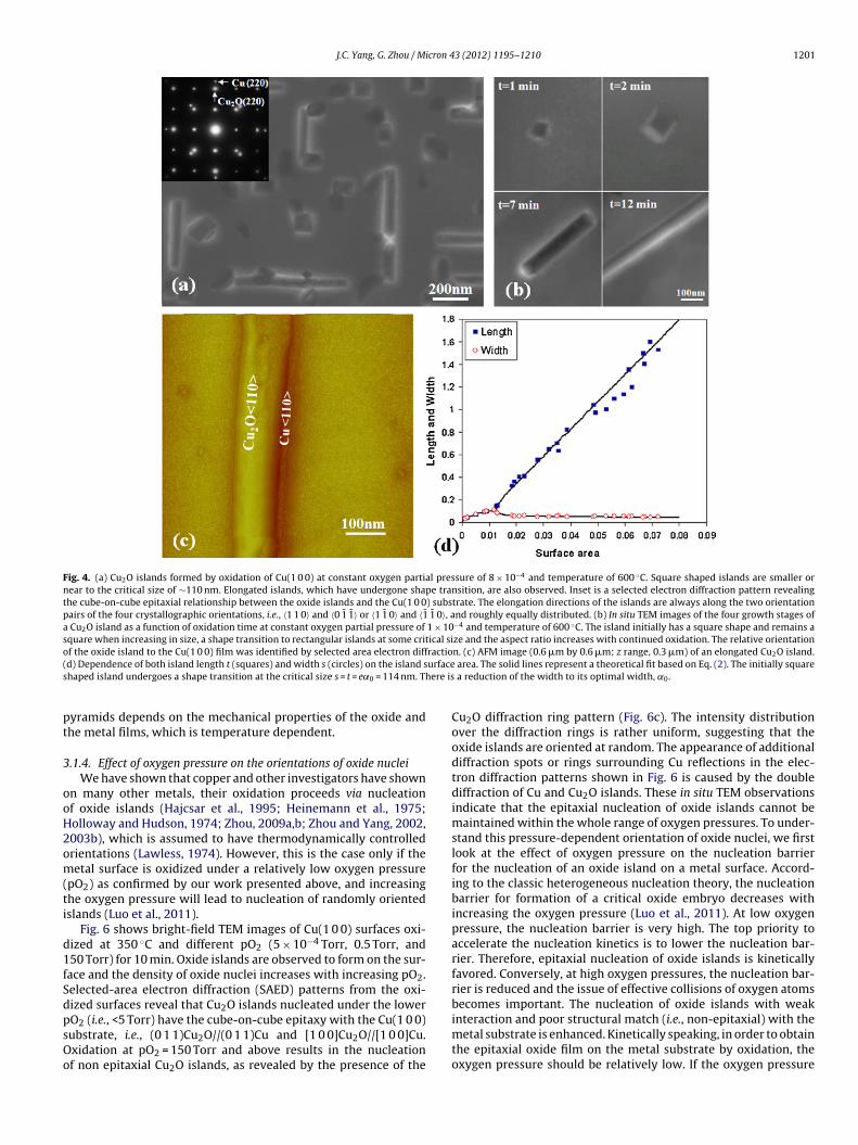
J.C. Yang, G. Zhou / Micron 43 (2012) 1195–1210 1201
Fig. 4. (a) Cu2O islands formed by oxidation of Cu(1 0 0) at constant oxygen partial pressure of 8 × 10⁻⁴ and temperature of 600 ◦C. Square shaped islands are smaller ornear to the critical size of ∼110 nm. Elongated islands, which have undergone shape transition, are also observed. Inset is a selected electron diffraction pattern revealingthe cube-on-cube epitaxial relationship between the oxide islands and the Cu(1 0 0) substrate. The elongation directions of the islands are always along the two orientationpairs of the four crystallographic orientations, i.e., 〈1 1 0〉 and 〈0 1 1〉 or 〈1 1 0〉 and 〈1 1 0〉, and roughly equally distributed. (b) In situ TEM images of the four growth stages ofa Cu2O island as a function of oxidation time at constant oxygen partial pressure of 1 × 10⁻⁴ and temperature of 600 ◦C. The island initially has a square shape and remains asquare when increasing in size, a shape transition to rectangular islands at some critical size and the aspect ratio increases with continued oxidation. The relative orientationof the oxide island to the Cu(1 0 0) film was identified by selected area electron diffraction. (c) AFM image (0.6 �m by 0.6 �m; z range, 0.3 �m) of an elongated Cu O island.( urfaces ere is
pt
3
ooH2om(ti
d1fSdpsOo
d) Dependence of both island length t (squares) and width s (circles) on the island shaped island undergoes a shape transition at the critical size s = t = e˛0 = 114 nm. Th
yramids depends on the mechanical properties of the oxide andhe metal films, which is temperature dependent.
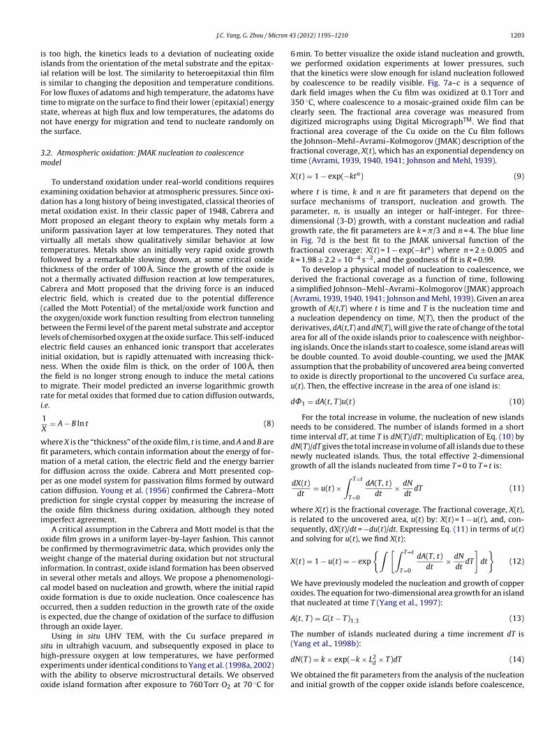
.1.4. Effect of oxygen pressure on the orientations of oxide nucleiWe have shown that copper and other investigators have shown
n many other metals, their oxidation proceeds via nucleationf oxide islands (Hajcsar et al., 1995; Heinemann et al., 1975;olloway and Hudson, 1974; Zhou, 2009a,b; Zhou and Yang, 2002,003b), which is assumed to have thermodynamically controlledrientations (Lawless, 1974). However, this is the case only if theetal surface is oxidized under a relatively low oxygen pressure
pO2) as confirmed by our work presented above, and increasinghe oxygen pressure will lead to nucleation of randomly orientedslands (Luo et al., 2011).
Fig. 6 shows bright-field TEM images of Cu(1 0 0) surfaces oxi-ized at 350 ◦C and different pO2 (5 × 10⁻⁴ Torr, 0.5 Torr, and50 Torr) for 10 min. Oxide islands are observed to form on the sur-ace and the density of oxide nuclei increases with increasing pO2.elected-area electron diffraction (SAED) patterns from the oxi-ized surfaces reveal that Cu2O islands nucleated under the lower
O2 (i.e., <5 Torr) have the cube-on-cube epitaxy with the Cu(1 0 0)ubstrate, i.e., (0 1 1)Cu2O//(0 1 1)Cu and [1 0 0]Cu2O//[1 0 0]Cu.xidation at pO2 = 150 Torr and above results in the nucleationf non epitaxial Cu2O islands, as revealed by the presence of the2
area. The solid lines represent a theoretical fit based on Eq. (2). The initially square a reduction of the width to its optimal width, ˛0.
Cu2O diffraction ring pattern (Fig. 6c). The intensity distributionover the diffraction rings is rather uniform, suggesting that theoxide islands are oriented at random. The appearance of additionaldiffraction spots or rings surrounding Cu reflections in the elec-tron diffraction patterns shown in Fig. 6 is caused by the doublediffraction of Cu and Cu2O islands. These in situ TEM observationsindicate that the epitaxial nucleation of oxide islands cannot bemaintained within the whole range of oxygen pressures. To under-stand this pressure-dependent orientation of oxide nuclei, we firstlook at the effect of oxygen pressure on the nucleation barrierfor the nucleation of an oxide island on a metal surface. Accord-ing to the classic heterogeneous nucleation theory, the nucleationbarrier for formation of a critical oxide embryo decreases withincreasing the oxygen pressure (Luo et al., 2011). At low oxygenpressure, the nucleation barrier is very high. The top priority toaccelerate the nucleation kinetics is to lower the nucleation bar-rier. Therefore, epitaxial nucleation of oxide islands is kineticallyfavored. Conversely, at high oxygen pressures, the nucleation bar-rier is reduced and the issue of effective collisions of oxygen atomsbecomes important. The nucleation of oxide islands with weak
interaction and poor structural match (i.e., non-epitaxial) with themetal substrate is enhanced. Kinetically speaking, in order to obtainthe epitaxial oxide film on the metal substrate by oxidation, theoxygen pressure should be relatively low. If the oxygen pressure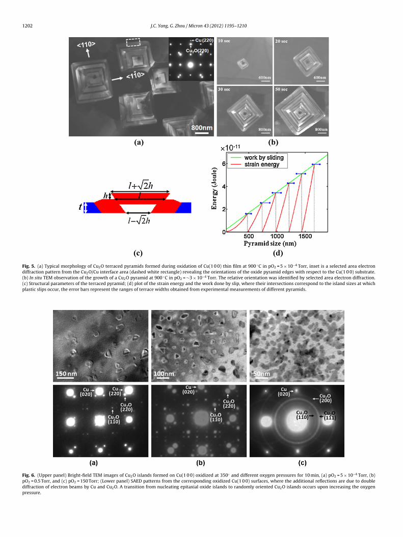
1202 J.C. Yang, G. Zhou / Micron 43 (2012) 1195–1210
Fig. 5. (a) Typical morphology of Cu2O terraced pyramids formed during oxidation of Cu(1 0 0) thin film at 900 ◦C in pO2 = 5 × 10⁻⁴ Torr, inset is a selected area electrondiffraction pattern from the Cu2O/Cu interface area (dashed white rectangle) revealing the orientations of the oxide pyramid edges with respect to the Cu(1 0 0) substrate.(b) In situ TEM observation of the growth of a Cu2O pyramid at 900 ◦C in pO2 = ∼3 × 10⁻⁴ Torr. The relative orientation was identified by selected area electron diffraction.(c) Structural parameters of the terraced pyramid; (d) plot of the strain energy and the work done by slip, where their intersections correspond to the island sizes at whichplastic slips occur, the error bars represent the ranges of terrace widths obtained from experimental measurements of different pyramids.
Fig. 6. (Upper panel) Bright-field TEM images of Cu2O islands formed on Cu(1 0 0) oxidized at 350◦ and different oxygen pressures for 10 min, (a) pO2 = 5 × 10⁻⁴ Torr, (b)pO2 = 0.5 Torr, and (c) pO2 = 150 Torr; (Lower panel) SAED patterns from the corresponding oxidized Cu(1 0 0) surfaces, where the additional reflections are due to doublediffraction of electron beams by Cu and Cu2O. A transition from nucleating epitaxial oxide islands to randomly oriented Cu2O islands occurs upon increasing the oxygenpressure.
cron 4
iiiiFtsnt
3m
edmMuvtftnCe(tbleinttri
wfimfpcpti
obwiicooit
shewo
J.C. Yang, G. Zhou / Mi
s too high, the kinetics leads to a deviation of nucleating oxideslands from the orientation of the metal substrate and the epitax-al relation will be lost. The similarity to heteroepitaxial thin films similar to changing the deposition and temperature conditions.or low fluxes of adatoms and high temperature, the adatoms haveime to migrate on the surface to find their lower (epitaxial) energytate, whereas at high flux and low temperatures, the adatoms doot have energy for migration and tend to nucleate randomly onhe surface.
.2. Atmospheric oxidation: JMAK nucleation to coalescenceodel
To understand oxidation under real-world conditions requiresxamining oxidation behavior at atmospheric pressures. Since oxi-ation has a long history of being investigated, classical theories ofetal oxidation exist. In their classic paper of 1948, Cabrera andott proposed an elegant theory to explain why metals form a
niform passivation layer at low temperatures. They noted thatirtually all metals show qualitatively similar behavior at lowemperatures. Metals show an initially very rapid oxide growthollowed by a remarkable slowing down, at some critical oxidehickness of the order of 100 A. Since the growth of the oxide isot a thermally activated diffusion reaction at low temperatures,abrera and Mott proposed that the driving force is an inducedlectric field, which is created due to the potential differencecalled the Mott Potential) of the metal/oxide work function andhe oxygen/oxide work function resulting from electron tunnelingetween the Fermi level of the parent metal substrate and acceptor
evels of chemisorbed oxygen at the oxide surface. This self-inducedlectric field causes an enhanced ionic transport that acceleratesnitial oxidation, but is rapidly attenuated with increasing thick-ess. When the oxide film is thick, on the order of 100 A, thenhe field is no longer strong enough to induce the metal cationso migrate. Their model predicted an inverse logarithmic growthate for metal oxides that formed due to cation diffusion outwards,.e.
1X
= A − B ln t (8)
here X is the “thickness” of the oxide film, t is time, and A and B aret parameters, which contain information about the energy of for-ation of a metal cation, the electric field and the energy barrier
or diffusion across the oxide. Cabrera and Mott presented cop-er as one model system for passivation films formed by outwardation diffusion. Young et al. (1956) confirmed the Cabrera–Mottrediction for single crystal copper by measuring the increase ofhe oxide film thickness during oxidation, although they notedmperfect agreement.
A critical assumption in the Cabrera and Mott model is that thexide film grows in a uniform layer-by-layer fashion. This cannote confirmed by thermogravimetric data, which provides only theeight change of the material during oxidation but not structural
nformation. In contrast, oxide island formation has been observedn several other metals and alloys. We propose a phenomenologi-al model based on nucleation and growth, where the initial rapidxide formation is due to oxide nucleation. Once coalescence hasccurred, then a sudden reduction in the growth rate of the oxides expected, due the change of oxidation of the surface to diffusionhrough an oxide layer.
Using in situ UHV TEM, with the Cu surface prepared initu in ultrahigh vacuum, and subsequently exposed in place to
igh-pressure oxygen at low temperatures, we have performedxperiments under identical conditions to Yang et al. (1998a, 2002)ith the ability to observe microstructural details. We observedxide island formation after exposure to 760 Torr O2 at 70 ◦C for
3 (2012) 1195–1210 1203
6 min. To better visualize the oxide island nucleation and growth,we performed oxidation experiments at lower pressures, suchthat the kinetics were slow enough for island nucleation followedby coalescence to be readily visible. Fig. 7a–c is a sequence ofdark field images when the Cu film was oxidized at 0.1 Torr and350 ◦C, where coalescence to a mosaic-grained oxide film can beclearly seen. The fractional area coverage was measured fromdigitized micrographs using Digital MicrographTM. We find thatfractional area coverage of the Cu oxide on the Cu film followsthe Johnson–Mehl–Avrami–Kolmogorov (JMAK) description of thefractional coverage, X(t), which has an exponential dependency ontime (Avrami, 1939, 1940, 1941; Johnson and Mehl, 1939).
X(t) = 1 − exp(−ktn) (9)
where t is time, k and n are fit parameters that depend on thesurface mechanisms of transport, nucleation and growth. Theparameter, n, is usually an integer or half-integer. For three-dimensional (3-D) growth, with a constant nucleation and radialgrowth rate, the fit parameters are k = �/3 and n = 4. The blue linein Fig. 7d is the best fit to the JMAK universal function of thefractional coverage: X(t) = 1 − exp(−ktn) where n = 2 ± 0.005 andk = 1.98 ± 2.2 × 10⁻⁴ s−2, and the goodness of fit is R = 0.99.
To develop a physical model of nucleation to coalescence, wederived the fractional coverage as a function of time, followinga simplified Johnson–Mehl–Avrami–Kolmogorov (JMAK) approach(Avrami, 1939, 1940, 1941; Johnson and Mehl, 1939). Given an areagrowth of A(t,T) where t is time and T is the nucleation time anda nucleation dependency on time, N(T), then the product of thederivatives, dA(t,T) and dN(T), will give the rate of change of the totalarea for all of the oxide islands prior to coalescence with neighbor-ing islands. Once the islands start to coalesce, some island areas willbe double counted. To avoid double-counting, we used the JMAKassumption that the probability of uncovered area being convertedto oxide is directly proportional to the uncovered Cu surface area,u(t). Then, the effective increase in the area of one island is:
d˚1 = dA(t, T)u(t) (10)
For the total increase in volume, the nucleation of new islandsneeds to be considered. The number of islands formed in a shorttime interval dT, at time T is dN(T)/dT; multiplication of Eq. (10) bydN(T)/dT gives the total increase in volume of all islands due to thesenewly nucleated islands. Thus, the total effective 2-dimensionalgrowth of all the islands nucleated from time T = 0 to T = t is:
dX(t)dt
= u(t) ×∫ T=t
T=0
dA(T, t)dt
× dN
dtdT (11)
where X(t) is the fractional coverage. The fractional coverage, X(t),is related to the uncovered area, u(t) by: X(t) = 1 − u(t), and, con-sequently, dX(t)/dt = −du(t)/dt. Expressing Eq. (11) in terms of u(t)and solving for u(t), we find X(t):
X(t) = 1 − u(t) = − exp
{∫ [∫ T=t
T=0
dA(T, t)dt
× dN
dtdT
]dt
}(12)
We have previously modeled the nucleation and growth of copperoxides. The equation for two-dimensional area growth for an islandthat nucleated at time T (Yang et al., 1997):
A(t, T) = G(t − T)1.3 (13)
The number of islands nucleated during a time increment dT is(Yang et al., 1998b):
dN(T) = k × exp(−k × L2d × T)dT (14)
We obtained the fit parameters from the analysis of the nucleationand initial growth of the copper oxide islands before coalescence,

1204 J.C. Yang, G. Zhou / Micron 43 (2012) 1195–1210
Fig. 7. Dark field images from the Cu2O reflection showing Cu2O island nucleation (a), growth (b) and then coalescence (c), when Cu(0 0 1) was oxidized at 0.1 Torr at3 e, wf ived si to the
taSrgae
oegtpatcdg
3
sgladt
50 ◦C; (d) experimental data of the Cu2O fractional area coverage, X(t), versus timormula, X(t) = 1 − exp(−ktn) (blue line), where k = 1.98 × 10⁻⁴ s−2, n = 2, and our dernterpretation of the references to color in this figure legend, the reader is referred
o calculate the saturation density, 1/L2d
= 0.31209, initial nucle-tion rate, k = 2.08 × 10⁻⁴, and the growth constant, G = 0.00849.ubstituting the above expressions for dN(T) and dA(T) into Eq. (13),esults in a double integral of a hypergeometric function, which wasraphically solved with MathematicaTM. The results of our analysisre shown in Fig. 7d where an excellent fit is noted (red line) to thexperimental data (circles) (Yang et al., 2002).
We have modeled the nucleation to coalescence of copperxides, in the framework of the JMAK equations, and noted anxcellent agreement between experiment and theory, where oxy-en surface diffusion is dominant. Since surface diffusion is critical,hen control of the surfaces is essential in controlling oxidationroperties. Extension of this surface model and experiments totmospheric pressures and comparison to classic data could leado a new paradigm of oxidation and passivation, where structuralhanges are crucial. Our results are based on in situ TEM of the oxi-ation of Cu thin films, but we suspect that these results can beeneralized to other metal systems that form epitaxial oxides.
.3. Cu alloy oxidation
Alloying commonly leads to new materials whose properties areubstantially changed with respect to pure metals. For example, aeneral strategy for the protection of bulk metals is alloying, which
eads to the formation of a uniform oxide scale layer which acts asdiffusion barrier to further oxidation attack over the alloy surfaceue to the preferential (selective) oxidation of one component ofhe alloy. The fundamental understanding of the complex atomistic
hen Cu(0 0 1) was oxidized at 0.1 Torr at 350 ◦C, and the comparison to the JMAKurface model (red line) using A(t) = Gt1.3 and N(t) = (1/L2
d) × [1 − exp(−kL2
dt)]. (For
web version of this article.)
mechanisms and the affects of surfaces, strain and dopants on theearly stages of binary alloy oxidation will lead to smarter designs ofcoatings that resist oxidation as well as templates to create specificoxide nanostructures.
Binary alloy oxidation is well-described by the Wagner theoryof oxidation, which predicts whether a continuous multi-layer oroxide precipitates, known as internal oxidation, form as a functionof the relative amount of the added secondary element. However,this is a macroscopic description of the oxide after it has devel-oped into a thick scale. The earlier stages of oxidation, which clearlyimpact the later stage oxide morphology and adhesion are notdescribed by these classical theories. Because of the authors exten-sive past experience with Cu thin films, Cu–Au (Section 3.3.1) andCu–Ni (Section 3.3.2) alloy systems were studied next, where the Cuoxidation provides a base-line for comparison. In addition to beingexcellent model systems, understanding the oxidation behavior ofCu-based alloys is also of practical interest. For example, Cu-basedalloys are presently used as an interconnect material in electron-ics applications. Since materials dimensions in electronic devicesare already in the nanometer regime and continually shrinking,understanding Cu alloy oxidation at the nanoscale is needed for theimproved designs of durable and corrosion-resistant interconnects.
3.3.1. Cu–Au(1 0 0) oxidation
As the next simplest model system to study, we selected Cu–Au,since only Cu oxidizes. Cu and Au are 100% solid soluble at ele-vated temperatures. For the case of Cu–Au, only one elementoxidizes (Cu). According to Vegard’s law, the lattice constant of

cron 4
C
atwbscpmA
baip(olittesoiet(sdotAoodpctwe
�
wmtw
dpoifoltStmihfif
J.C. Yang, G. Zhou / Mi
u1−xAux solution is aCu1−xAux = aAu × xAu + aCu × (1 − xAu), where
Cu = 3.61 A and aAu = = 4.02 A, aC2O = 4.27 Å , and xAu is mole frac-ion of Au in the alloy. The lattice constant of Cu–Au alloy increasesith the increase of Au mole fraction and the lattice mismatch
etween the oxide and substrate is correspondingly reduced. Thetrain between the metal alloy and oxide should decrease as the Auontent is increased. We noted in Section 3.1.3 that oxidation tem-erature influences interfacial strain that results in different oxideorphologies (e.g., nanorod, hollow pyramids); thus be altering theu content, we can systematically study strain effects on oxidation.
We find that, similar as the oxidation of Cu(1 0 0), oxide nucleiecome visible on the Cu–Au surface after some incubation period,nd their number increases with time and then saturates. Thencubation time for the island nucleation depends on the alloy com-osition, higher Au mole fraction leads to longer incubation timeZhou et al., 2007b). We speculate that Cu–Au alloys are Au richn the surface under vacuum conditions which may be a lead to aonger incubation time as Cu diffuses to the surface when oxygen isntroduced (Wang, 2005). The nucleation rate (from appearance ofhe first nuclei to saturation density) depends on the Au mole frac-ion in the alloy as well, and alloys with higher Au mole fractionsxhibit faster nucleation rates (Zhou et al., 2007a). Fig. 8a showsome examples of the oxide islands formed during Cu–Au(1 0 0)xidation at 600 ◦C in pO2 = 5 × 10⁻⁴ Torr. Fig. 8b shows the oxideslands formed during oxidation of (1 0 0) Cu–38 at.%Au at differ-nt temperatures in pO2 = 5 × 10⁻⁴ Torr. Similar to the Cu(1 0 0),he Cu2O islands are epitaxial with the underlying alloy substrateZhou et al., 2006, 2007b). These measurements indicate that theaturation density depends on the oxidation temperature, i.e., oxi-ation at a higher temperature results in a smaller number densityf islands. Fig. 8a also reveals that the number density is related tohe Au mole fraction in the alloys, i.e., oxidation of alloys with higheru mole fraction results in a larger saturation number density of thexide nuclei. The activation energy for nucleation of oxide islandsn the Cu–Au alloy surfaces can be deduced from the Arrheniusependence of the saturation island density on the oxidation tem-erature. Fig. 8c shows the EN values determined as a function ofomposition, which indicate that alloys with a larger Au mole frac-ion have a smaller EN. This observation reveals that alloying Cuith Au enhances the nucleation of oxide islands. The nucleation
nergy of the oxide island is given by:
G = �Gf V
V0+ (�0A0 − �mAm + �m/oAm/o) + E
1 − �ε2V (15)
here � are the surface and interface energies of the oxide andetal, �Gf is the Gibbs free energy of the oxide formation, and ε is
he strain energy. With increasing Au content, the strain is reducedhich may lower the nucleation energy barrier (Wang, 2005).
We also monitored the growth of oxide islands during the oxi-ation of CuAu(1 0 0) alloy (with 5, 10 and 15 at.% Au) at 600 ◦C inO2 = 5 × 10⁻⁴ Torr. The average individual island size (area) wasbserved to show a linear dependence on oxidation time, as shownn Fig. 8d, indicating that the growth depends on oxygen surface dif-usion (Yang et al., 1997). This growth behavior is different from thexidation of Cu(1 0 0) at 350 ◦C, where a slight deviation from theinear growth is noted and the non-linear oxide growth is ascribedo the oxygen surface diffusion plus oxygen direct impingement.ince the oxidation of the Cu–Au alloys is at higher temperatures,he oxygen surface diffusion can be therefore greatly enhanced. This
ay explain why the oxygen direct impingement is not significantn the oxide growth during the oxidation of the Cu–Au alloys at the
igher temperatures. The rate constants for oxide growth, obtainedrom the slope of the area vs. time curves in Fig. 8d, decrease withncreasing Au content. It is noted that alloy with a higher Au moleraction has a smaller rate constant for oxide growth for oxidation
3 (2012) 1195–1210 1205
at the same temperature, which may be due to the smaller amountof Cu available near the island.
We noted that the addition also changes the oxide morphology.Fig. 9a–c shows an in situ TEM observation of the growth of an oxideisland formed during oxidation of Cu–5 at.%Au(1 0 0) at 600 ◦C anda gradual transition occurs from initially compact island shapes to adendritic morphology as the islands grow. The oxide island nucle-ates with a square in cross section shape and retains this shapeduring the initial growth. The island then exhibit a gradual transi-tion to a dendritic shape as growth slows along the normal to theisland edges, i.e., along Cu2O 〈1 1 0〉 directions, while maintaininga faster growth rate along the directions of the island corners, i.e.,along Cu2O 〈1 0 0〉 directions. In contrast, the oxide islands formedduring oxidation of pure Cu(0 0 1) under the same conditions havean initially square shape that transforms to a rectangular morphol-ogy as growth proceeds (Fig. 4).
As seen from Fig. 9, a non-uniform dark contrast developsaround the island with the island growth, and the energy dispersiveX-ray spectroscopy (EDS) analysis reveals that the Au mole fractionis highly enhanced in the region with dark contrast (Zhou et al.,2007a). The contrast features in Fig. 9a–c reveal that the alloy filmregions adjacent to the island edges become Au-rich, while thereis almost no excess Au accumulation in the alloy film adjacent tothe island corners, as confirmed by the EDS analysis (Zhou et al.,2007b). Fig. 9 clearly reveals that the change in the local growthrate of the island boundary is closely related to this non-uniformpartition of Au atoms in the alloy film during the island growth,i.e., the growth rate of the boundary adjacent to the Au-rich zonegradually slows as the local Au concentration increases.
In the initial stages of metal oxidation under low oxygen pres-sures and high temperatures, the reaction rate is usually dominatedby the capture of oxygen. Our analysis of the growth of Cu2Oislands prior to the dendrite formation during oxidation of theCu–Au alloys demonstrate that the rate-limiting factor controllingthe oxide growth is oxygen surface diffusion. Growth of the com-pact shape is due to efficient diffusion of adatoms on the metalsurface and along island edges (Hwang et al., 1991; Li and Evans,2004). As oxidation progresses, a non-uniform partition of Au atomsdevelops in the alloy, and the local growth rate of the boundaryadjacent to the Au-rich regions is slowed due to depletion of Cuatoms. As a result, the rate-limiting factor in these regions becomesCu diffusion through the Au-rich zone, similar to reported behaviorfor oxidation of bulk Cu–Au alloys (Wagner, 1952). However, thegrowth of the dendritic tips is still controlled by fast oxygen sur-face diffusion because of the low Au accumulation in front of thetips. Therefore, boundary growth along the island perimeter hasdifferent rate-limiting factors that depend on the local concentra-tion of Au in the alloy. Fig. 9d is the schematic diagram showingour proposed model of Au segregation induced dendritic growth.According to this picture, increasingly ramified morphologies arepredicted as the oxidation proceeds and are also experimentallyobserved (Zhou et al., 2006). In comparison to Cu(0 0 1) oxidation,the addition of Au caused more rapid nucleation due to the reducedstrain between the metal and the oxide but slower growth kineticsdue to the Au build-up around the oxide island. The Au build uparound the island also lead to a dendritic oxide growth limiting thealloy’s ability to form a uniform protective oxide.
3.3.2. Cu–Ni(1 0 0) oxidationSimilar to Cu, oxidation studies of Cu–Ni alloys have a long his-
tory. Pilling and Bedworth investigated Cu–Ni oxidation by thermalgravimetric analysis to quantify the parabolic rate as function of
relative concentration (Pilling and Bedworth, 1925), but these clas-sic investigations did not provide structural information. Surfacescience methods, including in situ, examined oxygen interactionand strain induced NiO nucleation of CuNi, but focused on the
1206 J.C. Yang, G. Zhou / Micron 43 (2012) 1195–1210
Fig. 8. (a) Bright field TEM images of oxide islands formed during oxidation of Cu1−xAux(1 0 0) alloys (x = 0.05, 0.10, 0.2, 0.38) at 600 ◦C and pO2 = 5 × 10⁻⁴ Torr; (b) bright fieldTEM images of oxide islands formed during the oxidation of Cu–38 at.%Au at pO = 5 × 10⁻⁴ Torr, but with different temperatures; (c) nucleation activation energy determinedf rage ib rate c
v1d(tfbo
msshwcwo
Cttt
2
rom the oxidation of the Cu–Au alloys with different Au mole fractions; (d) the aveehavior is noted for the different alloys. The value of the slope corresponds to the
ery early stages (Brizuela et al., 2006; Bruekner and Baunack,999; Hono et al., 1991). An ex situ TEM study of Cu–Ni alloy oxi-ation revealed that both copper oxides and nickel oxides formHeinemann et al., 1975), but lacked temporal information neededo understand nucleation kinetics. Hence, limited literature existsor Cu–Ni oxidation studies, yet fundamental understanding ofinary alloy oxidation is the critical first step to controlling selectivexidation processes.
It is reasonable to expect that the Cu–Ni alloys will showore complex behavior, since the two components are 100% solid-
oluble down to ∼300 ◦C but Cu2O and NiO show very limitedolubility. Nickel oxide, which has the cubic NaCl crystal structure,as a more negative standard free energy of formation than Cu2O,hich is simple cubic, and is expected to form more readily. In this
ase, depending on pO2, either one or both components of the alloyill oxidize, thus enabling systematic determination of the effects
f compositional and phase development during oxidation.Irregular shaped oxide islands were observed to appear on
u–24 at.%Ni(0 0 1) tens of seconds to several minutes afterhe introduction of oxygen gas into the TEM. This incubationime (to) depended on the oxidation temperature, i.e. T = 500 ◦Co = 5 min, T = 600 ◦C to = 2 min, and T = 700 ◦C to = 40 s, which is
sland area vs. oxidation time during oxidation of the Cu–Au alloys, a linear growthonstant for the oxide growth.
reasonable to expect since at higher temperatures, the barrier tonucleation can be more easily overcome due to its temperature-dependency through the corresponding Boltzmann factor. Incomparison to Cu and Cu–Au oxidation, the incubation timebetween Cu and Cu–24 at.%Ni is quite similar, e.g. at T = 600 ◦Cand pO2 = 5 × 10⁻⁴ Torr, to(Cu–Ni) = 2 min, to(Cu) = 1.5 min andto(Cu–Au) = 4 min (Wang et al., 2006). We speculate that the longerincubation time before the onset of oxide nucleation for Cu–Au isdue to the higher Au concentration on the surface of Cu–Au, whichdoes not oxidize (Wang et al., 2006). The Au must migrate awayfrom the surface as Cu migrates toward the surface as thermo-dynamically driven to oxidize. For Cu and Cu–Ni, the surface isalready composed of elements that are thermodynamically drivento oxidize, and, hence, the oxide nucleation will occur faster incomparison to Cu–Au.
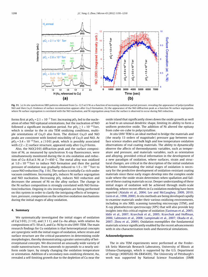
Fig. 10 are bright-field images taken in situ at T = 600 ◦C wherethe oxygen pressure is 5 × 10⁻⁴ Torr. We noted irregular-shapedelongated islands to nucleate initially, but after 15 min a second
rapid nucleation stage of compact oxide islands with higher islanddensity was observed. A similar behavior was noted at T = 700 ◦Cwhere irregular shaped square-in-cross-section islands nucleatedfirst, but after a couple of minutes a second nucleation of a high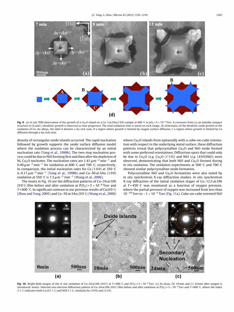
J.C. Yang, G. Zhou / Micron 43 (2012) 1195–1210 1207
Fig. 9. (a) In situ TEM observation of the growth of a Cu2O island on a Cu–5 at.%Au(1 0 0) sample at 600 ◦C in pO2 = 5 × 10⁻⁴ Torr. A crossover from (a) an initially compacts idatioo rowthd
dfwncN0Iio
(T(
Fi(
tructure to (b and c) dendritic growth is observed as time progresses. The total oxxidation of Cu–Au alloys, the label A denotes a Au-rich zone, B a region where giffusion through a Au-rich zone.
ensity of rectangular oxide islands occurred. The rapid nucleationollowed by growth supports the oxide surface diffusion modelhere the oxidation process can be characterized by an initialucleation rate (Yang et al., 1998b). The two-step nucleation pro-ess could be due to NiO forming first and then after the depletion ofi, Cu2O nucleates. The nucleation rates are 1.41 �m−2 min−1 and.49 �m−2 min−1 for oxidation at 600 ◦C and 700 ◦C, respectively.n comparison, the initial nucleation rates for Cu (1 0 0) at 350 ◦Cs 0.17 �m−2 min−1 (Yang et al., 1998b) and Cu–50 at.%Au (1 0 0)xidation at 550 ◦C is 7.3 �m−2 min−1 (Wang et al., 2006).
The insets in Fig. 10 are the diffraction patterns of Cu–24 at.%Ni0 0 1) film before and after oxidation at P(O2) = 5 × 10⁻⁴ Torr and
= 600 ◦C. In significant contrast to our previous results of Cu(0 0 1)Zhou and Yang, 2005) and Cu–50 at.%Au (0 0 1) (Wang et al., 2006)
ig. 10. Bright-field images of the in situ oxidation of Cu–24 at.%Ni (0 0 1) at T = 600 ◦Cntroduced. Insets: Selected area electron diffraction pattern of Cu–24 at.%Ni (0 0 1) film1 1 1) indicates both Cu2O(1 1 1) and NiO(1 1 1), similarly for (2 0 0) and (2 2 0).
n time is noted on each image. (b) Schematics of the dendritic oxide growth in the is limited by oxygen surface diffusion, C a region where growth is limited by Cu
where Cu2O islands form epitaxially with a cube-on-cube orienta-tion with respect to the underlying metal surface, these diffractionpatterns reveal that polycrystalline Cu2O and NiO oxide formedwith some preferred orientations. Diffraction spots that could onlybe due to Cu2O (e.g. Cu2O (1 1 0)) and NiO (e.g. (4 0 0)NiO) wereobserved, demonstrating that both NiO and Cu2O formed duringin situ oxidation. The oxidation experiments at 500 ◦C and 700 ◦Cshowed similar polycrystalline oxide formation.
Polycrystalline NiO and Cu2O formations were also noted byin situ synchrotron X-ray diffraction studies. In situ synchrotron
X-ray diffraction of the initial oxidation stages of Cu–12.5 at.%Niat T = 450 ◦C was monitored as a function of oxygen pressure,where the partial pressure of oxygen was increased from less than10−10 Torr to ∼1 × 10⁻⁵ Torr (Fig. 11a). Cube-on-cube oriented NiOand P(O2) = 5 × 10⁻⁴ Torr. (a) As-clean, (b) 10 min and (c) 24 min after oxygen is before and after oxidation at P(O2) = 5 × 10⁻⁴ Torr and T = 600 ◦C, where the index
1208 J.C. Yang, G. Zhou / Micron 43 (2012) 1195–1210
F ction oN ion; (bw ion aw
fafwpppw
tstapcvadttotd
4
ocrasmeofor
ig. 11. (a) In situ synchrotron XRD patterns obtained from Cu–12.5 at.% Ni as a funiO and then Cu2O. Evidence of surface reconstruction appears after Cu2O formathere Ni surface segregation is correlated with the NiO nucleation, and Ni segregat
orms first at pO2 = 2.1 × 10⁻⁷ Torr. Increasing pO2 led to the nucle-tion of other NiO epitaxial orientations, but the nucleation of NiOollowed a significant incubation period. For pO2 � 1 × 10⁻⁴ Torr,hich is similar to the in situ TEM oxidizing conditions, multi-le orientations of Cu2O also form. The distinct Cu2O and NiOeaks are consistent with limited miscibility of Cu2O and NiO. AtO2 = 3.4 × 10⁻⁴ Torr, a (1 0 0) peak, which is possibly associatedith c(2 × 2) surface structure, appeared only after Cu2O forms.
Also, the NiO(2 0 0) diffraction peak and the surface composi-ion of Ni, as measured by synchrotron X-ray fluorescence, wereimultaneously monitored during the in situ oxidation and reduc-ion of Cu–8.0 at.% Ni at T = 450 ◦C. The metal alloy was oxidizedt 1.0 × 10⁻⁶ Torr to induce NiO formation and then the partialressure of oxidation was gradually reduced to 1.5 × 10⁻⁷ Torr toause NiO reduction (Fig. 11b). The surface is initially Cu-rich underacuum conditions. Increasing pO2 induces Ni surface segregationnd NiO nucleation. Decreasing pO2 induces NiO reduction andecreases the amount of Ni on the alloy surface. The change inhe Ni surface composition is strongly correlated with NiO forma-ion/reduction. Ongoing in situ investigations are being performedn this system in order to clarify the intriguing effects of tempera-ure, pressure, composition on the selective oxidation mechanismsuring the initial stages of alloy oxidation.
. Summary
We systematically investigated the initial stages of oxidationf Cu(1 0 0), (1 1 0), and (1 1 1), and Cu–Au alloys, with relative Auompositions of 5–50 at.%, and Cu–Ni by in situ UHV-TEM. The mainesearch findings for Cu oxidation is that heteroepitaxial conceptsre synergistic with the initial stages of oxidation, where strain andurface structure are the critical parameters in determining oxideorphologies, thereby demonstrating a greater universality of het-
roepitaxial concepts. We discovered an unusually wide variety of
xide nanostructures, from nanorods to pyramids to a nearly uni-orm oxide layer, by simply changing the oxidation temperaturer orientation. Addition of a secondary non-oxidizing element, Au,evealed a self-limiting growth due to the depletion of Cu near thef increasing oxidation partial pressure, revealing the appearance of polycrystalline) the appearance of the NiO diffraction peak as a function Ni surface segregation,ay from the surface is observed to occur during NiO reduction.
oxide island that significantly slows down the oxide growth as wellas lead to an unusual dendritic shape, limiting its ability to form auniform protective oxide. The addition of Ni altered the epitaxyfrom cube-on-cube to polycrystalline.
In situ UHV TEM is an ideal method to bridge the materials and(the nearly 13 orders of magnitude) pressure gap between sur-face science studies and bulk high and low temperature oxidationobservations of real coating materials. The ability to dynamicallyobserve the affects of thermodynamic variables, such as temper-ature and pressure, and materials variables, such as orientationand alloying, provided critical information in the development ofa new paradigm of oxidation, where surfaces, strain and struc-tural changes, are critical in the description of the initial oxidationbehavior. Understanding the initial stages of oxidation is neces-sary for the predictive development of oxidation-resistant coatingmaterials since these early stages develop into the complex oxidescale where the oxide strain determines when spallation and fail-ure of these coating materials occur. Deeper understanding of theseinitial stages of oxidation will be achieved through multi-scalemodeling, where recent efforts in Cu oxidation modeling have beenreported (Alatalo et al., 2004; Lee and McGaughey, 2009, 2010;Liem et al., 1998, 2000). In situ methods are being developed rapidlyto examine materials under their various oxidizing environments,including in situ XRD, scanning tunneling microscopy (STM), andX-ray photoelectron spectroscopy (XPS) that will also provide keyinsights into this critical regime of oxidation (Eastman et al., 2005;Iddir et al., 2007; Kravchuk et al., 2005; Kravchuk and Hoffman,2006; Lahtonen et al., 2008; Lampimaki et al., 2007; Okada et al.,2007; Zhou et al., 2009). Oxidation exemplifies the fundamentalmaterials science significantly enabled by the recent advancementswith in situ characterization tools and theoretical simulations.
Acknowledgements
The in situ TEM experiments were performed at the Freder-
ick Seitz Materials Research Laboratory, University of Illinois atUrbana-Champaign, which is supported by the U.S. Departmentof Energy (#DEFG02-96-ER45439). The University of Pittsburgh’swork was supported by National Science Foundation (DMR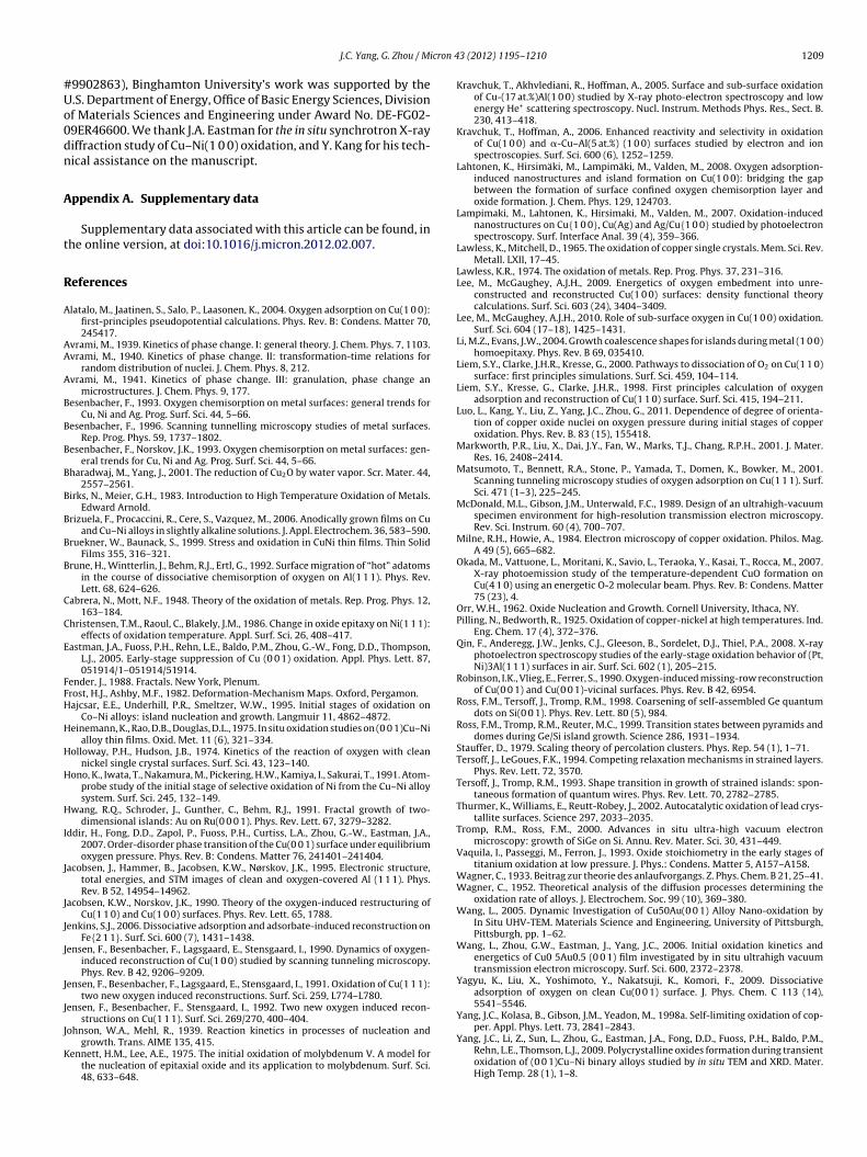
cron 4
#Uo0dn
A
t
R
A
AA
A
B
B
B
B
B
B
B
B
C
C
E
FFH
H
H
H
H
I
J
J
J
J
J
J
J
K
J.C. Yang, G. Zhou / Mi
9902863), Binghamton University’s work was supported by the.S. Department of Energy, Office of Basic Energy Sciences, Divisionf Materials Sciences and Engineering under Award No. DE-FG02-9ER46600. We thank J.A. Eastman for the in situ synchrotron X-rayiffraction study of Cu–Ni(1 0 0) oxidation, and Y. Kang for his tech-ical assistance on the manuscript.
ppendix A. Supplementary data
Supplementary data associated with this article can be found, inhe online version, at doi:10.1016/j.micron.2012.02.007.
eferences
latalo, M., Jaatinen, S., Salo, P., Laasonen, K., 2004. Oxygen adsorption on Cu(1 0 0):first-principles pseudopotential calculations. Phys. Rev. B: Condens. Matter 70,245417.
vrami, M., 1939. Kinetics of phase change. I: general theory. J. Chem. Phys. 7, 1103.vrami, M., 1940. Kinetics of phase change. II: transformation-time relations for
random distribution of nuclei. J. Chem. Phys. 8, 212.vrami, M., 1941. Kinetics of phase change. III: granulation, phase change an
microstructures. J. Chem. Phys. 9, 177.esenbacher, F., 1993. Oxygen chemisorption on metal surfaces: general trends for
Cu, Ni and Ag. Prog. Surf. Sci. 44, 5–66.esenbacher, F., 1996. Scanning tunnelling microscopy studies of metal surfaces.
Rep. Prog. Phys. 59, 1737–1802.esenbacher, F., Norskov, J.K., 1993. Oxygen chemisorption on metal surfaces: gen-
eral trends for Cu, Ni and Ag. Prog. Surf. Sci. 44, 5–66.haradwaj, M., Yang, J., 2001. The reduction of Cu2O by water vapor. Scr. Mater. 44,
2557–2561.irks, N., Meier, G.H., 1983. Introduction to High Temperature Oxidation of Metals.
Edward Arnold.rizuela, F., Procaccini, R., Cere, S., Vazquez, M., 2006. Anodically grown films on Cu
and Cu–Ni alloys in slightly alkaline solutions. J. Appl. Electrochem. 36, 583–590.ruekner, W., Baunack, S., 1999. Stress and oxidation in CuNi thin films. Thin Solid
Films 355, 316–321.rune, H., Wintterlin, J., Behm, R.J., Ertl, G., 1992. Surface migration of “hot” adatoms
in the course of dissociative chemisorption of oxygen on Al(1 1 1). Phys. Rev.Lett. 68, 624–626.
abrera, N., Mott, N.F., 1948. Theory of the oxidation of metals. Rep. Prog. Phys. 12,163–184.
hristensen, T.M., Raoul, C., Blakely, J.M., 1986. Change in oxide epitaxy on Ni(1 1 1):effects of oxidation temperature. Appl. Surf. Sci. 26, 408–417.
astman, J.A., Fuoss, P.H., Rehn, L.E., Baldo, P.M., Zhou, G.-W., Fong, D.D., Thompson,L.J., 2005. Early-stage suppression of Cu (0 0 1) oxidation. Appl. Phys. Lett. 87,051914/1–051914/51914.
ender, J., 1988. Fractals. New York, Plenum.rost, H.J., Ashby, M.F., 1982. Deformation-Mechanism Maps. Oxford, Pergamon.ajcsar, E.E., Underhill, P.R., Smeltzer, W.W., 1995. Initial stages of oxidation on
Co–Ni alloys: island nucleation and growth. Langmuir 11, 4862–4872.einemann, K., Rao, D.B., Douglas, D.L., 1975. In situ oxidation studies on (0 0 1)Cu–Ni
alloy thin films. Oxid. Met. 11 (6), 321–334.olloway, P.H., Hudson, J.B., 1974. Kinetics of the reaction of oxygen with clean
nickel single crystal surfaces. Surf. Sci. 43, 123–140.ono, K., Iwata, T., Nakamura, M., Pickering, H.W., Kamiya, I., Sakurai, T., 1991. Atom-
probe study of the initial stage of selective oxidation of Ni from the Cu–Ni alloysystem. Surf. Sci. 245, 132–149.
wang, R.Q., Schroder, J., Gunther, C., Behm, R.J., 1991. Fractal growth of two-dimensional islands: Au on Ru(0 0 0 1). Phys. Rev. Lett. 67, 3279–3282.
ddir, H., Fong, D.D., Zapol, P., Fuoss, P.H., Curtiss, L.A., Zhou, G.-W., Eastman, J.A.,2007. Order-disorder phase transition of the Cu(0 0 1) surface under equilibriumoxygen pressure. Phys. Rev. B: Condens. Matter 76, 241401–241404.
acobsen, J., Hammer, B., Jacobsen, K.W., Nørskov, J.K., 1995. Electronic structure,total energies, and STM images of clean and oxygen-covered Al (1 1 1). Phys.Rev. B 52, 14954–14962.
acobsen, K.W., Norskov, J.K., 1990. Theory of the oxygen-induced restructuring ofCu(1 1 0) and Cu(1 0 0) surfaces. Phys. Rev. Lett. 65, 1788.
enkins, S.J., 2006. Dissociative adsorption and adsorbate-induced reconstruction onFe{2 1 1}. Surf. Sci. 600 (7), 1431–1438.
ensen, F., Besenbacher, F., Lagsgaard, E., Stensgaard, I., 1990. Dynamics of oxygen-induced reconstruction of Cu(1 0 0) studied by scanning tunneling microscopy.Phys. Rev. B 42, 9206–9209.
ensen, F., Besenbacher, F., Lagsgaard, E., Stensgaard, I., 1991. Oxidation of Cu(1 1 1):two new oxygen induced reconstructions. Surf. Sci. 259, L774–L780.
ensen, F., Besenbacher, F., Stensgaard, I., 1992. Two new oxygen induced recon-structions on Cu(1 1 1). Surf. Sci. 269/270, 400–404.
ohnson, W.A., Mehl, R., 1939. Reaction kinetics in processes of nucleation andgrowth. Trans. AIME 135, 415.
ennett, H.M., Lee, A.E., 1975. The initial oxidation of molybdenum V. A model forthe nucleation of epitaxial oxide and its application to molybdenum. Surf. Sci.48, 633–648.
3 (2012) 1195–1210 1209
Kravchuk, T., Akhvlediani, R., Hoffman, A., 2005. Surface and sub-surface oxidationof Cu-(17 at.%)Al(1 0 0) studied by X-ray photo-electron spectroscopy and lowenergy He+ scattering spectroscopy. Nucl. Instrum. Methods Phys. Res., Sect. B.230, 413–418.
Kravchuk, T., Hoffman, A., 2006. Enhanced reactivity and selectivity in oxidationof Cu(1 0 0) and �-Cu–Al(5 at.%) (1 0 0) surfaces studied by electron and ionspectroscopies. Surf. Sci. 600 (6), 1252–1259.
Lahtonen, K., Hirsimäki, M., Lampimäki, M., Valden, M., 2008. Oxygen adsorption-induced nanostructures and island formation on Cu(1 0 0): bridging the gapbetween the formation of surface confined oxygen chemisorption layer andoxide formation. J. Chem. Phys. 129, 124703.
Lampimaki, M., Lahtonen, K., Hirsimaki, M., Valden, M., 2007. Oxidation-inducednanostructures on Cu{1 0 0}, Cu(Ag) and Ag/Cu{1 0 0} studied by photoelectronspectroscopy. Surf. Interface Anal. 39 (4), 359–366.
Lawless, K., Mitchell, D., 1965. The oxidation of copper single crystals. Mem. Sci. Rev.Metall. LXII, 17–45.
Lawless, K.R., 1974. The oxidation of metals. Rep. Prog. Phys. 37, 231–316.Lee, M., McGaughey, A.J.H., 2009. Energetics of oxygen embedment into unre-
constructed and reconstructed Cu(1 0 0) surfaces: density functional theorycalculations. Surf. Sci. 603 (24), 3404–3409.
Lee, M., McGaughey, A.J.H., 2010. Role of sub-surface oxygen in Cu(1 0 0) oxidation.Surf. Sci. 604 (17–18), 1425–1431.
Li, M.Z., Evans, J.W., 2004. Growth coalescence shapes for islands during metal (1 0 0)homoepitaxy. Phys. Rev. B 69, 035410.
Liem, S.Y., Clarke, J.H.R., Kresse, G., 2000. Pathways to dissociation of O2 on Cu(1 1 0)surface: first principles simulations. Surf. Sci. 459, 104–114.
Liem, S.Y., Kresse, G., Clarke, J.H.R., 1998. First principles calculation of oxygenadsorption and reconstruction of Cu(1 1 0) surface. Surf. Sci. 415, 194–211.
Luo, L., Kang, Y., Liu, Z., Yang, J.C., Zhou, G., 2011. Dependence of degree of orienta-tion of copper oxide nuclei on oxygen pressure during initial stages of copperoxidation. Phys. Rev. B. 83 (15), 155418.
Markworth, P.R., Liu, X., Dai, J.Y., Fan, W., Marks, T.J., Chang, R.P.H., 2001. J. Mater.Res. 16, 2408–2414.
Matsumoto, T., Bennett, R.A., Stone, P., Yamada, T., Domen, K., Bowker, M., 2001.Scanning tunneling microscopy studies of oxygen adsorption on Cu(1 1 1). Surf.Sci. 471 (1–3), 225–245.
McDonald, M.L., Gibson, J.M., Unterwald, F.C., 1989. Design of an ultrahigh-vacuumspecimen environment for high-resolution transmission electron microscopy.Rev. Sci. Instrum. 60 (4), 700–707.
Milne, R.H., Howie, A., 1984. Electron microscopy of copper oxidation. Philos. Mag.A 49 (5), 665–682.
Okada, M., Vattuone, L., Moritani, K., Savio, L., Teraoka, Y., Kasai, T., Rocca, M., 2007.X-ray photoemission study of the temperature-dependent CuO formation onCu(4 1 0) using an energetic O-2 molecular beam. Phys. Rev. B: Condens. Matter75 (23), 4.
Orr, W.H., 1962. Oxide Nucleation and Growth. Cornell University, Ithaca, NY.Pilling, N., Bedworth, R., 1925. Oxidation of copper-nickel at high temperatures. Ind.
Eng. Chem. 17 (4), 372–376.Qin, F., Anderegg, J.W., Jenks, C.J., Gleeson, B., Sordelet, D.J., Thiel, P.A., 2008. X-ray
photoelectron spectroscopy studies of the early-stage oxidation behavior of (Pt,Ni)3Al(1 1 1) surfaces in air. Surf. Sci. 602 (1), 205–215.
Robinson, I.K., Vlieg, E., Ferrer, S., 1990. Oxygen-induced missing-row reconstructionof Cu(0 0 1) and Cu(0 0 1)-vicinal surfaces. Phys. Rev. B 42, 6954.
Ross, F.M., Tersoff, J., Tromp, R.M., 1998. Coarsening of self-assembled Ge quantumdots on Si(0 0 1). Phys. Rev. Lett. 80 (5), 984.
Ross, F.M., Tromp, R.M., Reuter, M.C., 1999. Transition states between pyramids anddomes during Ge/Si island growth. Science 286, 1931–1934.
Stauffer, D., 1979. Scaling theory of percolation clusters. Phys. Rep. 54 (1), 1–71.Tersoff, J., LeGoues, F.K., 1994. Competing relaxation mechanisms in strained layers.
Phys. Rev. Lett. 72, 3570.Tersoff, J., Tromp, R.M., 1993. Shape transition in growth of strained islands: spon-
taneous formation of quantum wires. Phys. Rev. Lett. 70, 2782–2785.Thurmer, K., Williams, E., Reutt-Robey, J., 2002. Autocatalytic oxidation of lead crys-
tallite surfaces. Science 297, 2033–2035.Tromp, R.M., Ross, F.M., 2000. Advances in situ ultra-high vacuum electron
microscopy: growth of SiGe on Si. Annu. Rev. Mater. Sci. 30, 431–449.Vaquila, I., Passeggi, M., Ferron, J., 1993. Oxide stoichiometry in the early stages of
titanium oxidation at low pressure. J. Phys.: Condens. Matter 5, A157–A158.Wagner, C., 1933. Beitrag zur theorie des anlaufvorgangs. Z. Phys. Chem. B 21, 25–41.Wagner, C., 1952. Theoretical analysis of the diffusion processes determining the
oxidation rate of alloys. J. Electrochem. Soc. 99 (10), 369–380.Wang, L., 2005. Dynamic Investigation of Cu50Au(0 0 1) Alloy Nano-oxidation by
In Situ UHV-TEM. Materials Science and Engineering, University of Pittsburgh,Pittsburgh, pp. 1–62.
Wang, L., Zhou, G.W., Eastman, J., Yang, J.C., 2006. Initial oxidation kinetics andenergetics of Cu0 5Au0.5 (0 0 1) film investigated by in situ ultrahigh vacuumtransmission electron microscopy. Surf. Sci. 600, 2372–2378.
Yagyu, K., Liu, X., Yoshimoto, Y., Nakatsuji, K., Komori, F., 2009. Dissociativeadsorption of oxygen on clean Cu(0 0 1) surface. J. Phys. Chem. C 113 (14),5541–5546.
Yang, J.C., Kolasa, B., Gibson, J.M., Yeadon, M., 1998a. Self-limiting oxidation of cop-
per. Appl. Phys. Lett. 73, 2841–2843.Yang, J.C., Li, Z., Sun, L., Zhou, G., Eastman, J.A., Fong, D.D., Fuoss, P.H., Baldo, P.M.,Rehn, L.E., Thomson, L.J., 2009. Polycrystalline oxides formation during transientoxidation of (0 0 1)Cu–Ni binary alloys studied by in situ TEM and XRD. Mater.High Temp. 28 (1), 1–8.
1 icron 4
Y
Y
Y
Y
Y
Y
Y
Z
Z
Z
Z
Z
Z
Zhou, G., Yang, J.C., 2005. Initial oxidation kinetics of Cu(1 0 0), (1 1 0), and (1 1 1)
210 J.C. Yang, G. Zhou / M
ang, J.C., Tropia, L., Evan, D., 2002. From nucleation to coalescence of Cu2O islandsduring in situ oxidation of Cu(0 0 1). Appl. Phys. Lett. 81, 241–243.
ang, J.C., Yeadon, M., 1997. Oxygen surface diffusion in three-dimensional Cu2Ogrowth on Cu(0 0 1) thin films. App. Phys. Lett. 70 (26), 3522–3524.
ang, J.C., Yeadon, M., Kolasa, B., Gibson, J.M., 1997. Oxygen surface diffusion in 3-dCu2O growth on Cu(0 0 1) thin films. Appl. Phys. Lett. 70 (26), 3522–3524.
ang, J.C., Yeadon, M., Kolasa, B., Gibson, J.M., 1998b. Homogeneous nucleation ofCu2O on (0 0 1)Cu. Scr. Mater. 38 (8), 1237–1242.
ang, J.C., Yeadon, M., Kolasa, B., Gibson, J.M., 1998c. In situ UHV TEM investigationsof the initial oxidation stage of copper thin films. MRS Proc. 481, 557–562.
ang, J.C., Yeadon, M., Kolasa, B., Gibson, J.M., 1999. The limited role of surface defectsas nucleation sites for Cu2O on Cu(0 0 1). J. Electrochem. Soc. 146, 2103–2106.
oung, F., Cathcart, J., Gwathmey, A., 1956. The rates of oxidation of several facesof a single crystal of copper as determined with elliptically polarized light. ActaMetall. 4, 145–152.
hou, G.W., 2009a. Nucleation thermodynamics of oxide during metal oxidation.Appl. Phys. Lett. 94, 201905/1–201905/201905.
hou, G.W., 2009b. Stress-driven formation of terraced hollow oxide nanorods dur-ing metal oxidation. J. Appl. Phys. 105 (10), 104302–104306.
hou, G.W., 2009c. TEM investigation of interfaces during cuprous island growth.Acta Mater. 57 (15), 4432–4439.
hou, G.W., 2010. Nucleation-induced kinetic hindrance to the oxide formation dur-ing the initial oxidation of metals. Phys. Rev. B. 81 (19), 195440–195441–195447.
hou, G.W., Chen, X., Gallagher, D., Yang, J.C., 2008. Percolating oxide film growthduring Cu(1 1 1) oxidation. Appl. Phys. Lett. 93, 123104–123106.
hou, G.W., Eastman, J.A., Birtcher, R.C., Pearson, J.E., Baldo, P.E., Thompson, L.J.,2007a. Composition effects on the early-stage oxidation kinetics of (0 0 1)Cu–Aualloys. J. Appl. Phys. 101, 033521/1–033521/33521.
3 (2012) 1195–1210
Zhou, G.W., Fong, D.D., Wang, L., Fuoss, P.H., Baldo, P.M., Thompson, L.J., Eastman,J.A., 2009. Nanoscale duplex oxide growth during early stages of oxidation ofCu–Ni(1 0 0). Phys. Rev. B: Condens. Matter 80 (13), 134106/1–134106/134106.
Zhou, G.W., Slaughter, W.S., Yang, J.C., 2005a. Terraced hollow oxide pyramids. Phys.Rev. Lett. 94, 246101–246104.
Zhou, G.W., Wang, L., Yang, J.C., 2005b. Effect of surface topology on the formationof oxide islands on Cu surfaces. J. Appl. Phys. 97, 063509/1–063509/63509.
Zhou, G.W., Wang, L., Birtcher, R.C., Baldo, P.E., Pearson, J.E., Yang, J.C., Eastman, J.A.,2006. Cu2O island shape transition during Cu–Au oxidation. Phys. Rev. Lett. 96,226108–226111.
Zhou, G.W., Wang, L., Birtcher, R.C., Baldo, P.M., Pearson, J.E., Yang, J.C., Eastman, J.A.,2007b. Effect of composition on the early-stage oxidation of (1 0 0)Cu–Au alloys.J. Appl. Phys. 101, 033521–033526.
Zhou, G.W., Yang, J.C., 2002. Formation of quasi one-dimensional Cu2O structuresby in situ oxidation of Cu(0 0 1). Phys. Rev. Lett. 89, 106101–106104.
Zhou, G.W., Yang, J.C., 2003a. Initial oxidation kinetics of copper (1 1 0) film investi-gated by in situ UHV-TEM. Surf. Sci. 531, 359–367.
Zhou, G.W., Yang, J.C., 2003b. Temperature effect on the Cu2O oxide morphologycreated by oxidation of Cu(0 0 1) as investigated by in situ UHV-TEM. Appl. Surf.Sci. 210, 165–170.
Zhou, G.W., Yang, J.C., 2004. Temperature effects on the growth of oxide islands onCu (1 1 0). Appl. Surf. Sci. 222, 357–364.
thin films investigated by in situ UHV-TEM. J. Mater. Res. 20 (7), 1684–1694.Zhukov, V., Popova, I., Yates, J.T., 1999. Initial stages of Al(1 1 1) oxidation with oxy-
gen temperature dependence for the integral reactive sticking coefficient. Surf.Sci. 441 (2–3), 251–264.