in the diagnosis overview - ucsf cme jones iipjones.pdf · challenges in the diagnosis ... •...
TRANSCRIPT
5/12/2014
1
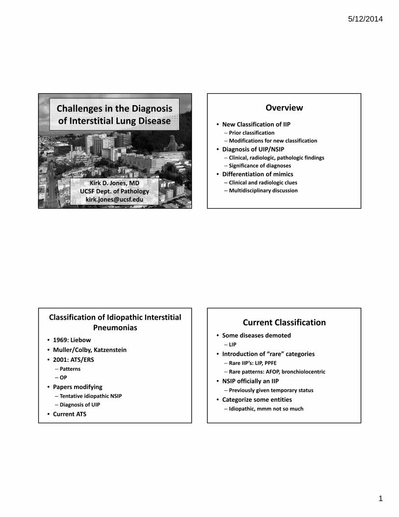
Challenges in the Diagnosis of Interstitial Lung Disease
Kirk D. Jones, MDUCSF Dept. of [email protected]
Overview
• New Classification of IIP– Prior classification
– Modifications for new classification
• Diagnosis of UIP/NSIP– Clinical, radiologic, pathologic findings
– Significance of diagnoses
• Differentiation of mimics– Clinical and radiologic clues
– Multidisciplinary discussion
Classification of Idiopathic Interstitial Pneumonias
• 1969: Liebow
• Muller/Colby, Katzenstein
• 2001: ATS/ERS
– Patterns
– OP
• Papers modifying
– Tentative idiopathic NSIP
– Diagnosis of UIP
• Current ATS
Current Classification
• Some diseases demoted
– LIP
• Introduction of “rare” categories
– Rare IIP’s: LIP, PPFE
– Rare patterns: AFOP, bronchiolocentric
• NSIP officially an IIP
– Previously given temporary status
• Categorize some entities
– Idiopathic, mmm not so much
5/12/2014
2
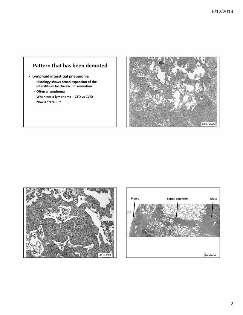
Pattern that has been demoted
• Lymphoid interstitial pneumonia
– Histology shows broad expansion of the interstitium by chronic inflammation
– Often a lymphoma
– When not a lymphoma – CTD vs CVID
– Now a “rare IIP”
LIP in CVID
LIP in CVID
Septal extensionPleura Mass
Lymphoma
5/12/2014
3
Added Entities
• Rare IIP
– Idiopathic pleuroparenchymal fibroelastosis
– LIP (as mentioned in demoted)
• Rare patterns
– Acute fibrinous organizing pneumonia
– Bronchiolocentric interstitial fibrosis
Pleuroparenchymal Fibroelastosis
• Pleural and subpleural fibrosis
• Upper lobes show consolidation with traction bronchiectasis
• Described in Japan by Amitani
• Progression in majority, death in 40%
• Unknown cause
• Don’t mistake an apical fibrous cap for PPFE!
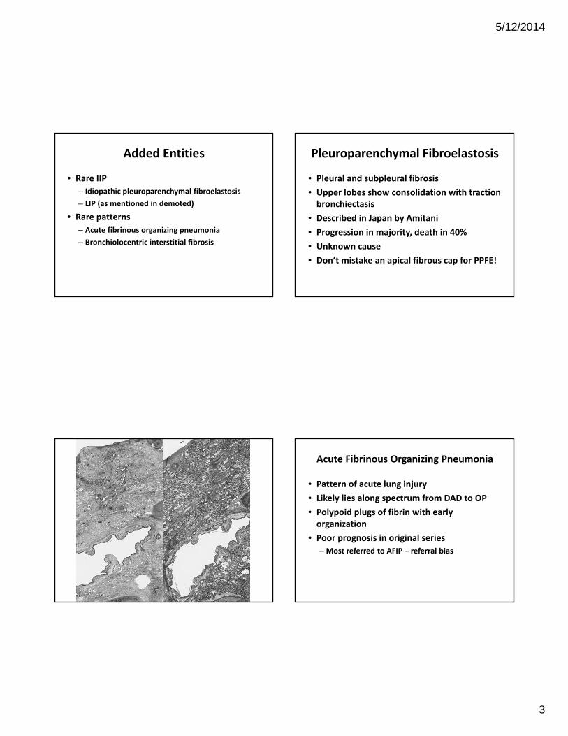
Acute Fibrinous Organizing Pneumonia
• Pattern of acute lung injury
• Likely lies along spectrum from DAD to OP
• Polypoid plugs of fibrin with early organization
• Poor prognosis in original series
– Most referred to AFIP – referral bias

5/12/2014
4
Bronchiolocentric Fibrosis
• Histologic changes with fibrosis centered on small airways
• “Bronchiolization” of alveolar ducts
• Many cases may have either HP or CTD
New Categorization
• Chronic fibrosing
– Usual interstitial pneumonia
– Non‐specific interstitial pneumonia
• Smoking‐related
– Desquamative interstitial pneumonia
– Respiratory bronchiolitis
• Acute/Subacute
– Diffuse alveolar damage
– Organizing pneumonia

5/12/2014
5
Interstitial fibrosis, difficult to classify
UIP
NSIP
DIP
OP
DAD
LIP
Elastotic fibrosis
RB
Travis WD, et al. Am J Respir Crit Care Med. 2013 Sep 15; 188(6): 733-48. PMID: 24032382.
Diagnosis of Usual Interstitial Pneumonia
• Hey, let’s be like radiologists!
Raghu G, et al. Am J Respir Crit Care Med. 2011 Mar 15; 183(6): 788-824. PMID: 21471066.
Spatial heterogeneity
Temporal heterogeneity
Fibrosis - with “temporal heterogeneity”
oneycomb fibrosisld collagenous fibrosis
• Pathologic Findings - Temporal Heterogeneity
– H
– O
– R
– N
ecent (fibroblastic) fibrosisormal lung
5/12/2014
6
Words to the clinician
• I don’t make a diagnosis of:
– Definite, Probable, Possible, Not…UIP
• I do put it in the comment:
– Reasons for – describing histology
– Reasons against – describing the features against
Significance of a UIP Diagnosis
• PANTHER Study
– Efficacy of Prednisone, Azathioprine, N‐acetylcysteine (NAC) vs. NAC alone vs. placebo
• Patients in the prednisone, aza, NAC arm
– Increased deaths (8 vs. 1)
– Increased hospitalization (23 vs. 7)
• NAC vs placebo still accumulating data
– mucolytic agent used often used in CF patients
Diagnosis of UIP
• Be aware of clinical and radiologic findings
– Idiopathic pulmonary fibrosis usually age 50+
• Some exceptions
• If younger, consider UIP pattern in CTD, HP, familial fibrosis, drug reaction
– UIP shows basilar and subpleural distribution
• If prominent upper lobe disease, consider PPFE, HP
• Look for classical histologic findings with spectrum from scarred to normal (HORN)
Diagnosis of Nonspecific Interstitial Pneumonia
• Clinical findings may be as nonspecific as its name:
– Dyspnea, cough
• May have some findings to suggest etiology
– Exposures, drugs, serologic studies, systemic symptoms
• Some radiologic clues
– Subpleural sparing
– Traction bronchiectasis without honeycombing
5/12/2014
7
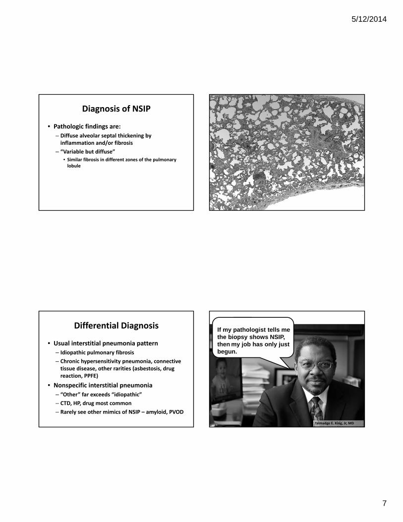
Diagnosis of NSIP
• Pathologic findings are:
– Diffuse alveolar septal thickening by inflammation and/or fibrosis
– “Variable but diffuse”
• Similar fibrosis in different zones of the pulmonary lobule
Differential Diagnosis
• Usual interstitial pneumonia pattern
– Idiopathic pulmonary fibrosis
– Chronic hypersensitivity pneumonia, connective tissue disease, other rarities (asbestosis, drug reaction, PPFE)
• Nonspecific interstitial pneumonia
– “Other” far exceeds “idiopathic”
– CTD, HP, drug most common
– Rarely see other mimics of NSIP – amyloid, PVOD
If my pathologist tells me the biopsy shows NSIP, then my job has only just begun.
Talmadge E. King, Jr, MD

5/12/2014
8
Case 1
• 50‐year‐old male with chief complaint of worsening shortness of breath over 1‐2 years
• Travels extensively with entertainment commitments
5/12/2014
9
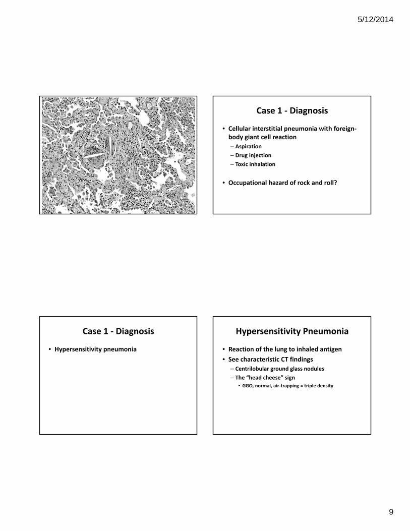
Case 1 ‐ Diagnosis
• Cellular interstitial pneumonia with foreign‐body giant cell reaction
– Aspiration
– Drug injection
– Toxic inhalation
• Occupational hazard of rock and roll?
Case 1 ‐ Diagnosis
• Hypersensitivity pneumonia
Hypersensitivity Pneumonia
• Reaction of the lung to inhaled antigen
• See characteristic CT findings
– Centrilobular ground glass nodules
– The “head cheese” sign
• GGO, normal, air‐trapping = triple density

5/12/2014
10
Courtesy of Rick Webb, MD
HP ‐ HistologyThe Four‐Part Triad
• Diffuse lymphoplasmacytic interstitial infiltrate
– With bronchiolocentric accentuation
• Poorly‐formed granulomas
• Foci of organizing pneumonia
Case 1 ‐ Diagnosis
• Traveled with same pillow for 15 years
– Down pillow
– Typical exposure
• Other cases we have observed:
– Feathers: Pets, Farm animal, Duvet, Pillow, Jacket.
– Molds: Work freezer, Man‐Cave, Sleep number mattress
– Mycobacteria: Indoor spa, shower
– ? Central valley: Almond dust?
Case 2
• 24‐year‐old woman with interstitial lung disease.
• Dry cough, Raynaud’s phenomenon, possible feather exposure, arthralgias.
• CT shows patchy ground glass opacities with a peripheral predominance.

5/12/2014
11
Case 2 ‐ Diagnosis
• Cellular and fibrosing interstitial pneumonia (non‐specific interstitial pneumonia pattern).
• Found to have a CK of 1108 (nl = 39‐189)
• Autoimmune myositis
• Improved with mycophenolate
• In our practice, patients with clinical symptoms get a large panel of serologic studies and likely won’t be biopsied.
Case 3
• 73‐year‐old woman with a six month history of shortness of breath.

5/12/2014
12
Case 3 ‐ Diagnosis
• Cellular nonspecific interstitial pneumonia with prominent lymphoid aggregates and organizing pneumonia
– I would probably be thinking connective tissue disease, but it looked like a prior case of a man with BPH.
Case 3 ‐ Continued
• Missing drug history.
– Medicine note: no drugs of concern.
– Surgeon’s pre‐op note: Nitrofurantoin.
• “It wasn’t me.”
• On nitrofurantoin for 1‐1/2 years.
– Stealth drug (post‐coital UTI’s)
• www.pneumotox.com
5/12/2014
13
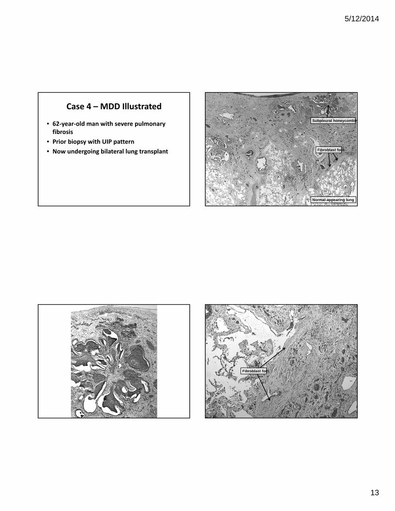
Case 4 – MDD Illustrated
• 62‐year‐old man with severe pulmonary fibrosis
• Prior biopsy with UIP pattern
• Now undergoing bilateral lung transplant
Subpleural honeycombin
Fibroblast foci
Normal-appearing lung
Fibroblast foci

5/12/2014
14
Pathologic Pattern
• Usual interstitial fibrosis
– Marked fibrosis with honeycombing
– Patchy involvement of lung
– Fibroblast foci present
– ?Features suggesting alternate diagnosis?
Bronchiolocentric Fibrosis
Poorly-formed granuloma
Pathologic Diagnosis
• Interstitial fibrosis, UIP pattern, with bronchiolocentric fibrosis and chronic inflammation, and poorly‐formed granulomas.
• Most consistent with chronic hypersensitivity pneumonia.
5/12/2014
15
Final Diagnosis
• Familial Interstitial Fibrosis
– Telomerase mutation (TERT gene)
• With superimposed hypersensitivity pneumonia
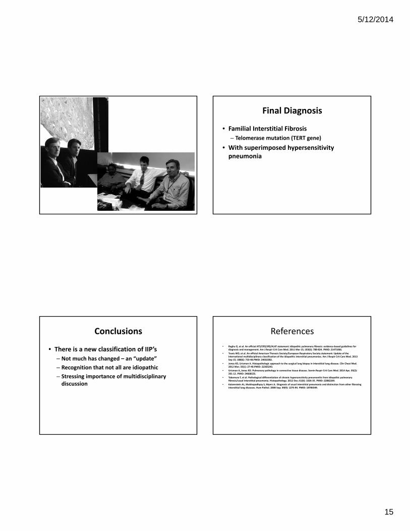
Conclusions
• There is a new classification of IIP’s
– Not much has changed – an “update”
– Recognition that not all are idiopathic
– Stressing importance of multidisciplinary discussion
References
• Raghu G, et al. An official ATS/ERS/JRS/ALAT statement: idiopathic pulmonary fibrosis: evidence‐based guidelines for diagnosis and management. Am J Respir Crit Care Med. 2011 Mar 15; 183(6): 788‐824. PMID: 21471066.
• Travis WD, et al. An official American Thoracic Society/European Respiratory Society statement: Update of the international multidisciplinary classification of the idiopathic interstitial pneumonias. Am J Respir Crit Care Med. 2013 Sep 15; 188(6): 733‐48.PMID: 24032382.
• Jones KD, Urisman A. Histopathologic approach to the surgical lung biopsy in interstitial lung disease. Clin Chest Med. 2012 Mar; 33(1): 27‐40.PMID: 22365243.
• Urisman A, Jones KD. Pulmonary pathology in connective tissue disease. Semin Respir Crit Care Med. 2014 Apr; 35(2): 201‐12. PMID: 24668535.
• Takemura T, et al. Pathological differentiation of chronic hypersensitivity pneumonitis from idiopathic pulmonary fibrosis/usual interstitial pneumonia. Histopathology. 2012 Dec; 61(6): 1026‐35. PMID: 22882269.
• Katzenstein AL, Mukhopadhyay S, Myers JL. Diagnosis of usual interstitial pneumonia and distinction from other fibrosing interstitial lung diseases. Hum Pathol. 2008 Sep; 39(9): 1275‐94. PMID: 18706349.