in vitro screening assay for teratogens using growth inhibition of
TRANSCRIPT

Proc. Natl. Acad. Sci. USAVol. 82, pp. 5791-5794, September 1985Developmental Biology
In vitro screening assay for teratogens using growth inhibition ofhuman embryonic cells
(cell proliferation/palate/human embryo/mesenchymal cell/metabolic activation)
ROBERT M. PRATT AND WILLIAM D. WILLIS
Experimental Teratogenesis Section, Laboratory of Reproductive and Developmental Toxicology, National Institute of Environmental Health Sciences,National Institutes of Health, P.O. Box 12233 (MD C4-02), Research Triangle Park, NC 27709
Communicated by Clement L. Markert, May 15, 1985
ABSTRACT We have tested 35 teratogenic and 20 nonter-atogenic chemicals or drugs in a short-term, in vitro assay thatidentifies teratogens by their ability to inhibit growth of anestablished line of human embryonic palatal mesenchymalcells. Only those chemicals that exhibited a dose-dependentinhibition of growth at concentrations less than 1 mM wereclassified as inhibitory. An Aroclor-induced rat liver S-9 systemwas effective in metabolizing cyclophosphamide to its terato-genic form in culture. We suggest that this assay, along with thecomplementary tumor cell-attachment assay of Braun et al.[Braun, A. G., Emerson, D. J. & Nichinson, B. B. (1979)Nature (London) 282, 507-509] may be useful as a short-termin vitro battery for assessment of the teratogenic potential inenvironmental agents and to prioritize those chemicals whichmerit further testing in vivo.
The application of in vitro systems to teratogenicity testinghas been of interest to teratologists and developmentalbiologists for many years (1, 2), with a dramatic increase inresearch in this area occurring after a workshop on thissubject was held in 1981 (3). It has become quite evident thatthe current whole-animal test procedures for teratogenesisare inadequate to screen the large number of chemicalsalready present as well as new ones being introduced into theenvironment each year (4). Whole animal tests suffer fromhigh cost, large number of animals used (mainly rodents andrabbits), and large amounts of chemical and time necessaryfor each full-scale determination.The use of short-term in vitro screening systems could
serve to establish priorities for the selection of chemicals thatshould be tested in vivo, could decrease the need for (butcertainly not replace) whole animal tests, and ultimatelycould accelerate the time in which potential teratogens ofhuman consequence can be evaluated. Various in vitrosystems, which have been used to investigate normalembryogenesis, have been proposed, including cell, organ,and whole-embryo cultures derived from vertebrates as wellas invertebrates (1-3). These systems reflect various embry-onic events that occur during organogenesis, including cel-lular migration, interactions, proliferation, and differentia-tion; these events are known to be especially sensitive toteratogenic insult (4). It is highly unlikely that any one singlein vitro system will suffice to reflect all of these events;instead a battery of complementary assays will be needed forteratogen screening.Rapid cellular proliferation is essential for the early phases
of organogenesis, and a number of chemicals and drugs,especially craniofacial teratogens, have been shown to inter-fere with this event in a specific manner (5). Several years
ago, an established cell line (HEPM) was derived by using themesenchymal cells of the secondary palate (roof of themouth) from a day 55 human embryonic abortus (6). Subse-quent studies ofours have examined the mechanism by whichspecific hormones and growth factors influence HEPM cellproliferation (7).The purpose of the present study was to examine the
potential for using teratogen-induced growth inhibition of theHEPM cells as a teratogen screening assay.
MATERIALS AND METHODS
Human embryonic palatal mesenchyme cells (HEPM) wereoriginally established in culture by R. M. Pratt and theAmerican Type Culture Collection (Rockville, MD) anddesignated as CRL 1486 (6). Passage 3 cells from theAmerican Type Culture Collection were carried in our lab topassage 6-7, refrozen, and stored under liquid nitrogen incomplete Dulbecco's modified Eagle's medium (DMEM)with 20% fetal calf serum and 10% dimethyl sulfoxide.Experiments in the present study were routinely performedwith cells of passage 9 through 12. HEPM cells were platedin 35-mm Falcon tissue culture dishes at 4-5 x 10⁴ cells perdish (4.2-5.2 x 10³ cells per cm2) in 2 ml ofDMEM (GIBCO430-1600) (pH 7.4) containing glutamine (0.6 mg/ml), peni-cillin G (50 units/ml), streptomycin sulfate (50 jsg/ml) (Sig-ma), and 10% fetal calf serum (GIBCO 200-6140, Hyclone lot100397). Cultures were incubated in humidified air containing7% C02, which maintained the pH at 7.3 ± 0.1. After 24 hrof culture, the medium was replaced (at 370C) with or withoutthe test chemical; the HEPM cells were maintained in culturefor an additional 72 hr without a medium change. The cellularplating efficiency determined at 24 hr was >95%.
Test chemicals were dissolved at their solubility limit inmedium, water, ethanol, or dimethyl sulfoxide. Ethanol anddimethyl sulfoxide final concentrations did not exceed 0.5%and 0.1%, respectively. A range-finding series of 4-5 con-centration points was performed for initial runs to determinethe upper concentration that almost completely inhibits cellgrowth without being toxic (loss of attachment or nonviableby the trypan blue exclusion test) and the lower concentra-tion that only inhibits cell growth by 10-15%. The final IC50(concentration that inhibits growth by 50%) concentration-finding run had at least 6 concentration points (mean of threeto four dishes). At the termination of the experiment (total of96 hr of culture), the cells were washed in calcium/magnesium-free phosphate-buffered saline and were de-tached in 0.05% trypsin/0.02% EDTA. Cell number wasdetermined using a Coulter ZBI Counter with three readingsfor each dish. Net growth was calculated by subtracting the
Abbreviations: HEPM cells, human embryonic palatal mesenchymecells; MOT assay, mouse ovarian tumor cell-attachment assay.
5791
The publication costs of this article were defrayed in part by page chargepayment. This article must therefore be hereby marked "advertisement"in accordance with 18 U.S.C. §1734 solely to indicate this fact.

5792 Developmental Biology: Pratt and Willis
EKE. 5
W a~~~~~~~~~~~~...:l.
I.*:..
i.
*N- . . ..N .
miFs' is; TAs cak sw~
FIG. 1. Phase-contrast micrographs ofHEPM cells (control) at 24 (a), 48 (b), 72 (c), and 96 (d) hr of culture. Cells were cultured as describedin the text. (Bar = 100 am.)
plating number at 24 hr from the final cell number. Theresulting percentages were plotted on semilogarithmic graphpaper against the test chemical concentration expressed asjig/ml or molarity; an IC50 for growth inhibition was theninterpolated. The test chemicals were all obtained fromSigma except for the following ones: 13-cis-retinoic acid(National Toxicology Program, National Institute of Envi-ronmental Health Sciences), chlorcyclizine and meclizine(Beth Horigan, National Institute of Dental Research,Bethesda, MD), trypan blue (Matheson Coleman and Bell,Norwood, OH), Nembutal (Abbott), and colchicine(GIBCO).Cyclophosphamide was chosen for metabolic activation
vinblostine colchicine amethopterin100
2- 80-
~60-40
studies because. this chemical has been used in a number ofstudies either in vivo or in whole embryo culture (8, 9). EitherAroclor- or noninduced rat liver S-9 prepared in KCl andobtained from Litton Bionetics or Microbiological Associateswas added directly to the HEPM cell cultures. Twenty-fourhours after plating, the growth medium was removed, and S-9and cofactors (10) in 0.5 ml of phosphate-buffered saline atpH 7.4 were added to fresh medium along with cyclophos-phamide at 0.25-100 ,ug/ml to provide final concentrations asfollows: 0.8 mg of S-9 protein per ml, 0.8 mM NADP, 10 mMglucose 6-phosphate, and 1.6mM MgCl2 in sodium phosphatebuffer (20 mM) at pH 7.4. After 4 hr at 370C, the reactionmixture was removed, cells were rinsed several times in
Molarity
FIG. 2. Semilogarithmic plot of teratogens from Table 1 that were positive in the HEPM assay and negative in the MOT assay. The horizontaldashed line is the IC50.
Proc. NatL Acad Sd USA 82 (1985)
s,zz
Proc. NatL Acad Sci USA 82 (1985) 5793
Table 1. Chemicals tested in the HEPM assay
IC50M ,ug/ml Mr
Vinblastine*Vincristine suColchicine*AmethopterinCycloheximid5-Fluorouracil6-Diazo-oxo-LArsenate6-AminonicotDiethylstilbesChlorcyclizinDexamethasoRetinoic acidDopamineMeclizine*HydroxyureaRetinoic acidTrypan blue*ActinomycinL-DopaHydrocortisoAtropine sulf,Chloramphen
Teratogens that are inhibitory in vitro1.1 x 10-9 0.0(
lfate 1.6 x 10⁻⁹ O.0c1.5 x 10-8 0.0C5.3 x 10-s 0.02
le 3.0 x 10⁻⁷ 0.011* 2.8 x 10-6 0.3,i-norleucine 3.2 x 10⁻⁶ 0.54
1.2 x 10-' 2.3:inamide 1.5 x 10⁻⁵ 2.0trol 1.8 x 10- 4.9e* 2.3 x 10-5 6.8ne 3.5 x 10⁻⁵ 14.0(trans) 6.8 x 10-' 20
7.2 x 10⁻⁵ 117.2 x 10⁻⁵ 289.9 X l0-5 7.5
(13-cis) 1.0 X 10⁻⁴ 301.2 x 10⁻⁴ 115
D* 1.3 x 10-4 1631.5 x 10-4 30
mne 2.2 x 10⁻⁴ 80ate 3.7 x 10⁻⁴ 250icol 6.2 x 10⁻⁴ 200
Nonteratogens that are inhibitoryOuabain 8.8 x 10⁻⁹Diphenhydramine 1.4 x 10⁻⁴Butylated hydroxyanisole 2.0 x 10-42,4-Dinitrophenol 2.9 x 10⁻⁴Tween 80 3.1 x 10⁻⁴Isoniazid 5.0 x 10⁻⁴Phenol 8.3 x 10⁻⁴Acetaminophen 9.9 x 10⁻⁴
0101506248574
in vitro0.0064403653
4106878150
Nonteratogens that are not inhibitory in vitroMetyrapone 1.2 x 10⁻³ 265Ascorbic acid 1.7 x 10⁻³ 300Cyclohexylamine 2.0 x 10⁻³ 200Pyridoxine HCO 4.0 x 10⁻³ 830Streptomycin sulfate 6.2 x 10⁻³ 10,000Ammonium chloride 8.4 x 10⁻³ 450Thiamine HCI 1.8 x 10⁻² 830Saccharin 2.6 x 10⁻² 5,350L-Lysine 2.7 x 10⁻² 5,000Cyclamic acid 3.2 x 10⁻² 6,500Sodium chloride 6.0 x 10⁻² 3,500L-Glutamine 6.6 x 10⁻² 9,700
Teratogens that are not inhibitory in vitroCaffeine 1.2 x 10⁻³ 225Nembutal 1.3 x 10⁻³ 320Amaranth 1.5 x 10⁻³ 890Theophylline* 2.0 x 10⁻³ 365Nicotine* 3.7 x 10⁻³ 600Acetazolamide 4.0 x 10⁻³ 900Aspirin 4.2 x 10⁻³ 755L-Phenylalanine 5.5 x 10⁻³ 910Allopurinol* 7.7 x 10⁻³ 1,050Penicillin G 8.1 x 10⁻³ 2,900Urethane 3.3 x 10⁻² 2,925Sucrose 6.3 x 10⁻² 21,500
909.1927.1399.4454.5281.3130.1171.2185.9137.1268.3300.8392.5300.4153.2391.076.1
300.4960.8
1,255.5197.2362.5676.8323.1
728.6291.8180.2184.1130.8137.294.1151.2
226.3176.199.2
205.61,619
53.5205.6205.2182.7201.258.4146.2
194.2248.3604.3180.2162.2222.2180.2165.2136.1356.489.1
342.3
00
I)d00
E
0U
00to0I-ku
102 F
100 F
lo-',10-' 100 lo1
ICO, mg/liter102 103
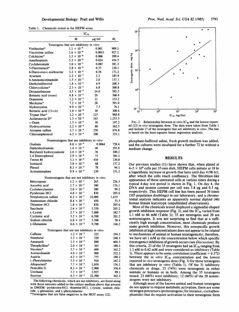
FIG. 3. Relationship between in vitro IC50 and the lowest report-ed (12) in vivo teratogenic dose. The data were taken from Table 1and include 17 of the teratogens that are inhibitory in vitro. The lineis based on the least-squares linear regression analysis.
phosphate-buffered saline, fresh growth medium was added,and the cultures were incubated for a further 72 hr without amedium change.
RESULTS
Our previous studies (11) have shown that, when plated at4-5 x 10⁴ cells per 35-mm dish, HEPM cells initiate at 18 hra logarithmic increase in growth that lasts until day 4 (96 hr),after which the cells reach confluency. The fibroblast-likeappearance of these untreated cells at various times during atypical 4-day test period is shown in Fig. 1. On day 4, theDNA and protein content per cell was 5.8 pg and 0.3 ng,respectively. This HEPM cell line has been passed 30 times(105 population doublings) in our laboratory, and a chromo-somal analysis indicates an apparently normal diploid (46)human female karyotype (unpublished observations).Most of the chemicals tested displayed a sigmoidal-shaped
growth inhibition response (Fig. 2), and the IC50 varied from1.1 nM to 66 mM (Table 1); 35 are teratogens and 20 arenonteratogens. It was not surprising to find that at a suffi-ciently high enough concentration, most chemicals producesome growth inhibition. However, this nonspecific growthinhibition at high concentrations does not appear to be relatedto mechanisms of animal or human teratogenicity; therefore,we have set 1 mM as the concentration below which specific(teratogenic) inhibition ofgrowth occurs (see Discussion). Bythis criteria, 23 of the 35 teratogens had an IC50 ranging from1.1 nM to 0.62 mM and were considered as inhibitory (Table1). There appears to be some correlation (coefficient = 0.572)between the in vitro IC50 concentration and the lowestreported in vivo teratogenic dose (Fig. 3) for those teratogensthat are inhibitory in vitro (Table 1). Of the 31 inhibitorychemicals or drugs, 23 (74%) were teratogenic in eitheranimals or humans or in both. Among the 35 teratogenstested, 23 (66%) were inhibitory; 12 (60%) of the 20 nonter-atogens were not inhibitory.Although most of the known animal and human teratogens
do not appear to require metabolic activation, there are someteratogen precursors (proteratogens; for instance, cyclophos-phamide) that do require activation to their teratogenic form
The following chemicals, which are not inhibitory, are listed alongwith those amounts added to the culture medium above that presentin DMEM: pyridoxine'HCl, thiamine HCl, L-lysine, sodium chlo-ride, L-glutamine, and L-phenylalanine.*Teratogens that are false negatives in the MOT assay (12).
Developmental Biology: Pratt and Willis
lo,
.
lo,F.

5794 Developmental Biology: Pratt and Willis
60-
o 40
20
10-, 100 lo lo2
Cyclophosphamide, jg/mlFIG. 4. Activation of cyclophosphamide in HEPM cell culture in
the presence (o) or absence (A) of Aroclor-induced rat liver S-9 andcofactors as described in the text. The IC50 for CP was 4.5 ,g/ml (17MM).
(phosphoramide mustard). We have devised conditions inculture such that a 4-hr exposure to Aroclor-induced rat liverS-9 at 24 hr of culture results in a concentration-dependentactivation of cyclophosphamide (IC50 = 4.5 ,g/ml or 17 ,uM)(Fig. 4). Cyclophosphamide by itself up to 100 ,ug/ml did notinhibit growth, nor did cyclophosphamide with unactivatedrat liver S-9 and cofactors.
DISCUSSIONThe results from the present study indicate that the growth-inhibition assay using HEPM cells is a highly reproducible,simple, and inexpensive assay for determining teratogen-induced growth inhibition. It is quite clear that most chem-icals we tested will produce growth inhibition if the concen-tration in the culture media is sufficiently high. These highconcentrations (>1 mM) appear to have little relevance toanimal or human teratogenesis and presumably representnonspecific effects to the cell, in part due to the alteredosmolarity. There is little information concerning the con-centrations at which teratogens are present in the embryounder conditions that result in malformations. We havechosen 1 mM as the critical concentration below whichgrowth inhibition is considered to reflect potential teratogen-esis. A similar concentration range limit has been used inevaluating data from various chemicals in several different invitro transformation assays including the mouse, rat, andhamster embryonic fibroblasts in culture (personal commu-
nication, Judson Spalding, National Toxicology Program,National Institute of Environmental Health Sciences).The 55 chemicals chosen for testing in our study were
mainly selected from the list of chemicals and drugs tested byBraun and co-workers (12-14) in their mouse ovarian tumorcell-attachment (MOT) assay. Most of the chemicals inBraun's list are animal teratogens that have been identified bya large data base; several of the chemicals (e.g., diethylstil-bestrol and 13-cis retinoic acid) are also known humanteratogens. We chose various representative chemicals fromthis list to test in the HEPM assay, including most of the falsenegatives in the MOT assay (Table 1); we suspected thatthese two assays, which measure different but essentialevents occurring during organogenesis, would be highlycomplementary. The results ofthe present study demonstratethat these two assays are highly complementary; the chem-icals that are false negative in the MOT assay are positive inthe HEPM assay. In both the HEPM and MOT assays, thereis a reasonable correlation between the in vitro IC50 concen-
tration and the lowest reported in vivo teratogenic dose forthose teratogens that are inhibitory in vitro. The overallpredictability in the combined MOT/HEPM assay is 90%,
and the rate of false negatives is 3%; these are very importantand desirable features because it is crucial that combinedscreening assays (batteries) do not miss potential humanteratogens such as thalidomide. Although thalidomide is notinhibitory under any condition so far tested in the HEPMassay, it is inhibitory in the MOT assay in the presence of anS-9 activation system (10).One of the advantages of the HEPM assay is that
proteratogens, such as cyclophosphamide can be convertedto their teratogenic form in a culture dish containing HEPMcells by using a limited (4 hr) exposure to an Aroclor-inducedrat liver (metabolizing) S-9 system. Under these conditions,a concentration-dependent inhibition of growth is observedwith an IC50 of 4.5 ttg/ml (17 jkM), which is nearly identicalto the concentration in whole-rodent embryo culture thatresults in growth inhibition and malformations. For thoseactivated proteratogens that may require continuous expo-sure in culture, the HEPM cells can be exposed for 4 hr to theS-9 activation system at 24-hr intervals during culture (un-published observations).
Furthermore, the diploid HEPM cells can be maintainedfor at least 30 passages (105 population doublings) in culture.These human embryonic cells are much more sensitive to thegrowth-inhibitory effects of glucocorticoids (i.e., dexameth-asone) in vitro as compared to adult skin fibroblasts (unpub-lished observations); in addition, these cells also may containunique sensitivities peculiar to the human embryo. Thesecells may represent an undifferentiated neural-crest-likemesenchymal cell that is thought to be a likely target for manyteratogens (5); therefore, we feel this cell line is unique andmay be especially valuable for teratogen screening.The MOT/HEPM battery has been tested with over 100
relevant chemicals to date (11-13). This does not imply thata final validation has been completed; however, the NationalToxicology Program is currently in the process of validatingthis battery with the 44 chemicals suggested by Smith et al.(15).
We are grateful to Drs. Andrew Braun and Kathleen Morgan fortheir help in the data analysis and to Ms. Susan DeBrunner for herexpert typing.
1. Wilson, J. G. (1978) J. Environ. Pathol. Toxicol. 2, 149-167.2. Johnson, E. M. (1981) Annu. Rev. Pharmacol. Toxicol. 21,
417-429.3. Kimmel, G. L., Smith, K., Kochhar, D. M. & Pratt, R. M.
(1982) Teratog. Carcinog. Mutagen. 2, 221-230.4. Shepard, T. H., Fantel, A. G., Merkes, P. E., Greenaway,
J. C., Faustman-Watts, E., Campbell, M. & Juchau, M. R.(1983) in Developmental Pharmacology (Liss, New York), pp.147-164.
5. Pratt, R. M. (1983) Trends Pharmacol. Sci. 4, 160-162.6. Yoneda, T. & Pratt, R. M. (1981) Science 213, 563-565.7. Pratt, R. M., Kim, C. S. & Grove, R. I. (1984) in Current
Topics in Developmental Biology, ed. Zimmerman, E. (Aca-demic, New York), Vol. 19, pp. 81-102.
8. Kitchin, K. T., Schmid, B. P. & Sanyal, M. K. (1981)Biochem. Pharmacol. 30, 59-64.
9. Fantel, A. G., Greenaway, J. C., Juchau, M. R. & Shepard,T. H. (1979) Life Sci. 25, 67-72.
10. Braun, A. G. & Weinreb, S. L. (1984) Biochem. Pharmacol.33, 1471-1477.
11. Pratt, R. M., Grove, R. I. & Willis, W. D. (1982) Teratog.Carcinog. Mutagen. 2, 313-318.
12. Braun, A. G., Buckner, C. A., Emerson, D. J. & Nichinson,B. B. (1982) Proc. Natl. Acad. Sci. USA 79, 2056-2060.
13. Braun, A. G., Emerson, D. J. & Nichinson, B. B. (1979)Nature (London) 282, 507-509.
14. Shepard, T. H. (1981) Catalog of Teratogenic Agents (JohnsHopkins University Press, JBaltimore).
15. Smith, M. K., Kimmel, G. L., Kochhar, D. M., Shepard,T. H., Spielberg, S. P. & Wilson, J. G. (1983) Teratog.Carcinog. Mutagen. 3, 461-480.
Proc. NatL Acad ScL USA 82 (1985)