in-vitro screening of acetylcholinesterase inhibitory...
TRANSCRIPT
Functional Foods in Health and Disease 2014; 4(9):381-400 Page 381 of 400
Research Article Open Access
In-vitro screening of acetylcholinesterase inhibitory activity of extracts from
Palestinian indigenous flora in relation to the treatment of Alzheimer’s
disease
Mohammed Saleem Ali-Shtayeh1 Rana Majed Jamous
1, Salam Yousef Abu Zaitoun
1, and
Iman Basem Qasem1
1Biodiversity and Biotechnology Research Unit, Biodiversity and Environmental Research
Center-BERC, P.O. Box 696, Til, Nablus, Palestine
Correspondence Author: Prof. Mohammed S Ali-Shtayeh, Biodiversity and Biotechnology
Research Unit, Biodiversity and Environmental Research Center-BERC, P.O. Box 696, Til, Nablus,
Palestine
Submission date: June 19, 2014; Acceptance date: August 31, 2014; Publication date: September
1, 2014
ABSTRACT:
Background: Cholinesterase inhibitory therapy serves as a strategy for the treatment of
Alzheimer’s disease (AD). Several acetylcholinesterase inhibitors (AChEIs) are used for the
symptomatic treatment of AD. These compounds have been reported to have adverse effects,
including gastrointestinal disturbances.
This study was therefore partly aimed at investigating in vitro possible AChEIs in herbal
medicines traditionally used in Palestine to treat cognitive disorders, and to point out the role of
these plants as potential sources for development of newly potent and safe natural therapeutic
agents of AD. Assay of AChE activity plays an important role in vitro characterization of drugs
including potential treatments for AD. The most widely used method, is based on Ellman’s
method. The reactant used in this method shows chemical reactivity with oxime antidots and
thiol leading to false positive reactions. A new alternative assay could be of high interest.
Methods: The effect on AChE activity of 92 extracts of 47 medicinal plants were evaluated
using a new micro-well plate AChE activity (NA-FB) and Ellman’s assays. In addition,
antioxidant activity using DPPH was determined.
Results: The main advantages of the new method (NA-FB) is that the colorimetric change is
better observable visually allowing spectrophotometric as well as colorimetric assay, and does
not show any chemical reactivity with thiol. 67.4% and 37% of extracts inhibited AChE by
>50% using the NA-FB and Ellman’s assays, respectively. Using NA-FB assay, 84 extracts
interacted reversibly with the enzyme, of which Mentha spicata (94.8%), Foeniculum vulgare
(89.81), and Oxalis pes-caprae (89.21) were most potent, and 8 showed irreversible inhibition of
Functional Foods in Health and Disease 2014; 4(9):381-400 Page 382 of 400
which leaves of Lupinus pilosus (92.02%) were most active. Antioxidant activity was
demonstrated by 73 extracts Majorana syriaca (IC50 0.21mg/ml), and Rosmarinus officinalis
(0.38) were the most active.
Conclusions: NA-FB assay has shown to be simple, accurate, sensitive, spectrophotometric and
colorimetric, and superior to Ellman’s, and therefore can be used efficiently for qualitative and
quantitative studies of AChEI activities of extracts. Palestinian flora have shown to be a rich
source for, new and promising agents (AChEIs) for the treatment of AD Further studies are
needed to isolate and identify the active compounds responsible for AChEI activities.
Keywords: Alzheimer's disease, ACh, medicinal plants, β-naphthyl acetate, micro-well plate
AChE activity Assay (NA-FB)
BACKGROUND:
Numerous medicinal plants have been used in Traditional Arabic Palestinian Herbal Medicine
(TAPHM) for the treatment of several diseases, including improvement of memory, Alzheimer’s
disease (AD) and old age related diseases [1, 2]. However, the use of medicinal plants is mainly
based on local tradition and not scientific knowledge.
AD is the most common form of dementia that affects more than 35 million people
worldwide and this number is believed to reach 65.7 million by 2030 [3]. It is one of the most
widespread neurodegenerative disorders that results in progressive loss of memory and
cognition, and deterioration of virtually all intellectual functions [3, 4]. AD has become the
fourth leading cause of death in the elderly population (over 65 years of age) as a result of
different biochemical pathways [5, 6]. The number of people with AD is expected to increase
substantially in the coming years as the proportion of the population aged 65 years or more rises
sharply [7].
A loss of acetylcholine (ACh) is considered to play a vital role in the learning and memory
deterioration of AD patients. Acetylcholine is an organic molecule released at nerve endings as a
neurotransmitter. It is produced by choline acetyltransferase which uses acetyl coenzyme-A and
choline as substrates for the formation of acetylcholine in specific cells known as cholinergic
neurons. Neurotransmitter disturbances and insufficient cholinergic functions are identified
among the pathological features in central nervous system disorders [8].
There are several strategies to improve cholinergic neurotransmission[9], although the one
that has been most successful so far is the “cholinergic hypothesis”, i.e., stimulation of
cholinergic receptors or increasing the availability of ACh released into the neuronal synaptic
cleft by inhibiting ACh hydrolysis by acetylcholinesterase (AChE) through the use AChE
inhibitors (AChEIs) [10, 11]. AChE is a membrane-bound enzyme found in excitable tissues,
such as synaptic junctions. The principle role of AChE is the termination of nerve impulse
transmission at the cholinergic synapses by rapid hydrolysis of the neurotransmitter ACh [12].
Thus, AChEIs (e.g., the drugs used for the AD therapy) promote an increase in the concentration
and duration of action of synaptic ACh [13, 14]. The therapy of early and moderate AD is
therefore mainly based on AChEIs such as synthetic galanthamine and donepezil isolated from
Functional Foods in Health and Disease 2014; 4(9):381-400 Page 383 of 400
the bulbs of daffodils [15]. However, these drugs are known to have limitations due to their
short-half-lives and/or unfavorable side effects (including gastrointestinal disturbances) and
problems associated with bioavailability [16-18], which necessitates the interest in finding better
AChEIs from natural resources [19-24].
In traditional practices of medicine, including TAPHM, plants have been used to enhance
cognitive function and to reduce other symptoms associated with AD [2, 12]. The search for
plant derived AChEI’s has accelerated in view of the benefits of these drugs in the treatment of
AD and other forms of dementia [25, 26]. Along with the prototype inhibitor of AChE
physostigmine, derived from the plant Phytostigma vevenosum, other molecules with high anti-
cholinesterase activity include galantamine, huperzine-A, alpha-viniferin and ursolic acid
obtained from Galanthus nivalis and Narcissus sp., Huperzia serrata, Caragana chamlague and
Origanum majorana, respectively.
Many synthetic anticholinesterase drugs take their origin from plant-derived substances and
belong to a diversity of classes of compounds and structures. The majority of these bioactive
substances are indole-, steroidal-, piperidine- and Amaryllidaceae alkaloids, glycosides,
coumarins, phenylpropanoids and terpenoids [12]. Since AD, the fourth cause of death
worldwide, has become a threat to public health, new treatment strategies based on medicinal
plants have become focused.
In addition, strong experimental evidences have indicated that reactive oxygen species are
associated with the pathogenesis of AD, as some cellular characteristics of this disease are either
causes or effects of oxidative stress theory (refers to the physiological condition at which the
capacity of the endogenous antioxidant system fails to cope with the damaging effects of free
radicals) of AD pathogenesis [27-30]. Generally, the physiological role of antioxidant
compounds is to attenuate the oxidation chain reactions by removing free-radical intermediates
[28]. Since strong experimental evidences demonstrate that oxidative stress is intimately
involved in age-related neurodegenerative diseases, there have been a number of studies which
have examined the positive effects of antioxidants in reducing or blocking neuronal death
occurring in the pathophysiology of these disorders [31]. Consequently, the use of antioxidants
has been explored in an attempt to slow AD progression and neuronal degeneration [11].
Determination of AChE activity has become an important tool in drug design and discovery
as well as in medicine and toxicology. A broad variety of methods have been developed over the
past decades for AChE inhibitory activity quantification [5, 32, 33]. The most common assay is
based on Ellman’s method [34] using the substrate acetylthiocholine iodide (ACTI) and 5,5’-
dithio-bis-2-nitrobenzoic acid (DTNB). The method is still used, generally with significant
modifications [35]. It has some disadvantages, including large interference of some compounds.
This method is particularly limited for testing antidots against organophosphorus AChEIs, or for
measuring AChE activity in samples of such treated individuals. The antidots contain reactive
oxime group splitting DTNB and provide false positive reaction in a process called oximolysis
[36].
In this work we present experiments to determine AChE activity assay using β-naphthyle
acetate as an alternative substrate, and fast blue B salt as the color reagent (absorbance at 600
nm), instead of DTNB. We introduced a new alternative protocol to the Ellman’s method, which
could be of high interest when DTNB generates unwanted side reactions [37].
Functional Foods in Health and Disease 2014; 4(9):381-400 Page 384 of 400
Therefore, the aims of this study were (1) to develop an economic, accurate, reproducible, and
convenient colorimetric micro-well plate assay for qualitative as well as quantitative
spectrophotometric analysis of phytochemical ingredients with activity against AChE; (2)
investigate in vitro possible AChEIs in Palestinian herbal medicines traditionally used in
TAPHM, and to point out the role of these plants as potential sources for the development of
newly potent and safe natural therapeutic agents of AD. Selection of the plants screened in this
study was based on their use as remedies for the central nervous system diseases, as antidotes for
human and animal poisoning or to improve memory and cognitive function.
METHODS:
Reagents and Chemicals
Acetylcholinesterase (AChE) type VI-S from an electric eel, Tris-HCl Tris(hydroxymethyl)
aminomethane hydrochloride], β-naphtyl acetate, bovine serum albumin (BSA), 3,3’-
dimethoxybiphenyl-4,4’-di(diazonium) zinc chloride (fast blue B salt), acetylthiocholine iodide
(ATCI), 5,5´-dithiobis [2- nitrobenzoic acid] (DTNB), galanthamine hydrobromide, 2,2-
diphenyl-1-picrylhydrazyl (DPPH),Gallic acid, butylated hydroxyanisole (BHA), ascorbic acid
were purchased from Sigma-Aldrich.
Plant Materials and Samples Preparation
Forty seven plant species were collected during 2014 from Nablus and Tulkarm districts in the
Northern part of Palestine (West Bank), mainly from their natural habitats or rarely from
“Attarin” shops. Voucher specimens (Table 1) were deposited at the Herbarium of Biodiversity
& Environmental Research Center-BERC, Nablus, Palestine. A total of 92 plant parts were
collected and ground to fine pieces using an electric mill (Phillips, France) and plant material
was exhaustively extracted with 60% Ethanol (2 ml/g), at room temperature for 24 hours. In all
cases, the solutions were filtered and concentrated to dryness under reduced pressure in a rotary
evaporator (45 ◦C). Dry extracts were stored at −20 ◦C until used.
Evaluation of AChE Inhibitory Activity Using Ellman’s Method
Inhibition of AChE activity was measured using a 96-well microplate reader (Biotek USA) based
on Ellman’s method [34]. The chemical principle of the reaction is depicted in Figure 1. The
enzyme hydrolyzes the substrate ATCI to thiocholine and acetic acid. Thiocholine is allowed to
react with DTNB, and this reaction resulted in the development of a yellow color. The color
intensity of the product is measured at 405 nm, and it is proportional to the enzyme activity.
In the 96-well plates, a reaction mixture of 25 µl of 15 mM ATCI in water, 125 µl of 3 mM
DTNB in buffer B and 25 µl of the plant extract were added, and the absorbance was measured
at 405 nm. Thereafter, 25 µl of AChE solution (0.22 U/ml) was added to the wells and the
microplate was read again at the same wavelength 10 times with 1 min intervals. Galanthamine
dissolved in methanol was used as standard drug at 1 mg/ml concentrations; a blank of methanol
in 50 mM Tris-HCl, (pH 8) was used. The percentage inhibition for each test solution was then
calculated using the following equation:
Inhibition (%) = 1- (Asample/Acontrol) X 100
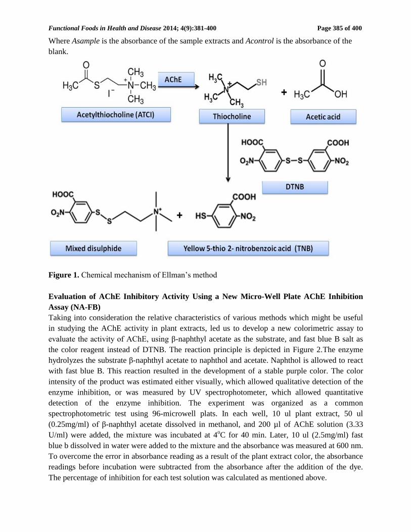
Functional Foods in Health and Disease 2014; 4(9):381-400 Page 385 of 400
Where Asample is the absorbance of the sample extracts and Acontrol is the absorbance of the
blank.
Figure 1. Chemical mechanism of Ellman’s method
Evaluation of AChE Inhibitory Activity Using a New Micro-Well Plate AChE Inhibition
Assay (NA-FB)
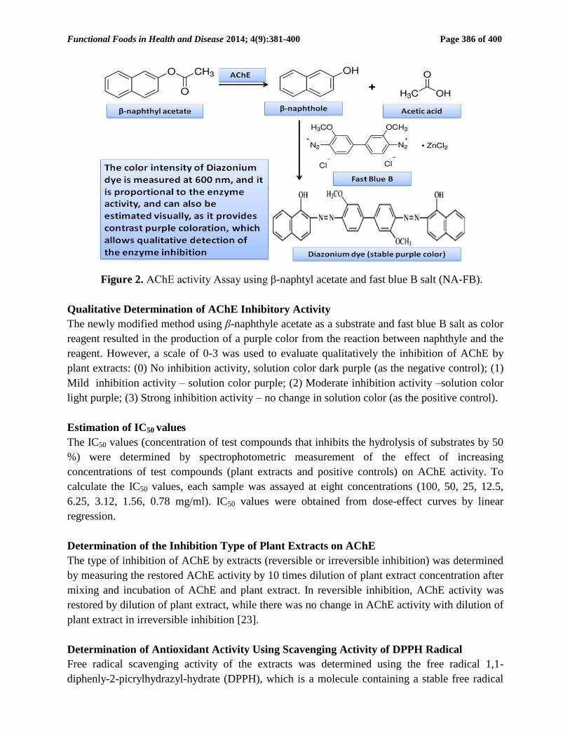
Taking into consideration the relative characteristics of various methods which might be useful
in studying the AChE activity in plant extracts, led us to develop a new colorimetric assay to
evaluate the activity of AChE, using β-naphthyl acetate as the substrate, and fast blue B salt as
the color reagent instead of DTNB. The reaction principle is depicted in Figure 2.The enzyme
hydrolyzes the substrate β-naphthyl acetate to naphthol and acetate. Naphthol is allowed to react
with fast blue B. This reaction resulted in the development of a stable purple color. The color
intensity of the product was estimated either visually, which allowed qualitative detection of the
enzyme inhibition, or was measured by UV spectrophotometer, which allowed quantitative
detection of the enzyme inhibition. The experiment was organized as a common
spectrophotometric test using 96-microwell plats. In each well, 10 ul plant extract, 50 ul
(0.25mg/ml) of β-naphthyl acetate dissolved in methanol, and 200 µl of AChE solution (3.33
U/ml) were added, the mixture was incubated at 4oC for 40 min. Later, 10 ul (2.5mg/ml) fast
blue b dissolved in water were added to the mixture and the absorbance was measured at 600 nm.
To overcome the error in absorbance reading as a result of the plant extract color, the absorbance
readings before incubation were subtracted from the absorbance after the addition of the dye.
The percentage of inhibition for each test solution was calculated as mentioned above.
Functional Foods in Health and Disease 2014; 4(9):381-400 Page 386 of 400
Figure 2. AChE activity Assay using β-naphtyl acetate and fast blue B salt (NA-FB).
Qualitative Determination of AChE Inhibitory Activity
The newly modified method using β-naphthyle acetate as a substrate and fast blue B salt as color
reagent resulted in the production of a purple color from the reaction between naphthyle and the
reagent. However, a scale of 0-3 was used to evaluate qualitatively the inhibition of AChE by
plant extracts: (0) No inhibition activity, solution color dark purple (as the negative control); (1)
Mild inhibition activity – solution color purple; (2) Moderate inhibition activity –solution color
light purple; (3) Strong inhibition activity – no change in solution color (as the positive control).
Estimation of IC50 values
The IC50 values (concentration of test compounds that inhibits the hydrolysis of substrates by 50
%) were determined by spectrophotometric measurement of the effect of increasing
concentrations of test compounds (plant extracts and positive controls) on AChE activity. To
calculate the IC50 values, each sample was assayed at eight concentrations (100, 50, 25, 12.5,
6.25, 3.12, 1.56, 0.78 mg/ml). IC50 values were obtained from dose-effect curves by linear
regression.
Determination of the Inhibition Type of Plant Extracts on AChE
The type of inhibition of AChE by extracts (reversible or irreversible inhibition) was determined
by measuring the restored AChE activity by 10 times dilution of plant extract concentration after
mixing and incubation of AChE and plant extract. In reversible inhibition, AChE activity was
restored by dilution of plant extract, while there was no change in AChE activity with dilution of
plant extract in irreversible inhibition [23].
Determination of Antioxidant Activity Using Scavenging Activity of DPPH Radical
Free radical scavenging activity of the extracts was determined using the free radical 1,1-
diphenly-2-picrylhydrazyl-hydrate (DPPH), which is a molecule containing a stable free radical
Functional Foods in Health and Disease 2014; 4(9):381-400 Page 387 of 400
[38]. In the presence of an antioxidant which can donate an electron to DPPH, the purple color
which is typical for free DPPH radical decays and the change in absorbance at 517 nm is
followed spectrophotometerically. The effect of the plant extracts on DPPH radical was
estimated using the method of Liyana-Pathirana and Shahidi [39] with minor modification. Twenty
five micro liter of plant extract were added to 175µl of 0.004% DPPH methanolic solution, in a
96-well plate. Appropriate blanks were prepared using the solvent only in addition to the same
amount of DPPH reagent to overcome any inherent solvent activity. The reaction mixture was
shaken well and allowed to stand at room temperature in the dark for 30 min, and then the
decrease in absorbance at 517 nm was measured against a control (methanol solution) by using
UV-vis spectrophotometer. The radical-scavenging activity of samples, expressed as percentage
inhibition of DPPH (I %), and it was calculated according to the formula:
% I = [(Acontrol-Asample) / Acontrol] X 100
Where Acontrol is the absorbance of DPPH radical
IC50 (concentration of the extract/compound producing 50% scavenging of DPPH radicals) was
determined using non-linear regression analysis of the dose-%I relationship. The extract
concentration providing 50% inhibition (IC50) was calculated from the graph of inhibition
percentage plotted against extract concentration (100, 50, 25, 12.5, 6.25, 3.12, 1.56, 0.78 mg/ml).
Antioxidant capacities of the extracts were compared with those of BHA, gallic acid and
ascorbic acid. Tests were carried out in triplicates.
Data Analysis
Tests were carried out were possible at least in duplicate on two different occasions. Results are
reported as mean ± standard deviation (S.D.). Standard curves were generated and calculation of
the 50% inhibitory concentration (IC50) values was done using Excel.
RESULTS:
Forty seven plant species were selected based on their uses as remedies for the central nervous
system diseases, as antidotes for human and animal poisoning or to improve memory and
cognitive function (Table 1). The inhibition effect of the 92 different extracts on AChE activity
was screened using the Ellman’s method and the new micro-well plate AChE inhibition assay,
NA-FB.
The results obtained by Ellman’s method and the NA-FB assay of all plant extracts are
shown in Table 1. The screenings were performed at a concentration of 100 mg/ml, and the
extracts were considered as active if they only inhibited the enzyme >50%.
Sixty two (67.4%) and 34 (37%) extracts inhibited AChE by > 50% using the NA-FB, and
Ellman’s assays, respectively. Also, some of the extracts such as Salvia fruticosa, Galium
pisiferum, Anemone coronaria, Juglans regia, Ornithogalum narbonense and the leaves of
Asphodeline lutea, which had lower activity against AChE using Ellman’s method, exhibited
much higher activity using the NA-FB (Figure 3). Therefore, AChEI analysis by Ellman’s
method was excluded from further discussion.
Functional Foods in Health and Disease 2014; 4(9):381-400 Page 388 of 400
Table 1. Biological activities of Palestinian plants against different bioassays related to
Alzheimer disease
No. Scientific name
Family Name
Voucher
No.
Plant
part
AChEI
on a
scale of
0-3
% of Inhibition IC50 (mg/ml)
NA-FB Ellman’s DPPH NA-FB Ellman’s DPPH
1. Allium neapolitanum Cirillo Liliaceae BERC-414 LE 2 63.4±2.26 6.21± 0.30 42.12±1.24 74.25 NA NA
2. FL 2 55.96±1.47 58.04±1.19 53.26±1.78 99.4 44.3 6.16
3. ST 1 47.72±3.22 28.75±2.33 58.36±2.32 NA NA NA
4. BU 1 33.28±0.40 48.46±3.05 73.4±0.85 NA NA NA
5. Anemone coronaria L. Ranunculaceae BERC-355 FL 3 83.36±0.91 35.67±2.02 68.94±1.33 71.5 NA 1.7
6. LE 2 76.04±1.36 33.33±.33 76.73±1.03 71.5 NA 60.17
7. Asphodeline lutea (L.) Rchb. Liliaceae BERC-371 LE 2 75±2.83 6.08±0.11 70.16±0.23 17.75 NA 100.21
8. BU 0 - 25.36±6.56 41.34±3.76 22.1±1.27 NA NA NA
9. Asphodelus aestivus Brot.
(Asphodelus microcarpus
Salzm. & Viv)
Liliaceae BERC-210 BU 1 49.7±0.42 4.64±0.91 53.55±2.05 NA NA NA
10. FL 0 - 6.42±0.82 1.73±0.38 25.3±0.99 NA NA NA
11. Bellevalia flexuosa Boiss. Liliaceae BERC-374 ST 1 51.27±5.28 2.47±0.75 14±1.41 97.3 NA NA
12. LE 2 59.88±1.24 11.38±0.54 52.3±0.99 31.75 NA 12.76
13. BU 1 49.33±0.95 22.37±0.52 65.34±0.93 NA NA NA
14. FR 2 64.08±1.30 10.96±1.36 82.69±1.85 97.9 NA 2.14
15. FL 2 62.36±2.32 19.75±1.06 25.3±1.5 73.5 NA NA
16. Chrysanthemum coronarium
L.
Asteraceae BERC-068 LE 0 20.1±4.10 38.91±1.29 22.4±0.2 NA NA NA
17. Conyza bonariensis
Cronquist.
Asteraceae BERC-259
LE 2 70.1±2.69 81.23±2.50 65.25±1.06 2.89 54.8 2.74
18. FL 3 74.092±2.70 83.12±2.66 83.94±0.08 3.45 0.35 2.74
19. Dodonaea viscosa L. Sapindaceae BERC-045 LE 1 31.96±2.77 40.27±0.38 138.83±0.24 NA NA NA
20. Erodium malacoides (l.)
L'Her.
Geraniaceae BERC-357 LE 0 - 4.10.57 33.69±0.98 70.5±0.71 NA NA NA
21. FL 0 - 7.51±0.69 1.11±1.26 74.25±1.06 NA NA NA
22. Eruca sativa Miller Brassicaceae BERC-202 LE 0 17.33±2.18 79.41±1.08 75.8±0.54 NA 36.7 NA
23. Euphorbia hierosolymitana
Boiss.
Euphorbiaceae BERC-170 LE 0 10.29±1.00 19.75±1.06 93.07±0.10 NA NA NA
24. FL 0 11.38±0.88 29.81±1.15 99.33±1.88 NA NA NA
25. Foeniculum vulgare Mill. Apiaceae BERC-030 LE 3 89.81±1.03 52.68±0 72.12±0.76 3.5 99.3 27.54
26. Fumaria capreolata L. Fumariaceae BERC-367 FR 3 84.36±1.92 97.37±0.52 37.08±1.30 0.025 0.34 NA
27. LE 3 82.26±2.46 90.27±0.38 69±1.41 0.035 0.55 1.34
28. FL 3 85.25±1.06 98.14±1.61 76.41±0.83 0.921 2.1 0.514
29. Fumaria densiflora DC. Fumariaceae BERC-154 LE 3 79.78±0.31 83.12±0.17 56±1.41 0.62 21 0.51
30. FL 3 80.81±0.27 82.31±0.44 73.66±1.90 10.5 3.25 0.68
31. Fumaria vaillantii loisel Fumariaceae BERC-396 LE 3 81.17±1.17 95.98±1.39 67.25±0.35 10.5 5.75 1.4
32. Galium pisiferum Boiss. Rubiaceae BERC-038 FL 2 73.64±1.92 13.38±0.54 60.5±0.71 8.45 NA 24.16
33. LE 3 77.84±0.23 18.04 ± 0.06 81.58±0.59 10.45 NA 45.53
34. Helichrysum sanguineum
(L.) Kostel.
Asteraceae BERC-091
LE 2 70.84±0.23 28.46 ±0.65 100±1.41 6.35 NA 44.5
35. FL 2 68.56±0.79 16.5±0.71 100±1.41 35.5 NA 53.5
36. Juglans regia L. Juglandaceae BERC-230 FR 3 78.562±2.39 11.36±1.50 78.69±0.20 8.52 NA 43.86
37. Lupinus pilosus L.
(L. varius L.)
Papilionaceae BERC-019 FL 3 85.24±1.07 53.98±1.39 44.23±1.09 17.75 23.5 NA
38. LE 3 92.02±1.39 70.52±0.74 88.16±1.19 3.49 2.23 1.15
39. FR 1 23.13±0.034 44.93±1.5 71.26±0.27 NA NA NA
40. Majorana syriaca
(L.) Rafin.
Lamiaceae BERC-026
LE 2 70.58±0.59 88.1±1.98 100±1.3 2.9 9.5 0.21
41. Mandragora autumnalis
Bertol
Solanaceae BERC-286
FR 2 71.43±0.81 98.12±0.17 27.21±0.30 10.06 8.5 NA
42. RT 3 74.21±1.12 97.46±0.65 48.08±1.30 48.1 49 NA
43. LE 2 64.4±0.85 94.4±0.57 68.91±0.13 5.1 1.48 1.17
44.
FR(ri
pen)
2 71.78±2.9 72.0±1.94 62.11±1.45 34.85 38.35 5.17
45. . Mentha spicita L. Labiatae BERC-116 LE 3 94.8±1.93 74.17±0.176 93.52±0.33 6.3 36.35 0.56
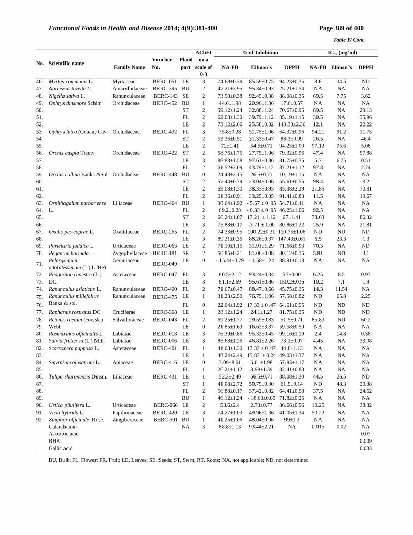
Functional Foods in Health and Disease 2014; 4(9):381-400 Page 389 of 400
Table 1/ Cont.
BU, Bulb, FL, Flower; FR, Fruit; LE, Leaves; SE, Seeds; ST, Stem; RT, Roots; NA, not applicable; ND, not determined
No. Scientific name
Family Name
Voucher
No.
Plant
part
AChEI
on a
scale of
0-3
% of Inhibition IC50 (mg/ml)
NA-FB Ellman’s DPPH NA-FB Ellman’s DPPH
46. Myrtus communis L. Myrtaceae BERC-051 LE 3 74.68±0.38 85.59±0.75 94.23±0.35 3.6 34.5 ND
47. Narcissus tazetta L. Amaryllidaceae BERC-395 BU 2 47.21±3.95 95.34±0.93 25.21±1.54 NA NA NA
48. Nigella sativa L. Ranunculaceae BERC-143 SE 2 73.58±0.38 92.49±0.38 88.08±0.35 69.5 7.75 3.62
49. Ophrys dinsmore Schltr Orchidaceae BERC-452 BU 1 44.6±1.98 20.96±1.36 17.6±0.57 NA NA NA
50. ST 2 59.12±1.24 32.88±1.24 70.67±0.95 89.5 NA 29.13
51. FL 2 62.08±1.30 39.79±1.12 85.19±1.15 30.5 NA 35.96
52. LE 2 73.12±2.66 25.58±0.82 143.33±2.36 12.1 NA 22.22
53. Ophrys lutea (Gouan) Cav Orchidaceae BERC-432 FL 3 75.8±0.28 51.75±1.06 64.32±0.96 94.21 91.2 11.75
54. ST 2 53.36±0.51 31.33±0.47 88.3±0.99 26.5 NA 46.4
55. LE 2 72±1.41 54.5±0.71 94.23±1.09 97.12 95.6 5.08
56. Orchis caspia Trautv Orchidaceae BERC-422 ST 2 68.76±1.75 27.75±1.06 79.32±0.96 47.4 NA 57.88
57. LE 3 88.88±1.58 97.61±0.86 81.75±0.35 5.7 6.75 0.51
58. FL 2 61.52±2.09 43.79±1.12 87.21±1.12 97.8 NA 2.74
59. Orchis collina Banks &Sol. Orchidaceae BERC-448 BU 0 24.48±2.15 26.5±0.71 10.19±1.15 NA NA NA
60. ST 2 57.44±0.79 23.04±0.06 55.61±0.55 98.4 NA 3.2
61. LE 2 69.08±1.30 38.33±0.95 85.38±2.29 21.85 NA 70.81
62. FL 2 61.36±0.91 33.25±0.35 91.41±0.83 11.5 NA 19.67
63. Ornithogalum narbonense
L.
Liliaceae BERC-464 BU 1 38.64±1.92 - 5.67 ± 0 .95 54.71±0.41 NA NA NA
64. FL 2 69.2±0.28 - 0.33 ± 0 .95 46.25±1.06 92.5 NA NA
65. ST 2 66.24±1.07 17.21 ± 1.12 67±1.41 78.63 NA 86.32
66. LE 3 75.88±0.17 -3.71 ± 1.00 80.86±1.22 25.9 NA 21.81
67. Oxalis pes-caprae L. Oxalidaceae BERC-265 FL 2 74.33±0.95 100.22±0.31 110.75±1.06 ND ND ND
68. LE 3 89.21±0.35 88.26±0.37 147.43±0.61 6.5 23.3 1.3
69. Parietaria judaica L. Urticaceae BERC-063 LE 2 71.19±1.15 31.91±1.29 71.66±0.93 70.3 NA ND
70. Peganum harmala L. Zygophyllaceae BERC-181 SE 2 50.85±0.21 81.06±0.08 80.12±0.15 5.81 ND 3.1
71. Pelargonium
odoratissimum (L.) L 'He'r
Geraniaceae BERC-049
LE 0 - 15.44±0.79 - 1.58±1.24 88.91±0.13 NA NA NA
72. Phagnalon rupestre (L.)
DC.
Asteraceae BERC-047 FL 3 80.5±2.12 93.24±0.34 57±0.00 6.25 8.5 0.93
73. LE 3 81.1±2.69 95.61±0.86 150.2±.036 10.2 7.1 1.9
74. Ranunculus asiaticus L. Ranunculaceae BERC-400 FL 2 71.67±0.47 88.47±0.66 45.75±0.35 14.3 11.54 NA
75. Ranunculus millefolius
Banks & sol.
Ranunculaceae BERC-475 LE 1 31.23±2.50 76.75±1.06 57.58±0.82 ND 65.8 2.25
76. FL 0 22.64±1.92 17.33 ± 0 .47 64.61±0.55 ND ND ND
77. Raphanus rostratus DC. Cruciferae BERC-368 LE 1 28.12±1.24 24.1±1.27 81.75±0.35 ND ND ND
78. Retama raetam (Forssk.)
Webb
Salvadoraceae BERC-043 FL 2 69.25±1.77 29.59±0.83 51.5±0.71 85.83 ND 60.2
79. LE 0 21.85±1.63 16.62±3.37 59.58±0.59 NA NA NA
80. Rosmarinus officinalis L. Labiatae BERC-018 LE 3 76.39±0.86 95.32±0.45 99.16±1.19 2.4 14.8 0.38
81. Salvia fruticosa (L.) Mill. Labiatae BERC-006 LE 3 85.68±1.26 46.81±2.26 73.1±0.97 4.45 NA 33.08
82. Scorzonera papposa L. Asteraceae BERC-401 FL 1 41.08±1.30 17.33 ± 0 .47 44.8±1.13 NA NA NA
83. LE 1 48.24±2.49 15.83 ± 0.24 49.03±1.37 NA NA NA
84. Smyrnium olusatrum L. Apiaceae BERC-416 LE 0 3.09±8.61 5.01±1.98 57.83±1.17 NA NA NA
85. FL 1 26.21±1.12 3.98±1.39 82.41±0.83 NA NA NA
86. Tulipa sharonensis Dinsm. Liliaceae BERC-431 LE 1 52.3±2.40 56.5±0.71 38.08±1.30 44.5 26.5 ND
87. ST 1 41.08±2.72 50.79±0.30 61.9±0.14 ND 48.3 20.38
88. FL 2 56.88±0.17 37.42±0.82 64.41±0.58 37.5 NA 24.62
89. BU 1 46.12±1.24 - 18.63±0.89 71.82±0.25 NA NA NA
90. Urtica pilulifera L. Urticaceae BERC-066 LE 2 58.6±2.4 2.73±0.77 86.66±0.96 10.25 NA 38.32
91. Vicia hybrida L. Papilionaceae BERC-420 LE 3 74.27±1.03 49.96±1.36 41.05±1.34 50.23 NA NA
92. Zingiber officinale Rose. Zingiberaceae BERC-501 BU 1 41.25±1.06 48.04±0.06 99±1.2 NA NA NA
Galanthamin NA 3 88.8±1.13 93.44±2.21 NA 0.015 0.02 NA
Ascorbic acid 0.07
BHA 0.009
Gallic acid 0.033
Functional Foods in Health and Disease 2014; 4(9):381-400 Page 390 of 400
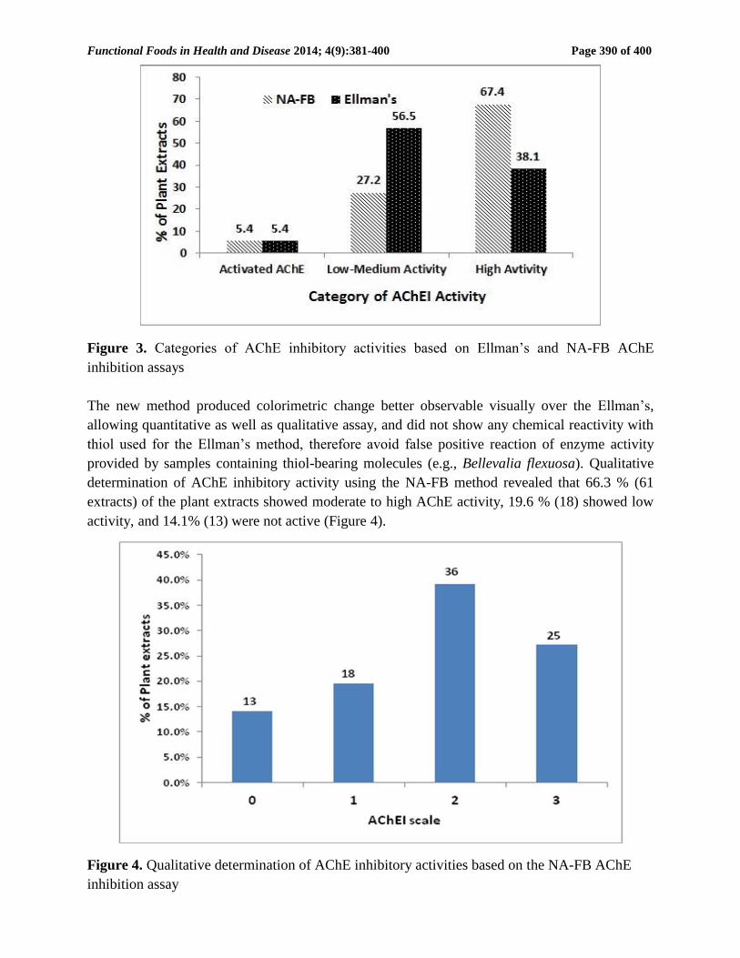
Figure 3. Categories of AChE inhibitory activities based on Ellman’s and NA-FB AChE
inhibition assays
The new method produced colorimetric change better observable visually over the Ellman’s,
allowing quantitative as well as qualitative assay, and did not show any chemical reactivity with
thiol used for the Ellman’s method, therefore avoid false positive reaction of enzyme activity
provided by samples containing thiol-bearing molecules (e.g., Bellevalia flexuosa). Qualitative
determination of AChE inhibitory activity using the NA-FB method revealed that 66.3 % (61
extracts) of the plant extracts showed moderate to high AChE activity, 19.6 % (18) showed low
activity, and 14.1% (13) were not active (Figure 4).
Figure 4. Qualitative determination of AChE inhibitory activities based on the NA-FB AChE
inhibition assay
Functional Foods in Health and Disease 2014; 4(9):381-400 Page 391 of 400
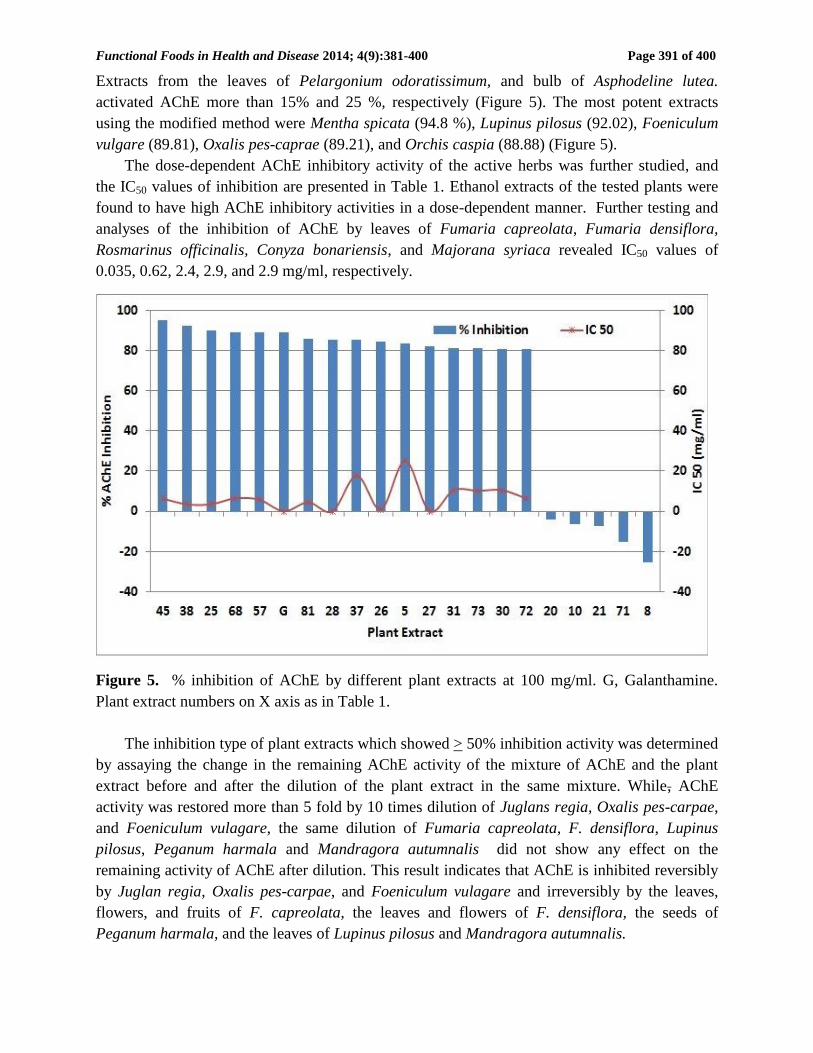
Extracts from the leaves of Pelargonium odoratissimum, and bulb of Asphodeline lutea.
activated AChE more than 15% and 25 %, respectively (Figure 5). The most potent extracts
using the modified method were Mentha spicata (94.8 %), Lupinus pilosus (92.02), Foeniculum
vulgare (89.81), Oxalis pes-caprae (89.21), and Orchis caspia (88.88) (Figure 5).
The dose-dependent AChE inhibitory activity of the active herbs was further studied, and
the IC50 values of inhibition are presented in Table 1. Ethanol extracts of the tested plants were
found to have high AChE inhibitory activities in a dose-dependent manner. Further testing and
analyses of the inhibition of AChE by leaves of Fumaria capreolata, Fumaria densiflora,
Rosmarinus officinalis, Conyza bonariensis, and Majorana syriaca revealed IC50 values of
0.035, 0.62, 2.4, 2.9, and 2.9 mg/ml, respectively.
Figure 5. % inhibition of AChE by different plant extracts at 100 mg/ml. G, Galanthamine.
Plant extract numbers on X axis as in Table 1.
The inhibition type of plant extracts which showed > 50% inhibition activity was determined
by assaying the change in the remaining AChE activity of the mixture of AChE and the plant
extract before and after the dilution of the plant extract in the same mixture. While, AChE
activity was restored more than 5 fold by 10 times dilution of Juglans regia, Oxalis pes-carpae,
and Foeniculum vulagare, the same dilution of Fumaria capreolata, F. densiflora, Lupinus
pilosus, Peganum harmala and Mandragora autumnalis did not show any effect on the
remaining activity of AChE after dilution. This result indicates that AChE is inhibited reversibly
by Juglan regia, Oxalis pes-carpae, and Foeniculum vulagare and irreversibly by the leaves,
flowers, and fruits of F. capreolata, the leaves and flowers of F. densiflora, the seeds of
Peganum harmala, and the leaves of Lupinus pilosus and Mandragora autumnalis.
Functional Foods in Health and Disease 2014; 4(9):381-400 Page 392 of 400
Table 1 shows the antioxidant results of the tested plant extracts. Seventy-three extracts
showed > 50% antioxidant activity, of these Phagnalon rupestre, Oxalis pes-caprae, Ophrys
dinsmor, Dodonaea viscosa, Helichrysum sanguineum, and Majorana syriaca were the most
active. The IC50 of the antioxidant activity for the plants extracts, which showed > 50 % AChE
inhibition activity using the NA-FB was determined (Table 1). Of these eight extracts; leaves of
M. syriaca (IC50 0.212mg/ml), leaves of Rosmarinus officinalis (0.377 mg/ml), leaves of
Fumaria densiflora (0.514 mg/ml), leaves of Orchis caspia (0.514 mg/ml), leaves of Mentha
apicata (0.56 mg/ml) flowers of Fumaria densiflora (0.678 mg/ml), flowers of Fumaria
capreolata (0.69 mg/ml), and flowers of Phagnalon rupestre (0.928 mg/ml) were particularly
strong antioxidants when compared to the reference radical scavengers (BHA, gallic acid, and
ascorbic acid) recording IC50’s < 1 mg/ml.
DISCUSSION:
Palestine is enriched with great plant diversity, and 368 of these plants have been reported to be
used in TAPHM for the treatment of several diseases [40]. However, the use of medicinal plants
is mainly based on local tradition and not scientific knowledge [41-43]. The chemical
constituents of most of these plants are unknown and may have dangerous effects on human
health. On the other hand, some plants, which are not reported to be used in herbal medicine,
might also possess potential activity.
The deficiency of ACh is one of characteristics of AD and responsible for most of its
symptoms, such as a decline in memory and cognition. AChE inhibitors such as tacrine,
donepezil, rivastigmine, and galantamine are currently used as anti-AD drugs [44]. The side
effects of these anti-AChE drugs, such as toxicity, tolerability, and loss of efficiency, have
interested the researchers to consider alternative natural anti-AD substances in place of current
synthetic medications [45].
In the present work, the selected extracts were screened for AChE inhibition using the
Ellman’s method and the NA-FB assay at 100 mg/ml dose. The Ellman’s method is the most
widely used AChE inhibitory assay [32]. This method has some advantages and disadvantages.
Its main advantages are simplicity, rapid processing of large numbers of samples, fast conversion
of ACTI comparing to other artificial substrates such as naphthyle acetate and relatively low cost
[46, 47]. On the other hand, Ellman’s method has some disadvantages, including the interference
of some compounds. The -SH groups in the plant extract may react with DTNB and ATCh, thus
the natural substrates are not identical from a kinetic point of view. False positive reaction of
enzyme activity can be provided by samples containing a lot of thiol-bearing molecules.
To overcome this problem an alternative method using β-naphthyl acetate as the substrate
and fast blue B as the color reagent (absorbance at 600 nm) instead of DTNB was developed in
this study. The main advantages of this method is that the colorimetric change is better
observable visually, allowing spectrophotometric as well as colorimetric assay, and does not
show any chemical reactivity with thiol, therefore avoid false positive reaction of enzyme
activity provided by samples containing thiol-bearing molecules. The NA-FB method can be
considered superior and more sensitive than the Ellman’s assay. In the present study, 67.4% of
plant extracts inhibited AChE by > 50% using the NA-FB method, while only 37% extracts
inhibited AChE by > 50% using Ellman’s assay. This result can be attributed to the accuracy and
Functional Foods in Health and Disease 2014; 4(9):381-400 Page 393 of 400
stability of the reaction using naphthyle acetate. Naphthyle acetate splits with lower turnover
rate, and does not show any chemical reactivity with thiol used for the Ellman’s method [37],
thus NA-FB can be advantageously used for accurate measurements of AChE activity.
Sixty two extracts belonging to 34 plant species (Table 1) have been identified to effectively
inhibit AChE enzymes, which is considered to be related to the mechanism of memory
dysfunction in this study. In the light of these findings, we can conclude that most of the plant
extracts screened showed inhibitory activity against AChE and could be considered worthwhile
in future studies in the treatment of AD. In particular, the species belonging to Apiaceae,
Papilionaceae, Oxalidaceae, Orchidaceae, Lamiaceae, Fumariaceae families had the highest
activity ranging between 94.8 and 85.25% at 100 mg/ml concentration against AChE. Since most
of the AChE inhibitors are known to contain nitrogen, the higher activity of these extracts may
be due to their rich alkaloidal content [9].
The most potent extracts were the leaves of Fumaria capreolata, Fumaria densiflora,
Rosmarinus officinalis, Conyza bonariensis, and Majorana syriaca with IC50 values of 0.035,
0.62, 2.4, 2.9, and 2.9 mg/ml respectively. At a concentration of 100 mg mL–1, they reduced the
enzymatic activity of AChE to 17.74%, 20.22, 23.61, 29.9 and 29.42 respectively. The inhibition
type of AChE varied among plant extracts, while 84 extracts showed reversible inhibition, 8
showed irreversible inhibitions. Although IC50 values of Fumaria capreolata, Fumaria
densiflora, are higher than that of Rosmarinus officinalis and Majorana syriaca, the inhibition
type in this study showed that Rosmarinus officinalis and Majorana syriaca reversibly inhibits
AChE and can be used for AD’s medication rather than Fumaria capreolata, F. densiflora which
inhibits irreversibly AChE. This recommendation was supported by the toxicity reports in
literature which indicated the higher safety margin of R. officinalis and M. syriaca as compared
to Fumaria species.
Rosmarinus officinalis (rosemary) contains the natural COX‑2 inhibitors (e.g. Apigenin,
carvacrol, eugenol, oleanolic acid, thymol, and ursolic acid, which can prevent Alzheimer’s
disease [48] . In addition, rosemary contains antioxidants and anti‑inflammatory compounds.
Some of the strongest antioxidant substances in the plant are carnosic acid and ferulic acid,
which have been reported to posses antioxidant activity much higher than the widely common
synthetic antioxidants butylated hydroxytoluene (BHT) and butylated hydroxyanisole (BHA) [3].
Conyza bonariensis is a medicinal plant, which has been reported to be used for constipation
and diarrhea, has been attributed to the spasmogenic and spasmolytic constitute of the plant [49].
The leaves and flowers of the plants have shown to possess high AChEI activity > 70% with IC50
value ranging between 2.89-3.45 mg/ml.
Majorana syriaca possesses an important food-flavouring ingredient in the Middle Eastern
culture, known commonly as Za'atar. The plant is used traditionally for the treatment of several
ailments and is associated as memory enhancer [2]. The main components of the plant extract
according to GS-MS analysis were thymol, and carvacrol. The remaining compounds comprise
flavonoids and phenolic acids that provide the antiradical and antioxidant activity [50]. The plant
has shown to have strong activity against AChE, ursolic acid which has been isolated from the
plant, has shown to be a potent AChE inhibitor in Alzheimer's Disease [51].
Functional Foods in Health and Disease 2014; 4(9):381-400 Page 394 of 400
Fumaria species have been used in traditional medicine as antihypertensives, diuretics,
hepatoprotectants and laxatives (to treat gastrointestinal disorders), as well as in the treatment of
rachis and conjunctivitis [52]. The plant has also been evaluated pharmacologically and shown to
possess antihelmintic, antipyretic and hypoglycemic properties [53-55]. The biological activities
of Fumaria species have been mainly associated with the presence of isoquinoline alkaloids [56].
The toxicity of the species have been evaluated, Fumaria capreolata has shown to be non-toxic
[56], while F. densiflora was reported to be toxic [57]. The AChEI activity of Fumaria species
has been reported by several researchers; the plant was reported to have strong AChEI activity
[9]. In this study, Fumaria were among the most active plant extracts against AChE activity,
however, the reaction was shown to be irreversible, thus the plant cannot be used for the
treatment of AD as the activity of the enzyme cannot be restored.
AChEI activity of the methanolic extract of Peganum harmala, has previously been reported
by Ali et al. [26]. The plant contains β-carboline alkaloids, which demonstrated potent activity
against AChE [58]. Harmaline, the major active constituent of P. harmala, is a common dihydro
β-carboline type; it possess interesting pharmacological activities and can interact with several
enzymes and neurotransmitters including topoisomerase I, and monoamine oxidase-A [59, 60].
Although, P. harmala has been used in traditional medicine, there are reports of severe
intoxication in cattle, donkeys, sheep and horses [61]. Digestive and nervous syndromes have
been reported in animals that consume a sub-lethal amount of the plant. Harmaline and harmine
are toxic alkaloids characterized in the seeds of P. harmala. Harmaline is almost twice as toxic
as harmine and in moderate doses cause tremors and clonic convulsions, but with no increase in
spinal reflex excitability [62]. The seeds of P. harmala were among the potent plant extracts
against AChE activity, however, the reaction have been shown to be irreversible, thus the plant
cannot be used for the treatment of AD.
Some insecticides including organophosphate and carbamates cause AChEI which lead to
the accumulation of ACh at neuromuscular junctions causing rapid twitching of voluntary
muscles and eventually paralysis of the insects. However, in this study, leaves and flowers of
Fumaria species, seeds of P. harmala, and the leaves of Lupinus pilosus and Mandragora
autumnalis, which have shown high irreversible AChEI activity, can be considered potent natural
insecticides.
Alzheimer’s appears to be caused to a large degree by oxidative damage [63]. Therefore,
antioxidants, in general, should have positive effects in both the prevention and treatment of
Alzheimer’s. A study found that antioxidants such as vitamin A, vitamin D, lycopene, and beta
carotene were all significantly lower in Alzheimer’s disease patients compared to controls [64].
Another study of 633 patients aged > 65 years found that high dose supplementation with
vitamin C decreased the risk of developing AD [65]. Therefore, the plant extracts which
demonstrated potent free radical scavenging properties are expected to play a vital role in
reducing the oxidative stress and this may explain their use in traditional medicine for
improvement of AD and/or ageing related diseases. It’s worth mentioning that some of the plant
extracts which have high antioxidant activity including M. spicata (93.52), Z. officinale (99), R.
officinalis (99.16), M. syriaca (100), and the leaves of O. pes-caprae (147.43), are wild edible
plants widely consumed among the Palestinian population [66]. Some of these plants have been
reported to be used traditionally for memory enhancement [2, 3, 23, 66].
Functional Foods in Health and Disease 2014; 4(9):381-400 Page 395 of 400
CONCLUSION:
The new micro-well plate AChE activity assay (NA-FB) has shown to be simple, accurate,
sensitive, spectrophotometric and colorimetric, and superior to the Ellman’s method, and
therefore can be used efficiently for qualitative and quantitative studies of AChE inhibitory
activities of plant extracts of a wide range of diverse plant species and to give high detection
rates from a range of plant parts. The extracts of R. officinalis, M. spicata, M. syriaca, and N.
sativa were proved to have a great potential and should be considered for further studies to
identify the constituents responsible for the AChE inhibitory activity, which can be eventually
utilized in the prevention and treatment of AD.
The pathophysiological process of AD is thought to begin many years before the diagnosis
of AD dementia. This long "preclinical" phase of AD would provide an important opportunity
for therapeutic intervention. It is hoped that plants with strong reversible AChEI and strong
antioxidant activities will aid in earlier intervention at a stage of AD when some disease-
modifying therapies may be most efficacious.
List of abbreviations:
TAPHM Traditional Arabic Palestinian Herbal Medicine
AD Alzheimer’s disease
ACh acetylcholine
AChE acetylcholinesterase
AChEIs AChE inhibitors
ACTI acetylthiocholine iodide
DTNB 5,5’-dithio-bis-2-nitrobenzoic acid
BSA bovine serum albumin
DPPH diphenyl-1-picrylhydrazyl
BHA Butylated hydroxyanisole
NA-FB New Micro-Well Plate AChE Inhibition Assay
Competing interests: The authors declared no conflict of interests with respect to the authorship
and/or publication of this paper. All authors contributed to this study.
Author’s contribution: All authors contributed to this article.
Acknowledgements and funding:
This research was funded by the European Union under the ENPI CBC MED Progamme and is a
collaborative international project ref. no. I-B/1.1/288.
REFERENCES:
1. Ali-Shtayeh MS, Jamous RM. Ethnobotany of Palestinian herbal medicine in the
Northern West Bank and Gaza Strip: Review and a comprehensive field study.
Biodiversity and Environmental Sciences Studies Series 2006, 4:1-122.
2. Ali-Shtayeh MS, Jamous RM: Traditional Arabic Palestinian Herbal Medicine,
TAPHM.Til, Nablus: Biodiversity & Environmental Research Center-BERC; 2008.
Functional Foods in Health and Disease 2014; 4(9):381-400 Page 396 of 400
3. Singhal AK, Naithani V, Bangar OP. Medicinal plants with a potential to treat
Alzheimer and associated symptoms. International Journal of Nutrition, Pharmacology,
Neurological Diseases 2012, 2(2):84-91.
4. Ferreira A, C. Proença C, Serralheiro MLM, Araújo MEM. The in vitro screening for
acetylcholinesterase inhibition and antioxidant activity of medicinal plants from
Portugal. Journal of Ethnopharmacology 2006, 108(1):31-37.
5. Abou-Donia AH, Darwish FA, Toaima SM, Shawky E, Takla SS. A new approach to
develop a standardized method for assessment of acetylcholinesterase inhibitory activity
of different extracts using HPTLC and image analysis. Journal of Chromatography B
2014, 955:50-57.
6. Koedam ELGE, Lauffer V, van der Vlies AE, van der Flier WM, Scheltens P,
Pijnenburg YAL. Early-versus late-onset Alzheimer's disease: more than age alone.
Journal of Alzheimer's Disease 2010, 19(4):1401-1408.
7. Vinutha B, Prashanth D, Salma K, Sreeja SL, Pratiti D, Padmaja R, Radhika S, Amit A,
Venkateshwarlu K, Deepak M. Screening of selected Indian medicinal plants for
acetylcholinesterase inhibitory activity. Journal of Ethnopharmacology 2007,
109(2):359-363.
8. Greenblatt HM, Kryger G, Lewis T, Silman I, Sussman JL. Structure of
acetylcholinesterase complexed with (−)-galanthamine at 2.3 resolution. Febs Letters
1999, 463(3):321-326.
9. Orhan I, Sener B, Choudhary MI, Khalid A. Acetylcholinesterase and
butyrylcholinesterase inhibitory activity of some Turkish medicinal plants. Journal of
Ethnopharmacology 2004, 91(1):57-60.
10. Lahiri DK, Farlow MR, Greig NH, Sambamurti K. Current drug targets for Alzheimer's
disease treatment. Drug Development Research 2002, 56(3):267-281.
11. Howes M-JR, Houghton PJ. Plants used in Chinese and Indian traditional medicine for
improvement of memory and cognitive function. Pharmacology Biochemistry and
Behavior 2003, 75(3):513-527.
12. Mukherjee PK, Kumar V, Mal M, Houghton PJ. Acetylcholinesterase inhibitors from
plants. Phytomedicine 2007, 14(4):289-300.
13. Heinrich M, Lee Teoh H. Galanthamine from snowdrop—the development of a modern
drug against Alzheimer’s disease from local Caucasian knowledge. Journal of
Ethnopharmacology 2004, 92(2):147-162.
14. Rollinger JM, Hornick A, Langer T, Stuppner H, Prast H. Acetylcholinesterase
inhibitory activity of scopolin and scopoletin discovered by virtual screening of natural
products. Journal of Medicinal Chemistry 2004, 47(25):6248-6254.
15. Shah RS, Lee H-G, Xiongwei Z, Perry G, Smith MA, Castellani RJ. Current approaches
in the treatment of Alzheimer's disease. Biomedicine & Pharmacotherapy 2008,
62(4):199-207.
16. Wszelaki N, Kuciun A, Kiss A. Screening of traditional European herbal medicines for
acetylcholinesterase and butyrylcholinesterase inhibitory activity. Acta Pharmaceutica
2010, 60(1):119-128.
Functional Foods in Health and Disease 2014; 4(9):381-400 Page 397 of 400
17. Sung SY, Kang SY, Lee KY, Park MJ, Kim JH. (+)-α-Viniferin, a stilbene Trimer from
Caranga chamlague inhibits acetylcholinesterase. Biological & Pharmaceutical Bulletin
2002, 25:125-127.
18. Knapp MJ, Knopman DS, Solomon PR, Pendlebury WW, Davis CS, Gracon SI, Apter
JT, Lazarus CN, Baker KE, Barnett M. A 30-week randomized controlled trial of high-
dose tacrine in patients with Alzheimer's disease. JAMA: the Journal of the American
Medical Association 1994, 271(13):985-991.
19. Nicolson GL. Lipid replacement as an adjunct to therapy for chronic fatigue, anti-aging
and restoration of mitochondrial function. Journal of the American Nutraceutical
Association 2003, 6(3):22-28.
20. Feitosa CM, Freitas RM, Luz NNN, Bezerra MZB, Trevisan MTS. Acetylcholinesterase
inhibition by somes promising Brazilian medicinal plants. Brazilian Journal of Biology
2011, 71(3):783-789.
21. Benamar H, Rached W, Derdour A, Marouf A. Screening of Algerian Medicinal Plants
for Acetylcholinesterase Inhibitory Activity. Journal of Biological Sciences 2010,
10(1):1-9.
22. Amessis-Ouchemoukh N, Madani K, Falé PL, Serralheiro ML, Araújo MEM.
Antioxidant capacity and phenolic contents of some Mediterranean medicinal plants and
their potential role in the inhibition of cyclooxygenase-1 and acetylcholinesterase
activities. Industrial Crops and Products 2014, 53:6-15.
23. Ali SK, Hamed AR, Soltan MM, Hegazy UM, Elgorashi EE, El-Garf IA, Hussein AA.
In-vitro evaluation of selected Egyptian traditional herbal medicines for treatment of
alzheimer disease. BMC Complementary and Alternative Medicine 2013, 13(1):121.
24. Adewusi EA, Moodley N, Steenkamp V. Antioxidant and acetylcholinesterase inhibitory
activity of selected southern African medicinal plants. South African Journal of Botany
2011, 77(3):638-644.
25. Erkinjuntti T, Kurz A, Gauthier S, Bullock R, Lilienfeld S, Damaraju CV. Efficacy of
galantamine in probable vascular dementia and Alzheimer's disease combined with
cerebrovascular disease: a randomised trial. The Lancet 2002, 359(9314):1283-1290.
26. Tang M, Wang Z, Zhou Y, Xu W, Li S, Wang L, Wei D, Qiao Z. A Novel Drug
Candidate for Alzheimer's Disease Treatment: gx-50 Derived from Zanthoxylum
Bungeanum. Journal of Alzheimer's Disease 2013, 34(1):203-213.
27. Konrath EL, Neves BM, Lunardi PS, Passos CdS, Simões-Pires A, Ortega MG,
Gonçalvesb CA, Cabrera JL, Moreira JCF, Henriques AT. Investigation of the in vitro
and ex vivo acetylcholinesterase and antioxidant activities of traditionally used
Lycopodium species from South America on alkaloid. Journal of Ethnopharmacology
2012, 139(1):58-67.
28. Liu Y, Nair MG. An efficient and economical MTT assay for determining the
antioxidant activity of plant natural product extracts and pure compounds. Journal of
Natural Products 2010, 73(7):1193-1195.
29. Sultana R, Poon HF, Cai J, Pierce WM, Merchant M, Klein JB, Markesbery WR,
Butterfield DA. Identification of nitrated proteins in Alzheimer's disease brain using a
redox proteomics approach. Neurobiology of Disease 2006, 22(1):76-87.
Functional Foods in Health and Disease 2014; 4(9):381-400 Page 398 of 400
30. Zhu Z, Zheng T, Homer RJ, Kim Y-K, Chen NY, Cohn L, Hamid Q, Elias JA. Acidic
mammalian chitinase in asthmatic Th2 inflammation and IL-13 pathway activation.
Science 2004, 304(5677):1678-1682.
31. Ramassamy C. Emerging role of polyphenolic compounds in the treatment of
neurodegenerative diseases: a review of their intracellular targets. European Journal of
Pharmacology 2006, 545(1):51-64.
32. Miao Y, He N, Zhu J-J. History and new developments of assays for cholinesterase
activity and inhibition. Chemical reviews 2010, 110(9):5216-5234.
33. Pohanka M. Alzheimer's disease and related neurodegenerative disorders: implication
and counteracting of melatonin. Journal of Applied Biomedicine 2011, 9(4):185-196.
34. Ellman GL, Courtney KD, Featherstone RM. A new and rapid colorimetric
determination of acetylcholinesterase activity. Biochemical Pharmacology 1961,
7(2):88-95.
35. Pohanka M, Skládal P. Electrochemical biosensors--principles and applications. Journal
of Applied Biomedicine 2008, 6(2):57-64.
36. Šinko G, Čalić M, Bosak A, Kovarik Z. Limitation of the Ellman method:
Cholinesterase activity measurement in the presence of oximes. Analytical Biochemistry
2007, 370(2):223-227.
37. Pohanka M, Hrabinova M, Kuca K, Simonato J-P. Assessment of Acetylcholinesterase
Activity Using Indoxylacetate and Comparison with the Standard Ellman’s Method.
International Journal of Molecular Sciences 2011, 12(4):2631-2640.
38. Sharma OP, Bhat TK. DPPH antioxidant assay revisited. Food Chemistry 2009,
113(4):1202-1205.
39. Liyana-Pathirana CM, Shahidi F. Antioxidant activity of commercial soft and hard
wheat (Triticum aestivum L.) as affected by gastric pH conditions. Journal of
agricultural and food chemistry 2005, 53(7):2433-2440.
40. Ali-Shtayeh MS, Jamous RM, Abu-Zeitoun SY: BERC 2014 "National list of
Medicinal Plants in Palestine - West Bank and Gaza Strip".Til, Nablus: Biodiversity and
Environmental Research Center (BERC); 2014.
41. Ali-Shtayeh MS, Jamous RM, Jamous RM. Herbal preparation use by patients suffering
from cancer in Palestine. Complementary Therapies in Clinical Practice 2011,
17(4):235-240.
42. Ali-Shtayeh MS, Jamous RM, Jamous RM. Complementary and alternative medicine
use amongst Palestinian diabetic patients. Complementary Therapies in Clinical Practice
2012, 18(1):16-21.
43. Ali-Shtayeh MS, Yaniv Z, Mahajna J. Ethnobotanical survey in the Palestinian area: a
classification of the healing potential of medicinal plants. Journal of Ethnopharmacology
2000, 73(1):221-232.
44. Mehta SH, Sudarshi D, Srikrishnan AK, Celentano DD, Vasudevan CK, Anand S,
Kumar MS, Latkin C, Solomon S, Solomon SS. Factors associated with injection
cessation, relapse and initiation in a community―based cohort of injection drug users
in Chennai, India. Addiction 2012, 107(2):349-358.
Functional Foods in Health and Disease 2014; 4(9):381-400 Page 399 of 400
45. Gauthier S, Emre M, Farlow MR, Bullock R, Grossberg GT, Potkin SG. Strategies for
continued successful treatment of Alzheimer's disease: switching cholinesterase
inhibitors. Current Medical Research and Opinion 2003, 19(8):707-714.
46. Pohanka M, Vlček V, Žďárová K, Cabal J, Fusek J. Acetylcholinesterase Based
Colorimetric Dipsticks for Military Performance: Principles and Construction. Advances
in Military Technology 2012, 7(1).
47. Rakonczay Z, Brimijoin S. Monoclonal antibodies to rat brain acetylcholinesterase:
comparative affinity for soluble and membrane-associated enzyme and for enzyme from
different vertebrate species. Journal of Neurochemistry 1986, 46(1):280-287.
48. Duke J, McEvoy M, Sibbritt D, Guest M, Smith W, Attia J. Vibrotactile threshold
measurement for detecting peripheral neuropathy: defining variability and a normal
range for clinical and research use. Diabetologia 2007, 50(11):2305-2312.
49. Bukhari IA, Shah AJ, Khan RA, Meo SA, Khan A, Gilani AH. Gut modulator effects of
Conyza bonariensis explain its traditional use in constipation and diarrhea. European
Review for Medical and Pharmacological Sciences 2013, 17(4):552-558.
50. Ghada Al-Bandak G, Tsironi T, Taoukis P, Oreopoulou V. Antimicrobial and
antioxidant activity of Majorana syriaca in Yellowfin tuna. International Journal of Food
Science & Technology 2009, 44(2):373-379.
51. Chung Y-K, Heo H-J, Kim E-K, Kim H-K, Huh T-L, Lim Y, Kim S-K, Shin D-H.
Inhibitory Effect of Ursolic Acid Purified from Origanum majorana L. on the
Acetylcholinesterase. Molecules & Cells 2001, 11(2):137-143.
52. Stübing G, Peris JB: Plantes Medicinales de la Comunidad Valenciana.Valencia:
Generalitat, Valenciana; 1988.
53. Hordgen P, Hertzberg H, Heilmann Jr, Langhans W, Maurer V. The anthelmintic
efficacy of five plant products against gastrointestinal trichostrongylids in artificially
infected lambs. Veterinary Parasitology 2003, 117(1):51-60.
54. Khattak SG, Gilani SN, Ikram M. Antipyretic studies on some indigenous Pakistani
medicinal plants. Journal of Ethnopharmacology 1985, 14(1):45-51.
55. Akhtar MS, Khan QM, Khaliq T. Effect of Euphorbia prostrate and Fumaria indica in
normoglycemic and alloxan-treated hyperglycemic rabbits. Planta Medica 1984, 50:140-
142
56. Noureddine B, Yacine B, Fadila M-B. Evaluation of Erythrocytes Toxicity And
Antioxidant Activiry Of Alkaloids Of Fumaria Capreolata International Journal of
Pharma & Bio Sciences 2013, 4(2):770-776.
57. Erdoğan TF. Brine Shrimp Lethality Bioassay of Fumaria densiflora Dc. and Fumaria
officinalis L. Extracts. Hacettepe University Journal of the Faculty of Pharmacy 2009,
28(2):125-132.
58. Cao R, Peng W, Wang Z, Xu A. beta-Carboline alkaloids: biochemical and
pharmacological functions. Current Medicinal Chemistry 2007, 14(4):479-500.
59. Herraiz T, D. G, Ancín-Azpilicueta C, Arán VJ, Guillén H. β-Carboline alkaloids in
Peganum harmala and inhibition of human monoamine oxidase (MAO). Food and
Chemical Toxicology 2010, 48(3):839-845.
Functional Foods in Health and Disease 2014; 4(9):381-400 Page 400 of 400
60. Sobhani AM, Ebrahimi SA, Mahmoudian M. An in vitro evaluation of human DNA
topoisomerase I inhibition by Peganum harmala L. seeds extract and its beta-carboline
alkaloids. Journal of Pharmacy and Pharmaceutical Sciences 2002, 5(1):19-23.
61. Bailey M. Major poisonous plant problems in cattle. Bovine Pract 1979, 14:169-175.
62. Budavari S, Neil M: The Merck Index.New Jersey: CRC Press; 1996.
63. Pratico D, Delanty N. Oxidative injury in diseases of the central nervous system: focus
on Alzheimer's disease. American Journal of Medicine 2000, 109(7):577-585.
64. Foy CJ, Passmore AP, Vahidassr MD, Young IS, Lawson JT. Plasma chain-breaking
antioxidants in Alzheimer's disease, vascular dementia and Parkinson's disease. QJM:
An International Journal of Medicine 1999, 92(1):39-45.
65. Morris JS, Friston KJ, Büchel C, Frith CD, Young AW, Calder AJ, Dolan RJ. A
neuromodulatory role for the human amygdala in processing emotional facial
expressions. Brain 1998, 121(1):47-57.
66. Ali-Shtayeh MS, Jamous RM, Al-Shafie JH, Elgharabah WA, Kherfan FA, Qarariah
KH, Isra'S K, Soos IM, Musleh AA, Isa BA. Traditional knowledge of wild edible plants
used in Palestine (Northern West Bank): a comparative study. Journal of Ethnobiology
and Ethnomedicine 2008, 4(1):13.