in vitro somatic embryogenesis in … culture laboratory, department of studies in botany,...
TRANSCRIPT

Int J Pharm Bio Sci 2014 Jan; 5(1): (B) 103 - 111
This article can be downloaded from www.ijpbs.net
B - 103
Research Article Biotechnology
International Journal of Pharma and Bio Sciences ISSN
0975-6299
IN VITRO SOMATIC EMBRYOGENESIS IN ERYNGIUM
FOETIDUM L. – A MEDICINAL HERB.
M.S.SUDARSHANA*, AKSHATHA, B.H, MAHENDRA, C AND GURU, C
Tissue Culture Laboratory, Department of Studies in Botany, Manasagangotri, University of Mysore.
ABSTRACT
High frequency somatic embryogenesis and plant regeneration was achieved on calli developed from internodal and flower buds of E. foetidum. The explants were cultured on Murashige and Skoog medium supplemented with different concentrations and combinations of BA and IAA. The nodal explants produced multiple adventitious shoots while the internodal and flower buds induced callus, from which multiple shoot buds originated. Both direct and indirect shoots elongated within 4-6 weeks of culture. The well- organized shoots were individually separated and transferred to half strength MS liquid medium containing NAA or IAA at different concentrations. Rhizogenesis was observed within two weeks of transfer. They are transferred to polycups containing vermiculite for acclimatization and finally transplanted to soil. Histology of embryogenic calli revealed the presence of various types of embryoid and shoot buds both in the internodal and inflorescence calli. Upto fifty-five percent of the plants survived under field conditions.
KEY WORDS: Callus culture, Histology, Organogenesis, Eryngium foetidum.
*Corresponding author
M.S.SUDARSHANA
Tissue Culture Laboratory, Department of Studies in Botany,
Manasagangotri, University of Mysore.
Int J Pharm Bio Sci 2014 Jan; 5(1): (B) 103 - 111
This article can be downloaded from www.ijpbs.net
B - 104
INTRODUCTION
Eryngium foetidum L. (Apiaceae) is an endemic, aromatic herb distributed in tropical and temperate regions. It grows in Kashmir and Western Himalayas at altitudes of 5000- 6000 ft. It is cultivated for its leafy vegetable and essential oils, which is of high economic value in international trade1, the oil has applications in perfumery and cosmetic industry2. The main constituent of the oil is 2-didecen-1-al. The roots are considered tonic and aphrodisiac and ashes of the plant are recommended for haemorrhoids3. The roots contain saponin4. Saenz et al. 5 have demonstrated the anti-inflammatory and analgesic properties in the leaves of E. foetidum. In India the plant E. foetidum is cultivated in certain regions of Tamil Nadu, Kerala, Karnataka, Assam, Andaman and Nicobar islands. Propagation is hampered by a low germination seed rate and low seed viability. The flower buds are minute, conventional hybridization is tedious. Hence its utility for commercial application is greatly hindered. Large scale propagation is a prerequisite to meet the pharmaceutical needs and to conserve this medicinal species plant regeneration through tissue culture could be considered as the simple method for mass propagation. The in vitro production of plants from leaf and shoot tissue has been reported, 6 however documentation of regeneration through somatic embryogenesis from internodal calli and flower bud calli is not available. Hence in the present investigation, an attempt was made to regenerate through internodal and flower bud cultures of E. foetidum.
MATERIALS AND METHODS
Plant material Mature plants of E. foetidum were collected from Sakaleshpur regions of the Western Ghats and were maintained in the Botanical garden, Manasa Gangotri, Mysore, India. Internodal segments and young inflorescences without scapes were excised from plants, washed in running tap water followed by thorough washing in sterile double distilled water. They were
surface disinfected with 0.1% (L/C) mercuric chloride solution for 7-10 min (stem segments) and for 4-5 min (flower buds), then washed 4-5 times in sterile distilled water. The internodal segments were cut into 5 to 10mm segments with a sterile knife and were placed horizontally on MS medium7 containing 30gL-1 sucrose and 9gL-1 bacteriological grade agar supplemented with 2,4-D, Kn, TDZ, NAA, IBA, BA and IAA at different concentrations and combinations. The pH was adjusted to 5.8 before autoclaving. Twenty milliliter of the medium was dispensed into culture vessels, autoclaved at 121°C and 15 lb pressure for 20 min. All the experiments were conducted with ten replicates per treatment and repeated three times. Each replicate represents 2-3 internodal segments as well as young inflorescences were inoculated aseptically and plugged with non- absorbent cotton. All the cultures were maintained at 21±1º C under white fluorescent light (40µM m2 51) with 16 hr photoperiod.
After 6 weeks of initial culture, callus cultures were transferred to fresh MS medium with different concentrations of TDZ, Kn or BA with or without IAA or NAA. Experiment was repeated twice with ten replicates per treatment. To ascertain the embryogenic nature of the globular structures, they were subjected to histological studies at different stages after plating. Embryogenic calli were fixed in FAA (1:1:8), then dehydrated in ethanol- xylol series, embedded in paraffin wax, sectioned to 11µ thickness and stained with 2% Heidenhein’s haematoxylin and mounted in DPX. For cytological studies, flower buds and roots of regenerated plants after fixation in carnoy’s fixative were squashed in acetocarmine for meiotic stages and roots in acetoorcein for mitotic stages respectively. The experimental data were analyzed statistically by one way analysis of variance (ANOVA) to determine the variation in the number of multiple shoots and roots within the treatment. The percentage of explants producing callus was calculated by dividing the number of explants producing callus after four weeks of incubation with the total
Int J Pharm Bio Sci 2014 Jan; 5(1): (B) 103 - 111
This article can be downloaded from www.ijpbs.net
B - 105
number of cultured explants and multiplying by 100.
RESULTS AND DISCUSSION Callus induction Both the explants produced callus on MS medium fortified with 1.0 - 8.0 mgL-1 2, 4-D alone or in combination of IAA (0.5 mgL-1) and BA (1.0 mgL-1). After four weeks, the explants with primary calluses were transferred to the medium of same composition. Their effect has been recorded in table 1. The cultures were subcultured in fresh medium once in two weeks to sustain growth. MS medium has been the favoured medium for tissue culture of E. foetidum8. Martin 9 reported micropropagation of E. foetidum from root, leaf, stem disc and scape explants. They also recorded that a combination of 4.65µm Kn and 2.22µm BA induced the greatest number of shoots on all types of explants. Scape explants were the best for shoot formation and developed a mean of 37.2 shoots per explant on the combination of Kn and BA. Besides, micropropagation of E. foetidum10 was demonstrated on MS medium supplemented with BA. These reports suggest that BA is the preferred cytokinin to generate multiple shoots in
E. foetidum, although a low frequency of regeneration was obtained as compared to results of the present study. The high rate of shoot regeneration obtained in this study could be due to the synergistic effects of BA (3.0 mgL-
1) and IAA (1.0 mgL-1). The internodal and young inflorescence
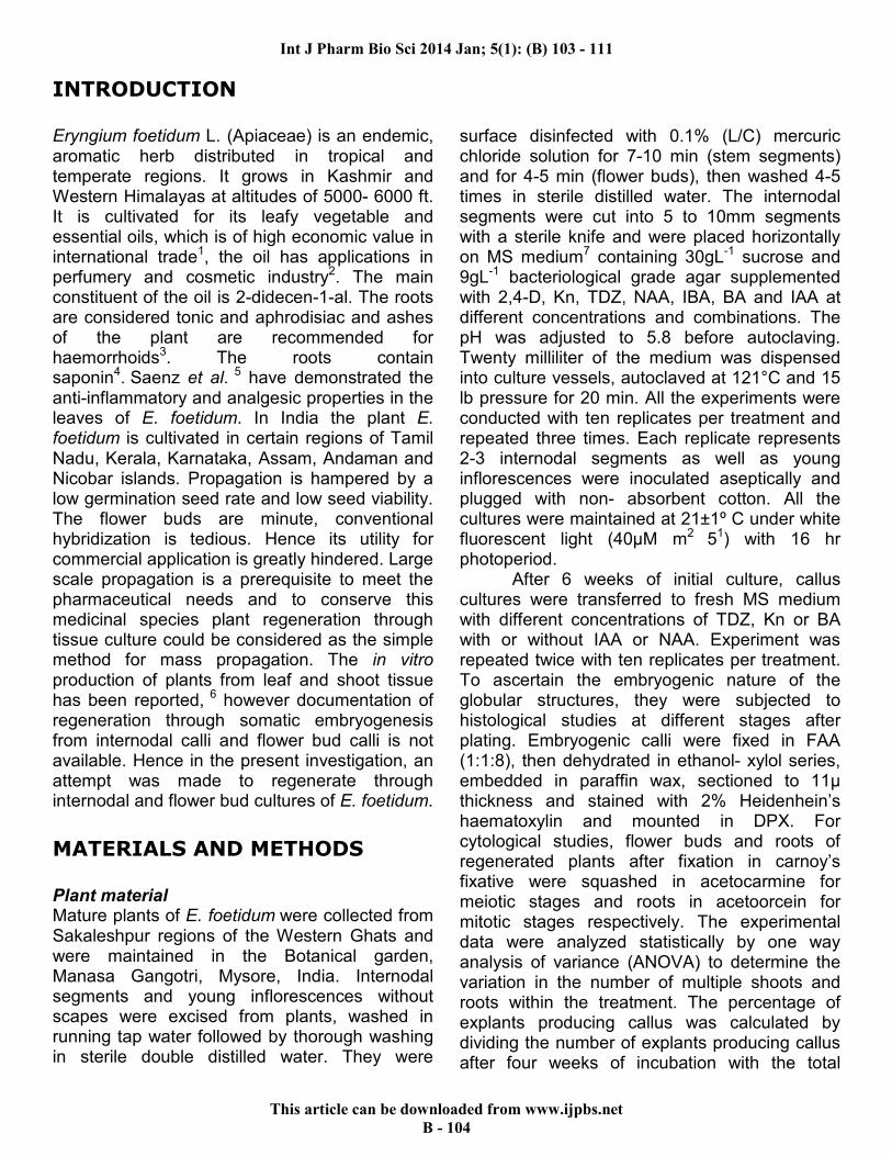
explants cultured on MS medium supplemented with 1.0 mgL-1 2,4–D produced 65 and 80% of calli (fig: 1) respectively as compared to 53 and 68% on medium containing 2.0 mgL-1 2,4-D. Supplementation of the medium with 1.0 mgL-1
BA with0.5 mgL-1 IAA generated non-friable callus. Therefore, callus generated on MS medium supplemented with 1.0 mgL-1 2, 4-D was serially subcultured onto MS medium containing different concentrations of 2, 4-D to obtain friable callus. The friable callus obtained after two consecutive subcultures in MS medium containing 2.0 mgL-1 2, 4-D was used to initiate the embryoids and shoot buds on MS medium containing various concentrations and combinations of BA and IAA. Both types of explants showed considerable decrease in the fresh weight as the concentration of BA and IAA was increased. Maximum callus proliferation was observed at 1-3 mgL-1 BA and 0.5 to 1.0 mgL-1
IAA.
Figure 1 Inflorescence callus.
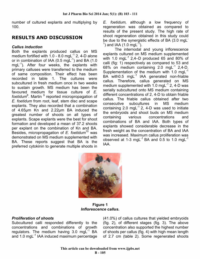
Proliferation of shoots Subcultured calli responded differently to the concentrations and combinations of growth regulators. The medium having 3.0 mgL-1 BA and 1.0 mgL-1 IAA induced maximum percentage
(41.0%) of callus cultures that yielded embryoids (fig. 2), of different stages (fig. 3). The above concentration also supported the highest number of shoots per callus (fig. 4) with high mean length of 2.7 cm (table 2). Some regenerated shoots
Int J Pharm Bio Sci 2014 Jan; 5(1): (B) 103 - 111
This article can be downloaded from www.ijpbs.net
B - 106
produced well developed roots (fig.5) even in semi-solid medium. The combination of BA and IAA was generally more effective than Kn and IAA combination in inducing shoots from the
callus, as has been shown in several medicinal plant species, including Lippia alba11, Hemidesmus indicus12 and Holostemma ada Kodien13.
Figure: 2 Embryogenic callus. Figure: 3 Stages of embryogenesis.
Figure: 4 Multiple shoots. Figure: 5 Shoots showing rhizogenesis.
Table 1
Effect of growth regulators on initiation of callus from internodal and young inflorescence explants of E. foetidum.
Plant hormone (mgL
-1)
Responsive explants (%) Callus Internodal segments Immature inflorescence bits
Basal 0 0 0
2,4 – D (1.0) 65 80 ++
2,4 – D (2.0) 53 68 +
2,4 – D (3.0) 34 39 +
2,4 – D (5.0) 15 19 +
2,4 – D (8.0) 6 5 +
BA (1.0)+ IAA (0.5) 78 87 +++
BA (2.0)+ IAA (1.0) 59 71 +++
BA (3.0)+ IAA (1.0) 42 45 ++
BA (5.0)+ IAA (2.0) 16 18 +
BA (8.0)+IAA (3.0) 4 5 +
‘0’ – No callus; ‘+’ - Scanty callus; ‘++’ - Proliferating callus; ‘+++’ - Luxuriant callus.
Int J Pharm Bio Sci 2014 Jan; 5(1): (B) 103 - 111
This article can be downloaded from www.ijpbs.net
B - 107
Table 2 Effect of plant growth regulators on differentiation of embryoids from the callus
Plant growth regulator (mgL
-1) Callus cultures with shoot
regeneration (%) Mean number of shoots per callus
Mean shoot length (cm) BA Kn IAA
0.0 0.0 0.0 0.00±0.0 0.0±0.0 0.0±0.0
0.5 0.0 0.1 5.00±0.1 2.3±0.3 2.0±0.2
1.0 0.0 0.5 22.8±2.3 2.6±0.3 2.3±0.2
2.0 0.0 1.0 31.7±0.4 3.2±0.4 2.5±0.2
3.0 0.0 1.0 41.0±0.1 4.1±0.5 2.7±0.2
0.0 0.5 0.1 0.00±0.0 0.0±0.0 0.0±0.0
0.0 1.0 0.5 19.1±0.1 2.3±0.3 1.8±0.2
0.0 2.0 1.0 25.0±0.1 3.0±0.4 2.5±0.2
0.0 3.0 1.0 13.0±1.0 1.4±0.2 1.2± 0.0
The results represent the mean ± standard error of the mean of replicated experiments after 6 weeks of culture.
Table 3
Effect of auxins on root induction from regenerated shoots of E. foetidum on MS medium.
Plant growth regulator (mgL-1)
Rooting (%) Average number of roots per shoot
Root length (cm) IAA IBA NAA
0 0 0 0 0 0
0.5 0 0 51 1.8 ±0.6 2.6 ±0.3
1.0 0 0 76 1.4± 0.4 4.0 ±0.5
2.0 0 0 65 1.3± 0.3 3.0 ±1.1
0 0.5 0 70 1.6 ±0.3 4.0 ±1.1
0 1.0 0 84 1.6 ±0.3 2.0± 0.5
0 2.0 0 67 1.3± 0.3 3.0 ±1.0
0 0 0.5 41 1.3 ±0.3 1.0 ±0.5
0 0 1.0 53 1.4± 0.4 1.5± 0.2
0 0 2.0 46 1.3± 0.4 1.1± 0.3
Observation were taken 45 days after culture. The values represent the mean (±SE) of three independent experiments. At least 24 cultures were raised in each experiment.
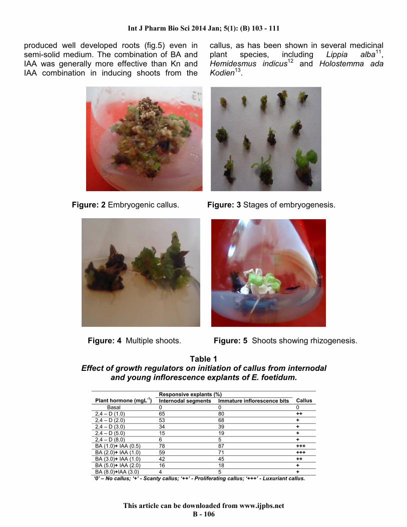
Rhizogenesis and plant establishment Rooting of in vitro derived shoots were carried out by transferring the healthy, well elongated shoots to rooting media that composed of half strength MS medium with different concentrations of auxins (NAA, IAA, IBA). In all cases 1.0 mgL-1 of IBA was found to be more effective in producing roots. Most of shoots produced roots within two weeks after placing on
rooting medium. The effect of auxins on rhizogenesis in regenerated shoots has been presented in table 3.The rooted plantlets were successfully hardened inside the culture room in planting substrate for 4 weeks. The plantlets were then subsequently transferred to pots (fig. 6) containing garden soil and finally to the field. About 55% of the plantlets are survived under the field conditions.
Figure 7 Acclimatization of regenerated plantlet.
Int J Pharm Bio Sci 2014 Jan; 5(1): (B) 103 - 111
This article can be downloaded from www.ijpbs.net
B - 108
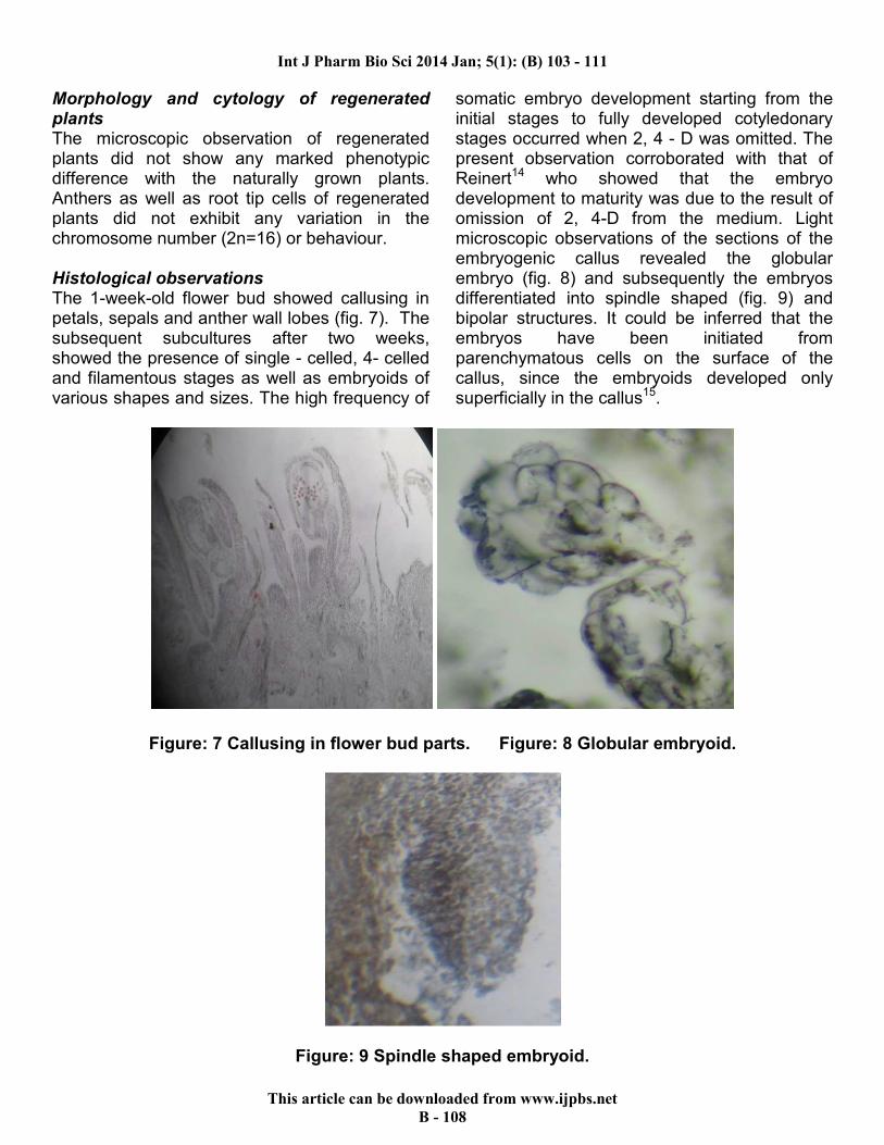
Morphology and cytology of regenerated plants The microscopic observation of regenerated plants did not show any marked phenotypic difference with the naturally grown plants. Anthers as well as root tip cells of regenerated plants did not exhibit any variation in the chromosome number (2n=16) or behaviour. Histological observations The 1-week-old flower bud showed callusing in petals, sepals and anther wall lobes (fig. 7). The subsequent subcultures after two weeks, showed the presence of single - celled, 4- celled and filamentous stages as well as embryoids of various shapes and sizes. The high frequency of
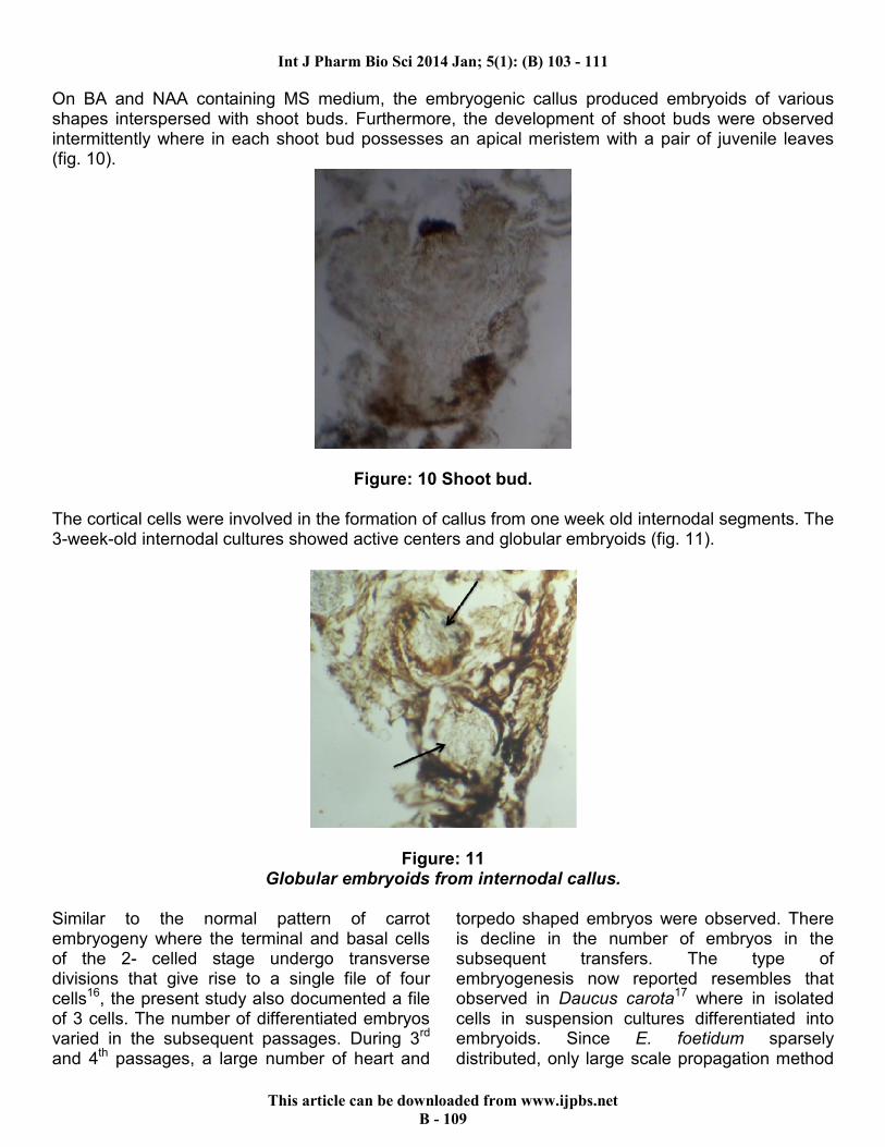
somatic embryo development starting from the initial stages to fully developed cotyledonary stages occurred when 2, 4 - D was omitted. The present observation corroborated with that of Reinert14 who showed that the embryo development to maturity was due to the result of omission of 2, 4-D from the medium. Light microscopic observations of the sections of the embryogenic callus revealed the globular embryo (fig. 8) and subsequently the embryos differentiated into spindle shaped (fig. 9) and bipolar structures. It could be inferred that the embryos have been initiated from parenchymatous cells on the surface of the callus, since the embryoids developed only superficially in the callus15.
Figure: 7 Callusing in flower bud parts. Figure: 8 Globular embryoid.
Figure: 9 Spindle shaped embryoid.
Int J Pharm Bio Sci 2014 Jan; 5(1): (B) 103 - 111
This article can be downloaded from www.ijpbs.net
B - 109
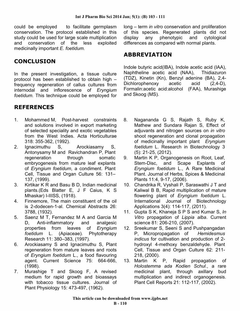
On BA and NAA containing MS medium, the embryogenic callus produced embryoids of various shapes interspersed with shoot buds. Furthermore, the development of shoot buds were observed intermittently where in each shoot bud possesses an apical meristem with a pair of juvenile leaves (fig. 10).
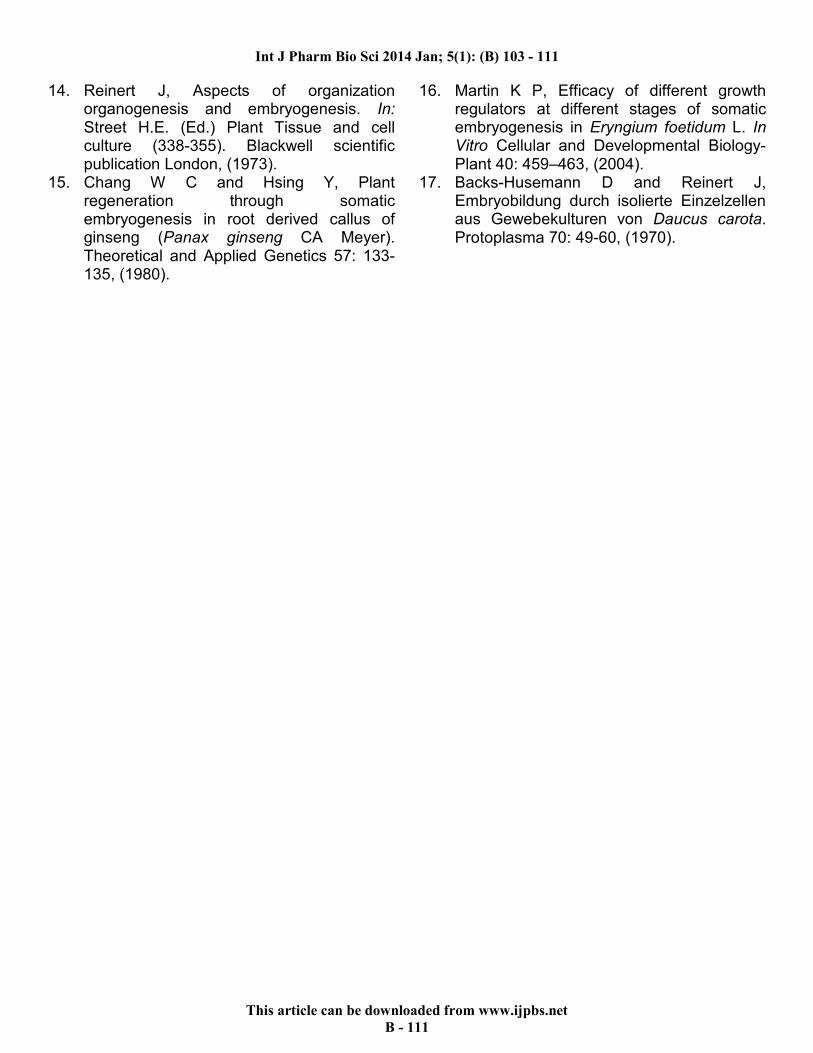
Figure: 10 Shoot bud. The cortical cells were involved in the formation of callus from one week old internodal segments. The 3-week-old internodal cultures showed active centers and globular embryoids (fig. 11).
Figure: 11 Globular embryoids from internodal callus.
Similar to the normal pattern of carrot embryogeny where the terminal and basal cells of the 2- celled stage undergo transverse divisions that give rise to a single file of four cells16, the present study also documented a file of 3 cells. The number of differentiated embryos varied in the subsequent passages. During 3rd and 4th passages, a large number of heart and
torpedo shaped embryos were observed. There is decline in the number of embryos in the subsequent transfers. The type of embryogenesis now reported resembles that observed in Daucus carota17 where in isolated cells in suspension cultures differentiated into embryoids. Since E. foetidum sparsely distributed, only large scale propagation method
Int J Pharm Bio Sci 2014 Jan; 5(1): (B) 103 - 111
This article can be downloaded from www.ijpbs.net
B - 110
could be employed to facilitate germplasm conservation. The protocol established in this study could be used for large scale multiplication and conservation of the less exploited medicinally important E. foetidum.
CONCLUSION
In the present investigation, a tissue culture protocol has been established to obtain high – frequency regeneration of callus cultures from internodal and inflorescence of Eryngium foetidum. This technique could be employed for
long – term in vitro conservation and proliferation of this species. Regenerated plants did not display any phenotypic and cytological differences as compared with normal plants.
ABBREVIATION
Indole butyric acid(IBA), Indole acetic acid (IAA), Naphtheline acetic acid (NAA), Thidiazuron (TDZ), Kinetin (Kn), Benzyl adenine (BA), 2,4- Dichlorophenoxy acetic acid (2,4-D), Formalin:acetic acid:alcohol (FAA), Murashige and Skoog (MS).
REFERENCES
1. Mohammed M, Post-harvest constraints and solutions involved in export marketing of selected speciality and exotic vegetables from the West Indies. Acta Horticulturae 318: 355-362, (1992).
2. Ignacimuthu S, Arockiasamy S, Antonysamy M and Ravichandran P, Plant regeneration through somatic embryogenesis from mature leaf explants of Eryngium foetidum, a condiment. Plant Cell, Tissue and Organ Culture 56: 131–137, (1999).
3. Kirtikar K R and Basu B D, Indian medicinal plants.(Eds Blatter E, J F Caius, K S Mhaskar) I-IIISS, (1918).
4. Finnemore, The main constituent of the oil is 2-dodecen-1-al. Chemical Abstracts 26: 3788, (1932).
5. Saenz M T, Fernandez M A and Garcia M D, Anti-inflammatory and analgesic properties from leaves of Eryngium foetidum L. (Apiaceae). Phytotherapy Research 11: 380–383, (1997).
6. Arockiasamy S and Ignacimuthu S, Plant regeneration from mature leaves and roots of Eryngium foetidum L., a food flavouring agent. Current Science 75: 664-666, (1998).
7. Murashige T and Skoog F, A revised medium for rapid growth and bioassays with tobacco tissue cultures. Journal of Plant Physiology 15: 473-497, (1962).
8. Nagananda G S, Rajath S, Ruby K, Mathew and Sundara Rajan S, Effect of adjuvants and nitrogen sources on in vitro shoot regeneration and clonal propagation of medicinally important plant Eryngium foetidum L. Research in Biotechnology 3 (5): 21-25, (2012).
9. Martin K P, Organogenesis on Root, Leaf, Stem-Disc, and Scape Explants of Eryngium foetidum L., A Rare Medicinal Plant. Journal of Herbs, Spices & Medicinal Plants 11:4, 9-17, (2006).
10. Chandrika R, Vyshali P, Saraswathi J T and Kaliwal B B, Rapid multiplication of mature flowering plant of Eryngium foetidum L. International Journal of Biotechnology Applications 3(4): 114-117, (2011).
11. Gupta S K, Khaneja S P S and Kumar S, In Vitro propagation of Lippia alba. Current science 81: 206-210, (2007).
12. Sreekumar S, Seeni S and Pushpangadan P, Micropropagation of Hemidesmus indicus for cultivation and production of 2- hydroxyl 4-methoxy benzaldehyde. Plant Cell, Tissue and Organ Culture 62: 211-218, (2000).
13. Martin K P, Rapid propagation of Holostemma ada Kodien Schul., a rare medicinal plant, through axillary bud multiplication and indirect organogenesis. Plant Cell Reports 21: 112-117, (2002).
Int J Pharm Bio Sci 2014 Jan; 5(1): (B) 103 - 111
This article can be downloaded from www.ijpbs.net
B - 111
14. Reinert J, Aspects of organization organogenesis and embryogenesis. In: Street H.E. (Ed.) Plant Tissue and cell culture (338-355). Blackwell scientific publication London, (1973).
15. Chang W C and Hsing Y, Plant regeneration through somatic embryogenesis in root derived callus of ginseng (Panax ginseng CA Meyer). Theoretical and Applied Genetics 57: 133-135, (1980).
16. Martin K P, Efficacy of different growth regulators at different stages of somatic embryogenesis in Eryngium foetidum L. In Vitro Cellular and Developmental Biology-Plant 40: 459–463, (2004).
17. Backs-Husemann D and Reinert J, Embryobildung durch isolierte Einzelzellen aus Gewebekulturen von Daucus carota. Protoplasma 70: 49-60, (1970).