in vitro study of secreted aspartyl proteinases sap1 to ... · et al. , 1994 and 1998; chen et al....
TRANSCRIPT

Polish Journal of Microbiology2012, Vol. 61, No 4, 247–256
ORGINAL PAPER
Introduction
!e opportunistic fungal pathogen Candida albicans possesses a repertoire of virulence attributes including adhesion to host tissue, the ability to undergo reversible morphogenetic transition, the secretion of extracellular hydrolases, and rapid switching between di"erent phe-notypic forms (Argimón et al., 2007; Naglik et al., 2008; Dalle et al., 2010; Hayek et al., 2010). Among the hydro-lytic enzymes, aspartic proteases (Saps) are considered to be key virulence determinants of C. albicans which contribute to the adhesive and invasion capabilities of strains from this species (Raška et al., 2007; Tongchusak et al., 2008; Dalle et al., 2010). Saps are the products of a family of 10 SAP genes divided into subfamilies based on amino acid sequence homology alignment (SAP1 to SAP3, SAP4 to SAP6, SAP9, and SAP10). Further-
more, SAP7 and SAP8 are divergent and are not repre-sented as subfamily members (Hube et al., 1994; Monod et al., 1994 and 1998; Chen et al., 2002; Correia et al., 2010). Expression of the SAP genes varies according to the type and stage of the disease (Schaller et al., 2001; Fradin et al., 2003; Taylor et al., 2005; Jackson et al., 2007; Correia et al., 2010; Naglik et al., 2008; Abegg et al., 2011). Moreover, SAP genes expression is also reg-ulated during the morphological transition (Argimón et al., 2007; Décanis et al., 2011). Candida albicans is able to grow in di"erent forms (blastoconidia, germ tubes, pseudohyphae and true hyphae), a phenomenon defined as pleomorphism (Whiteway and Bachewich, 2007). Pleomorphic forms enable C. albicans to colo-nize and invade human tissues (Morrison et al., 2003; Kumamoto and Vinces, 2005a; 2005b; Raška et al., 2007; Barnett, 2008). It was found (Gow et al., 2002) that the
In vitro Study of Secreted Aspartyl Proteinases Sap1 to Sap3and Sap4 to Sap6 Expression in Candida albicans Pleomorphic Forms
MONIKA STANISZEWSKA1, MAŁGORZATA BONDARYK1, KATARZYNA SIENNICKA2, ANNA KUREK3,
JACEK ORŁOWSKI4, MARTIN SCHALLER4 and WIESŁAW KURZĄTKOWSKI1
1 Independent Laboratory of Streptomyces and Fungi Imperfecti,National Institute of Public Health-National Institute of Hygiene, Warsaw, Poland
2 Warsaw University of Life Science, Warsaw, Poland3 Department of Bacterial Genetics, Institute of Microbiology, University of Warsaw, Warsaw, Poland
4 Laboratory of Fungal Glycobiology, Institute of Biochemistry and Biophysics,Polish Academy of Sciences, Warsaw, Poland
4Department of Dermatology, Eberhard-Karls-University Tübingen, Tübingen, Germany
Received 13 January 2012, revised 20 August 2012, accepted 1 September 2012
A b s t r a c t
Transition from round budding cells to long hyphal forms and production of secreted aspartic proteases (Saps) are considered virulence-
associated factors of Candida albicans. Although plenty of data dealing with Saps involvement in the infection process have been published,
Saps expression by the di"erent pleomorphic forms as well as the capacity of C. albicans filaments to express Sap1-6 under serum in+uence
are poorly investigated. In this study, we used immuno+uorescence and immunoelectron microscopy for the detection of Sap1-6 isoen-
zymes in C. albicans pleomorphic cells (blastoconidia, germ tubes, pseudohyphae, true hyphae) grown in Sap-inductive human serum
and Sap non-inductive medium – yeast extract-peptone-glucose (YEPD). Isoenzymes were below the detection level in all blastoconidial
cells grown in YEPD for 18 h. Sap1-6 expression was hardly detected in C. albicans cells cultivated in serum for 20 min. Increasing level
of Sap1-6 expression was observed when C. albicans was incubated for 2, 6 and 18 h in serum corresponding to the development of germ
tubes, pseudohyphae and true hyphae. !e expression of Sap1-3 in pseudohyphae and true hyphae was more intensive compared to Sap4-6.
!us, we could show that human serum induced hyphae formation and the expression of Sap1-6 were co-regulated.
K e y w o r d s: Candida albicans, aspartic protease expression, isoenzymes 1-3, isoenzymes 4-6, morphotypes
* Corresponding author: M. Staniszewska, Independent Laboratory of Streptomyces and Fungi Imperfecti, Chocimska 24, 00791,
Warsaw, Poland; phone: +48 54 21 228; fax: +48 22 84 97 484; e-mail: [email protected]

Staniszewska M. et al. 4248
morphogenetic response (transition from budding to hyphal cells) of C. albicans as well as expression of SAP genes are triggered by factors existing in the environ-ment of the host (pH, temperature, serum).
Previous studies (Schaller et al., 2000; Felk et al., 2002; Naglik et al., 2003; 2008; Lermann and Morschhäuser, 2008; Gropp et al., 2009; Dalle et al., 2010), indicate that the expression of Sap isoenzymes, for the mor-phogenesis of C. albicans varies strongly, depending on the experimental setup having a significant impact on the dependence on protease activity. Felk et al. (2002) showed that in vivo (in tissue from infected mice) expression of Sap1-3 was detected on the surface of both yeast and hyphal of wild-type cells. In contrast, the Sap4-6 antigens were identified mostly on penetrating hyphal cells (Schaller et al., 2000; 2001; Copping et al., 2005; Hornbach et al., 2009). !ose authors suggested that Sap4-6 are the hyphal-associated proteins which is in striking contrast to the results obtained by Lermann and Morschhäuser (2008), who suggested that none of the SAP1-6 genes is required for invasion of vaginal RHE by hyphal morphologies. Moreover, according to Correia et al. (2010) Sap1-6 do not play a signifi-cant role in C. albicans virulence in a murine model of hematogenously disseminated candidiasis and that, in this model, Sap1-3 are not necessary for successful C. albicans infection.
In contrast, previous reports (Naglik et al., 1999; Schaller et al., 2000; Hube, 2004; Hornbach et al., 2009) showed that the Sap4-6 subfamily produced in high level by hyphal cells plays a role in immune evasion and protection from phagocytic killing by murine macro-phages. Gropp et al. (2009) showed that Sap1-3 degrade and inactivate the central human complement compo-nents C3b, C4b as well as C5 and block the damaging e"ects of the activated complement system.
Although plenty of data dealing with the Saps invol-ved in the infection process have been published the expression of Saps in particular morphotype is not suf-ficiently described. Given the role of serum in hyphae formation (Lermann and Morschhäuser, 2008; Gropp et al., 2009) and as SAP1-3 play essential role in the growth of C. albicans in medium consisting proteins (Felk et al., 2002; Lermann and Morschhäuser, 2008; Naglik et al., 2008; Gropp et al., 2009) as well as SAP4-6 are hypha-related genes (Lermann and Morschhäuser,
2008; Naglik et al. 2008; Gropp et al., 2009), we asked whether Sap1-3 or Sap4-6 are expressed in each mor-photype under human serum in+uence in vitro. In addi-tion, we analyzed whether any di"erences in expression between these two subfamilies exist. To investigate the expression of the Sap1-3 and Sap4-6 proteins, we stud-ied the media, pH, and temperature shi>s. Following to previous conclusion (Hube et al., 1994; Naglik et al., 1999; Wise et al., 2007; Gropp et al., 2009), that di"er-ent expression profiles of Saps are regulated by pH of the maintenance medium, we studied pH, and tempera-ture shi>s during expression of the Sap1-3 and Sap4-6. In contrast to all other members of the Sap family, the proteases Sap9-10 monitored under conditions in vitro and in vivo are independent of pH and morpho-type (Hornbach et al., 2009; Schild et al., 2011). Moreo-ver, the expressions of Sap7 and Sap8 do not correlate with virulence (Hornbach et al., 2009). !at is why we did not include Sap7, Sap8, Sap9 and Sap10 respectively in our study.
!e aim of this study was to: (i) examine the expres-sion of aspartic proteases (Sap1 to Sap3 and Sap4 to Sap6) in vitro in neutral pH during morphogenesis under human serum in+uence by immuno+uores-cence and immunoelectron microscopy; (II) estab-lish the relationship between isoenzymes expression and pleomorphism; (iii) determine the localization of Sap1-3 and Sap4-6 in pleomorphic cells of C. albicans by immunoelectron microscopy.
Experimental
Materials and Methods
Strains and growth conditions. !e Candida strains used in this study are listed in Table I. !e clinical iso-late of C. albicans (strain 82) was recovered from the blood of 3-year-old patient being treated for an anaplas-tic ependymoma. In the study, we used ATCC SC5314 reference strain to analyse the conceivable di"erences in Sap1-3 and Sap4-6 expression profile appearing between pleomorphic forms and various C. albicans strains. !e stock culture of examined strains was stored on ceramic beads (MicrobankTM, Pro-Lab Diagnostics, Canada) at –70°C. Prior to the respective examinations,
Wild type, clinical isolate 82 URA3/URA3 Staniszewska et al. (2011a)
Wild type, reference strain ATCC 5314 URA3/URA3 Gillum et al. (1984)
Wild type, reference strain ATCC MYA 581 URA3/URA3 Sullivan et al. (1995)
Table I
Candida strains used in this study
Designation Clone or strainRelevant characteristics
or genotypeReference

Saps expression in C. albicans pleomorphic forms4 249
routine culturing of strains for growth was conducted at 30°C for 18 h in extract-peptone-glucose broth medium YEPD [10 g yeast extract, 20 g peptone (BBL Trypticase Peptone, Becton Dickinson) and 20 g glucose, pH 5.7] (Ness et al., 2010).
Phenotypic and biochemical characterization. !e presumptive identification of the clinical strain 82 was conducted using CHROMagar Candida medium (Becton Dickenson, Sparks, MD, USA), as described previously Staniszewska et al. (2011b). Colors of the colonies were compared in reference to the C. albicans SC5314 strain. !e assimilation pattern of the isolate and the reference strains SC5314 was determined using the API 20C AUX identification system (BioMérieux, France). !e API 20C AUX test was used according to the manufacturer’s instructions. Results of the test were obtained based on the numerical profile read-out (Analytical Profile Index; BioMérieux) (Staniszewska et al., 2011a).
Molecular examination. DNA was extracted from blastoconidial cells of the clinical isolate as well as C. dubliniensis ATCC MYA 581 and C. albicans SC 5314 according to Yeast DNA Miniprep Protocol as described by Amberg and Burke (2005), and DNA quantification was performed using the NanoDrop ND 1000 spec-trophotometer at an absorbance of 260 nm. Ribosomal DNA region including a fragment of the 5.8S rDNA gene (GenBank) was amplified by standard PCR. !e primers (CALB1, CALB2) were used for species-spe-cific PCR (Luo and Mitchell, 2002). !e species-spe-cific PCR products were electrophoresed as previously described Luo and Mitchell (2002). DNA bands were visualized using a transilluminator SYNGENE (Divi-sion of Synoptics LTD) under UV 260 nm. Results were documented by using the GenSnap program.
In vitro study of aspartic protease enzyme expres-sion. !e expression of Sap isoenzymes in C. albicans strain cultivated in YEPD medium and, subsequently, in filtered undiluted human serum was studied. Expres-sion of Sap1-3 and Sap4-6 was monitored in blastoco-nidial cells, germ tubes, pseudohyphal and true hyphal forms.
Induction of pleomorphic cells. Blastoconidial cells were grown as described Staniszewska et al. (2011a). Cells were observed under a phase-contrast microscope (Docuval, Carl Zeiss, Germany). !en, blastoconidia were harvested, washed three times with distilled water, pelleted by centrifugation (300 g for 10 min), and stored at –70°C for 96 h.
To induce remaining pleomorphic forms the blasto-conidial cells suspensions in YEPD (50 µl) were trans-ferred to 500 µl of filtrated undiluted human serum (pH 7.2–7.4) and incubated separately for 20 min (pre-incubation), 2 h (to induce germ tubes), 6 h (to induce pseudohyphae), 18 h (to induce true hyphae) at 37°C.
!en, pleomorphic forms were harvested, washed, pelleted and stored as described above. Particular mor-photypes were observed under a phase-contrast micro-scope (Docuval, Carl Zeiss, Germany). To examine cell morphology, the pleomorphic cells were fixed in 2.5% glutaraldehyde (Serva, Heidelberg, Germany), dehydrated in graded ethanol, critical point dried in CO
2, coated with gold and viewed in FEI Quanta
200 Scanning Electron microscope (Czech Republic) (Staniszewska et al., 2011a).
Immuno!uorescence microscopy (IFM) Leica TCS SP (Leica, Wetz lar, Germany). !e specific anti-Sap2, anti-Sap3 and anti-Sap6 rabbit polyclonal pri-mary antibodies generated by Chen et al. (2002) were used in the study. !en, each anti-Sap rabbit antibody was separately mixed with Candida cell wall suspen-sion to prevent unspecific labelling. Subsequently, each antibody mixture was centrifuged at 10 000 g for 5 min.
For immuno+uorescence staining of Saps the cryo-section (Frigocut, model 2700, Reichert-Jung) of pleo-morphic forms were blocked with donkey serum (1:20; Merck & Kollegen, Ochsenhausen, Germany) and incubated with anti-Sap polyclonal rabbit antibodies (1:100), followed by positive human Candida serum (1:60; Merck & Kollegen, Ochsenhausen, Germany). Antibodies were directed against Sap1-3 and Sap4-6. Samples were then incubated with donkey-anti-rabbit Cy5 (1:500; Merck & Kollegen, Ochsenhausen, Ger-many) and donkey-anti-human serum Cy3 (1:500; Merck & Kollegen, Ochsenhausen, Germany), respec-tively. !is was followed by subsequent nucleus staining with Yopro (1:2000; Invitrogen, Karlsruhe, Germany).
Immunoelectron microscopy (IEM). Electron microscopy and postembedding immunogold label-ling of sections of pleomorphic forms were performed as described by Schaller et al. (1998; 1999). In brief, each pellet was fixed in periodate-lysine-paraformal-dehyde (PLP) and a>er embedding in Lowicryl K4M, the blocks were cut using an ultramicrotome (Ultracut; Reichert, Vienna Austria). Ultrathin sections (30 nm) were mounted on formvar-coated (Serva, Heidelberg, Germany) nickel or cooper grids (Stork Veco, Eerbeek, Netherlands) and incubated with anti-Sap polyclonal rabbit antibodies, directed against Sap1 to Sap3 or against Sap4 to Sap6 followed by 10 nm or 5 nm-gold-conjugated goat-anti-rabbit IgG (Dianova, Hamburg, Germany). In control samples, the primary antibody was omitted. For examination of Sap immunogold labelling, the transmission electron microscope Zeiss Libra 120 (Zeiss, Oberkochen, Germany) operating at 80 kV was used. Evaluation of data obtained from 250 randomly chosen cells was done by determining the intensity of staining with gold particles using a plus scale ranging from 1 (+) to 4 (++++), lack of staining was mark as minus (–).

Staniszewska M. et al. 4250
Results
Identification of isolated strain. Colony color of the clinical strain 82 grown on CHROMagar® Candida medium was determined to be light green, which is indicative of C. albicans species. Results of this assay indicated that the strain was beta-N-acetylhexosami-nidase positive. It was capable of assimilating treha-lose (TRE) a>er 48 h and 72 h of incubation. D-Xylose (XYL), DL-lactate (LAC) and alpha-methyl-D-gluco-side (MDG) assimilation was observed a>er 72 h of incubation. It utilized sucrose as a carbon source for growth, which is another typical feature of C. albicans. !e clinical strain 82 and SC5314 reference strain were phenotypically similar in all experimental system described in this study.
Application of C. albicans specific primers (CALB1, CALB2) allowed detecting the expected product size (273 bp) of C. albicans isolate. Subsequently, DNA of strain 82, ATCC SC5314 strain, excluding C. dubliniensis MYA 581, gave readily a PCR product with CALB pri- mers. Primers used in this study were determined to discri- minate between C. albicans and C. dubliniensis (Fig. 1).
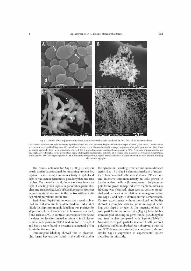
Detection of Sap1-3 and Sap4-6 antigens by immuno!uorescence and immunoelectron stain-ing techniques. Fractions of pleomorphic forms that were examined for their Saps expression are shown in Fig. 2. For strain 82, 100% of cells were found to form blastoconidia a>er 18 h of incubation in YEPD medium. In undiluted human serum, germ tube for-mation was observed at 2 h induction and approached 100%. Hyphae were formed as pseudohyphae (chains of elongated blastoconidial cells) at 6 h induction, with addition of budding blastoconidial cells and uniformly elongated the hyphal cells. True hyphal forms appeared
as homogeneous fraction a>er incubation for 18 h in serum. No di"erences were observed between the clin-ical strain 82 and SC 5314 reference strain (data not shown) in the ability to form pleomorphic cells.
IFM and IEM were carried out for intracellular detection of Sap1-3 and Sap4-6 in C. albicans pleomor-phic cells grown in Sap-inductive human serum and Sap non-inductive media – YEPD. Yeast cells cultivated in YEPD medium for 18 h as well as cells transferred to undiluted human serum for 20 min showed a lack of blue +uorescence protein of Sap1-3 and Sap4-6 express-ing signal. A>er 2 h of incubation in Sap-inductive undiluted human serum, Sap labelling became more distinct. Analysis of the di"erent pleomorphic forms cultivated in vitro demonstrated almost similar results for all tested Sap antigens (Table II).
Blastoconidia1 – 22N – 10N – 4N1
– 4 N1
Blastoconidia2 + 12 + 13 + 5 + 2
Germ tube3 ++ 29 +++ 13 ++ 5 +++ 5
Pseudohyphae4 ++++ 30 +++ 15 ++++ 7 +++ 2
True hyphae5 ++++ 24 +++ 36 ++++ 8 +++ 4
Table II
Expression of aspartic protease (Sap) isoenzymes by pleomorphic forms of Candida albicans
1 blastoconidia cultivated in YEPD medium (pH 5.7) for 18 h at 30°C 2 blastoconidia transferred to undiluted human serum (pH 7.2–7.4) for 20 min at 37°C 3 germ tubes grown in human serum for 2 h at 37°C4 pseudohyphae grown in human serum for 6 h at 37°C5 true hyphae grown in human serum for 18 h at 37°C N number of analyzed cellsN
1number of analyzed images
– lack of Sap immunogold or immuno+uorescence staining; from + to ++++ the intensity of Sap
staining with gold particles or immuno+uorescence. Cell and images were observed by three persons
independently. !e experiment repeated three times gave similar results.
Pleomorphic
cells
Immunogold labelling intensity
(immunoelectron microscopy)
Immuno+uorescence labelling intensity
(+uorescence microscopy)
Sap1-3 Sap4-6 Sap4-6Sap1-3
Fig. 1. Candida albicans identification based on PCR amplifica-
tion of the 5.8S rDNA gene fragment using the CALB1
and CALB2 primers.
(MM) Molecular marker mass; (lane 1) analysis of C. dubliniensis ATCC
MYA 581 showed absence of PCR product size (273 bp); (lane 2) PCR
product size (273 bp) of C. albicans ATCC 5314; (lane 3) PCR product
size (273 bp) of C. albicans clinical strain 82. PCR products were sepa-
rated on an agarose gel (0.8%) and stained with ethidium bromide.
Saps expression in C. albicans pleomorphic forms4 251
Fig. 2. Candida albicans pleomorphic forms. (A) Blastoconidial cells incubated at 30°C for 18 h in YEPD medium.
Oval-shaped blastoconidial cells exhibiting bipolarly-located bud scars (arrows). Fragile blastoconidial septa are seen (open arrow). Blastoconidial
poles are the privileged budding areas. (B) In undiluted human serum blastoconidial cells undergo the process of unipolar germination. A>er 2 h of
incubation germ tube forms were abundantly observed. (C) 6 h of cultivation in undiluted human serum at 37°C. A mixture of pseudohyphae and
true hyphae, pseudohyphae forms are visible as chains of elongated blastoconidial cells (ph1, ph
2). Fragile septa junctions are typical for pseudohyphal
forms (arrows). (D) True hyphae grown for 18 h. Uniformly elongated true hyphal forms exhibit lack of constrictions at the solid septum. Scanning
electron micrographs
!e results obtained for Sap1-3 (Fig. 3) express nearly similar data obtained for remaining proteins i.e., Sap4-6. !e increasing immunoreactivity of Sap1-3 and Sap4-6 was seen in germ tubes, pseudohyphae and true hyphae. On the other hand, there was more intensive Sap1-3 labelling than Sap4-6 in germ tubes, pseudohy-phae and true hyphae. Lack of blue +uorescence protein expressing signal was seen in the control without anti-Sap rabbit polyclonal antibodies.
Sap1-3 and Sap4-6 immunoreactivity results obta-ined for IEM were similar as described for IFM studies (Table II). Sap immunogold labelling was positive for all pleomorphic cells incubated in human serum for 2, 6 and 18 h at 30°C. In contrast, isoenzymes were below the detection level (estimated as minus –) in all blasto-conidial cells grown in YEPD medium for 18 h. Sap1-3 and Sap4-6 were found to be active at a neutral pH in Sap-inductive medium.
Immunogold labelling showed that in pleomor-phic forms Sap localizes mainly in the cell wall and in
the cytoplasm. Labelling with Sap antibodies directed against Sap1-3 or Sap4-6 demonstrated lack of reactiv-ity in blastoconidial cells cultivated in YEPD medium and intensive immunoreactivity in cells grown in Sap-inductive medium (human serum). In pleomor-phic forms grown in Sap-inductive medium, intensive labelling was observed, o>en seen as vesicles-associ-ated gold particles. A correlation between germination and Sap1-3 and Sap4-6 expression was demonstrated. Control experiments without polyclonal antibodies showed a complete absence of immunogold label-ling with Sap1-3 or Sap4-6. !e intensity of Sap1-3 gold particles immunoreactivity (Fig. 4.) bears higher immunogold labelling in germ tubes, pseudohyphae and true hyphae compared with Sap4-6 (Table II). No evidence of gold particles in control cells (without polyclonal rabbit antibodies) was observed. Strain 82 and SC5314 reference strain (data not shown) showed similar Sap1-6 expression in experimental system described in this study.

Staniszewska M. et al. 4252
Discussion
In this study, the expression of Sap1-6 during C. albi-cans morphogenesis in undiluted human serum was evaluated. Our data indicate that (1) Sap1-3 and Sap4-6 are the isoenzymes whose expression was observed in germ tubes, pseudohyphae and true hyphae of C. albi-cans (2) Sap1-3 antigens expression was significantly raised during hyphae formation compared with Sap4-6.
Recent studies (Taylor et al., 2000; Leinberger et al., 2005; Okawa et al., 2007) demonstrated that conven-tional biochemical tests can misidentify clinical iso-lates. !at is why, in this study, sequence analysis of the 5.8 rDNA region amplified by using the species-spe-cific primer pair (CALB1 CALB2) (Luo and Mitchell, 2002) confirmed that the examined isolate belongs to the C. albicans. In our study, identification of the clini-cal isolate (strain 82) based on genotypic di"erences confirmed results obtained through phenotypic studies.
Many authors (Lermann and Morschhäuser, 2008; Naglik et al., 2008; Gropp et al., 2009; Dalle et al., 2010), included the reference strain SC5314 and its mutants in studying the roles of secreted Sap hydrolases in the pathogenesis process in humans. We selected the clini-cal isolate in purpose in view of inconciliable results referring to strain SC5314, which are presented below.
In this study, we compared Saps expression profile of pleomorphic forms of the C. albicans clinical isolate recovered from blood samples as well as SC5314, which was similar (data not shown).
Taylor et al. (2000) showed that C. albicans strain SC5314 well known from animal experiments is a poor colonizer and invader of mammalian epithelia. On the contrary, it was established (Schaller et al., 2000; Felk et al., 2002; Dalle et al., 2010), that this strain was able to invade the host tissues, which was followed by systemic dissemination, as well as it caused damage in an in vitro model. It may be said that the virulence (in view of the place of recovering) of the clinical isolate and the refer-ence strain is comparable; both strains are virulent by intravenous challenge. !e clinical strain was chosen because it caused candidaemia in the patient, proving that it develops virulence factors.
Previously, we demonstrated (Staniszewska et al., 2011a) that the clinical isolate showed virulence determi- nants, it produced germ tubes, pseudohyphae, and true hyphae in undiluted human serum. Furthermore, strain 82 showed distinct di"erences in activity profiles of hydrolytic enzymes between hyphae and blastoconidia by using the api®ZYM test (Staniszewska et al., 2011b).
In the current work, the antibodies generated by Chen et al. (2002) were used. !e authors highlighted
Fig. 3. Visual estimation of Sap1-3 immunolabelling in pleomorphic forms of Candida albicans.
(A) Blastoconidial cells incubated in YEPD (yeast extract-peptone-glucose, YEPD-grown blastoconidial cells) at 30°C for 18 h, (–) lack of blue
+uorescence of Sap1-3 expressing signal. (B) YEPD-grown blastoconidial cells incubated in undiluted human serum for 20 min, (+) weak Sap1-3
immunoreactivity. (C) Germ tube forms developed from YEPD-grown blastoconidial cells by incubation in undiluted human serum for 2 h at 37°C,
(++) increased content of Sap1-3. (D) Pseudohyphae developed from YEPD-grown blastoconidial cells by incubation in undiluted human serum for
6 h at 37°C, (++++) very high content of Sap1-3. (E) True hyphae developed from YEPD-blastoconidial cells by incubation in undiluted human serum
for 18 h at 37°C, (++++) very high immunoreactivity of Sap1-3. !e immunoreactivity of Sap4-6 was of low intensity at (+++) compared with Sap1-3
in germ tubes, pseudohyphae or true hyphae (Table II)

Saps expression in C. albicans pleomorphic forms4 253
Fig. 4. Immunoelectron microscopy (IEM).
Detection of Sap1-3 in pleomorphic cells of Candida albicans using polyclonal rabbit anti-Sap2 serum and goat-anti-rabbit IgG conjugated to
5 nm gold particles. (A) Cells cultivated in Sap non-inductive medium YEPD and (B-E) in Sap-inductive undiluted human serum for (B) 20 min,
(C) 2 h, (D) 6 h and (E) 18 h. (A) For blastoconidial cells, the gold particle labelling intensity is evaluated as (–). !e gold labelling was not visible in
the cytoplasm neither cell wall. (B) Blastoconidial cells cultivated for 20 min in human serum. !e gold particle labelling density is estimated as (+).
In comparison to the cell wall (cw), labelling is seen mainly in the cytoplasm (arrows). (C) Germ tube forms. !e gold particle labelling density is
estimated as (++). Labelling is seen mainly in the cell wall (cw) (arrow). (D) Pseudohyphae. Note the cytoplasm-located clusters of the enzyme marker
surrounded by a membrane-like structure (arrows). (E) True hyphae. In comparison to the cytoplasm, labelling is seen mainly in the cell wall (arrow).
Enhanced clusters of gold particles in the cell wall are seen . !e gold particle labelling density is estimated as (++++). (F) !e immunoreactivity
of Sap4-6 was less intensive compared with that of Sap1-3 (Table II)
the difficulty with generating specific and sensitive antibodies against each Sap. It was mentioned (Chen et al., 2002) that antibodies against Sap3 showed cross reactivity with Sap2 or Sap4. !e cross reactivity of anti-Sap3 might have a"ected results obtained in the present study. !at is why in our study, additionally anti-Sap2 and anti-Sap6 were used which reacted specifically with Sap1-3 or Sap4-6, respectively (Chen et al., 2002).
!e immuno+uorescence microscopy studies reve-aled that the pattern of enzyme expression in blastoco-nidia grown in YEPD medium di"ers significantly from other C. albicans forms cultivated in the human serum, possibly due to the fact that YEPD medium contains no suitable substrate for Sap. Higher level of Sap1-6 expression was correlated with the course of germina-tion process and germ tubes, pseudohyphae and true hyphae appearance during incubation blastoconidial cells respectively for 2, 6 and 18 hours in human serum. Analysis using microscopy techniques determine that
Sap expression profiles demonstrate significant di"er-ences between particular pleomorphic forms grown in human serum. Sap1-3 expression gradually increased in cells during germination and was much more intensive than Sap4-6. A previous report showed (Hube et al., 1994) that deletion of SAP4-6 did not result in di"er-ences in hyphae formation both in vitro and in vivo.
Moreover, Felk et al. (2002) showed that SAP4-6 expression is associated with, but not required for hyphal morphology. Our findings are in line with pre vious data (Felk et al., 2002), showing that Sap4-6 expression is rela- ted with germination process. Additionally, we showed that Sap4-6 are expressed in each morphotype (includ-ing blastoconidial cells) under human serum in+uence.
!ese results were consistent with the in vivo expres-sion pattern (Staib et al., 2001) and suggested that expression of Sap1-6 proteins is regulated by factors that also regulate C. albicans morphology in human serum. Yet, information on secretion of particular Sap

Staniszewska M. et al. 4254
isoenzymes by C. albicans germ tubes and pseudohy-phae is still missing in the literature. In our studySap1-6 expression was observed in both morphotypes (germ tubes; pseudohyphae).
Our data allow postulating that there is a correlation between human serum induced hyphae growth in vitro and expression of Sap1-6. Here, we showed that the presence of high level of Sap1-3 expression through-out 18-h-incubation in human serum supports the view that Sap1-3 are probably key proteases that pro-mote cell growth and may be dependent on morphol-ogy. Although Sap1-3 expression level was higher than Sap4-6, the latter were consistently detected in germ tubes, pseudohyphae and true hyphae at steady levels, which may also support a contributory role of Sap4-6 in C. albicans cell growth and fitness. Our observation is not in line with results published previously (Correia et al., 2010), which suggested that yeast growth may be protease independent when C. albicans cells were deliv-ered directly into the bloodstream. On the contrary, Gropp et al. (2009) and Staib et al. (2001) showed that Sap2 is essential for growth when the protein is the only nitrogen source. Another support for our findings may be the previous report (Gropp et al., 2009) showing the strong complement inhibitory activity of Sap1-3 in human plasma. In the present study, it was shown that the Sap1-3 expression was highly induced in C. albicans hyphal cells upon host body +uid (serum) in+uence suggesting that also in vivo each of the three Sap pro-teins may contribute to blood infection, complement inactivation and immune evasion (Gropp et al., 2009). Here, we have provided evidence that the human serum plays a important role in hyphae formation and Sap1-3 expression. However, more investigations about the expression of SAP1-3 as well as SAP4-6 under human serum in+uence by using RT-PCR are going to be done.
In the current study, increasing Sap1-3 and Sap4-6 expression was detected during hyphae formation induced by shi> of temperature (from 30°C to 37°C) and pH (from 5.7 to 7.2) in human serum. !e same results were obtained for another clinical isolate (Taylor et al., 2000) as well as SC5314 reference strain (data not shown). We did not observe di"erences between C. albicans strains due to pH changes. !ere was no Sap1-3 or Sap4-6 activity in yeast cells grown in medium at pH 5.7, while germ tubes, pseudohyphae and true hyphae expressed these isoenzymes at a neu-tral pH (7.2–7.4). !ese characteristics may indicate the ability of C. albicans to survive and cause infection in a variety of host tissues.
!e results obtained for the expression pattern of the immuno+uorescence study were confirmed by immuno electron microscopy studies (IEM). !e use of antibodies raised against Sap1-3 or Sap4-6 made it possible to determine the precise location of these
proteins in the pleomorphic forms that have been pre-cultured in human serum. Sap proteins were detected inside the cytoplasm and within the cell wall of blasto-conidia, germ tubes, pseudo- and true hyphae forms, but not in the surrounding of the cell wall, which was confirmed by the negative control (non-antigens in the surrounding of the cell wall). !e Sap1-3 and Sap4-6 were observed to be organized in groups and packed with the vesicles localized in the cytoplasm. !e results of our study are in line with those of Stringaro et al. (1997) demonstrating that during murine vaginitis Sap antigen is located within the cell wall of hyphal cells. Similar results were observed in the in vitro model of experimental oral candidiasis (Stringaro et al., 1997) and reconstituted human epidermis (Schaller et al., 2000).
Our studies may support the data of Brown (2002) that both blastoconidia and filamentous forms of C. albi-cans are pathogenic and both contribute to di"erent stages in the establishment and progress of the infec-tion. Sap1-6 proteins tested in the above-mentioned study were determined to be expressed in pleomor-phic forms during incubation in human serum which mimics in vivo conditions encountered during systemic blood infections. !ese data may indicate an impor-tant role of Sap1-6 proteins during blood infections and immune evasion (Hornbach et al., 2009). We therefore conclude that there is a correlation between the increased expression of Sap1-6 proteins and germ tubes, pseudohyphae and true hyphae formation.
Acknowledgement
Martin Schaller was supported by the Deutsche Forschungs-
gemeinscha> (Sch 897/3, SFB773 Z2, graduate college 685), the
BMBF (MedSys 0315409B) and by a NIDCR grant R01DE017514-01.
Monika Staniszewska and Wiesław Kurzątkowski were sup-
ported by grant N N404 113639 from the Ministry of Science and
Higher Education.
We thank Birgit Fehrenbacher, Renate Nordin, Helga Möller and
Hannelore Bischof, Univer-sity of Tübingen, for excellent technical
assistance.
We thank Prof. D.D. Dzierżanowska-Madalińska for providing
C. albicans clinical isolate.
Literature
Abegg M.A., R. Lucietto, P.V.G. Alabarse, M.F.A. Mendes and
M.S. Benfato. 2011. Di"erential Resistance to oxidants and produc-
tion of hydrolytic enzymes in Candida albicans. Mycopathologia.
171: 35–41.
Amberg D.C. and D.J. Burke. 2005. Yeast DNA isolation, tech-
niques and protocols 3, p.17. In: Amberg D.C. and D.J. Burke (eds.),
Methods in yeast genetics. Cold Spring Harbor Laboratory Press,
Cold Spring Harbor.
Argimón S., J.A. Wishart, R. Leng, S. Macaskill, A. Mavor,
T. Alexandris, S. Nicholls, A.W. Knight, B. Enjalbert, R. Walmsley,
F.C. Odds, N.A.R. Gow and A.J. Brown. 2007. Developmental
regulation of an adhesin gene during cellular morphogenesis in the
fungal pathogen Candida albicans. Eukaryotic Cell. 6: 682–692.

Saps expression in C. albicans pleomorphic forms4 255
Barnett J.A. 2008. A history of research on yeasts 12: medical yeasts part I, Candida albicans. Yeast 25: 385–417.Brown A.J.P. 2002. Expression of growth form-specific factor dur-ing morphogenesis in Candida albicans, pp. 87–93. In: Calderone R.A. (eds.), Candida and candidiasis, ASM Press, Washington, DC.Chen Y.C., C.C. Wu, W.L. Chung and F.J.S. Lee. 2002. Di"erential secretion of Sap4-6 proteins in Candida albicans during hyphae for-mation. Microbiol. 148: 3743–3754.Copping V.M.S., C.J. Barelle, B. Hube, N.A.R. Gow, A.J.P. Brown
and F. Odds. 2005. Exposure of Candida albicans to antifungal agents a"ects expression of SAP2 and SAP9 secreted proteinase genes. J. Antimicrob. Chemother. 55: 645–654.Correia A., U. Lerman, L. Teixeira, F. Cerca, S. Botelho, R.M.G. da
Costa, P. Sampaio, F. Gärtner, J. Morshhäuser, M. Vilanova and
others. 2010. Limited role of secreted aspartyl proteinases Sap1 to Sap6 in Candida albicans virulence and host immune response in murine hematogenously disseminated candidiasis. Infect. Immun. 78: 4839–4849.Dalle F., B. Wächter, C. L’Ollivier, G. Holland, N. Bannert, D. Wil-
son, C. Labruére, A. Bonnin and B. Hube. 2010. Cellular inter-actions of Candida albicans with human oral epithelial cells and enterocytes. Cell Microbiol. 12: 248–71.Décanis N., N. Tazi, A. Correia, M. Vilanova and M. Rouabhia.
2011. Farnesol, a fungal quorum-sensing molecule triggers Candida albicans morphological changes by downregulating the expression of di"erent secreted aspartyl proteinase genes. !e Open Microbiol-ogy Journal. 5: 119–126.Felk A., M. Kretschmar, A. Albrecht, M. Schaller, S. Beinhauser,
T. Nichterlein, D. Sanglard, H.C. Korting, W. Schäfer and
B. Hube. 2002. Candida albicans hyphal formation and the expression of the Efg1-regulated proteinases Sap4 to Sap6 are required for the invasion of parenchymal organs. Infect. Immun. 70: 3689–3700.Fradin Ch., M. Kretschmar, T. Nitchterlein, C. Gaillardin,
Ch. d’Enfert and B. Hube. 2003. Stage-specific gene expres- sion of Candida albicans in human blood. Mol. Microbiol. 47: 1523–1543.Gillum A.M., E.Y. Tsay and D.R. Kirsch. 1984. Isolation of the Candida albicans gene for ortodine-5’-phosphate decarboxylase by complementation of Saccharomyces cerevisiae ura3 and Eschericha coli pyrF mutations. Mol. Gen. Genet. 198: 179–182.Gow N.A.R. 2002. Cell biology and cell cycle of Candida, pp. 145–158. In: Calderone R.A. (eds.), Candida and Candidiasis, ASM Press, Washington D.C.Gropp K., L. Schild, S. Schindler, B. Hube, P.F. Zipfel and
C. Skerka. 2009. !e yeast Candida albicans evades human comple-ment attack by secretion of aspartic proteases. Mol. Microbiol. 47: 465–475.Hayek P., L. Dib, P. Yazbeck, B. Beyrouthy, R.A. Khalaf. 2010. Characterization of Hwp2, a Candida albicans putative GPI-anchored cell wall protein necessary for invasive growth. Microbiol Res. 165: 250–258. Hornbach A., A. Heyken, L. Schild, B. Hube, J. Lö*er and
O. Kurzai. 2009. !e glycosylphosphatidylinositol-anchored prote-ase Sap9 modulates the interaction of Candida albicans with human neutrophils. Infect. Immune. 77: 1–9.Hube B., M. Monod, D.A. Schofield, A.J. Brown and N.A.R. Gow. 1994. Expression of seven members of the gene family encoding secretory aspartic proteinases in Candida albicans. Mol. Microbiol. 14: 87–99.Hube B. 2004. From commensal to pathogen: stage- and tissue-specific gene expression of Candida albicans. Curr. Opin. Microbiol. 7: 336–341.Jackson B.E., K.R. Wilhelmus and B. Hube. 2007. !e role of secreted aspartyl proteinases in Candida albicans keratitis. IOVS. 48: 3559–3565.
Kumamoto C.A.and M.D. Vinces. 2005a. Alternative Candida albi-cans lifestyles: Growth on surface. Annu. Rev. Microbiol. 59: 113–130.Kumamoto C.A. and M.D. Vinces. 2005b. Contributions of hyphae and hypha-co-regulated genes to Candida albicans virulence. Annu. Rev. Microbiol. 7: 1546–1554.Leinberger D.M., U. Schumacher, I.B. Autenrieth and T.T. Bach-
mann. 2005. Development of a DNA Microarray for Detection and Identification of Fungal Pathogens Involved in Invasive Mycoses. J. Clin. Microbiol. 43: 4943–4953.Lermann U. and J. Morschhäuser. 2008. Secreted aspartic proteases are not required for invasion of reconstituted human epithelia by Candida albicans. Microbiol. 154: 3281–3295.Luo G. and T.G. Mitchell. 2002. Rapid identification of pathogenic fungi directly from cultures by using multiplex PCR. J. Clin. Micro-biol. 40: 2860–2865.Monod M., G. Togni, B. Hube, and D. Sanglard. 1994. Multiplicity of genes encoding secreted aspartic proteases in Candida species. Mol. Microbiol. 13: 357–368.Monod M., Togni, B. Hube, D. Heß, D. Sanglard. 1998. Cloning, sequencing, and expression of two new members of the secreted aspartic proteinase family of Candida albicans. Microbiology. 144: 2731–2737. Morrison C.J., S.F. Hurst and E. Reiss. 2003. Competitive binding enzyme-linked immunosorbent assay that uses the secreted aspartyl proteinase of Candida albicans as an antigenic marker for diagnosis of disseminated candidiasis. Clin. Diagn. Lab. Immunol. 10: 835–848.Naglik J.R., G. Newport, T.C. White, L.L. Fernandes-Naglik,
J.S. Greenspan, D. Greenspan, S.P. Sweet, S.J. Challacombe and
N. Agabian. 1999. In vivo analysis of secreted aspartyl proteinases expression in human oral candidiasis. Infect Immun. 67: 2482–2490.Naglik J.R., S.J. Challacombe and B. Hube. 2003. Candida albicans secreted aspartyl proteases in virulence and pathogenesis. Microbiol. Mol. Biol. Rev. 67: 400–428.Naglik J.R., D. Moyes, J. Makwana, P. Kanzaria, E. Tsichlaki,
G. Weindl, A.R. Tappuni, C.A. Roggers, A.J. Woodman, S.J. Chal-
lacombe and others. 2008. Quantitative expression of the Candida albicans secreted aspartyl proteinase gene family in human oral and vaginal candidiasis. Microbiol. 154: 3266–3280.Ness F., V. Prouzet-Mauleon, A. Vieillemard, F. Lefebvre, T Noёl,
M. Crouzet, F. Doignon and D. /oraval. 2010. !e Candida albi-cans Rgd1 is a RhoGAP protein involved in the control of filamen-tous growth. Fungal Genet Biol 47: 1001–1011.Okawa Y., M. Miiiyauchi, S. Takahashi and H. Kobayashi. 2007. Comparison of pathogenicity of various Candida albicans and C. stellatoidea strains. Biol. Pharm. Bull. 30: 1870–1873.Raška M., J. Běláková, M. Křupka and E. Weigl. 2007. Candidia-sis – do we need to fight or to tolerate the Candida fungus? Folia Microbiol. 52: 297–312.Schaller M., W. Schafer, C. Korting and B. Hube. 1998. Di"erential expression of secreted aspartyl proteinases in model of human oral candidiosis and in patient samples from oral cavity. Mol. Microbiol. 29: 605–615.Schaller M., H.C. Korting, W. Schäfer, J. Bastert, W. Chen and
B. Hube. 1999. Secreted aspartic proteinase (Sap) activity contrib-utes to tissue damage in a model of human oral candidiasis. Mol. Microbiol. 34: 169–180.Schaller M., C. Schackert, H.C. Korting, E. Janusche and B. Hube.
2000. Invasion of Candida albicans correlates with expression of secreted aspartic proteinases during experimental infection of human epidermis. J. Invest. Dermatol. 114: 712–717.Schaller M., E. Januschke, C. Schackert, B. Woerle and H.C. Kor-
ting. 2001. Di"erent isoforms of secreted aspartyl proteinases (Sap)
are expressed by Candida albicans during oral and cutaneous can-
didiosis in vivo. J. Med. Microbiol. 50: 743–747.
Schild L., A. Heyken, P.W.J. de Groot, E. Hiller, M. Mock, C. de
Koster, U. Horn, S. Rupp and B. Hube. 2001. Proteolytic cleavage
Staniszewska M. et al. 4256
of covalently linked cell wall proteins by Candida albicans Sap9 and
Sap10. Eukaryot Cell. 10: 98–109.
Staib P., S. Wirsching, A. Strauß and J. Morschhäuser. 2001. Gene
regulation and host adaptation mechanisms in Candida albicans. Int.
J. Med. Microbiol. 291: 183–188.
Staniszewska M., M. Bondaryk and W. Kurzątkowski. 2011a.
Morphotypes of Candida albicans. Phase-contrast microscopy. Med.
Mycol. 18: 5–10.
Staniszewska M., D. Rabczenko and W. Kurzątkowski. 2011b. Dis-
crimination between the enzymatic activities of Candida albicans
pleomorphic forms determined using the api® ZYM test. Mycoses 54:
e744–e750. doi: 10.1111/j.1439–0507.2010.02011.x
Stringaro A., P. Crateri, G. Pellegrini, G. Arancia, A. Cassone
and F. De Bernardis. 1997. Ultrastructural localization of the secre-
tory aspartyl proteinase in Candida albicans cell wall in vitro and
in experimentally infected rat vagina. Mycopathologia 137: 95–105.
Sullivan D.J., T.J. Westerneng, K.A. Haynes, D.E. Bennett and
D.C. Coleman. 1995. Candida dubliniensis sp. nov.: Phenotypic and
molecular characterization of a novel species associated with oral
candidosis in HIV-infected individuals. Microbiology 141: 1507–1521.
Taylor B.N., C. Fichtenbaum, M. Saavedra, J. Slavinsky III,
R. Swoboda, K. Wozniak, A. Arribas, W. Powderly and P.L. Fidel Jr.
2000. In vivo virulence of Candida albicans isolates causing mucosal
infections in people infected with the human immunodeficiency
virus. J. Infect. Dis. 182: 955–959.
Taylor B.N., H. Hannemann H., M. Sehnal, A. Biesemeier,
A. Schwei zer, M. Röllingho9 and K. Schröppel. 2005. Induction
of SAP7 correlates with virulence in an intravenous infection model
of candidiasis but not in a vaginal infection model in mice. Infect
Immun. 73: 7061–7063.
Tongchusak S., V. Brusic and S.C. Chaiyaroj. 2008. Promiscuous
T cell epitope prediction of Candida albicans secretory aspartyl pro-
teinase family of proteins. Infect. Genetics Evolution. 8: 467–473.
Whiteway M. and C. Bachewich. 2007. Morphogenesis in Candida
albicans. Annu. Rev. Microbiol. 61: 529–553.
Wise M.G., M. Healy, K. Reece, R. Smith, D. Walton, W. Dutch,
A. Renwick, J. Huong, S. Young, J. Tarrand and others. 2007. Spe-
cies identification and strain di"erentiation of clinical Candida iso-
lates using the DiversiLab system of automated repetitive sequence-
based PCR. J. Med. Microbiol. 56: 778–787.